Review

Aβ-Targeting Bifunctional Chelators (BFCs) for Potential Therapeutic and PET Imaging Applications

1
Department of Materials Science of Semiconductors and Dielectrics, National University of Science and Technology (MISIS), Leninskiy Prospect 4, 119049 Moscow, Russia
2
Chemistry Department, Lomonosov Moscow State University, Leninskie Gory 1-3, 119991 Moscow, Russia
*
Author to whom correspondence should be addressed.
Int. J. Mol. Sci. 2023, 24(1), 236; https://doi.org/10.3390/ijms24010236
Submission received: 28 November 2022 / Revised: 13 December 2022 / Accepted: 17 December 2022 / Published: 23 December 2022
(This article belongs to the Special Issue Molecular Imaging in Precision Medicine: A Challenging Alliance)

Abstract

:
Currently, more than 55 million people live with dementia worldwide, and there are nearly 10 million new cases every year. Alzheimer’s disease (AD) is the most common neurodegenerative disease resulting in personality changes, cognitive impairment, memory loss, and physical disability. Diagnosis of AD is often missed or delayed in clinical practice due to the fact that cognitive deterioration occurs already in the later stages of the disease. Thus, methods to improve early detection would provide opportunities for early treatment of disease. All FDA-approved PET imaging agents for Aβ plaques use short-lived radioisotopes such as 11C (t1/2 = 20.4 min) and 18F (t1/2 = 109.8 min), which limit their widespread use. Thus, a novel metal-based imaging agent for visualization of Aβ plaques is of interest, due to the simplicity of its synthesis and the longer lifetimes of its constituent isotopes. We have previously summarized a metal-containing drug for positron emission tomography (PET), magnetic resonance imaging (MRI), and single-photon emission computed tomography (SPECT) imaging of Alzheimer’s disease. In this review, we have summarized a recent advance in design of Aβ-targeting bifunctional chelators for potential therapeutic and PET imaging applications, reported after our previous review.

1. Introduction

Alzheimer’s disease (AD) is a multifactorial neurodegenerative disorder, which is characterized by a number of hallmarks, such as cerebral deposition of amyloid β-protein (Aβ) and intracellular neurofibrillary tangles (NFTs) formed by tau protein, neuroinflammation and loss of cholinergic neurons [1,2]. Aβ is produced from amyloid precursor protein (APP), which is formed from cleavages by β-secretase and γ-secretase, which leads to the formation of two predominant Aβ alloforms, Aβ40 and Aβ42 [3]. Thus, Aβ42/Aβ40 blood level is widely used as a biomarker of PET status of AD patients [4]. In addition, soluble Aβ oligomers have been shown to be involved in the synapse loss and neuronal injury [5]. The formation of Aβ-metal conjugates is often accompanied by the generation of reactive oxygen species (ROS) through Fenton chemistry, which in turn leads to enhanced oxidative stress [6].
Among the various imaging modalities such as magnetic resonance imaging (MRI) and computerized tomography (CT), positron emission tomography (PET) and single photon emission computed tomography (SPECT) are extensively used in the diagnosis of neurological disorders [7]. MRI and PET are the most frequently used imaging techniques in clinical settings. However, MRI has low detection sensitivity and can only visualize the larger plaques or tangles (>50 μm) with long acquisition time [8]. Compared with MRI, radiolabeled PET and SPECT probes have high sensitivity and can visualize most interactions between physiological targets and ligands [9]. In addition, optical imaging of Aβ plaques is of high interest due to several undeniable advantages, such as being non-invasive, non-radioactive, and inexpensive [10,11]. However, optical imaging is still limited by weak penetration, especially considering the fact that Aβ plaques and tau proteins are buried inside the brain [12].
The first PET in vivo imaging of Aβ in an AD patient was performed in 2002 with the 11C-labeled Pittsburgh compound B ([11C]PIB, Figure 1), a radiolabeled PET traced based on Aβ staining agent thioflavin-T (ThT) [13]. To date, [11C]PIB is still a gold standard for non-invasive amyloid imaging in humans. However, the short half-life of the 11C isotope (T1/2 = 20 min, β+ ≈ 100%, Emax = 0.96 MeV) was a stimulus for the design of a novel PET-tracers labeled with longer-lived nuclides. Widely used in clinical practice, the 18F isotope possesses the longer half-life (T1/2 = 110 min, β+ = 97%, Emax = 0.63 MeV), which greatly simplifies both the synthesis of radiopharmaceuticals based on it and its clinical use.
Therefore, there are several 18F-based radioligands with favorable binding and imaging properties, [18F]florbetapir ([18F]AV-45), [18F]florbetaben ([18F]AV-1, [18F]BAY-94-9172), and [18F]flutemetamol ([18F]GE-067) that have also been approved by the United States Food and Drug Administration (FDA) for clinical diagnosis of AD [14,15,16,17,18,19] (Figure 1).
However, these PET imaging agents are still labeled with short-lived radioisotopes, and a production of these isotopes makes PET diagnostics dependent on cyclotron location and limits the use of radiopharmaceuticals [20,21]. In addition, radiolabeling schemes of 11C and 18F complexes often require complex multistep synthesis.
Among the Aβ imaging products being developed, special attention is paid to coordinating copper compounds for PET imaging of amyloid plaques. Copper cations seem to be one of the main cationic elements in Aβ plaque formation, and Cu2+ has been shown to stabilize soluble neurotoxic Aβ species [22]. One copper radionuclide, 64Cu (t1/2 = 12.7 h, β+ = 17%, β = 39%, electron capture EC = 43%, and Emax = 0.656 MeV) has a unique decay profile and can be used for positron emission tomography imaging and radionuclide therapy. The well-established coordination chemistry of copper allows for its reaction with different types of chelator systems [23]. Thus, several 64Cu-based coordination compounds were successfully used in vivo for the PET imaging and diagnosis of tumors [24] and hypoxia [25].
In addition, 68Ga (T1/2 = 68 min, β+ = 89%, Emax = 1.92 MeV) is a generator produced positron-emitting radionuclide, thus allowing for the distribution of PET imaging agents independent of on-site cyclotrons [26]. Further., the complex formation reaction is simple, does not require the synthesis of radiolabeled ligands, and allows convenient introduction of a radioactive label at the last stage of the synthesis, which favorably distinguishes metal-containing radiopharmaceuticals from those based on 11C and 18F.
We have previously summarized a metal-containing drug for positron emission tomography (PET), magnetic resonance imaging (MRI), and single-photon emission computed tomography (SPECT) imaging of Alzheimer’s disease [27]. In this review, we summarize a recent advance in design of Aβ-targeting bifunctional chelators for potential therapeutic and PET imaging applications.

2. Bifunctional Chelators for Visualization of Aβ Plaques

Aβ aggregates possess amphiphilic properties, including hydrophobic cores and water-soluble hydrophilic regions [28]. Thus, a conjugation of hydrophilic moieties to hydrophobic Aβ fibril-binding fragments is an effective strategy to design Aβ-targeted ligands, as such an amphiphilic molecule can interact with both the hydrophobic regions and the hydrophilic residues of the soluble Aβ oligomers. In addition, as AD is a complex disorder with multiple pathogenic factors, a novel paradigm for AD treatment is the design of multifunctional compounds (MFCs). Thus, both for PET imaging agent design and for anti-AD drugs, a common approach is a development of bifunctional chelators (BFCs) via bioconjugation of a metal chelator that forms highly stable complexes with Aβ-targeting aromatic moiety [29,30,31].
For Aβ-affinic aromatic moiety, a number of fibril-specific dyes are commonly used, such as Congo Red (CR) or ThT. Despite the fact that neither CR nor ThT are suitable for in vivo application, they serve as the promising scaffolds for development of improved imaging agents to detect amyloid accumulation [32].
For copper chelators, cyclic chelators such as 2,4,7-triazacyclononane (TACN), 1,4,7-triazacyclononane-1,4,7-triacetic acid (NOTA), 1,4,8,11-tetraazacyclotetradecane-N,N′,N′′,N′′′-tetraacetic acid (TETA), and 2,2′,2′′,2′′′-(1,4,7,10-tetraazacyclododecane-1,4,7,10-tetrayl)tetraacetic acid (DOTA) are usually used [33,34,35,36] (Figure 2).
As non-cyclic chelators, ethylenediaminetetraacetic acid (EDTA), diethylenetriamine pentaacetate (DTPA), dithiocarbamatebisphosphonate (DTCBP) derivatives dithiocarbamate-based ligands such as bis(thiosemicarbazone), and ATSM are also commonly used [34,37,38,39] (Figure 3).
Below, we summarize the bifunctional compounds claimed as agents for the imaging of Aβ or treatment of AD by binding to Aβ and influencing metal homeostasis published since December 2020 (Table 1).

2.1. BFCs Based on (2-Formyl-5-Furanyl)-3-Hydroxymethylbenzofuran

Cho et al. reported a BFCs based on 2-(2-formyl-5-furanyl)-3-hydroxymethylbenzofuran scaffold with NOTA as copper chelating moiety [40] (Figure 4). Importantly, this molecular structure has not been used previously for developing 64Cu-based PET imaging agents for the Aβ aggregates relevant to AD.
To evaluate the affinity of these compounds toward amyloid plaques, a staining of nonradioactive Cu complexes 5-Cu–8-Cu with brain sections of 3-month-old 5xFAD mice was performed and showed that complexes 5-Cu–8-Cu bind specifically to the amyloid plaques. In addition, immunostaining with the AF594-conjugated HJ3.4 antibody (AF594-HJ3.4) revealed a good colocalization of 6-Cu and 7-Cu with antibody-labeled Aβ plaques.
Further, a comparison of autoradiography images of the 5xFAD mouse brain sections incubated with the divalent 6-Cu–7-Cu and the monovalent 2-Cu–3-Cu compounds showed that the signal intensities of the divalent compounds were higher than those of the monovalent compounds; these results support the multivalent strategy in our BFC.
Cytotoxicity of the nonradioactive Cu complexes 5-Cu–8-Cu on neuroblastoma Neuro-2a cells was evaluated, and no cytotoxicity up to 10 μM was revealed. A brain uptake and in vivo biodistribution of the 64Cu complexes 5-Cu–8-Cu in WT mice (CD-1) was also evaluated: complexes 6-Cu and 8-Cu exhibited high brain uptake at 2 and 60 min, with low nonspecific accumulation in the major organs. A comparison of PET/CT images of WT and 5xFAD mice injected with∼3-MBq doses of 6-Cu and 8-Cu showed lower intensity of signal in WT mouse brains than in the 5xFAD mouse brains, and a statistically significant higher brain uptake in the 5xFAD mice was observed for 6-Cu.

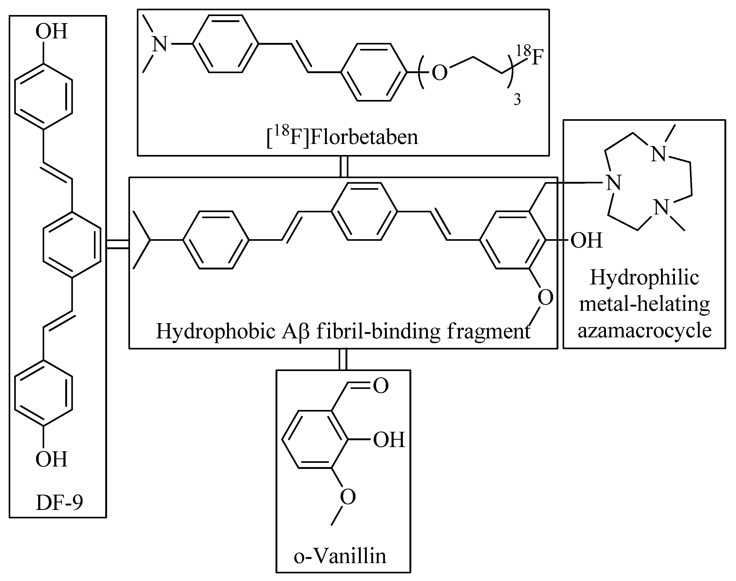
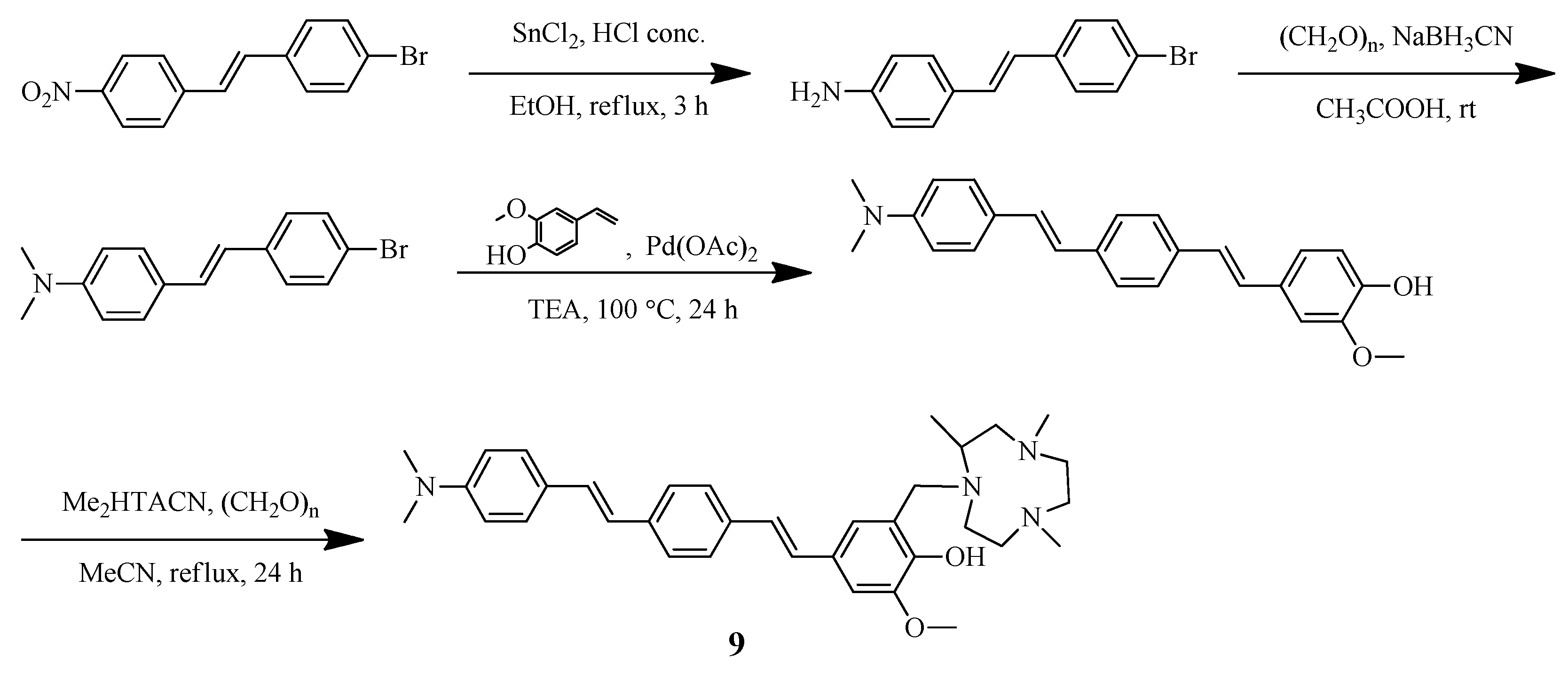
2.2. Distyrylbenzene-Vanilin BFC

Sun et al. reported [41] a distyrylbenzene-based hybrid 9 with a hydrophilic triazamacrocycle chelating moiety (Figure 5 and Figure 6). An asymmetric distyrylstilbene was designed as an FDA-approved PET imaging agent [18F]florbetaben. The symmetric distyrylbenzene structure of previously described compound DF-9 have been widely used in detecting amyloid plaques [49] as well as the 2-methoxy-phenol fragment reminiscent of o-vanillin that was shown to inhibit the formation of Aβ oligomers and also exhibit antioxidant properties [50].
Antioxidant ability of hybrid 9 was confirmed by trolox-equivalent antioxidant capacity (TEAC) assay. Both 9 and Cu(II) coordination compound based on Cu-9 showed the fluorescence turn-on effect in the presence of Aβ species, especially in the presence of soluble Aβ42 oligomers. Importantly, in the absence of the hydrophilic azamacrocycle fragment, the binding affinity of Pre-9 toward the amyloid species dramatically decreased.
A nanomolar affinity of 9 for Aβ42 oligomers (Kd = 50 ± 9 nM) and Aβ fibrils (Kd = 58 ± 15 nM) was established. In addition, in the presence of both Aβ42 and Cu2+, hybrid 9 proved the ability to rescue the viability of N2a cells and significantly alleviate the neurotoxicity of Cu2+-42 species. While monitoring of kinetics of Aβ42 aggregation in the presence of chelator 9 and complex Cu-9, an unusual behavior of ligand 9 and complex Cu-9 was observed. Thus, hybrid 9 was found to detect the “on-pathway” Aβ42 oligomers, that is, monomeric Aβ42 aggregates, and a decrease in its fluorescence was detected when Aβ42 fibrils were formed in solution. This is an important result, as high-soluble Aβ oligomers have been shown to be involved in synapse loss and neuronal injury [51].
Fluorescence staining of chelator 9 with brain sections from 7-month-old 5xFAD mice was also performed, with Congo Red dye, HJ3.4. antibody, or Aβ oligomer-specific monoclonal antibody (OMAB), which specifically binds to Aβ oligomers as controls. Both 9 and Cu-9 showed excellent colocalization with the immunofluorescence with both OMAB and HJ3.4, thus proving an ability of 9 to bind both the Aβ oligomers and fibrils in AD brain sections. In addition, a successive treatment of Aβ fibrils with Cu2+ and 9 lead to a significant inhibition of ascorbate consumption when compared to Aβ fibrils threated with Cu2+ only. Hybrid 9 found to reduce the neurotoxicity of Cu2+-Aβ42 species.
In vivo BBB permeability of 9 was also confirmed. Thus, after administration of 9 daily (1 mg/kg) to 7-month-old 5xFAD mice for 10 days via intraperitoneal injection, a strong fluorescence of mouse brain sections was detected, which was in a good colocalization with Congo Red fluorescence, and both HJ3.4 and OMAB antibodies. To assess therapeutic efficacy, 5xFAD mice were treated with 9, a significant reduction of both amyloid plaques and associated p-tau aggregates was detected, and microglia activation was also reduced. Finally, a radiolabeled 64Cu-9 was synthesized, and a series of PET imaging and biodistribution studies were performed. The results obtained proved 64Cu-9 complex can cross the BBB and binds to the amyloid plaques. What is more important, 64Cu-9 proved to accumulate to a significantly larger extent in the 5xFAD mice brains vs. the WT controls.
Finally, the effect of chelator 9 on the aggregation of p-tau protein and the activation of microglia as a neuroinflammatory response was assessed using fluorescently labeled AT8 antibody, which is specific to p-tau aggregates. The total amount of p-tau aggregates surrounding the amyloid plaques was decreased in the 9-treated vs. vehicle-treated 5xFAD mice. The level of activated microglia cells in AD mice was assessed using CF594-labeled ionized calcium-binding adapter molecule 1 (Iba1) antibody, and the ability of chelator 9 to suppress the activation of microglia cells to alleviate the neuroinflammation was revealed. Docking studies of binding of chelator 9 to both soluble Aβ oligomers and Aβ fibrils showed an ability of 9 to efficiently restrict the fibril formation in vivo, probably due to the preferential binding to the fibril ends of 9 to mitigate the Aβ elongation process.

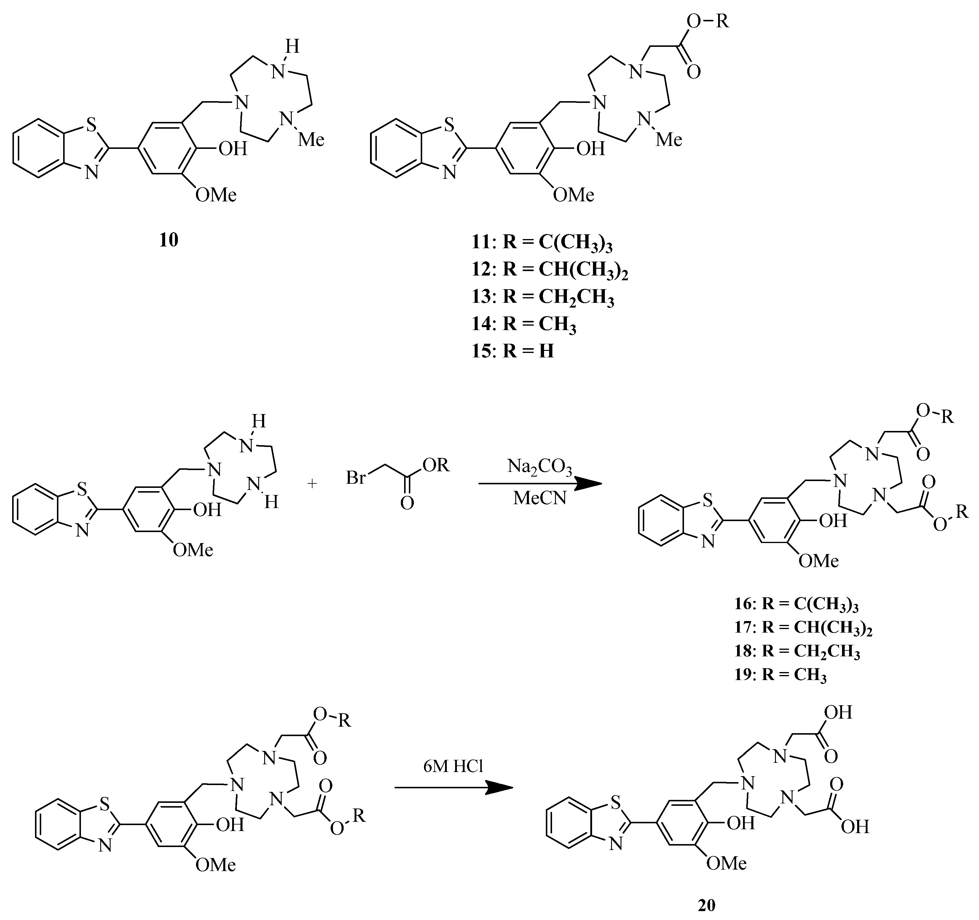
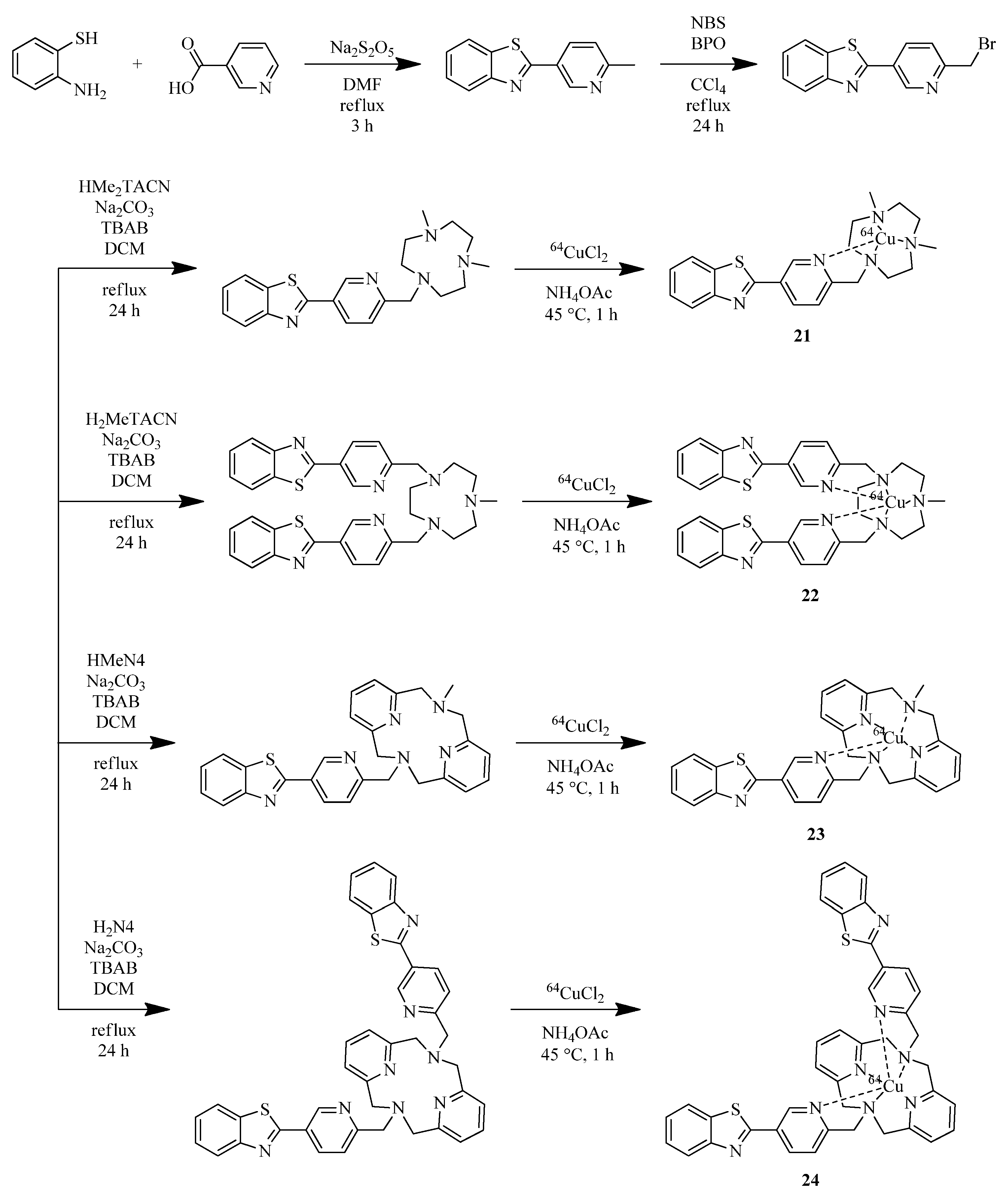
2.3. Benzothiazole-Based BFCs

Wang et al. reported five benzothiazole-based BFCs 11–15 with ester derivatives of TACN and non-ester derivative 10 [42] (Figure 7). Ester derivatives of the carboxylate pendant arm were conjugated with TACN moiety in order to increase the lipophilicity of the bifunctional chelators and facilitate brain uptake. Spectrophotometric titrations were used to quantify a stability constant of the complexes (log Ks); the results show that a carboxylic acid or ester moieties in TACN scaffold increases the log K by 3−4 orders of magnitude versus the parent TACN derivative.
Fluorescence imaging of amyloid plaques in 5xFAD mouse brain sections as well as immunostaining with HJ3.4 antibody revealed a specific binding of BFCs 11, 13, 14, and their Cu(II) complexes to Aβ species. A specific binding of ligands and their Cu(II) complexes with amyloid plaques was confirmed by staining with Congo Red dye on brain sections collected from 11-month-old 5xFAD mice. In addition, good colocalization of both ligands and their Cu(II) complexes was shown on brain sections from six-month-old 5xFAD mouse with HJ3.4 antibody (AF594- HJ3.4), especially for BFCs 11, 13, 14.
Autoradiography studies were performed on brain sections from 11-month-old 5xFAD and aged-matched WT mice. The results obtained strongly suggest that the 64Cu-labeled BFCs exhibited the ability to detect Aβ species ex vivo, and TACN esters show more specific binding to Aβ plaques than corresponding acids. In vivo biodistribution experiments in CD-1 mice were also performed to investigate the pharmacokinetics and revealed some brain uptake of complexes. The highest brain uptake was shown by 64Cu-14 of 0.46 ± 0.21% ID/g at 2 min post-injection.
In addition, the same scientific group reported five benzothiazole-based complexes with TACN chelator with two ester moieties [43] (Figure 7). A direct binding of 20 with Aβ42 fibrils was confirmed by fibril titration with solution of 20, a saturation behavior was observed, and a binding constant was calculated (Kd = 121 ± 44 nM).
A co-staining with a brain sections of 11-month-old 5xFAD transgenic mice with Congo Red dye revealed affinity of BFCs 17 and 18 and their Cu(II) complexes 17-Cu and 18-Cu toward Aβ species, and the specific staining of Cu-20 with AF594-conjugated HJ3.4 antibody (AF594-HJ3.4) also exhibited a strong colocalization with the antibody stained regions. Autoradiography studies of 11-month-old 5xFAD and age-matched WT mice revealed an increased intensity that 64Cu-20 exhibits in 5xFAD mice compared to WT.
Huang et al. reported Benzothiazole-based complexes with copper-chelating TACN and 2,11-diaza [3.3]-(2,6)pyridinophane (N4) moieties 21–24 [44] (Figure 8).
EPR spectra of complex Cu-22 suggest that the complex remains mononuclear in solution. Fluorescence imaging studies on 5xFAD mouse brain sections treated with 21–24, and Cu(I,II) complexes based on them revealed a specific binding of ones to Aβ plaques, which was confirmed by co-staining CF594-conjugated HJ3.4 antibody (CF594-HJ3.4), affinic to a wide range of Aβ species. Autoradiography studies of 64Cu-labeled 21–24 complexes revealed a specific binding of the complexes to amyloid plaques, which was also confirmed by blocking with the nonradioactive blocking agent B1. A great contrast between the intensity of WT and 5xFAD mice brains for all radiolabeled complexes was shown, especially for 64Cu-22. In addition, an incubation of 64Cu-22 and64Cu-23 with human serum at 37 °C for up to 24 h showed the stability of the complexes. To evaluate the ability of radiolabeled coordination compounds to cross the BBB in vivo, a biodistribution in normal CD-1 mice was evaluated. The highest brain uptake for 64Cu-22 complex was shown and was approximately ~ 0.4% both after 2 min post injection and after 24 h, which indicates the rapid penetration of the complex into the brain and its retention.
Wang et al. investigated a series of BFCs with an Aβ-binding 2-(4-hydroxyphenyl)-benzothiazole moiety and metal-chelating 1,4,7-triazacyclononane (TACN) ligands and gallium coordination compounds based on them [45] (Figure 9).
Histological staining of 5xFAD mouse brain sections with compounds 25–28 showed a good affinity of BFCs 25, 26, 28 for the amyloid aggregates, which correlated well with Congo Red or HJ3.4 antibody controls. In contrast, BFCs 27 exhibited weak Congo Red colocalization, thus indicating that introduction of extra amyloid β targeting moieties are able to increase the affinity of BFCs to amyloid plaques. Autoradiography studies with radiolabeled complexes [68Ga]25–28 revealed a specific binding with brain sections of 5xFAD and WT mice, with the highest non-specific binding of 68Ga-labeled bivalent complexes.
Recently, the same scientific group reported a series of BFCs containing two Aβ-targeting fragments and a TACN macrocyclic ligand and novel derivatives with carboxylate ester arms (Figure 10) [46]. ThT competition assays revealed binding of BFCs to Aβ plaques with most active hybrids 30, 31. In ex vivo autoradiography studies of 64Cu-radiolabeled BFCs with brain sections from 11-month-old 5xFAD and aged-matched WT mice, BFCs 29, 32, 33 exhibited ∼4-fold increase for 5xFAD vs. WT brain sections, with hybrid 36 exhibiting the highest overall intensity. Finally, 64Cu-30 showed the most promising brain uptake in CD-1 mice, with a maximum %ID/g of 0.47 ± 0.12 at 2 min post-injection.

2.4. Azo-Stilbene-Based BFCs

Rana et al. reported unusual bifunctional compounds that include the amyloid binding properties from stilbene and the staining characteristics of Congo Red, a commonly used Aβ-specific dye, conjugated with strong metal-binding arms [47]. These BFCs were designed to target metal-mediated neurotoxicity, but may also be considered as a perspective of organic scaffolds for design of metal-based drugs for PET Aβ imaging. Azo-stilbene-derived compounds with N,N,O and N,N,N,O donor metal chelation moiety were designed and thoroughly investigated (Figure 11).
An ability of BFCs 35, 36 to bind Aβ plaques was confirmed using ThT competition assay as well as UV−Vis spectroscopy. Inhibition of Aβ42 aggregation by BFCs 35, 36, as well as Cu-35 and Cu-36 was monitored by a decrease in ThT fluorescence. Aβ42 monomers showed low ThT fluorescence and a striking increase in fluorescence during aggregation. Both compounds 35 and 36 reduced the fluorescence of Aβ42 aggregates as well as Aβ42 aggregates pretreated with Cu2+ or Zn2+. Inhibition of Aβ42 metal-free and metal−Aβ aggregation was also confirmed by TEM images. Thus, in the presence of BFCs 35, 36, the morphology was quite different from that with Aβ42 alone. In addition, Aβ42 aggregation in the presence of both Cu2+ or Zn2+ and chelators 35 and 36 led to lesser aggregates of amorphous morphology, differing from that of Aβ42 alone.
Docking interactions of 35 and 36 with the Aβ40 fibrillar structure revealed their positioning near the KLVFF hydrophobic region of the peptide, π−π interactions of BFCs 35, 36 with both with Aβ40 and Aβ42. In addition, molecular docking with acetylcholinesterase AChE showed an interaction of BFCs 35, 36 with catalytic active site (CAS) and peripheral anionic site (PAS) of AChE. An ability of cholinesterase inhibition was also confirmed (Table 2), as well as the ability of BFCs 35, 36 to inhibit AChE-induced Aβ42 aggregation confirmed by ThT fluorescence assay.
An antioxidant property of BFCs 35, 36 was confirmed using 6-hydroxy-2,5,7,8-tetramethylchroman-2-carboxylic acid (Trolox) (TEAC). Finally, compound 35 showed low neurotoxicity on Neuro2A cells, in contrast to 36, thus suggest that extra pyridine groups may lead to higher cell toxicity.

2.5. Styrylpyridyl-Based BFCs

Spyrou et al. reported a three tetradentate ligand based on styrylpyridyl scaffolds with pyridyl, amide, amine, and thiol chelating moieties and charge-neutral complexes [Tc=O]3+ and [Re=O]3+ based on it [48] (Figure 12).
The ability of BFCs 37–39 to interact with Aβ1−40 fibrils was investigated with a competition assay between each rhenium complex and ThT. Each of the complexes showed an ability to displace ThT from the fibrils and had significant affinity for Aβ1−40 with Ki ∼ 240−260 nM. In addition, excellent colocalization of the complexes with Aβ plaques of human brain tissue was revealed by immunohistochemistry with a Aβ-specific 1E8 antibody as a control. Radiolabeled [99mTc][TcO 37–39] were obtained, and biodistribution of [99mTc][TcO 37–39] in wild-type mice was determined. Unfortunately, brain uptake values of the complexes were too low for SPECT imaging.

3. Conclusions

Summarizing the above data, one can conclude that metal-containing imagining agents are a promising alternative to clinically used radiopharmaceuticals based on short-lived 11C and 18F isotopes. This review provides examples of the successful design of ligands and coordinating compounds based on them, capable of crossing the blood-brain barrier and successfully binding to amyloid plaques in an AD brain. Radiolabeled complex 6-64Cu showed a significant higher brain uptake in the 5xFAD mice than in WT; this testifies to the thoughtful drug design and confirms the ability of a Cu-based coordination compounds to act as imaging agents for Aβ plaques.
In addition, the successful design of an effective and selective bifunctional chelator 9 and a coordination compound Cu-9 based on it shows great potential of copper-containing coordination compounds as drugs for imaging of Alzheimer’s disease. In addition, hybrid 9 showed the ability to act on soluble Aβ oligomers, which is an extremely promising result due to the high toxicity of the latter, as well as an acute shortage of drugs capable of acting on them. During several attempts to create coordination compounds with an ester or carboxyl group, radiolabeled coordination compound 64Cu-20 showed increased brain uptake in 5xFAD mice compared to WT. It should also be noted that the ability of bifunctional ligands 35, 36 to inhibit acetylcholinesterase suggests that the developed bifunctional compounds can be not only effective imaging agents, but also have therapeutic anti-AD efficacy.
Thus, bifunctional compounds with an amyloid affinity fragment together with a chelating fragment are able to visualize both Aβ plaques and soluble Aβ oligomers. Their ability to influence metal homeostasis and Aβ aggregation opens up opportunities for creating not only visualizing but also theranostic agents for AD.

Funding

The work was financially supported by the Ministry of Education and Science of the Russian Federation, Agreement No. 075-15-2022-264 (unique scientific facility “Scanning ion-conductance microscope with a confocal module” (registration number 2512530)).

Conflicts of Interest

The authors declare no conflict of interest.

Abbreviations

AD—Alzheimer’s disease, AChE—acetylcholinesterase, TEAC—trolox-equivalent antioxidant capacity, WT—wild type mice, SPECT—single-photon emission computerized tomography, BFCs—bifunctional compounds, CR—Congo Red, CAS—catalytic active site, PAS—peripheral anionic site, PET—positron emission tomography, MRI—magnetic resonance imaging, TACN—2,4,7-triazacyclononane, NOTA—1,4,7-triazacyclononane-1,4,7-triacetic acid, TETA—1,4,8,11-tetraazacyclotetradecane-N,N′,N′′,N′′′-tetraacetic acid, DOTA—2,2′,2′′,2′′′-(1,4,7,10-tetraazacyclododecane-1,4,7,10-tetrayl)tetraacetic acid

References

  1. Kepp, K.P. Bioinorganic Chemistry of Alzheimer’s Disease. Chem. Rev. 2012, 112, 5193–5239. [Google Scholar] [CrossRef] [PubMed] [Green Version]
  2. Lee, N.; Yoo, D.; Ling, D.; Cho, M.H.; Hyeon, T.; Cheon, J. Iron Oxide Based Nanoparticles for Multimodal Imaging and Magnetoresponsive Therapy. Chem. Rev. 2015, 115, 10637–10689. [Google Scholar] [CrossRef] [PubMed]
  3. O’Brien, R.J.; Wong, P.C. Amyloid Precursor Protein Processing and Alzheimer’s Disease. Annu. Rev. Neurosci. 2011, 34, 185–204. [Google Scholar] [CrossRef] [Green Version]
  4. Doecke, J.D.; Pérez-Grijalba, V.; Fandos, N.; Fowler, C.; Villemagne, V.L.; Masters, C.L.; Pesini, P.; Sarasa, M. Total Aβ(42)/Aβ(40) Ratio in Plasma Predicts Amyloid-PET Status, Independent of Clinical AD Diagnosis. Neurology 2020, 94, e1580–e1591. [Google Scholar] [CrossRef] [PubMed] [Green Version]
  5. Atrián-Blasco, E.; Gonzalez, P.; Santoro, A.; Alies, B.; Faller, P.; Hureau, C. Cu and Zn Coordination to Amyloid Peptides: From Fascinating Chemistry to Debated Pathological Relevance. Coord. Chem. Rev. 2018, 375, 38–55. [Google Scholar] [CrossRef]
  6. Yang, T.; Li, S.; Xu, H.; Walsh, D.M.; Selkoe, D.J. Large Soluble Oligomers of Amyloid β-Protein from Alzheimer Brain Are Far Less Neuroactive Than the Smaller Oligomers to Which They Dissociate. J. Neurosci. 2017, 37, 152–163. [Google Scholar] [CrossRef] [Green Version]
  7. Mueller, S.P.; Polak, J.F.; Kijewski, M.F.; Holman, B.L. Collimator Selection for SPECT Brain Imaging: The Advantage of High Resolution. J. Nucl. Med. 1986, 27, 1729–1738. [Google Scholar]
  8. Holliger, P.; Hudson, P.J. Engineered Antibody Fragments and the Rise of Single Domains. Nat. Biotechnol. 2005, 23, 1126–1136. [Google Scholar] [CrossRef]
  9. Lu, F.-M.; Yuan, Z. PET/SPECT Molecular Imaging in Clinical Neuroscience: Recent Advances in the Investigation of CNS Diseases. Quant. Imaging Med. Surg. 2015, 5, 433–447. [Google Scholar] [CrossRef]
  10. Dunyan, S.; Wei, D.; Jie, L.; Lili, P.; Xiaoyang, Z.; Xiaoai, W.; Wuyu, M. Strategic Design of Amyloid-β Species Fluorescent Probes for Alzheimer’s Disease. ACS Chem. Neurosci. 2022, 13, 5–540. [Google Scholar]
  11. Soloperto, A.; Quaglio, D.; Baiocco, P.; Romeo, I.; Mori, M.; Ardini, M.; Presutti, C.; Sannino, I.; Ghirga, S.; Iazzetti, A.; et al. Rational design and synthesis of a novel BODIPY-based probe for selective imaging of tau tangles in human iPSC-derived cortical neurons. Sci. Rep. 2022, 12, 5257. [Google Scholar] [CrossRef] [PubMed]
  12. Yang, J.; Zeng, F.; Ge, Y.; Peng, K.; Li, X.; Li, Y.; Xu, Y. Development of Near-Infrared Fluorescent Probes for Use in Alzheimer’s Disease Diagnosis. Bioconj.Chem. 2020, 31, 2–15. [Google Scholar] [CrossRef] [PubMed]
  13. Klunk, W.E.; Engler, H.; Nordberg, A.; Wang, Y.; Blomqvist, G.; Holt, D.P.; Bergström, M.; Savitcheva, I.; Huang, G.-F.; Estrada, S.; et al. Imaging Brain Amyloid in Alzheimer’s Disease with Pittsburgh Compound-B. Ann. Neurol. 2004, 55, 306–319. [Google Scholar] [CrossRef] [PubMed]
  14. Barthel, H.; Gertz, H.-J.; Dresel, S.H.; Peters, O.; Bartenstein, P.; Buerger, K.; Hiemeyer, F.; Wittemer-Rump, S.; Seibyl, J.; Reininger, C.; et al. Cerebral Amyloid-β PET with Florbetaben (18F) in Patients with Alzheimer’s Disease and Healthy Controls: A Multicentre Phase 2 Diagnostic Study. Lancet Neurol. 2011, 10, 424–435. [Google Scholar] [CrossRef]
  15. Lister-James, J.; Pontecorvo, M.J.; Clark, C.; Joshi, A.D.; Mintun, M.A.; Zhang, W.; Lim, N.; Zhuang, Z.; Golding, G.; Choi, S.R.; et al. Florbetapir F-18: A Histopathologically Validated Beta-Amyloid Positron Emission Tomography Imaging Agent. Semin. Nucl. Med. 2011, 41, 300–304. [Google Scholar] [CrossRef]
  16. Curtis, C.; Gamez, J.E.; Singh, U.; Sadowsky, C.H.; Villena, T.; Sabbagh, M.N.; Beach, T.G.; Duara, R.; Fleisher, A.S.; Frey, K.A.; et al. Phase 3 Trial of Flutemetamol Labeled With Radioactive Fluorine 18 Imaging and Neuritic Plaque Density. JAMA Neurol. 2015, 72, 287–294. [Google Scholar] [CrossRef]
  17. Serdons, K.; Terwinghe, C.; Vermaelen, P.; Van Laere, K.; Kung, H.; Mortelmans, L.; Bormans, G.; Verbruggen, A. Synthesis and Evaluation of 18F-Labeled 2-Phenylbenzothiazoles as Positron Emission Tomography Imaging Agents for Amyloid Plaques in Alzheimer’s Disease. J. Med. Chem. 2009, 52, 1428–1437. [Google Scholar] [CrossRef]
  18. Choi, S.R.; Golding, G.; Zhuang, Z.; Zhang, W.; Lim, N.; Hefti, F.; Benedum, T.E.; Kilbourn, M.R.; Skovronsky, D.; Kung, H.F. Preclinical Properties of 18F-AV-45: A PET Agent for Aβ Plaques in the Brain. J. Nucl. Med. 2009, 50, 1887–1894. [Google Scholar] [CrossRef] [Green Version]
  19. Uzuegbunam, B.C.; Librizzi, D.; Hooshyar Yousefi, B. PET Radiopharmaceuticals for Alzheimer’s Disease and Parkinson’s Disease Diagnosis, the Current and Future Landscape. Molecules 2020, 25, 977. [Google Scholar] [CrossRef] [Green Version]
  20. Alauddin, M.M. Positron Emission Tomography (PET) Imaging with (18)F-Based Radiotracers. Am. J. Nucl. Med. Mol. Imaging 2012, 2, 55–76. [Google Scholar]
  21. Kuijpers, W.H.A.; Kaspersen, F.M.; Veeneman, G.H.; Van Boeckel, C.A.A.; Bos, E.S. Specific Recognition of Antibody-Oligonucleotide Conjugates by Radiolabeled Antisense Nucleotides: A Novel Approach for Two-Step Radioimmunotherapy of Cancer. Bioconjug. Chem. 1993, 4, 94–102. [Google Scholar] [CrossRef] [PubMed]
  22. Bagheri, S.; Squitti, R.; Haertlé, T.; Siotto, M.; Saboury, A.A. Role of Copper in the Onset of Alzheimer’s Disease Compared to Other Metals. Front. Aging Neurosci. 2018, 9, 446. [Google Scholar] [CrossRef] [PubMed] [Green Version]
  23. Zhou, Y.; Li, J.; Xu, X.; Zhao, M.; Zhang, B.; Deng, S.; Wu, Y. 64Cu-Based Radiopharmaceuticals in Molecular Imaging. Technol. Cancer Res. Treat. 2019, 18, 1533033819830758. [Google Scholar] [CrossRef] [PubMed]
  24. Anderson, C.J.; Ferdani, R. Copper-64 radiopharmaceuticals for PET imaging of cancer: Advances in preclinical and clinical research. Cancer Biother. Radiopharm. 2009, 24, 379–393. [Google Scholar] [CrossRef] [PubMed]
  25. Nie, X.; Laforest, R.; Elvington, A.; Randolph, G.; Zheng, J.; Voller, N.; Abendschein, D.; Lapi, S.; Woodard, K. PET/MRI of Hypoxic Atherosclerosis Using 64Cu-ATSM in a Rabbit Model. J. Nuc. Med. 2016, 57, 2006–2011. [Google Scholar] [CrossRef] [PubMed] [Green Version]
  26. De Silva, R.A.; Kumar, D.; Lisok, A.; Chatterjee, S.; Wharram, B.; Venkateswara Rao, K.; Mease, R.; Dannals, R.F.; Pomper, M.G.; Nimmagadda, S. Peptide-Based 68Ga-PET Radiotracer for Imaging PD-L1 Expression in Cancer. Mol. Pharm. 2018, 15, 3946–3952. [Google Scholar] [CrossRef] [Green Version]
  27. Krasnovskaya, O.; Spector, D.; Zlobin, A.; Pavlov, K.; Gorelkin, P.; Erofeev, A.; Beloglazkina, E.; Majouga, A. Metals in Imaging of Alzheimer’s Disease. Int. J. Mol. Sci. 2020, 21, 9190. [Google Scholar] [CrossRef]
  28. Ciudad, S.; Puig, E.; Botzanowski, T.; Meigooni, M.; Arango, A.S.; Do, J.; Mayzel, M.; Bayoumi, M.; Chaignepain, S.; Maglia, G.; et al. Aβ(1-42) Tetramer and Octamer Structures Reveal Edge Conductivity Pores as a Mechanism for Membrane Damage. Nat. Commun. 2020, 11, 3014. [Google Scholar] [CrossRef]
  29. Sharma, A.K.; Schultz, J.W.; Prior, J.T.; Rath, N.P.; Mirica, L.M. Coordination Chemistry of Bifunctional Chemical Agents Designed for Applications in 64Cu PET Imaging for Alzheimer’s Disease. Inorg. Chem. 2017, 56, 13801–13814. [Google Scholar] [CrossRef] [Green Version]
  30. Storr, T.; Merkel, M.; Song-Zhao, G.X.; Scott, L.E.; Green, D.E.; Bowen, M.L.; Thompson, K.H.; Patrick, B.O.; Schugar, H.J.; Orvig, C. Synthesis, Characterization, and Metal Coordinating Ability of Multifunctional Carbohydrate-Containing Compounds for Alzheimer’s Therapy. J. Am. Chem. Soc. 2007, 129, 7453–7463. [Google Scholar] [CrossRef]
  31. Sharma, A.K.; Pavlova, S.T.; Kim, J.; Finkelstein, D.; Hawco, N.J.; Rath, N.P.; Kim, J.; Mirica, L.M. Bifunctional Compounds for Controlling Metal-Mediated Aggregation of the Aβ42 Peptide. J. Am. Chem. Soc. 2012, 134, 6625–6636. [Google Scholar] [CrossRef] [PubMed] [Green Version]
  32. Ono, M.; Watanabe, H.; Kimura, H.; Saji, H. BODIPY-Based Molecular Probe for Imaging of Cerebral β-Amyloid Plaques. ACS Chem. Neurosci. 2012, 3, 319–324. [Google Scholar] [CrossRef] [PubMed] [Green Version]
  33. Wu, N.; Kang, C.S.; Sin, I.; Ren, S.; Liu, D.; Ruthengael, V.C.; Lewis, M.R.; Chong, H.-S. Promising Bifunctional Chelators for Copper 64-PET Imaging: Practical (64)Cu Radiolabeling and High in Vitro and in Vivo Complex Stability. J. Biol. Inorg. Chem. 2016, 21, 177–184. [Google Scholar] [CrossRef] [PubMed]
  34. Tosato, M.; Dalla Tiezza, M.; May, N.V.; Isse, A.A.; Nardella, S.; Orian, L.; Verona, M.; Vaccarin, C.; Alker, A.; Mäcke, H.; et al. Copper Coordination Chemistry of Sulfur Pendant Cyclen Derivatives: An Attempt to Hinder the Reductive-Induced Demetalation in 64/67Cu Radiopharmaceuticals. Inorg. Chem. 2021, 60, 11530–11547. [Google Scholar] [CrossRef] [PubMed]
  35. Lever, S.Z.; Lydon, J.D.; Cutler, C.S.; Jurisson, S.S. Radioactive Metals in Imaging and Therapy; McCleverty, J.A., Meyer, T.J.B.T.-C.C.C.I.I., Eds.; Pergamon: Oxford, UK, 2003; pp. 883–911. ISBN 978-0-08-043748-4. [Google Scholar]
  36. Guillou, A.; Lima, L.M.P.; Esteban-Gómez, D.; Le Poul, N.; Bartholomä, M.D.; Platas-Iglesias, C.; Delgado, R.; Patinec, V.; Tripier, R. Methylthiazolyl Tacn Ligands for Copper Complexation and Their Bifunctional Chelating Agent Derivatives for Bioconjugation and Copper-64 Radiolabeling: An Example with Bombesin. Inorg. Chem. 2019, 58, 2669–2685. [Google Scholar] [CrossRef] [PubMed]
  37. Hogarth, G.; Onwudiwe, D.C. Copper dithiocarbamates: Coordination chemistry and applications in materials science, biosciences and beyond. Inorganics 2021, 9, 70. [Google Scholar] [CrossRef]
  38. Wang, J.; Guan, H.; Liang, Q.; Ding, M. Construction of Copper (II) Affinity- DTPA Functionalized Magnetic Composite for Efficient Adsorption and Specific Separation of Bovine Hemoglobin from Bovine Serum. Compos. Part B Eng. 2020, 198, 108248. [Google Scholar] [CrossRef]
  39. Calvary, C.A.; Hietsoi, O.; Hofsommer, D.T.; Brun, H.C.; Costello, A.M.; Mashuta, M.S.; Spurgeon, J.M.; Buchanan, R.M.; Grapperhaus, C.A. Copper Bis(Thiosemicarbazone) Complexes with Pendent Polyamines: Effects of Proton Relays and Charged Moieties on Electrocatalytic HER. Eur. J. Inorg. Chem. 2021, 2021, 267–275. [Google Scholar] [CrossRef]
  40. Cho, H.-J.; Huynh, T.T.; Rogers, B.E.; Mirica, L.M. Design of a Multivalent Bifunctional Chelator for Diagnostic 64Cu PET Imaging in Alzheimer’s Disease. Proc. Natl. Acad. Sci. USA 2020, 117, 30928–30933. [Google Scholar] [CrossRef]
  41. Sun, L.; Cho, H.-J.; Sen, S.; Arango, A.S.; Huynh, T.T.; Huang, Y.; Bandara, N.; Rogers, B.E.; Tajkhorshid, E.; Mirica, L.M. Amphiphilic Distyrylbenzene Derivatives as Potential Therapeutic and Imaging Agents for Soluble and Insoluble Amyloid β Aggregates in Alzheimer’s Disease. J. Am. Chem. Soc. 2021, 143, 10462–10476. [Google Scholar] [CrossRef]
  42. Wang, Y.; Huynh, T.T.; Cho, H.-J.; Wang, Y.-C.; Rogers, B.E.; Mirica, L.M. Amyloid β-Binding Bifunctional Chelators with Favorable Lipophilicity for 64Cu Positron Emission Tomography Imaging in Alzheimer’s Disease. Inorg. Chem. 2021, 60, 12610–12620. [Google Scholar] [CrossRef] [PubMed]
  43. Wang, Y.; Huynh, T.T.; Bandara, N.; Cho, H.-J.; Rogers, B.E.; Mirica, L.M. 2-(4-Hydroxyphenyl)Benzothiazole Dicarboxylate Ester TACN Chelators for 64Cu PET Imaging in Alzheimer’s Disease. Dalt. Trans. 2022, 51, 1216–1224. [Google Scholar] [CrossRef] [PubMed]
  44. Huang, Y.; Huynh, T.T.; Sun, L.; Hu, C.-H.; Wang, Y.-C.; Rogers, B.E.; Mirica, L.M. Neutral Ligands as Potential 64Cu Chelators for Positron Emission Tomography Imaging Applications in Alzheimer’s Disease. Inorg. Chem. 2022, 61, 4778–4787. [Google Scholar] [CrossRef] [PubMed]
  45. Huynh, T.T.; Wang, Y.; Terpstra, K.; Cho, H.-J.; Mirica, L.M.; Rogers, B.E. 68Ga-Labeled Benzothiazole Derivatives for Imaging Aβ Plaques in Cerebral Amyloid Angiopathy. ACS Omega 2022, 7, 20339–20346. [Google Scholar] [CrossRef] [PubMed]
  46. Terpstra, K.; Wang, Y.; Huynh, T.; Bandara, N.; Cho, H.; Rogers, B.; Mirica, L. Divalent 2-(4-Hydroxyphenyl)benzothiazole Bifunctional Chelators for 64Cu Positron Emission Tomography Imaging in Alzheimer’s Disease. Inorg. Chem. 2022, 50, 20326–20336. [Google Scholar] [CrossRef]
  47. Rana, M.; Cho, H.-J.; Arya, H.; Bhatt, T.K.; Bhar, K.; Bhatt, S.; Mirica, L.M.; Sharma, A.K. Azo-Stilbene and Pyridine–Amine Hybrid Multifunctional Molecules to Target Metal-Mediated Neurotoxicity and Amyloid-β Aggregation in Alzheimer’s Disease. Inorg. Chem. 2022, 61, 10294–10309. [Google Scholar] [CrossRef]
  48. Spyrou, B.; Hungnes, I.N.; Mota, F.; Bordoloi, J.; Blower, P.J.; White, J.M.; Ma, M.T.; Donnelly, P.S. Oxorhenium(V) and Oxotechnetium(V) Complexes of N3S Tetradentate Ligands with a Styrylpyridyl Functional Group: Toward Imaging Agents to Assist in the Diagnosis of Alzheimer’s Disease. Inorg. Chem. 2021, 60, 13669–13680. [Google Scholar] [CrossRef]
  49. Flaherty, D.P.; Kiyota, T.; Dong, Y.; Ikezu, T.; Vennerstrom, J.L. Phenolic Bis-Styrylbenzenes as β-Amyloid Binding Ligands and Free Radical Scavengers. J. Med. Chem. 2010, 53, 7992–7999. [Google Scholar] [CrossRef] [Green Version]
  50. Necula, M.; Kayed, R.; Milton, S.; Glabe, C.G. Small Molecule Inhibitors of Aggregation Indicate That Amyloid Beta Oligomerization and Fibrillization Pathways Are Independent and Distinct. J. Biol. Chem. 2007, 282, 10311–10324. [Google Scholar] [CrossRef] [Green Version]
  51. Ferreira, S.; Lourenco, M.; Oliveira, M.; De Felice, F. Soluble Amyloid-b Oligomers as Synaptotoxins Leading to Cognitive Impairment in Alzheimer’s Disease. Front. Cell. Neurosci. 2015, 9, 191. [Google Scholar] [CrossRef]
Figure 1. FDA-approved drugs for PET-imaging of amyloid plaques: Pittsburgh Compound-B ([11C]PIB), [18F]flutemetamol ([18F]GE-067), [18F]NAV-4694 (AZD-4694), [18F]florbetaben ([18F]AV-1, [18F]BAY-94-9172), and [18F]florbetapir ([18F]AV-45).
Ijms 24 00236 g001
Figure 2. Commonly used cyclic copper chelators TACN, NOTA, DOTA, and TETA.
Ijms 24 00236 g002
Figure 3. Commonly used acyclic copper chelators EDTA, DTPA, DTCBP, dithiocarbamate, and bis(thiosemicarbazone) derivatives.
Ijms 24 00236 g003
Figure 4. BFCs based on 2-(2-formyl-5-furanyl)-3-hydroxymethylbenzofuran scaffold with NOTA as copper chelating moiety 1–8 and Cu(II) complexes 1-Cu–8-Cu based on them reported by Cho et al. [40].
Ijms 24 00236 g004
Figure 5. Design strategy and structure of the amphiphilic compound 9.
Ijms 24 00236 g005
Figure 6. Distyrylbenzene-based bifunctional chelator 9 reported by Sun et al. [41].
Ijms 24 00236 g006
Figure 7. Benzothiazole-based BFCs 11–15 with ester derivatives of TACN, non-ester derivative 10 reported; BFCs 16–20 with two ester moieties of TACN reported by Wang et al. [42,43].
Ijms 24 00236 g007
Figure 8. Benzothiazole-based BFCs 21–24 and Cu(II) complexes based on them, 21-Cu–24-Cu, reported by Huang et al. [44].
Ijms 24 00236 g008
Figure 9. Benzothiazole-based BFCs 25–28 reported by Wang et al. [45].
Ijms 24 00236 g009
Figure 10. Benzothiazole-based BFCs 29–34 reported by Terpstra et al. [46].
Ijms 24 00236 g010
Figure 11. Azo-stilbene-based BFCs 35, 36 with two ester moieties of TACN reported by Rana et al. [47].
Ijms 24 00236 g011
Figure 12. Styrylpyridyl-based BFCs 37–39, reported by Spyrou et al. [48].
Ijms 24 00236 g012
Table 1. Multifunctional chelators for visualization of Aβ plaques.
BFCsMetalImaging MethodAmyloid-Binding MoietyChelatorBrain Uptake, ID/g **, Time Post InjectionRef.
1–8CuPET *BenzofuranNOTA2-, 60-, and 240-min p.i. ***
1
0.65 ± 0.23
0.10 ± 0.03
0.05 ± 0.00
2
0.76 ± 0.03
0.35 ± 0.10
0.08 ± 0.00
3
0.38 ± 0.04
0.13 ± 0.02
0.08 ± 0.01
4
0.83 ± 0.14
0.27 ± 0.05
0.09 ± 0.02
[40]
9CuPETFlorbetaben + VanilinTACNWT: 0.75 ± 0.10% ID/g
2 min
18 ± 0.02% ID/g
1 h
AD mice: 0.79 ± 0.06%ID/g 2 min
0.39 ± 0.02% ID/g (1 h)
[41]
10–15CuPETBenzothiazoleTACN with one alkyl carboxylate ester pendant arms2 min, 1 h, 4 h
11
0.35 ± 0.01
0.04 ± 0.01
0.03 ± 0.01
12
0.23 ± 0.06
0.02 ± 0.01
0.01 ± 0.00
13
0.32 ± 0.02
0.02 ± 0.00
0.01 ± 0.00
14
0.46 ± 0.21
0.14 ± 0.00
0.18 ± 0.02
15
0.23 ± 0.05
0.02 ± 0.02
0.02 ± 0.00
[42]
16–20CuPETBenzothiazoleTACN with two alkyl carboxylate ester pendant arms-[43]
21–24CuPETBenzothiazole1,4,7-triazacyclononane (TACN) and 2,11-diaza [3.3]-(2,6)pyridinophane (N4)Cu-23: 0.2% ID/g at 2 min, yet an increased brain accumulation of ∼0.4% ID/g was observed after 4 h[44]
25–28GaPET2-(4-hydroxyphenyl)-benzothiazoleTACN0.10 ± 0.03
0.05 ± 0.02
(2 h)
0.26 ± 0.12
0.07 ± 0.02
0.03 ± 0.00
0.33 ± 0.12
0.01 ±0.009
(2 h)
[45]
29–34CuPETBenzothiazoleTACN0.47 ± 0.12 (2 min)[46]
35, 36--Azo-stilbenePyridine-[47]
37–39TcSPECT ****StyrylpyridylDiamide−thiol, Monoamide−monoamine−
thiol
Diamine−thiol
WT: *****
[99mTc][TcO-38]
2 min 0.15 ± 0.06%
35 min 0.17 ± 0.01%
[99mTc][TcO-39]
2 min 0.36 ± 0.09%
35 min 0.15 ± 0.02%
[48]
* PET—positron emission tomography, ** ID/g—injected dose per gram of tissue, p.i. ***—post-injection, **** SPECT—single-photon emission computerized tomography, ***** WT—wild-type mice.
Table 2. In vitro AChE inhibition by BfCs 35, 36.
AChE3536RivastigmineDopenezil
IC50 (µM)4.18 ± 0.153.86 ± 0.1311.02 ± 1.260.06 ± 1.13
Disclaimer/Publisher’s Note: The statements, opinions and data contained in all publications are solely those of the individual author(s) and contributor(s) and not of MDPI and/or the editor(s). MDPI and/or the editor(s) disclaim responsibility for any injury to people or property resulting from any ideas, methods, instructions or products referred to in the content.

