
Radiobiological and Treatment-Related Aspects of Spatially Fractionated Radiotherapy


Abstract
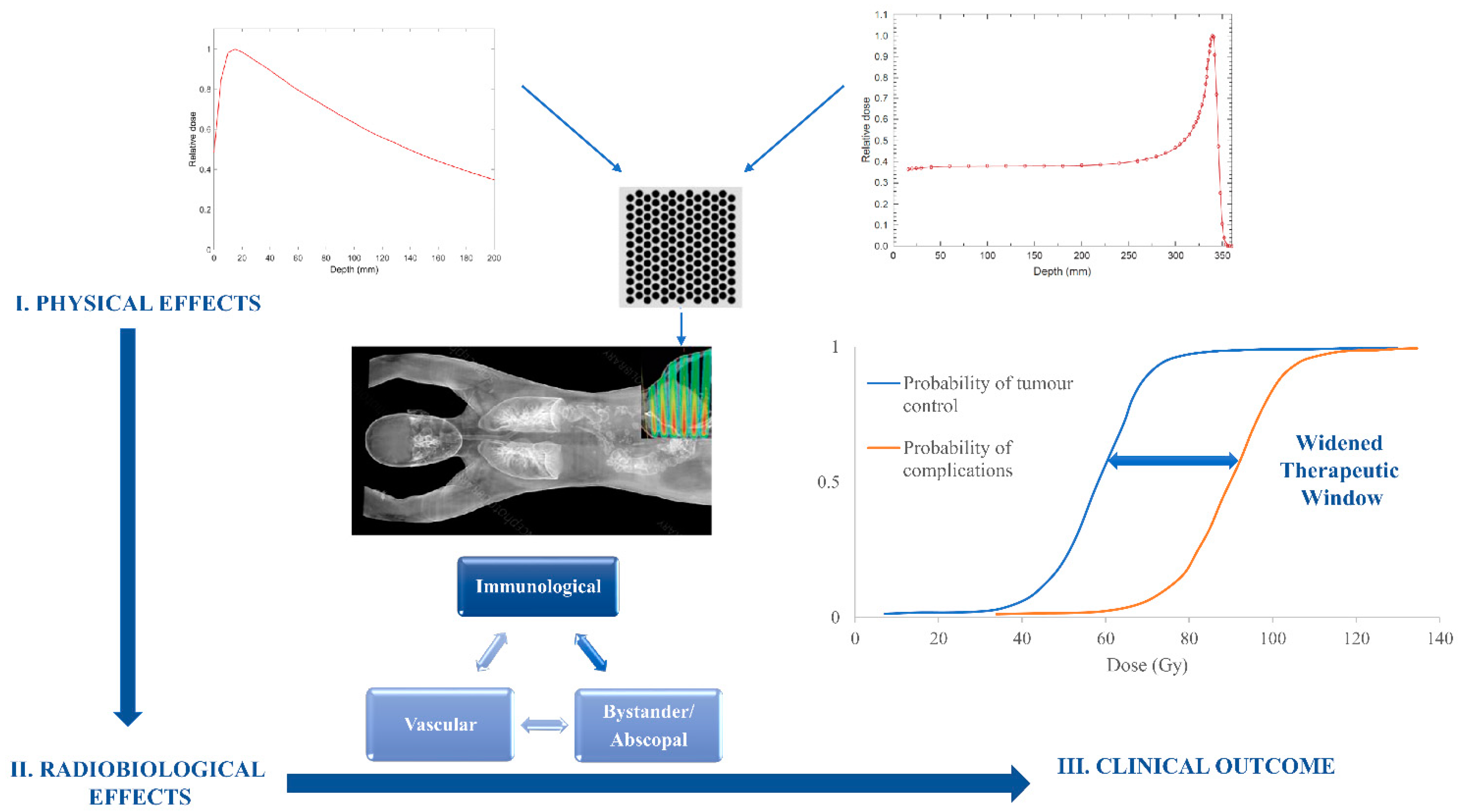
:1. Introduction to Non-Conventional Radiotherapy Delivery Approaches
2. Molecular and Radiobiological Mechanisms of Spatially Fractionated Radiotherapy
2.1. Vascular Effects
2.2. Immunological Effects
2.3. Bystander and Abscopal Effects
2.4. Proton SFRT
3. Preclinical and Clinical Evidence with Spatially Fractionated Radiotherapy
3.1. Spatially Fractionated Radiotherapy with Photons
3.1.1. Response Modelling in GRID RT with Photons
3.1.2. Mathematical-Model-Based Studies
3.1.3. Clinical Studies
3.2. Spatially Fractionated Radiotherapy with Protons
3.2.1. Animal Studies
3.2.2. Clinical Studies
3.2.3. Heavy-Ion Minibeam Therapy
4. Conclusions
- Spatially fractionated radiotherapy aims to widen the therapeutic window by better sparing of normal structures;
- While not all mechanisms behind this technique are fully elucidated, some potential biological mechanisms include: differential vascular effects between cancer and normal tissues, bystander or abscopal effects, and immunological effects;
- To date, both low-LET and high-LET radiation-based therapies have been investigated in vitro, as well as in vivo;
- Clinical studies are limited, nevertheless with promising results; further multidisciplinary research is required to clarify the radiobiological and physico-chemical mechanisms to understand and explore the full potential of spatially fractionated radiotherapy, in particular for aggressive and recurrent cancers.
Author Contributions
Funding
Institutional Review Board Statement
Informed Consent Statement
Conflicts of Interest
References
- Borras, J.M.; Lievens, Y.; Dunscombe, P.; Coffey, M.; Malicki, J.; Corral, J.; Gasparotto, C.; Defourny, N.; Barton, M.; Verhoeven, R.; et al. The optimal utilization proportion of external beam radiotherapy in European countries: An ESTRO-HERO analysis. Radiother. Oncol. 2015, 116, 38–44. [Google Scholar] [CrossRef] [PubMed] [Green Version]
- Billena, C.; Khan, A.J. A current review of spatial fractionation: Back to the future? Int. J. Radiat. Oncol. Biol. Phys. 2019, 104, 177–187. [Google Scholar] [CrossRef] [PubMed]
- Jiang, L.; Li, X.; Zhang, J.; Li, W.; Dong, F.; Chen, C.; Lin, Q.; Zhang, C.; Zheng, F.; Yan, W.; et al. Combined High-Dose LATTICE Radiation Therapy and Immune Checkpoint Blockade for Advanced Bulky Tumors: The Concept and a Case Report. Front. Oncol. 2021, 10, 3270. [Google Scholar] [CrossRef] [PubMed]
- Tubin, S.; Popper, H.H.; Brcic, L. Novel stereotactic body radiation therapy (SBRT)-based partial tumor irradiation targeting hypoxic segment of bulky tumors (SBRT-PATHY): Improvement of the radiotherapy outcome by exploiting the bystander and abscopal effects. Radiat. Oncol. 2019, 14, 21. [Google Scholar] [CrossRef]
- Tubin, S.; Grusch, M.; Popper, H.H.; Brcic, L.; Jeremic, B. SBRT-Partial Tumor Irradiation for The Treatment of Unresectable Bulky NSCLC. Int. J. Radiat. Oncol. Biol. Phys. 2020, 108, E111. [Google Scholar] [CrossRef]
- Grotzer, M.A.; Schultke, E.; Brauer-Krisch, E.; Laissue, J.A. Microbeam radiation therapy: Clinical perspectives. Phys. Med. 2015, 31, 564–567. [Google Scholar] [CrossRef] [Green Version]
- Romano, M.; Bravin, A.; Mittone, A.; Eckhardt, A.; Barbone, G.E.; Sancey, L.; Dinkel, J.; Bartzsch, S.; Ricke, J.; Alunni-Fabbroni, M.; et al. A Multi-Scale and Multi-Technique Approach for the Characterization of the Effects of Spatially Fractionated X-ray Radiation Therapies in a Preclinical Model. Cancers 2021, 13, 4953. [Google Scholar] [CrossRef]
- Duriseti, S.; Kavanaugh, J.A.; Szymanski, J.; Huang, Y.; Basarabescu, F.; Chaudhuri, A.; Henke, L.; Samson, P.; Lin, A.; Robinson, C.; et al. LITE SABR M1: A phase I trial of Lattice stereotactic body radiotherapy for large tumors. Radiother. Oncol. 2021, 167, 317–322. [Google Scholar] [CrossRef]
- Fukunaga, H.; Butterworth, K.; McMahon, S.; Prise, K. A brief overview of the preclinical and clinical radiobiology of microbeam radiotherapy. Clin. Oncol. 2021, 33, 705–712. [Google Scholar] [CrossRef]
- Griffin, R.J.; Ahmed, M.M.; Amendola, B.; Belyakov, O.; Bentzen, S.M.; Butterworth, K.T.; Chang, S.; Coleman, C.N.; Djonov, V.; Formenti, S.C.; et al. Understanding high-dose, ultra-high dose rate, and spatially fractionated radiation therapy. Int. J. Radiat. Oncol. Biol. Phys. 2020, 107, 766–778. [Google Scholar] [CrossRef]
- Fukunaga, H. Stem Cell Migration: A Possible Mechanism for the Tissue-Sparing Effect of Spatially Fractionated Radiation. Radiat. Res. 2021, 196, 680–685. [Google Scholar] [CrossRef] [PubMed]
- Steel, H.; Brüningk, S.C.; Box, C.; Oelfke, U.; Bartzsch, S.H. Quantification of differential response of tumour and normal cells to microbeam radiation in the absence of FLASH effects. Cancers 2021, 13, 3238. [Google Scholar] [CrossRef] [PubMed]
- Schültke, E.; Balosso, J.; Breslin, T.; Cavaletti, G.; Djonov, V.; Esteve, F.; Grotzer, M.; Hildebrandt, G.; Valdman, A.; Laissue, J. Microbeam radiation therapy—Grid therapy and beyond: A clinical perspective. Br. J. Radiol. 2017, 90, 20170073. [Google Scholar] [CrossRef] [PubMed]
- Dilmanian, F.A.; Button, T.M.; Le Duc, G.; Zhong, N.; Peña, L.A.; Smith, J.A.; Martinez, S.R.; Bacarian, T.; Tammam, J.; Ren, B.; et al. Response of rat intracranial 9L gliosarcoma to microbeam radiation therapy. Neuro-Oncology 2022, 4, 26–38. [Google Scholar] [CrossRef]
- Klein, D. The tumor vascular endothelium as decision maker in cancer therapy. Front. Oncol. 2018, 8, 367. [Google Scholar] [CrossRef] [PubMed]
- Schaaf, M.B.; Garg, A.D.; Agostinis, P. Defining the role of the tumor vasculature in antitumor immunity and immunotherapy. Cell Death Dis. 2018, 9, 1–14. [Google Scholar] [CrossRef] [Green Version]
- Bouchet, A.; Serduc, R.; Laissue, J.A.; Djonov, V. Effects of microbeam radiation therapy on normal and tumoral blood vessels. Phys. Med. 2015, 31, 634–641. [Google Scholar] [CrossRef] [Green Version]
- Bouchet, A.; Lemasson, B.; Le Duc, G.; Maisin, C.; Bräuer-Krisch, E.; Siegbahn, A.; Renaud, L.; Khalil, E.; Rémy, C.; Poillot, C.; et al. Preferential Effect of Synchrotron Microbeam Radiation Therapy on Intracerebral 9L Gliosarcoma Vascular Networks. Int. J. Radiat. Oncol. 2010, 78, 1503–1512. [Google Scholar] [CrossRef] [Green Version]
- Brönnimann, D.; Bouchet, A.; Schneider, C.; Potez, M.; Serduc, R.; Bräuer-Krisch, E.; Graber, W.; von Gunten, S.; Laissue, J.A.; Djonov, V. Synchrotron microbeam irradiation induces neutrophil infiltration, thrombocyte attachment and selective vascular damage in vivo. Sci. Rep. 2016, 6, 1–10. [Google Scholar] [CrossRef] [Green Version]
- Fuks, Z.; Kolesnick, R. Engaging the vascular component of the tumor response. Cancer Cell 2005, 8, 89–91. [Google Scholar] [CrossRef] [Green Version]
- Rao, S.S.; Thompson, C.; Cheng, J.; Haimovitz-Friedman, A.; Powell, S.N.; Fuks, Z.; Kolesnick, R.N. Axitinib sensitization of high Single Dose Radiotherapy. Radiother. Oncol. 2014, 111, 88–93. [Google Scholar] [CrossRef] [PubMed] [Green Version]
- Haimovitz-Friedman, A.; Kan, C.C.; Ehleiter, D.; Persaud, R.S.; McLoughlin, M.; Fuks, Z.; Kolesnick, R.N. Ionizing radiation acts on cellular membranes to generate ceramide and initiate apoptosis. J. Exp. Med. 1994, 180, 525–535. [Google Scholar] [CrossRef] [PubMed] [Green Version]
- Sathishkumar, S.; Boyanovski, B.; A Karakashian, A.; Rozenova, K.A.; Giltiay, N.V.; Kudrimoti, M.; Mohiuddin, M.; Ahmed, M.M.; Nikolova-Karakashian, M. Elevated sphingomyelinase activity and ceramide concentration in serum of patients undergoing high dose spatially fractionated radiation treatment: Implications for endothelial apoptosis. Cancer Biol. Ther. 2005, 4, 979–986. [Google Scholar] [CrossRef] [PubMed]
- Dutt, S.; Ahmed, M.M.; Loo, B.W., Jr.; Strober, S. Novel radiation therapy paradigms and immunomodulation: Heresies and hope. Semin. Radiat. Oncol. 2020, 30, 194–200. [Google Scholar] [CrossRef]
- Trappetti, V.; Fazzari, J.; Fernandez-Palomo, C.; Scheidegger, M.; Volarevic, V.; Martin, O.; Djonov, V. Microbeam Radiotherapy—A Novel Therapeutic Approach to Overcome Radioresistance and Enhance Anti-Tumour Response in Melanoma. Int. J. Mol. Sci. 2021, 22, 7755. [Google Scholar] [CrossRef] [PubMed]
- Potez, M.; Fernandez-Palomo, C.; Bouchet, A.; Trappetti, V.; Donzelli, M.; Krisch, M.; Laissue, J.; Volarevic, V.; Djonov, V. Synchrotron Microbeam Radiation Therapy as a New Approach for the Treatment of Radioresistant Melanoma: Potential Underlying Mechanisms. Int. J. Radiat. Oncol. 2019, 105, 1126–1136. [Google Scholar] [CrossRef] [Green Version]
- Eling, L.; Bouchet, A.; Ocadiz, A.; Adam, J.-F.; Kershmiri, S.; Elleaume, H.; Krisch, M.; Verry, C.; Laissue, J.; Balosso, J.; et al. Unexpected Benefits of Multiport Synchrotron Microbeam Radiation Therapy for Brain Tumors. Cancers 2021, 13, 936. [Google Scholar] [CrossRef]
- Bouchet, A.; Sakakini, N.; El Atifi, M.; Le Clec’H, C.; Brauer, E.; Moisan, A.; Deman, P.; Rihet, P.; Le Duc, G.; Pelletier, L. Early Gene Expression Analysis in 9L Orthotopic Tumor-Bearing Rats Identifies Immune Modulation in Molecular Response to Synchrotron Microbeam Radiation Therapy. PLoS ONE 2013, 8, e81874. [Google Scholar] [CrossRef] [Green Version]
- Sprung, C.N.; Cholewa, M.; Usami, N.; Kobayashi, K.; Crosbie, J.C. DNA damage and repair kinetics after microbeam radiation therapy emulation in living cells using monoenergetic synchrotron X-ray microbeams. J. Synchrotron Radiat. 2011, 18, 630–636. [Google Scholar] [CrossRef]
- Zhou, H.; Ivanov, V.N.; Gillespie, J.; Geard, C.R.; Amundson, S.A.; Brenner, D.J.; Yu, Z.; Lieberman, H.B.; Hei, T.K. Mechanism of radiation-induced bystander effect: Role of the cyclooxygenase-2 signaling pathway. Proc. Natl. Acad. Sci. USA 2005, 102, 14641–14646. [Google Scholar] [CrossRef] [Green Version]
- Yan, W.; Khan, M.K.; Wu, X.; Simone, C.B., 2nd; Fan, J.; Gressen, E.; Zhang, X.; Limoli, C.L.; Bahig, H.; Tubin, S.; et al. Spatially fractionated radiation therapy: History, present and the future. Clin. Transl. Radiat. Oncol. 2020, 20, 30–38. [Google Scholar] [CrossRef] [PubMed] [Green Version]
- Bazyar, S.; O’Brien, E.T.; Benefield, T.; Roberts, V.R.; Kumar, R.J.; Gupta, G.P.; Zhou, O.; Lee, Y.Z. Immune-Mediated Effects of Microplanar Radiotherapy with a Small Animal Irradiator. Cancers 2021, 14, 155. [Google Scholar] [CrossRef] [PubMed]
- Ferini, G.; Valenti, V.; Tripoli, A.; Illari, S.; Molino, L.; Parisi, S.; Cacciola, A.; Lillo, S.; Giuffrida, D.; Pergolizzi, S. Lattice or Oxygen-Guided Radiotherapy: What If They Converge? Possible Future Directions in the Era of Immunotherapy. Cancers 2021, 13, 3290. [Google Scholar] [CrossRef] [PubMed]
- Johnsrud, A.J.; Jenkins, S.V.; Jamshidi-Parsian, A.; Quick, C.M.; Galhardo, E.P.; Dings, R.P.; Vang, K.B.; Narayanasamy, G.; Makhoul, I.; Griffin, R.J. Evidence for Early Stage Anti-Tumor Immunity Elicited by Spatially Fractionated Radiotherapy-Immunotherapy Combinations. Radiat. Res. 2020, 194, 688–697. [Google Scholar] [CrossRef]
- Peng, V.; Suchowerska, N.; Rogers, L.; Mackonis, E.C.; Oakes, S.; McKenzie, D.R. Grid therapy using high definition multileaf collimators: Realizing benefits of the bystander effect. Acta Oncol. 2017, 56, 1048–1059. [Google Scholar] [CrossRef] [Green Version]
- Girst, S.; Marx, C.; Bräuer-Krisch, E.; Bravin, A.; Bartzsch, S.; Oelfke, U.; Greubel, C.; Reindl, J.; Siebenwirth, C.; Zlobinskaya, O.; et al. Improved normal tissue protection by proton and X-ray microchannels compared to homogeneous field irradiation. Phys. Med. 2015, 31, 615–620. [Google Scholar] [CrossRef] [Green Version]
- Mohiuddin, M.; Lynch, C.; Gao, M.; Hartsell, W. Early clinical results of proton spatially fractionated GRID radiation therapy (SFGRT). Br. J. Radiol. 2020, 93, 20190572. [Google Scholar] [CrossRef]
- Griffin, R.J.; Prise, K.M.; McMahon, S.J.; Zhang, X.; Penagaricano, J.; Butterworth, K.T. History and current perspectives on the biological effects of high-dose spatial fractionation and high dose-rate approaches: GRID, Microbeam & FLASH radiotherapy. Br. J. Radiol. 2020, 93, 20200217. [Google Scholar]
- Marcu, L.G.; Bezak, E.; Peukert, D.D.; Wilson, P. Translational Research in FLASH Radiotherapy—From Radiobiological Mechanisms to In Vivo Results. Biomedicines 2021, 9, 181. [Google Scholar] [CrossRef]
- Prezado, Y. Divide and conquer: Spatially fractionated radiation therapy. Expert Rev. Mol. Med. 2022, 24, e3. [Google Scholar] [CrossRef]
- Laissue, J.A.; Blattmann, H.; Slatkin, D.N. Alban Köhler (1874–1947): Erfinder der Gittertherapie. Z. Für Med. Phys. 2012, 22, 90–99. [Google Scholar] [CrossRef] [PubMed]
- Asur, R.; Butterworth, K.T.; Penagaricano, J.A.; Prise, K.M.; Griffin, R.J. High dose bystander effects in spatially fractionated radiation therapy. Cancer Lett. 2015, 356, 52–57. [Google Scholar] [CrossRef] [PubMed] [Green Version]
- Asur, R.S.; Sharma, S.; Chang, C.-W.; Penagaricano, J.; Kommuru, I.M.; Moros, E.G.; Corry, P.M.; Griffin, R.J. Spatially Fractionated Radiation Induces Cytotoxicity and Changes in Gene Expression in Bystander and Radiation Adjacent Murine Carcinoma Cells. Radiat. Res. 2012, 177, 751–765. [Google Scholar] [CrossRef] [PubMed] [Green Version]
- Mackonis, E.C.; Suchowerska, N.; Zhang, M.; Ebert, M.; McKenzie, D.R.; Jackson, M. Cellular response to modulated radiation fields. Phys. Med. Biol. 2007, 52, 5469–5482. [Google Scholar] [CrossRef]
- Poh, S.S.; Chua, M.L.K.; Wee, J.T.S. Why we should give spatially fractionated radiation therapy (GRID) a second look—Especially in nasopharyngeal carcinoma. Ann. Nasopharynx Cancer 2018, 2, 12–19. [Google Scholar] [CrossRef]
- Sathishkumar, S.; Dey, S.; Meigooni, A.S.; Regine, W.F.; Kudrimoti, M.; Ahmed, M.M.; Mohiuddin, M. The Impact of TNF-α Induction on Therapeutic Efficacy following High Dose Spatially Fractionated (GRID) Radiation. Technol. Cancer Res. Treat. 2002, 1, 141–147. [Google Scholar] [CrossRef] [Green Version]
- Lee, Y.; Auh, S.L.; Wang, Y.; Burnette, B.; Meng, Y.; Beckett, M.; Sharma, R.; Chin, R.; Tu, T.; Weichselbaum, R.R.; et al. Therapeutic effects of ablative radiation on local tumor require CD8+ T cells: Changing strategies for cancer treatment. Blood J. Am. Soc. Hematol. 2009, 114, 589–595. [Google Scholar] [CrossRef]
- Griffin, R.J.; A Koonce, N.; Dings, R.; Siegel, E.; Moros, E.; Bräuer-Krisch, E.; Corry, P.M. Microbeam radiation therapy alters vascular architecture and tumor oxygenation and is enhanced by a galectin-1 targeted anti-angiogenic peptide. Radiat. Res. 2012, 177, 804–812. [Google Scholar] [CrossRef] [Green Version]
- Martínez-Rovira, I.; Puxeu-Vaqué, J.; Prezado, Y. Dose evaluation of Grid Therapy using a 6 MV flattening filter-free (FFF) photon beam: A Monte Carlo study. Med. Phys. 2017, 44, 5378–5383. [Google Scholar] [CrossRef]
- Reiff, J.E.; Huq, M.S.; Mohiuddin, M.; Suntharalingam, N. Dosimetric properties of megavoltage grid therapy. Int. J. Radiat. Oncol. 1995, 33, 937–942. [Google Scholar] [CrossRef]
- Trapp, J.V.; Warrington, A.P.; Partridge, M.; Philps, A.; Glees, J.; Tait, D.; Ahmed, R.; Leach, M.; Webb, S. Measurement of the three-dimensional distribution of radiation dose in grid therapy. Phys. Med. Biol. 2004, 49, N317–N323. [Google Scholar] [CrossRef] [PubMed]
- Costlow, H.N.; Zhang, H.; Das, I.J. A treatment planning approach to spatially fractionated megavoltage grid therapy for bulky lung cancer. Med. Dosim. 2014, 39, 218–226. [Google Scholar] [CrossRef] [PubMed]
- Zhang, H.; Johnson, E.L.; Zwicker, R.D. Dosimetric validation of the MCNPX Monte Carlo simulation for radiobiologic studies of megavoltage grid radiotherapy. Int. J. Radiat. Oncol. 2006, 66, 1576–1583. [Google Scholar] [CrossRef] [PubMed]
- Zhang, H.; Wu, X.; Zhang, X.; Chang, S.X.; Megooni, A.; Donnelly, E.D.; Ahmed, M.M.; Griffin, R.J.; Welsh, J.S.; Simone, C.B., 2nd; et al. Photon GRID radiation therapy: A physics and dosimetry white paper from the radiosurgery society (RSS) GRID/LATTICE, microbeam and FLASH radiotherapy working group. Radiat. Res. 2020, 194, 665–677. [Google Scholar] [CrossRef]
- Ha, J.K.; Zhang, G.; Naqvi, S.A.; Regine, W.F.; Yu, C.X. Feasibility of delivering grid therapy using a multileaf collimator. Med. Phys. 2005, 33, 76–82. [Google Scholar] [CrossRef]
- Jin, J.-Y.; Zhao, B.; Kaminski, J.M.; Wen, N.; Huang, Y.; Vender, J.; Chetty, I.J.; Kong, F.-M. A MLC-based inversely optimized 3D spatially fractionated grid radiotherapy technique. Radiother. Oncol. 2015, 117, 483–486. [Google Scholar] [CrossRef]
- Stathakis, S.; Esquivel, C.; Gutiérrez, A.N.; Shi, C.; Papanikolaou, N. Dosimetric evaluation of multi-pattern spatially fractionated radiation therapy using a multi-leaf collimator and collapsed cone convolution superposition dose calculation algorithm. Appl. Radiat. Isot. 2009, 67, 1939–1944. [Google Scholar] [CrossRef]
- Almendral, P.; Mancha, P.J.; Roberto, D. Feasibility of a simple method of hybrid collimation for megavoltage grid therapy. Med. Phys. 2013, 40, 051712. [Google Scholar] [CrossRef]
- Hodapp, N. ICRU83: Recording, and reporting photon-beam intensity-modulated radiation therapy (IMRT). Strahlenther. Onkol. 2010, 83, 27–40. [Google Scholar]
- Nestle, U.; De Ruysscher, D.; Ricardi, U.; Geets, X.; Belderbos, J.; Pöttgen, C.; Dziadiuszko, R.; Peeters, S.; Lievens, Y.; Hurkmans, C.; et al. ESTRO ACROP guidelines for target volume definition in the treatment of locally advanced non-small cell lung cancer. Radiother. Oncol. 2018, 127, 1–5. [Google Scholar] [CrossRef]
- Niemierko, A. Reporting and analyzing dose distributions: A concept of equivalent uniform dose. Med. Phys. 1997, 24, 103–110. [Google Scholar] [CrossRef] [PubMed]
- Brenner, D.J. The Linear-Quadratic Model Is an Appropriate Methodology for Determining Isoeffective Doses at Large Doses Per Fraction. Semin. Radiat. Oncol. 2008, 18, 234–239. [Google Scholar] [CrossRef] [PubMed] [Green Version]
- Chapman, J.D.; Gillespie, C.J. The power of radiation biophysics—Let’s use it. Int. J. Radiat. Oncol. Biol. Phys. 2012, 84, 309–311. [Google Scholar] [CrossRef] [PubMed]
- Guerrero, M.; Li, X.A. Extending the linear–quadratic model for large fraction doses pertinent to stereotactic radiotherapy. Phys. Med. Biol. 2004, 49, 4825–4835. [Google Scholar] [CrossRef]
- Kirkpatrick, J.P.; Meyer, J.J.; Marks, L.B. The linear-quadratic model is inappropriate to model high dose per fraction effects in radiosurgery. Semin. Radiat. Oncol. 2008, 18, 240–243. [Google Scholar] [CrossRef]
- Zhang, H.; Zhong, H.; Barth, R.F.; Cao, M.; Das, I.J. Impact of dose size in single fraction spatially fractionated (grid) radiotherapy for melanoma. Med. Phys. 2014, 41, 21727. [Google Scholar] [CrossRef]
- Zwicker, R.; Meigooni, A.; Mohiuddin, M. Radiobiological advantage of megavoltage grid therapy. Int. J. Radiat. Oncol. 2001, 51, 401. [Google Scholar] [CrossRef]
- Zwicker, R.D.; Meigooni, A.; Mohiuddin, M. Therapeutic advantage of grid irradiation for large single fractions. Int. J. Radiat. Oncol. 2004, 58, 1309–1315. [Google Scholar] [CrossRef]
- Zhang, H.; Wang, J.Z.; Mayr, N.; Kong, X.; Yuan, J.; Gupta, N.; Lo, S.; Grecula, J.; Montebello, J.; Martin, D.; et al. Fractionated Grid Therapy in Treating Cervical Cancers: Conventional Fractionation or Hypofractionation? Int. J. Radiat. Oncol. 2008, 70, 280–288. [Google Scholar] [CrossRef]
- Mohiuddin, M.; Curtis, D.L.; Grizos, W.T.; Komarnicky, L. Palliative treatment of advanced cancer using multiple nonconfluent pencil beam radiation: A pilot study. Cancer 1990, 66, 114–118. [Google Scholar] [CrossRef]
- Mohiuddin, M.; Fujita, M.; Regine, W.F.; Megooni, A.S.; Ibbott, G.S.; Ahmed, M.M. High-dose spatially-fractionated radiation (GRID): A new paradigm in the management of advanced cancers. Int. J. Radiat. Oncol. 1999, 45, 721–727. [Google Scholar] [CrossRef]
- Mohiuddin, M.; Stevens, J.H.; Reiff, J.E.; Huq, M.S.; Suntharalingam, N. Spatially fractionated (GRID) radiation for palliative treatment of advanced cancer. Radiat. Oncol. Investig. Clin. Basic Res. 1996, 4, 41–47. [Google Scholar] [CrossRef]
- Kudrimoti, M.; Regine, W.; Huhn, J.; Meigooni, A.; Ahmed, M.; Mohiuddin, M. Spatially fractionated radiation therapy (SFR) in the palliation of large bulky (>8 cm) melanomas. Int. J. Radiat. Oncol. 2002, 54, 342–343. [Google Scholar] [CrossRef]
- Huhn, J.L.; Regine, W.F.; Valentino, J.P.; Meigooni, A.S.; Kudrimoti, M.; Mohiuddin, M. Spatially fractionated GRID radiation treatment of advanced neck disease associated with head and neck cancer. Technol. Cancer Res. Treat. 2006, 5, 607–612. [Google Scholar] [CrossRef] [PubMed] [Green Version]
- Somaiah, N.; Warrington, J.; Taylor, H.; Ahmad, R.; Tait, D.; Glees, J. High Dose Spatially Fractionated Radiotherapy (SFR) using a Megavoltage GRID in Advanced Lung Tumors: Preliminary Experience in UK. Int. J. Radiat. Oncol. 2008, 72, S490. [Google Scholar] [CrossRef]
- Mohiuddin, M.; Miller, T.; Ronjon, P.; Malik, U. Spatially Fractionated Grid Radiation (SFGRT): A Novel Approach in the Management of Recurrent and Unresectable Soft Tissue Sarcoma. Int. J. Radiat. Oncol. 2009, 75, S526. [Google Scholar] [CrossRef]
- Peñagarícano, J.A.; Moros, E.G.; Ratanatharathorn, V.; Yan, Y.; Corry, P. Evaluation of Spatially Fractionated Radiotherapy (GRID) and Definitive Chemoradiotherapy With Curative Intent for Locally Advanced Squamous Cell Carcinoma of the Head and Neck: Initial Response Rates and Toxicity. Int. J. Radiat. Oncol. 2010, 76, 1369–1375. [Google Scholar] [CrossRef]
- Neuner, G.; Mohiuddin, M.M.; Walde, N.V.; Goloubeva, O.; Ha, J.; Yu, C.X.; Regine, W.F. High-Dose Spatially Fractionated GRID Radiation Therapy (SFGRT): A Comparison of Treatment Outcomes With Cerrobend vs. MLC SFGRT. Int. J. Radiat. Oncol. 2012, 82, 1642–1649. [Google Scholar] [CrossRef]
- Kaiser, A.; Mohiuddin, M.M.; Jackson, G.L. Dramatic response from neoadjuvant, spatially fractionated GRID radiotherapy (SFGRT) for large, high-grade extremity sarcoma. J. Radiat. Oncol. 2012, 2, 103–106. [Google Scholar] [CrossRef] [Green Version]
- Mohiuddin, M.; Memon, M.; Nobah, A.; Elsebaie, M.; Al Suhaibani, A.; Pant, R.; Shaheen, M.; Alyamani, M.; Al Dayal, F. Locally advanced high-grade extremity soft tissue sarcoma: Response with novel approach to neoadjuvant chemoradiation using induction spatially fractionated GRID radiotherapy (SFGRT). J. Clin. Oncol. 2014, 32, 10575. [Google Scholar] [CrossRef]
- Edwards, J.; Shah, P.; Huhn, J.; Clair, W.S.; Regine, W.; Mohiuddin, M.; Kudrimoti, M. Definitive GRID and Fractionated Radiation in Bulky Head and Neck Cancer Associated With Low Rates of Distant Metastasis. Int. J. Radiat. Oncol. 2015, 93, E334. [Google Scholar] [CrossRef]
- Choi, J.I.; Daniels, J.; Cohen, D.; Li, Y.; Ha, C.S.; Eng, T.Y. Clinical Outcomes of Spatially Fractionated GRID Radiotherapy in the Treatment of Bulky Tumors of the Head and Neck. Cureus 2019, 11, e4637. [Google Scholar] [CrossRef] [Green Version]
- Snider, J.W.; Molitoris, J.; Shyu, S.; Diwanji, T.; Rice, S.; Kowalski, E.; Decesaris, C.; Remick, J.S.; Yi, B.; Zhang, B.; et al. Spatially Fractionated Radiotherapy (GRID) Prior to Standard Neoadjuvant Conventionally Fractionated Radiotherapy for Bulky, High-Risk Soft Tissue and Osteosarcomas: Feasibility, Safety, and Promising Pathologic Response Rates. Radiat. Res. 2020, 194, 707–714. [Google Scholar] [CrossRef]
- Grams, M.P.; Owen, D.; Park, S.S.; Petersen, I.A.; Haddock, M.G.; Jeans, E.B.; Finley, R.R.; Ma, D.J. VMAT Grid Therapy: A Widely Applicable Planning Approach. Pr. Radiat. Oncol. 2021, 11, e339–e347. [Google Scholar] [CrossRef]
- Tajiki, S.; Gholami, S.; Kazemian, A.; Haddad, P.; Esfahani, M.; Ghalehtaki, R.; Rastjoo, A.; Pakniyat, F.; Meigooni, A.; Nevada, L.V.C.C.C.O. Management of bulky high-grade pleomorphic sarcoma using grid therapy technique. Int. J. Radiat. Res. 2021, 19, 239–242. [Google Scholar] [CrossRef]
- Wang, D.; Harris, J.; Kraybill, W.G.; Eisenberg, B.L.; Kirsch, D.G.; Kane, J.M.; Ettinger, D.S.; Spiro, I.J.; Trotti, A.; Freeman, C.R.; et al. Pathologic complete response and survival outcomes in patients with localized soft tissue sarcoma treated with neoadjuvant chemoradiotherapy or radiotherapy: Long-term update of NRG Oncology RTOG 9514 and 0630. J. Clin. Oncol. 2017, 35, 11012. [Google Scholar] [CrossRef]
- Pokhrel, D.; Halfman, M.; Sanford, L.; Chen, Q.; Kudrimoti, M. A novel, yet simple MLC-based 3D-crossfire technique for spatially fractionated GRID therapy treatment of deep-seated bulky tumors. J. Appl. Clin. Med. Phys. 2020, 21, 68–74. [Google Scholar] [CrossRef]
- Murphy, N.L.; Philip, R.; Wozniak, M.; Lee, B.H.; Donnelly, E.D.; Zhang, H. A simple dosimetric approach to spatially fractionated GRID radiation therapy using the multileaf collimator for treatment of breast cancers in the prone position. J. Appl. Clin. Med Phys. 2020, 21, 105–114. [Google Scholar] [CrossRef]
- Charyyev, S.; Artz, M.; Szalkowski, G.; Chang, C.; Stanforth, A.; Lin, L.; Zhang, R.; Wang, C.C. Optimization of hexagonal-pattern minibeams for spatially fractionated radiotherapy using proton beam scanning. Med. Phys. 2020, 47, 3485–3495. [Google Scholar] [CrossRef]
- De Marzi, L.; Nauraye, C.; Lansonneur, P.; Pouzoulet, F.; Patriarca, A.; Schneider, T.; Guardiola, C.; Mammar, H.; Dendale, R.; Prezado, Y. Spatial fractionation of the dose in proton therapy: Proton minibeam radiation therapy. Cancer/Radiothérapie 2019, 23, 677–681. [Google Scholar] [CrossRef]
- Henry, T.; Ureba, A.; Valdman, A.; Siegbahn, A. Proton Grid Therapy: A Proof-of-Concept Study. Technol. Cancer Res. Treat. 2016, 16, 749–757. [Google Scholar] [CrossRef] [PubMed] [Green Version]
- Mazal, A.; Prezado, Y.; Ares, C.; de Marzi, L.; Patriarca, A.; Miralbell, R.; Favaudon, V. FLASH and minibeams in radiation therapy: The effect of microstructures on time and space and their potential application to protontherapy. Br. J. Radiol. 2020, 93, 20190807. [Google Scholar] [CrossRef]
- Zlobinskaya, O.; Girst, S.; Greubel, C.; Hable, V.; Siebenwirth, C.; Walsh, D.W.M.; Multhoff, G.; Wilkens, J.J.; Schmid, T.E.; Dollinger, G. Reduced side effects by proton microchannel radiotherapy: Study in a human skin model. Radiat. Environ. Biophys. 2012, 52, 123–133. [Google Scholar] [CrossRef]
- Girst, S.; Greubel, C.; Reindl, J.; Siebenwirth, C.; Zlobinskaya, O.; Walsh, D.W.; Ilicic, K.; Aichler, M.; Walch, A.; Wilkens, J.J.; et al. Proton Minibeam Radiation Therapy Reduces Side Effects in an In Vivo Mouse Ear Model. Int. J. Radiat. Oncol. 2015, 95, 234–241. [Google Scholar] [CrossRef] [PubMed] [Green Version]
- Sammer, M.; Zahnbrecher, E.; Dobiasch, S.; Girst, S.; Greubel, C.; Ilicic, K.; Reindl, J.; Schwarz, B.; Siebenwirth, C.; Walsh, D.W.M.; et al. Proton pencil minibeam irradiation of an in-vivo mouse ear model spares healthy tissue dependent on beam size. PLoS ONE 2019, 14, e0224873. [Google Scholar] [CrossRef] [PubMed] [Green Version]
- Prezado, Y.; Jouvion, G.; Hardy, D.; Patriarca, A.; Nauraye, C.; Bergs, J.; González, W.; Guardiola, C.; Juchaux, M.; Labiod, D.; et al. Proton minibeam radiation therapy spares normal rat brain: Long-Term Clinical, Radiological and Histopathological Analysis. Sci. Rep. 2017, 7, 1–7. [Google Scholar] [CrossRef]
- Prezado, Y.; Jouvion, G.; Patriarca, A.; Nauraye, C.; Guardiola, C.; Juchaux, M.; Lamirault, C.; Labiod, D.; Jourdain, L.; Sebrie, C.; et al. Proton minibeam radiation therapy widens the therapeutic index for high-grade gliomas. Sci. Rep. 2018, 8, 1–10. [Google Scholar] [CrossRef]
- Lamirault, C.; Doyère, V.; Juchaux, M.; Pouzoulet, F.; Labiod, D.; Dendale, R.; Patriarca, A.; Nauraye, C.; Le Dudal, M.; Jouvion, G.; et al. Short and long-term evaluation of the impact of proton minibeam radiation therapy on motor, emotional and cognitive functions. Sci. Rep. 2020, 10, 1–14. [Google Scholar] [CrossRef]
- Sammer, M.; Dombrowsky, A.C.; Schauer, J.; Oleksenko, K.; Bicher, S.; Schwarz, B.; Rudigkeit, S.; Matejka, N.; Reindl, J.; Bartzsch, S.; et al. Normal Tissue Response of Combined Temporal and Spatial Fractionation in Proton Minibeam Radiation Therapy. Int. J. Radiat. Oncol. 2020, 109, 76–83. [Google Scholar] [CrossRef]
- Prezado, Y.; Jouvion, G.; Guardiola, C.; Gonzalez, W.; Juchaux, M.; Bergs, J.; Nauraye, C.; Labiod, D.; De Marzi, L.; Pouzoulet, F.; et al. Tumor Control in RG2 Glioma-Bearing Rats: A Comparison Between Proton Minibeam Therapy and Standard Proton Therapy. Int. J. Radiat. Oncol. 2019, 104, 266–271. [Google Scholar] [CrossRef]
- Bertho, A.; Ortiz, R.; Juchaux, M.; Gilbert, C.; Lamirault, C.; Pouzoulet, F.; Polledo, L.; Liens, A.; Warfving, N.; Sebrie, C.; et al. First Evaluation of Temporal and Spatial Fractionation in Proton Minibeam Radiation Therapy of Glioma-Bearing Rats. Cancers 2021, 13, 4865. [Google Scholar] [CrossRef] [PubMed]
- Gao, M.; Mohiuddin, M.M.; Hartsell, W.F.; Pankuch, M. Spatially fractionated (GRID) radiation therapy using proton pencil beam scanning (PBS): Feasibility study and clinical implementation. Med. Phys. 2018, 45, 1645–1653. [Google Scholar] [CrossRef] [PubMed]
- Lansonneur, P.; Mammar, H.; Nauraye, C.; Patriarca, A.; Hierso, E.; Dendale, R.; Prezado, Y.; De Marzi, L. First proton minibeam radiation therapy treatment plan evaluation. Sci. Rep. 2020, 10, 1–8. [Google Scholar] [CrossRef] [PubMed]
- Martínez-Rovira, I.; González, W.; Brons, S.; Prezado, Y. Carbon and oxygen minibeam radiation therapy: An experimental dosimetric evaluation. Med. Phys. 2017, 44, 4223–4229. [Google Scholar] [CrossRef]
- Schneider, T.; Patriarca, A.; Prezado, Y. Improving the dose distributions in minibeam radiation therapy: Helium ions vs. protons. Med. Phys. 2019, 46, 3640–3648. [Google Scholar] [CrossRef]


| Author (Year) | Number of Patients | Histology | Dose (Gy) | RT Regimen | RT Goal | Overall Response Rate% (CR%,PR%) | Complications |
|---|---|---|---|---|---|---|---|
| Mohiuddin (1990) [70] | 22 | Diverse | 10–15 | GRID | Palliative | 91 (26,67) | 5 mild acute, 1 mild late toxicities |
| Mohiuddin (1996) [72] | 61 | Diverse | 10–20 | GRID | Palliative | 91 (27,64) | No severe acute toxicity/morbidity |
| Mohiuddin (1999) [71] | 63 | Diverse | 10–20 GRID; 50–70 FRT | GRID and GRID + FRT | Palliative | 91 (16,75) | 1 Grade 3 mucositis, 1 acute morbidity |
| 8 | H&N | GRID + FRT | Curative | 100 (63,37) | No Grade 3 or higher | ||
| Kudrimoti (2002) [73] | 19 | Melanoma | 12–20 GRID; NI | GRID and GRID + FRT | Palliative | 80 (37,47) | No Grade 3 or higher |
| Sathishkumar (2002) [46] | 34 | Diverse | 12–20 GRID; NI | GRID + FRT | Curative | 81 (32,49) | - |
| Sathishkumar (2005) [23] | 11 | Diverse | 15 GRID; 60 FRT | GRID + FRT | Curative | 74 | - |
| Huhn (2006) [74] | 14 | H&N | 15–20 GRID; 54–79 FRT | GRID + FRT | Curative | Neck control: 93% | Acute and late toxicities of Grades 1–2 |
| 13 | GRID + FRT + Surgery | Neck control: 92% 100 (85,15) | |||||
| Somaiah (2008) [75] | 10 | NSCLC | 15 GRID; 60 FRT | GRID + FRT | Palliative | (71.4,28.5) | No Grade 3 or higher |
| Mohiuddin (2009) [76] | 33 | Sarcoma | 12–20 GRID; 22–70 FRT | GRID and GRID + FRT | Palliative | 76 (26,50) | Mild acute and late toxicities; 2 Grade 3 acute skin reaction |
| Penagaricano (2010) [77] | 14 | H&N | 20 GRID; 54–66 IMRT | GRID + Chemo + IMRT | Curative | 79 | Acute and late toxicities of Grades 1–3, 1 death carotid blow-in |
| Neuner (2012) [78] | 39 cerrobend) | Diverse | 10–20 GRID; 12–70 FRT | GRID and GRID + FRT | Palliative+ Curative | Pain: 75 (25,50) Mass: 67 (17,50) | Acute and late toxicities of Grades 1–2; 2 acute Grades 3–4 |
| 40 (MLC) | Pain: 74 (30,44) Mass: 73 (9,64) | Acute and late toxicities of Grade 1–2; 6 acute and 3 late Grades 3–4 | |||||
| Kaiser (2013) [79] | 1 | Sarcoma | 18 GRID; 32 FRT | GRID + FRT | Curative | 90% tumour regression | No skin toxicity |
| Mohiuddin (2014) [80] | 14 | Sarcoma | 18 GRID; 50 FRT | GRID + FRT + Surgery | Curative | pCR (>90% necrosis): 65% | 1 Grade 3 acute skin reaction; 2 late wound healing |
| Edwards (2015) [81] | 53 | H&N (T4 & N3) | 15 GRID; 48–79.2 FRT | GRID + FRT | Curative | 91% clinical local control | 4% > Grade 3 toxicity; 2 requiring feeding tube |
| Choi (2019) [82] | 7 | H&N | 15–20 GRID; Variable | GRID + FRT | Palliative | 70 | 1 Grade 3, 4 Grade 4 acute toxicity |
| 8 | Curative | 87.5 (44.4,13) | |||||
| Snider (2020) [83] | 26 | Sarcoma | 15 GRID; 45–50.4 FRT | GRID + FRT | Curative | pCR (>80% necrosis): 35.3% | 27% > Grade 3 acute skin toxicity |
| Grams (2020) [84] | 2 | Pancreas; Abdominal leiomyosarcoma | 20 GRID; 20–30 FRT | GRID + FRT | Palliative | marked reduction in tumour size; symptomatic relief | NI |
| Tajiki (2021) [85] | 1 | Sarcoma | 15 GRID; 50 FRT | GRID + FRT |
| Study Aim [Ref] | Treatment Protocol | Observations |
|---|---|---|
| Normal tissue toxicity evaluation after mouse ear irradiation | ||
| Comparative study of mouse ear irradiation [94] | (1) Homogenous 20 MeV proton field of overall 60 Gy (2) Minibeam (4 × 4 minibeams, 0.18 × 0.18 mm2, 1.8 mm distance) of 6000 Gy peak dose | Up to 4-times greater extent of ear swelling, erythema, and desquamation in the homogenous group vs. minibeam. Hair loss and damage to the sebaceous glands observed only in the homogenously exposed group. |
| Minibeam size dependence of normal tissue effects in mouse ear [95] | 20 MeV minibeam (4 × 4 minibeams, 1.8 mm centre-to-centre beam distance) of a 6 kGy peak dose, with various beam sizes | The ideal minibeam size for minimal side effects should be < 0.1 mm. Still, any spatial fractionation with a beam size < 1 mm leads to superior normal tissue protection as compared to homogenous irradiation. |
| Normal tissue toxicity and tumour control evaluation after high-grade glioma treatment in rats | ||
| The effect of proton minibeam vs. standard proton therapy on rat high-grade gliomas [96] | (1) Standard proton therapy of 100 MeV with 2 Gy/min at a 1 cm depth of 25 Gy in one fraction (2) Proton minibeam of 400 μm-wide slits and 3200 μm centre-to-centre distance, 57 Gy peak dose at a 1 cm depth | Main aim: to evaluate the reduction in neurotoxicity with proton minibeam. Rats under standard proton therapy developed significant skin damage and long-term brain damage (after 6 mo of evaluation) as compared to minimal toxicity in the minibeam group. |
| Tumour control of high-grade gliomas in rats with proton minibeam [97] | 100 MeV proton minibeam of 400 μm-wide slits and a 3200 μm centre-to-centre distance, 70 Gy peak dose at a 1 cm depth; one dose fraction | 22% disease-free long-term survival (6 mo follow-up) in the treated group. Mean survival time of the control group (untreated): 18 d; mean survival time of minibeam group: 32.5 d. |
| Evaluation of cerebral functions in rats with high-grade gliomas after proton minibeam [98] | 100 MeV proton minibeam with a 57 Gy peak dose at a 1 cm depth. Minibeam width at 1 cm was 1100 ± 50 μm. | No locomotory, behavioural, or cognitive differences observed between proton minibeam and control (unirradiated) groups. Small growth perturbation was observed in the treated group (12.5% lower body weight and size). |
Publisher’s Note: MDPI stays neutral with regard to jurisdictional claims in published maps and institutional affiliations. |
© 2022 by the authors. Licensee MDPI, Basel, Switzerland. This article is an open access article distributed under the terms and conditions of the Creative Commons Attribution (CC BY) license (https://creativecommons.org/licenses/by/4.0/).
Share and Cite
Moghaddasi, L.; Reid, P.; Bezak, E.; Marcu, L.G. Radiobiological and Treatment-Related Aspects of Spatially Fractionated Radiotherapy. Int. J. Mol. Sci. 2022, 23, 3366. https://doi.org/10.3390/ijms23063366
Moghaddasi L, Reid P, Bezak E, Marcu LG. Radiobiological and Treatment-Related Aspects of Spatially Fractionated Radiotherapy. International Journal of Molecular Sciences. 2022; 23(6):3366. https://doi.org/10.3390/ijms23063366
Chicago/Turabian StyleMoghaddasi, Leyla, Paul Reid, Eva Bezak, and Loredana G. Marcu. 2022. "Radiobiological and Treatment-Related Aspects of Spatially Fractionated Radiotherapy" International Journal of Molecular Sciences 23, no. 6: 3366. https://doi.org/10.3390/ijms23063366