Article

Expression of the AHPND Toxins PirAvp and PirBvp Is Regulated by Components of the Vibrio parahaemolyticus Quorum Sensing (QS) System

1
International Center for the Scientific Development of Shrimp Aquaculture, National Cheng Kung University, Tainan 701, Taiwan
2
Graduate Institute of Translational Medicine, College of Medical Science and Technology, Taipei Medical University, Taipei 110, Taiwan
3
Translational Medicine Division, Graduate Institute of Biomedical Informatics, College of Medical Science and Technology, Taipei Medical University, Taipei 110, Taiwan
4
The PhD Program for Translational Medicine, College of Medical Science and Technology, Taipei Medical University and Academia Sinica, Taipei 115, Taiwan
5
School of Medical Laboratory Science and Biotechnology, College of Medical Science and Technology, Taipei Medical University, Taipei 110, Taiwan
6
Core Laboratory of Antibody Generation and Research, Taipei Medical University, Taipei 110, Taiwan
7
Graduate Institute of Biomedical Informatics, College of Medical Science and Technology, Taipei Medical University, Taipei 110, Taiwan
*
Authors to whom correspondence should be addressed.
These authors contributed equally to this work.
Int. J. Mol. Sci. 2022, 23(5), 2889; https://doi.org/10.3390/ijms23052889
Submission received: 27 January 2022 / Revised: 2 March 2022 / Accepted: 5 March 2022 / Published: 7 March 2022

Abstract

:
Acute hepatopancreatic necrosis disease (AHPND) in shrimp is caused by Vibrio strains that harbor a pVA1-like plasmid containing the pirA and pirB genes. It is also known that the production of the PirA and PirB proteins, which are the key factors that drive the observed symptoms of AHPND, can be influenced by environmental conditions and that this leads to changes in the virulence of the bacteria. However, to our knowledge, the mechanisms involved in regulating the expression of the pirA/pirB genes have not previously been investigated. In this study, we show that in the AHPND-causing Vibrio parahaemolyticus 3HP strain, the pirAvp and pirBvp genes are highly expressed in the early log phase of the growth curve. Subsequently, the expression of the PirAvp and PirBvp proteins continues throughout the log phase. When we compared mutant strains with a deletion or substitution in two of the quorum sensing (QS) master regulators, luxO and/or opaR (luxOD47E, ΔopaR, ΔluxO, and ΔopaRΔluxO), our results suggested that expression of the pirAvp and pirBvp genes was related to the QS system, with luxO acting as a negative regulator of pirAvp and pirBvp without any mediation by opaRvp. In the promoter region of the pirAvp/pirBvp operon, we also identified a putative consensus binding site for the QS transcriptional regulator AphB. Real-time PCR further showed that aphBvp was negatively controlled by LuxOvp, and that its expression paralleled the expression patterns of pirAvp and pirBvp. An electrophoretic mobility shift assay (EMSA) showed that AphBvp could bind to this predicted region, even though another QS transcriptional regulator, AphAvp, could not. Taken together, these findings suggest that the QS system may regulate pirAvp/pirBvp expression through AphBvp.

1. Introduction

Vibrio parahaemolyticus is an opportunistic marine pathogen often found in the ocean and estuary environment [1]. In general, V. parahaemolyticus is recognized as an etiologic agent that causes acute gastroenterocolitis and diarrhea after human consumption of contaminated seafood [2,3]. Consequently, most earlier studies were on the pathogenesis of V. parahaemolyticus in humans and focused on virulence factors such as its hemolysins, T3SSs, and T6SSs [4,5,6]. However, several strains of V. parahaemolyticus were also identified as causative agents of the newly emergent acute hepatopancreatic necrosis disease (AHPND) in shrimp [7]. Since the first outbreak in China in 2009, AHPND has rapidly spread across southeast Asia and reached as far as central and south America, leading to huge losses in the aquaculture industry [8,9,10]. AHPND induces early mortality, usually within 35 to 45 days after stocking post-larvae shrimp in cultivation ponds [11]. Characteristic AHPND symptoms in shrimp include a pale and atrophied hepatopancreas (HP) with an empty stomach and midgut [7,9]. Histological examination has further shown that AHPND causes sloughing of the HP tubule epithelial cells into the HP tubule lumens [7,9], and this has become the main diagnostic criterion to confirm an AHPND infection. AHPND-causing strains harbor a 70-kbp plasmid (pVA1) that produces the “Photorhabdus insect-related” (Pir)-like binary toxins, PirAvp and PirBvp [12]. These two AHPND-associated toxins form a complex that is structurally homologous to the insecticidal Cry toxin [12,13,14], and they have been confirmed as the key factors that drive AHPND pathogenesis [12]. However, while previous studies have increased our understanding of the structural and functional characteristics of PirAvp and PirBvp, the mechanism by which the pirAvp and pirBvp genes are regulated still remains unknown. Here, we investigate this mechanism and show that the quorum sensing (QS) system may play an important role.
QS is a cell density-dependent process that regulates the expression of a number of genes in both Gram-positive and Gram-negative bacteria [15]. The QS system achieves this regulation by a series of control factors, including LuxO, OpaR (a homolog of LuxR), and AphA [16,17]. QS-regulated genes are involved in many important physiological activities, such as biofilm formation, bioluminescence, virulence factor production, conjugation, plasmid transfer, antibiotic production, cell mobility, and sporulation [15]. The importance of the QS system in AHPND pathogenicity was also recently demonstrated: extract from V. alginolyticus BC25 contained the anti-QS compounds Cyclo-(L-Leu-L-Pro) and Cyclo-(L-Phe-L-Pro), both of which had anti-QS activity, and pre-treatment with V. alginolyticus BC25 reduced mortality after challenge with the AHPND-causing strain V. parahaemolyticus PSU5591 [18]. However, this study did not investigate the mechanism by which the QS system regulates the virulence of AHPND-causing V. parahaemolyticus.
Here we show how LuxOvp, which is an important regulator of QS in Vibrio spp., affects the gene expression of the key AHPND pathogenic factors pirAvp and pirBvp. First, by monitoring the gene/protein expression of PirAvp and PirBvp during the growth of V. parahaemolyticus, we found the appearance of these two toxins is cell density-dependent. We then confirmed that, at the low cell density stage (LCD, OD600 ≈ 0.6), the deletion of LuxOvp significantly increased the gene/protein expression of PirAvp and PirBvp in V. parahaemolyticus. Next, in a PirAvp/PirBvp operon analysis, a possible DNA binding site for the Vibrio regulator AphB was identified. AphB is known as an activator, which plays a central role in virulence gene expression in both Vibrio cholerae and Vibrio alginolyticus [19,20]. It also plays a regulatory role as a QS control factor of LuxR [20]. Here, using EMSA (electrophoretic mobility shift assay), we confirmed that V. parahaemolyticus AphB (AphBvp) bound to the proposed promoter region of the PirAvp/PirBvp operon. Using real-time polymerase chain reactions, we also found a correlation between the expression levels of LuxOvp and AphBvp. At low cell density, the expression of AphBvp was increased by 1.7 fold in LuxOvp-deleted V. parahaemolyticus, and this increase was positively correlated to the gene/protein expression of PirAvp and PirBvp under the same conditions. Taken together, our results show that the pirAvp/pirBvp genes are regulated by components of the QS system, particularly by AphBvp.
This is the first report to investigate the influence of the bacterial physiological system on the pirAvp/pirBvp genes. Our findings will be helpful for the development of APHND prevention and/or control strategies in the future.

2. Results

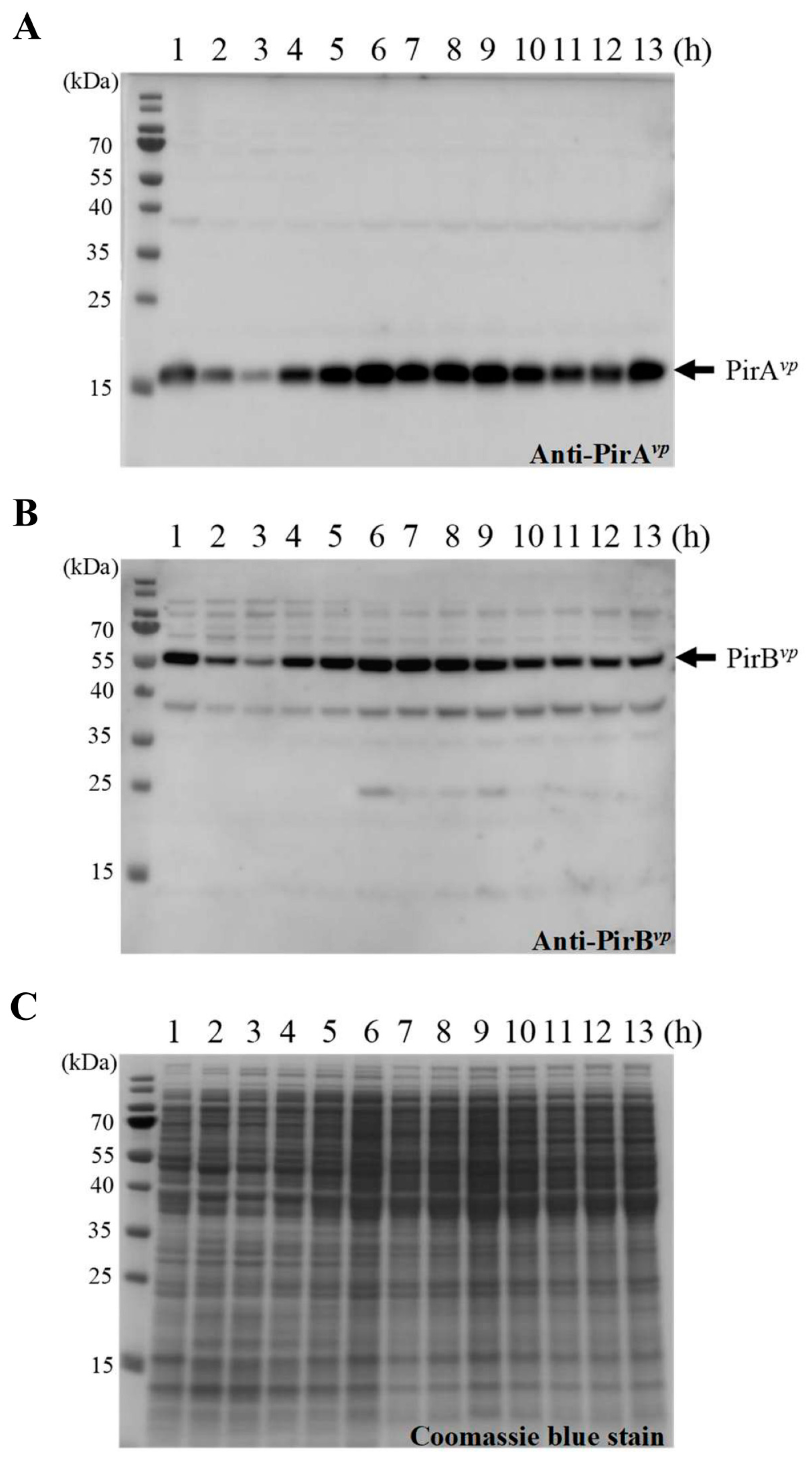
2.1. The Expression Levels of pirAvp/pirBvp during Different Growth Phases of V. parahaemolyticus

In Vibrio, since QS is involved in the regulation of many physiological processes, including several virulence-related systems, we first wanted to determine if the expression level of pirAvp/pirBvp was cell density-dependent. We, therefore, recorded the growth curve of the wild type V. parahaemolyticus, 3HP strain, and analyzed the gene expression patterns of pirAvp and pirBvp. As shown in Figure 1A, the lag phase lasted until about 3.5 h (OD600 from 0.01 to ~0.6), the log phase ran until 9 h (OD600 from 0.6 to ~7.7), and the stationary phase ran from 9~13 h (OD600 from 7.7 to ~8.8). The expression of pirAvp and pirBvp remained low until the curve entered the log phase (Figure 1B). Expression levels reached their peaks at 5 h, then declined again at 6 h and continued to remain low (Figure 1B). To determine the protein expression patterns, we used specific anti-PirAvp and anti-PirBvp antibodies to detect the PirAvp and PirBvp proteins at each time point. As shown in Figure 2, the PirAvp and PirBvp expression levels were low in the lag phase except for 1 h. The over-presence at this time point might be due to carry-over from 15 h culture prior to inoculation. The expression levels were high in the log phase through to the early part of the stationary phase (4~10 h), after which they returned to lower levels until the end of the recording.

2.2. LuxO Is a Negative Regulator for the Expression of pirAvp and pirBvp

To better understand whether QS regulation is involved in the expression of pirAvp and pirBvp, isogenic mutants of opaR and luxO were derived from the AHPND-causing strain 3HP. These mutants had a deletion in opaRopaR) or luxOluxO), or in both opaR and luxOopaRΔluxO), and they were constructed by allelic gene exchange as described previously [21]. Successful deletion in the different mutants was confirmed by using opaR-specific and luxO-specific primer sets (Figure 3). In addition, we also mutated the 47th amino acid of LuxOvp from aspartic acid (D) to glutamic acid (E) to mimic the permanently active form of LuxOvp (luxOD47E). All mutants exhibited similar growth rates to that of the wild-type strain in LB+ medium (Figure S1). As shown in Figure 4, the gene expression levels of pirAvp and pirBvp were down-regulated to about 60% when the active form of LuxOvp was mimicked, while the expression levels were up-regulated by about 2 folds with the luxOvp deletion mutants (i.e., ΔluxO and ΔopaRΔluxO). By contrast, the opaRvp deletion mutant had only a small, statistically insignificant effect on the expression of pirAvp and pirBvp, suggesting that although opaRvp is one of the core regulators of the QS system, unlike luxOvp, it plays only a minor role in the expression of pirAvp and pirBvp. Similar effects were also seen in the protein expression levels of PirAvp and PirBvp, although we note that, despite the reduction in mRNA expression, protein levels were not reduced by the luxOD47E mimic (Figure 4A,B, upper panels). Taken together, these results suggest that the expression of pirAvp and pirBvp is negatively regulated by luxOvp, but not by opaRvp.

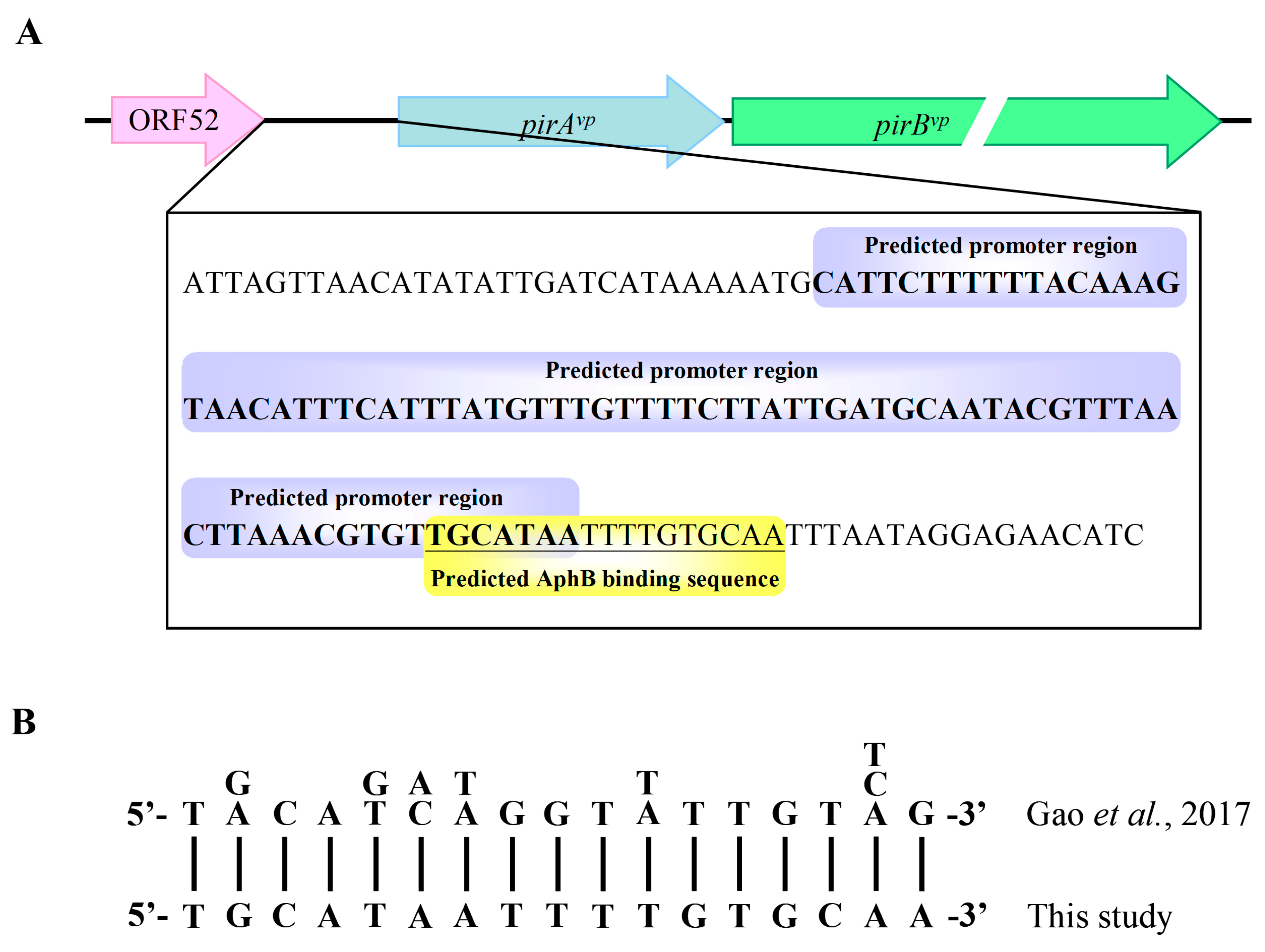
2.3. The QS Transcription Factor AphBvp Is a Possible Regulator for pirAvp and pirBvp

Since the key QS transcription factor, OpaR is evidently not involved in the regulation of pirAvp and pirBvp, we further analyzed the predicted promoter region of the pirAvp/pirBvp operon, and found that there was a sequence (5′-TGCATAATTTTGTGCAA-3′), which was similar to the consensus sequence of the AphB binding site, 5′-T-G/A-C-A-G/T-A/C-T/A-G-G-T-T/A-T-T-G-T-T/C/A-G-3′ [20] (Figure 5). Using real-time PCR, we found that for aphBvp, the elevated gene expression levels of the luxOvp deletion mutants ΔluxO and ΔopaRΔluxO were similar to those seen for pirAvp and pirBvp (Figure 6B). Conversely, the expression levels of another important QS transcription factor, aphAvp, did not correspond so closely to the expression patterns of pirAvp and pirBvp (Figure 6A). Taken together, these results suggest that AphBvp, but not AphAvp, may be important for the expression of pirAvp and pirBvp.

2.4. His-AphBvp Binds with the Predicted Promoter Region of pirAvp/pirBvp

In order to verify whether AphBvp could bind directly to the predicted AphBvp binding site located upstream of pirAvp/pirBvp, we amplified the fragment 300 bp upstream of the pirAvp/pirBvp operon by PCR, and mixed it with the indicated concentrations (0, 0.5, 1, 2, 5, 10, 20, and 40 μM) of the recombinant His-tagged Aph proteins His-AphBvp and His-AphAvp. As shown in Figure 7B, the DNA fragments shifted upward at His-AphBvp concentrations of 10 μM and above, showing that His-AphBvp was able to bind to this DNA fragment. By contrast, no DNA band shift was seen for His-AphAvp (Figure 7A), suggesting that binding to the predicted promoter region of pirAvp/pirBvp is His-AphBvp-specific.
To further verify the importance of the putative AphB binding sequence in the predicted promoter region of pirAvp/pirBvp, we truncated the DNA fragment into Fragment 1 (136 bp), which contained the complete AphB binding sequence, and Fragment 2 (116 bp), which contained only half of the AphB binding sequence (Figure 8A). After incubation with the indicated concentrations of His-AphBvp, we observed shifting with Fragment 1 at concentrations of 5~10 μM and above, whereas there were no shifts observed at any concentration with Fragment 2 (Figure 8B). These results confirmed the importance of the predicted sequence for AphBvp binding and thus for its potential role in the regulation of pirAvp and pirBvp expression.

3. Discussion

PirAvp and PirBvp have been confirmed as critical pathogenic factors of AHPND [12]. In a previous study, we further showed that PirAvp and PirBvp formed a heterotetramer in solution, and based on this tetramer’s structural similarity to the Cry toxin, we proposed that the binary toxin may destroy host cells by means of a mechanism similar to that used by Cry [14]. In particular, we suggested that the role of the PirAvp component is to recognize the glycan of its receptor on the host cells, after which PirBvp penetrates the cell membrane to form an unregulated channel, ultimately leading to critical cell damage [14]. Other reports have further shown that both PirAvp and PirBvp can bind directly to the receptor LvAPN1, and that PirBvp was translocated to the cytoplasm and nucleus of hemocytes [22,23], suggesting that the PirAvp and PirBvp toxins might be involved in other, additional pathogenic mechanisms. Until the present study, however, the mechanisms that might regulate expression of the pirA and pirB genes themselves had remained unknown. Here, we found a relationship between cell density and high expression levels of pirAvp and pirBvp in the early log phase (Figure 1). In addition, starting at around the same time as the peak of the gene expression levels, we also observed sustained elevated levels of PirAvp and PirBvp protein throughout the entire log phase and on into the stationary phase (Figure 1 and Figure 2).
It is already known that QS system is involved in the regulation of many virulence factors in Vibrio, including the genes of the type-III secretion systems (T3SS1 and T3SS2), type-VI secretion systems (T6SS1 and T6SS2), and the thermostable direct hemolysin genes tdh1 and tdh2 [24,25,26]. Here, by using strains with mutations in key molecules of the QS system, we found that the expression of pirAvp and pirBvp was also regulated by components of the QS system and that it was negatively regulated by LuxOvp (Figure 4).
Although the upstream region of the pirAvp/pirBvp operon did not include a consensus LuxO binding sequence (5′-TTGCAW3TGCAA-3′, where W stands for A or T; [27]), we found a possible AphB binding site as shown in Figure 5. In addition, we also found that the expression of aphBvp was affected by mutation of the QS component luxOvp (Figure 6). The upregulated expression pattern of aphBvp was similar to the pattern seen for pirAvp and pirBvp under the same conditions (Figure 4). The possible AphB binding sequence we observed (5′-TGCATAATTTTGTGCAA-3′) diverges from other established binding motifs, such as those of tcpP (5′-TGCAAN7TTGCA-3′), toxR (5′-TGCAAN7ATGGA-3′), aphB (5′-TGCAAN7TGTCA-3′), and a consensus sequence (5′-TGCAGN7TGTTG-3′), as well as cadC I (5′-TTAAAN7ACTTA-3′) and cadC II (5′-TACGTN7GGCTA-3′) [20]. Nevertheless, despite this high divergence, our EMSA results showed that the binding between AphBvp and the predicted AphB binding site was both direct and specific (Figure 7 and Figure 8). AphBvp (but not AphAvp) thus appears to act as an enhancer of pirAvp and pirBvp. Given AphBvp’s role as a regulator of QS, we hypothesize that the QS system regulates the expression of pirAvp and pirBvp via AphBvp (Figure 9, upper left panel). We also found that pirAvp and pirBvp were down-regulated by the permanently active luxOvp mutant even though there was no significant change in aphBvp. This suggests that the regulation of the PirAvp/PirBvp genes may not be controlled only by AphBvp, but that other LuxOvp-related effectors may also be involved (Figure 9, upper center panel). The proposed regulatory mechanisms and outcomes for LuxOvp, AphBvp, and PirAvp/PirBvp in the wild type (3HP) and mutant strains (luxOD47E, ΔopaR, ΔluxO, and ΔopaRΔluxO) are shown in Figure 9.
While AphA is a well-studied QS regulator [28,29], there are relatively few studies on AphB. These studies include its regulation of virulence [19,20] and the role it plays in survival under particular conditions [30]. AphB is also a positive regulator of LuxR/OpaR activity, and it activates the expression of the exotoxin Asp [20]. These studies, together with the recent finding that anti-QS compounds may reduce AHPND pathogenicity [18], all suggest that the QS system might be critically important for regulating the virulence of AHPND-causing bacteria.
To this body of evidence, we now add the results of the present study, which suggests that the QS system might be modulating virulence by regulating expression of the pirAvp/pirBvp genes through AphBvp. This new insight into the pathogenic mechanisms of AHPND points toward the QS system as a possible target for therapeutics that might one day be able to control the virulence of AHPND-causing bacteria and prevent AHPND.

4. Materials and Methods

4.1. Growth Curve Measurement and Sample Collection

All V. parahaemolyticus strains (3HP, LuxOD47E, ΔopaR, ΔluxO, and ΔopaR ΔluxO) were activated by culturing on LB+ agar plates (that is, LB agar plates that contained 2% NaCl) at 30 °C for 16 h. From these plates, 3 single colonies were transferred into 5 mL LB+ medium (LB medium that contained 2% NaCl) and incubated at 30 °C for 3 h to OD600~1.0. The culture was then diluted 100-fold, transferred into 50 mL fresh LB+ medium and cultured at 30 °C for 15 h with shaking at 200 rpm. Dilutions (1:1000) of these overnight cultures were further sub-cultured into 500 mL LB+ medium in a 2L flask, incubated at 30 °C with shaking at 200 rpm, and the OD600 was measured every hour for a total of 13 h. At the same time, 3HP cells were collected from part of the culture every hour, and temporarily stored at −80 °C prior to subsequent RNA and protein extractions. Other batches of sample cells of 3HP, LuxOD47E, ΔopaR, ΔluxO, and ΔopaR ΔluxO were collected at low cell density (LCD; OD600~0.6) and temporarily stored at −80 °C for later use.

4.2. RNA Extraction and Real-Time PCR

Total bacterial RNA was extracted using RareRNA reagent (Blossom Biotechnologies, Inc.; Taipei, ROC). DNaseI (Invitrogen) was used to digest any DNA contamination. From 1 µg of total RNA, cDNA was synthesized by using M-MLV reverse transcriptase (Promega; Madison, WI, USA) with random hexamers. Using specific primers (Table 1), real-time PCR was carried out to quantify the expression levels of target genes (pirAvp, pirBvp, aphAvp, and aphBvp) and an internal control gene (gryBvp, a housekeeping gene). The reaction mixture contained 1 μL cDNA, 0.2 μM forward primer, 0.2 μM reverse primer, 1× ChamQ Universal SYBR qPCR Master Mix (Vazyme Biotech Co. Ltd.; Nanjing, China) in a total volume of 20 μL, and thermal cycling was performed using a LightCycler® 96 System (Roche; Basel, Schweiz) as follows: 95 °C for 2 min followed by 40 cycles of 95 °C for 10 s and 60 °C for 30 s, and an extension cycle of 95 °C for 10 s, 65 °C for 60 s and 97 °C for 60 s.
To calculate ΔCt, the threshold cycle values of the internal control gene (Ct gyrB) were first subtracted from the threshold cycle values of the target genes. Next, either the initial ΔCt for the “1-h” timepoint was subtracted from the ΔCts of the other timepoints (i.e., 2–13 h), or else the ΔCt for the wild-type 3HP strain was subtracted from the ΔCt of all the groups (3HP, luxOD47E, ΔopaR, ΔluxO, and ΔopaR ΔluxO) to obtain ΔΔCt values. The fold change of the gene expression levels was then expressed as 2−ΔΔCt [31], and the data presented as mean ± SD. Statistically significant differences were tested using an unpaired Student’s t-test (p < 0.05).

4.3. Protein Extraction and Western Blots

Two batches of bacterial samples were removed from storage at −80 °C and lysed immediately using B-PER™ Bacterial Protein Extraction Reagent (ThermoFisher Scientific; Waltham, MA, USA) containing 1 mM PMSF, 1 mM EDTA, and 120 μg/mL DNaseI. The suspensions were inverted at room temperature for 10 min, and the cell debris was removed by centrifuging at 13,000× g for 10 min. Two micrograms of cell lysates were separated by 12.5% SDS-PAGE, transferred onto a PVDF membrane, and blocked with 5% skim milk at 4 °C overnight. The blots were then hybridized with chicken anti-PirAvp or chicken anti-PirBvp polyclonal antibodies (1:5000 diluted with 5% skim milk). After 1 hour of incubation at room temperature, the blots were washed 3 times with PBST solution (1× PBS contained 0.1% Tween-20). The blots were further incubated with donkey anti-chicken-HRP conjugated secondary antibody (Jackson; West Grove, PA; 1:10,000 diluted with 5% skim milk) at room temperature for 1 h. Following 3 more washes, the protein bands were visualized using a chemiluminescence reagent (GE Healthcare; Chicago, IL, USA) and detected with an Amersham Imager 600 (GE Healthcare; Chicago, IL, USA). For loading controls, 8 μg of cell lysates were separated with another 12.5% SDS-PAGE, and stained with Coomassie blue.

4.4. Construction of the ΔopaR, ΔluxO, ΔopaRΔluxO, and luxOD47E Mutants

The in-frame ΔopaR, ΔluxO, and ΔopaR ΔluxO mutants were constructed by in vivo allelic exchange as described previously [21]. Briefly, DNA fragments from the down- and up-stream regions of opaR and luxO were amplified, respectively, with the primer sets opaR-1/opaR-2 and opaR-3/opaR-4, and luxO-1/luxO-2 and luxO-3/luxO-4 (Table 1). These fragments were cloned into pGEM-T® Easy vector (Promega; Madison, WI, USA) in the correct orientation to generate a recombinant fragment containing a 627 bp- and a 1323 bp-deletion in opaR and luxO, respectively. The DNA fragments were removed from the pGEMT®-easy vector by enzyme digestion with SacI and SalI, respectively, and then cloned into the suicide vector pDS132. The suicide plasmids containing either the ΔopaR or ΔluxO fragment were transformed into Escherichia coli S17-1λpir [21], and then transferred into V. parahaemolyticus 3HP by conjugation to facilitate allelic exchange to produce the mutants. The double mutant was obtained by introducing the ΔluxO fragment into an already-constructed ΔopaR mutant by the method described above. For the luxOD47E mutant, a 2340-bp fragment amplified by the primers luxO-1 and luxO-16 was given a single nucleotide mutation (GAT to GAG) using a QuikChange® Site-Directed Mutagenesis Kit (Stratagene; La Jolla, CA, USA). This luxOD47E-containing fragment was introduced into V. parahaemolyticus 3HP by allelic exchange to generate the mutant.

4.5. Confirmation of the ΔopaR, luxO, ΔopaRΔluxO and luxOD47E Mutants by PCR

Genomic DNA was extracted from 3HP and the mutants using a Genomic DNA Extraction Kit (Bioman; New Taipei City, ROC) according to the manufacturer’s instructions. The opaR and luxO genes from all of the isolated mutants were then checked by PCR with the opaR- and luxO-specific primers opaR-5/opaR-6 and luxO-5/luxO-6 (Table 1), respectively. The DNA sequences were also determined to confirm that the deletion was in-frame, or, in the case of luxOD47E, that the single nucleotide mutation was correct.

4.6. Plasmid Construction for Recombinant Protein Expression

The codons in the coding sequences of aphAvp (CP045794; region 937101-937640) and aphBvp (WP069541384) were optimized, synthesized, and cloned into pET21b vector (Novagen; Madison, WI, USA) by GenScript Inc. (Piscataway, NJ, USA). Using the resulting plasmids as templates, the aphAvp and aphBvp genes were amplified with the primer sets AphA-F-NdeI/AphA-R-XhoI and AphB-F-NdeI/AphB-R-XhoI (Table 1), respectively, and then subcloned into pET28a vector (Novagen; Madison, WI, USA). The resulting plasmids were named aphAvp-pET28a and aphBvp-pET28a, respectively.

4.7. Expression and Purification of Recombinant His-AphA and His-AphB

To express the recombinant AphAvp and AphBvp, the aphAvp-pET28a and aphBvp-pET28a plasmids were, respectively, transformed into E. coli strain BL21 (DE3) cells. For AphAvp, the transformed cells were inoculated into 50 mL of fresh LB medium and grown at 37 °C for 12–14 h. Three ml of this overnight culture was then added to 500 mL of fresh LB medium in a 2L flask, and grown at 37 °C until the OD600 of the culture reached 0.4. IPTG was then added to a final concentration of 0.4 mM, and the culture was incubated at 16 °C for 20 h. The cells were then collected, resuspended in binding buffer (20 mM Tris-base, 500 mM NaCl, 20 mM imidazole, pH 8.0) containing 1 mM PMSF, 100 μg/mL lysozyme and 10 μg/mL DNase I, and homogenized by sonication on ice. After the cell debris was removed by centrifugation, the supernatant was filtrated using a 0.45 μm filter and loaded onto a 5 mL HisTrap HP column (GE Healthcare; Chicago, IL, USA). The column was washed with 100 mL of binding buffer and then eluted with a 20–500 mM imidazole gradient. The eluted recombinant protein was concentrated and loaded onto a Superdex 75 gel filtration column (GE Healthcare; Chicago, IL, USA) using 20 mM Tris-base, 500 mM NaCl, pH 8.0 as a running buffer. The protein concentration was measured by the Bradford method. For AphBvp, all of the culture and purification processes were the same as for AphAvp, except that for the subculture step, 10 mL of the overnight culture was inoculated into the 500 mL of fresh LB+ medium.

4.8. Electrophoretic Mobility Shift Assay (EMSA)

ProOpDB (Prokaryotic Operon DataBase) was used to predict possible promoter sequences in the upstream region of the pirAvp/pirBvp operon [32]. The DNA fragment that contained this predicted promoter region was further amplified by PCR using the primer set pirAB promoter-F1-NdeI/pirAB promoter-R1-XhoI, pirAB promoter-F2-NdeI/pirAB promoter-R2-XhoI (for Fragment 1, which included the complete predicted AphB binding sequence) or pirAB promoter-F3/pirAB promoter-R3-XhoI (for Fragment 2, which included only a partial predicted AphB binding sequence) (Table 1). For EMSA, recombinant His-AphA or His-AphB was mixed with the DNA fragment in a reaction buffer (20 mM Tris, pH 8.0, 100 mM NaCl) to final concentrations of 0, 0.5, 1, 2, 5, 10, 20, 40 µM (proteins), and 135 nM (DNA), and incubated at 25 °C for 20 min. The reactants were analyzed with 2% agarose gels, and stained by SYBR® Green I nucleic acid gel stain (Sigma-Aldrich; Burlington, MA, USA).

Supplementary Materials

The following supporting information can be downloaded at: https://www.mdpi.com/article/10.3390/ijms23052889/s1.

Author Contributions

Conceptualization, C.-F.L. and H.-C.W.; data curation, S.-J.L. and H.-C.W.; formal analysis, S.-J.L. and H.-C.W.; funding acquisition, S.-J.L., C.-F.L. and H.-C.W.; investigation, S.-J.L., J.-Y.H., P.-T.L., C.-T.L. and H.-C.W.; methodology, C.-F.L. and H.-C.W.; project administration, C.-F.L. and H.-C.W.; resources, C.-C.C., Y.-Y.Y., E.C.-Y.S., C.-F.L. and H.-C.W.; supervision, C.-F.L. and H.-C.W.; validation, S.-J.L., C.-F.L. and H.-C.W.; writing—original draft, S.-J.L. and H.-C.W.; writing—review and editing, S.-J.L., C.-F.L. and H.-C.W. All authors have read and agreed to the published version of the manuscript.

Funding

This research was funded by Ministry of Science and Technology (grant number MOST 109-2313-B-038-001-MY3, MOST 110-2313-B-006-001-) and the University System of Taipei Joint Research Program (NTOU-TMU-109-01).

Institutional Review Board Statement

Not applicable.

Informed Consent Statement

Not applicable.

Data Availability Statement

Not applicable.

Acknowledgments

We are indebted to Paul Barlow for his helpful comments on the MS.

Conflicts of Interest

The authors declare no conflict of interest.

References

  1. Baker-Austin, C.; Trinanes, J.; Gonzalez-Escalona, N.; Martinez-Urtaza, J. Non-Cholera vibrios: The microbial barometer of climate change. Trends Microbiol. 2017, 25, 76–84. [Google Scholar] [CrossRef] [Green Version]
  2. McCarter, L. The multiple identities of Vibrio parahaemolyticus. J. Mol. Microbiol. Biotechnol. 1999, 1, 51–57. [Google Scholar]
  3. Newton, A.; Kendall, M.; Vugia, D.J.; Henao, O.L.; Mahon, B.E. Increasing rates of vibriosis in the United States, 1996–2010: Review of surveillance data from 2 systems. Clin. Infect. Dis. 2012, 54, S391–S395. [Google Scholar] [CrossRef]
  4. Broberg, C.A.; Calder, T.J.; Orth, K. Vibrio parahaemolyticus cell biology and pathogenicity determinants. Microbes Infect. 2011, 13, 992–1001. [Google Scholar] [CrossRef] [Green Version]
  5. Park, K.S.; Ono, T.; Rokuda, M.; Jang, M.H.; Okada, K.; Iida, T.; Honda, T. Functional characterization of two type III secretion systems of Vibrio parahaemolyticus. Infect. Immun. 2004, 72, 6659–6665. [Google Scholar] [CrossRef] [Green Version]
  6. Salomon, D.; Gonzalez, H.; Updegraff, B.L.; Orth, K. Vibrio parahaemolyticus type VI secretion system 1 is activated in marine conditions to target bacteria, and is differentially regulated from system 2. PLoS ONE 2013, 8, e61086. [Google Scholar] [CrossRef]
  7. Tran, L.; Nunan, L.; Redman, R.M.; Mohney, L.L.; Pantoja, C.R.; Fitzsimmons, K.; Lightner, D.V. Determination of the infectious nature of the agent of acute hepatopancreatic necrosis syndrome affecting penaeid shrimp. Dis. Aquat. Organ. 2013, 105, 45–55. [Google Scholar] [CrossRef]
  8. NACA-FAO. Quarterly Aquatic Animal Disease Report (Asia and Pacific Region), April–June 2011; NACA: Bangkok, Thailand, 2011. [Google Scholar]
  9. Lightner, D.V.; Redman, R.M.; Pantoja, C.R.; Noble, B.L.; Tran, L. Early mortality syndrome affects shrimp in Asia. Glob. Aquac. Advocate 2012, 15, 40. [Google Scholar]
  10. Nunan, L.; Lightner, D.; Pantoja, C.; Gomez-Jimenez, S. Detection of acute hepatopancreatic necrosis disease (AHPND) in Mexico. Dis. Aquat. Organ. 2014, 111, 81–86. [Google Scholar] [CrossRef] [Green Version]
  11. Thitamadee, S.; Prachumwat, A.; Srisala, J.; Jaroenlak, P.; Salachan, P.V.; Sritunyalucksana, K.; Flegel, T.W.; Itsathitphaisarn, O. Review of current disease threats for cultivated penaeid shrimp in Asia. Aquaculture 2016, 452, 69–87. [Google Scholar] [CrossRef]
  12. Lee, C.T.; Chen, I.T.; Yang, Y.T.; Ko, T.P.; Huang, Y.T.; Huang, J.Y.; Huang, M.F.; Lin, S.J.; Chen, C.Y.; Lin, S.S.; et al. The opportunistic marine pathogen Vibrio parahaemolyticus becomes virulent by acquiring a plasmid that expresses a deadly toxin. Proc. Natl. Acad. Sci. USA 2015, 112, 10798–10803. [Google Scholar] [CrossRef] [PubMed] [Green Version]
  13. Lin, S.J.; Hsu, K.C.; Wang, H.C. Structural insights into the cytotoxic mechanism of Vibrio parahaemolyticus PirAvp and PirBvp toxins. Mar. Drugs 2017, 15, 373. [Google Scholar] [CrossRef] [PubMed] [Green Version]
  14. Lin, S.J.; Chen, Y.F.; Hsu, K.C.; Chen, Y.L.; Ko, T.P.; Lo, C.F.; Wang, H.C.; Wang, H.C. Structural insights to the heterotetrameric interaction between the Vibrio parahaemolyticus PirAvp and PirBvp toxins and activation of the Cry-like pore-forming domain. Toxins 2019, 11, 233. [Google Scholar] [CrossRef] [PubMed] [Green Version]
  15. Miller, M.B.; Bassler, B.L. Quorum sensing in bacteria. Annu. Rev. Microbiol. 2001, 55, 165–199. [Google Scholar] [CrossRef] [Green Version]
  16. Rutherford, S.T.; van Kessel, J.C.; Shao, Y.; Bassler, B.L. AphA and LuxR/HapR reciprocally control quorum sensing in vibrios. Genes Dev. 2011, 25, 397–408. [Google Scholar] [CrossRef] [Green Version]
  17. Kalburge, S.S.; Carpenter, M.R.; Rozovsky, S.; Boyd, E.F. Quorum sensing regulators are required for metabolic fitness in Vibrio parahaemolyticus. Infect. Immun. 2017, 85, e00930-16. [Google Scholar] [CrossRef] [Green Version]
  18. Paopradit, P.; Tansila, N.; Surachat, K.; Mittraparp-Arthorn, P. Vibrio alginolyticus influences quorum sensing-controlled phenotypes of acute hepatopancreatic necrosis disease-causing Vibrio parahaemolyticus. PeerJ 2021, 9, e11567. [Google Scholar] [CrossRef]
  19. Kovacikova, G.; Skorupski, K. Binding site requirements of the virulence gene regulator AphB: Differential affinities for the Vibrio cholerae classical and El Tor tcpPH promoters. Mol. Microbiol. 2002, 44, 533–547. [Google Scholar] [CrossRef]
  20. Gao, X.; Liu, Y.; Liu, H.; Yang, Z.; Liu, Q.; Zhang, Y.; Wang, Q. Identification of the regulon of AphB and its essential roles in LuxR and exotoxin Asp expression in the pathogen Vibrio alginolyticus. J. Bacteriol. 2017, 199, e00252-17. [Google Scholar] [CrossRef] [Green Version]
  21. Lee, C.T.; Amaro, C.; Wu, K.M.; Valiente, E.; Chang, Y.F.; Tsai, S.F.; Chang, C.H.; Hor, L.I. A common virulence plasmid in biotype 2 Vibrio vulnificus and its dissemination aided by a conjugal plasmid. J. Bacteriol. 2008, 190, 1638–1648. [Google Scholar] [CrossRef] [Green Version]
  22. Luangtrakul, W.; Boonchuen, P.; Jaree, P.; Kumar, R.; Wang, H.C.; Somboonwiwat, K. Cytotoxicity of Vibrio parahaemolyticus AHPND toxin on shrimp hemocytes, a newly identified target tissue, involves binding of toxin to aminopeptidase N1 receptor. PLoS Pathog. 2021, 17, e1009463. [Google Scholar] [CrossRef] [PubMed]
  23. Zheng, Z.; Li, R.; Aweya, J.J.; Yao, D.; Wang, F.; Li, S.; Tuan, T.N.; Zhang, Y. The PirB toxin protein from Vibrio parahaemolyticus induces apoptosis in hemocytes of Penaeus vannamei. Virulence 2021, 12, 481–492. [Google Scholar] [CrossRef] [PubMed]
  24. Henke, J.M.; Bassler, B.L. Quorum sensing regulates type III secretion in Vibrio harveyi and Vibrio parahaemolyticus. J. Bacteriol. 2004, 186, 3794–3805. [Google Scholar] [CrossRef] [PubMed] [Green Version]
  25. Wang, L.; Zhou, D.; Mao, P.; Zhang, Y.; Hou, J.; Hu, Y.; Li, J.; Hou, S.; Yang, R.; Wang, R.; et al. Cell density- and quorum sensing-dependent expression of type VI secretion system 2 in Vibrio parahaemolyticus. PLoS ONE 2013, 8, e73363. [Google Scholar] [CrossRef] [Green Version]
  26. Zhang, Y.; Hu, L.; Qiu, Y.; Osei-Adjei, G.; Tang, H.; Zhang, Y.; Zhang, R.; Sheng, X.; Xu, S.; Yang, W.; et al. QsvR integrates into quorum sensing circuit to control Vibrio parahaemolyticus virulence. Environ. Microbiol. 2019, 21, 1054–1067. [Google Scholar] [CrossRef]
  27. Tu, K.C.; Bassler, B.L. Multiple small RNAs act additively to integrate sensory information and control quorum sensing in Vibrio harveyi. Genes Dev. 2007, 21, 221–233. [Google Scholar] [CrossRef] [Green Version]
  28. Wang, L.; Ling, Y.; Jiang, H.; Qiu, Y.; Qiu, J.; Chen, H.; Yang, R.; Zhou, D. AphA is required for biofilm formation, motility, and virulence in pandemic Vibrio parahaemolyticus. Int. J. Food Microbiol. 2013, 160, 245–251. [Google Scholar] [CrossRef]
  29. Haycocks, J.R.J.; Warren, G.Z.L.; Walker, L.M.; Chlebek, J.L.; Dalia, T.N.; Dalia, A.B.; Grainger, D.C. The quorum sensing transcription factor AphA directly regulates natural competence in Vibrio cholerae. PLoS Genet. 2019, 15, e1008362. [Google Scholar] [CrossRef] [Green Version]
  30. Kovacikova, G.; Lin, W.; Skorupski, K. The LysR-type virulence activator AphB regulates the expression of genes in Vibrio cholerae in response to low pH and anaerobiosis. J. Bacteriol. 2010, 192, 4181–4191. [Google Scholar] [CrossRef] [Green Version]
  31. Livak, K.J.; Schmittgen, T.D. Analysis of relative gene expression data using real-time quantitative PCR and the 2−ΔΔCt method. Methods 2001, 25, 402–408. [Google Scholar] [CrossRef]
  32. Taboada, B.; Ciria, R.; Martinez-Guerrer, C.E.; Merino, E. ProOpDB: Prokaryotic Operon DataBase. Nucleic Acids Res. 2021, 40, D627–D631. [Google Scholar] [CrossRef] [PubMed] [Green Version]
Figure 1. Growth curve and pirAvp and pirBvp expression levels in V. parahaemolyticus strain 3HP. (A) V. parahaemolyticus strain 3HP was cultured in LB (2% NaCl), and the growth curve was recorded every hour until 13 h after inoculation. (B) Relative mRNA expression of pirAvp and pirBvp in the strain 3HP during different growth phases. Total RNA was extracted from V. parahaemolyticus 3HP collected at the indicated time points (OD600 0.01–8.82), and analyzed by real-time qPCR with specific primers for pirAvp and pirBvp, respectively. Expression levels are shown relative to those of the gyrB reference gene.
Ijms 23 02889 g001
Figure 2. Protein expression levels of endogenous PirAvp and PirBvp in V. parahaemolyticus wild type strain (3HP) during different growth phases. Cell lysate (2 μg) was separated, transferred onto a PVDF membrane, and reacted with (A) chicken anti-PirAvp and (B) anti-PirBvp polyclonal antibodies to recognize the endogenous PirAvp and PirBvp, respectively. (C) Loading control (8 μg/lane).
Ijms 23 02889 g002
Figure 3. Confirmation of ΔopaR, ΔluxO, ΔopaRΔluxO, and luxOD47E mutants by PCR. DNAs from the wild-type strain and the mutants were analyzed by PCR with the primers specific to opaR (A) and luxO (B). The bands corresponding to the wild type opaR and luxO, and the deleted opaR and luxOopaR and ΔluxO, respectively) are indicated.
Ijms 23 02889 g003
Figure 4. The gene and protein expression levels of (A) pirAvp and (B) pirBvp in the wild type strain (3HP) compared to those in the luxOD47E, ∆opaR, ∆luxO, and ∆opaRluxO mutants. Soluble proteins were extracted from cells collected at OD600~0.6 and analyzed by immunoblotting with anti-PirAvp or anti-PirBvp antibodies (upper panels). Total RNA was extracted from the same batch of bacterial samples, and real-time RT-PCR was carried out using specific primer sets for pirAvp and pirBvp (lower panels), respectively. The housekeeping gene, gyrB served as an internal control. *: p < 0.05.
Ijms 23 02889 g004
Figure 5. Schematic representation of the predicted AphB binding site in the pirAvp/pirBvp promoter region. (A) The predicted pirAvp/pirBvp promoter region is bolded and shaded blue. The putative AphB binding sequence, 5′-TGCATAATTTTGTGCAA-3′, is underlined and shaded yellow. (B) Alignment of the AphB binding sequence on the V. alginolyticus chromosome [20] with the predicted AphBvp binding sequence in the predicted pirAvp/pirBvp promoter region (this study).
Ijms 23 02889 g005
Figure 6. The gene expression levels of (A) aphAvp (B) aphBvp in the wild type strain (3HP), luxOD47E, ∆opaR, ∆luxO, and ∆opaRluxO mutants at OD600~0.6. The expression pattern of aphBvp was similar to the expression patterns of pirAvp and pirBvp. By contrast, the expression pattern of aphAvp was not a good match. The housekeeping gene, gyrB served as an internal control. *: p < 0.05.
Ijms 23 02889 g006
Figure 7. AphBvp binds to the predicted pirAvp/pirBvp promoter region in an electrophoretic mobility shift assay (EMSA). (A) As the concentration of His-AphAvp increased, the DNA fragments produced by PCR amplification of the predicted promoter region remained unshifted. (B) By contrast, an upward shift was seen for His-AphBvp at concentrations of 10 μM and above.
Ijms 23 02889 g007
Figure 8. The predicted AphBvp binding sequence is important for binding AphBvp. (A) The pirAvp/pirBvp promoter region was truncated into fragments that included either the full (Fragment 1) or partial (Fragment 2) predicted AphBvp binding sequence. The head-tail sequences of these fragments are shown in the box, and the nucleotides in the predicted AphBvp binding sequence are shaded orange. (B) The EMSA results show that AphBvp bound only to the full binding sequence (Fragment 1).
Ijms 23 02889 g008
Figure 9. The relationships between LuxOvp, AphBvp and PirAvp/PirBvp at OD600~0.6. In the first three strains (i.e., wild type strain 3HP, the activated mimic strain luxOD47E, and the OpaR deletion strain ΔopaR), the phosphorylated LuxOvp acted to limit the free expression of AphBvp and its downstream genes, pirAvp/pirBvp. Conversely, when LuxO was deleted (strains ΔluxO and ΔopaRΔluxO), AphBvp was significantly up-regulated, and this further promoted the expression of PirAvp/PirBvp.
Ijms 23 02889 g009
Table 1. Primers used in this study.
Primer NamePrimer Sequence (5′–3′)Usage
opaR-1GAGACCGTTGAAGCATCGMutant construction
opaR-2CAGGTACCGAGTCCATATCCATTTMutant construction
opaR-3CAGGTACCCGAACACTAAAGCTCAMutant construction
opaR-4CAGAGCTCGGGTACGGTTTACCACMutant construction
opaR-5GTTCTAGAGTGGGTTGAGGTAGGTMutant selection
opaR-6GGTCTAGAGTTGGTACTAACGGTGMutant selection
luxO-1GAGAGCTCCGTATTCGTGCCGCCAAAGMutant construction
luxO-2CTGGTACCGCTGTATCCTCAACCATCMutant construction
luxO-3GAGGTACCAGAAGAGCGGCAGAAGGTGMutant construction
luxO-4CTGGTACCCGACCGCTGGATGCAATCMutant construction
luxO-5GCTCTAGACGGCTGAGAAGCGTGATGMutant selection
luxO-6GGTCTAGAGAGTCCAAGAGCGATACGMutant selection
pirAQFTTAGCCACTTTCCAGCCGCqPCR
pirAQRCCGGAAGTCGGTCGTAGTGTqPCR
pirBQFTCGTTATCAGCCCACGCAGqPCR
pirBQRTTTCACCGATTCTGATGTGCAqPCR
aphAQFGAAACTTATGGCTTGTGCTGqPCR
aphAQRGCGGCTTCAATTTCTTTGTAqPCR
aphBQFTGGGATGTTATTTTCCGTGTqPCR
aphBQRCTGCTAGATAGTCTTGGCTGqPCR
gyrB-1GAAGGTGGTATTCAAGCGTTCGqPCR
gyrB-2GAGATGCCGTCTTCACGTTCTqPCR
AphA-F-NdeIAATGCCCCATATGAGCCTGCCGCACGTGProtein expression
AphA-R-XhoICCGCTCGAGTTAGCCAATAACTTCCAGCTCGProtein expression
AphB-F-NdeIAAGGCCCCATATGAAGCTGGACGATCTGAACCProtein expression
AphB-R-XhoIGCCGCTCGAGTTAGTGGATGTTATACGCAATAACAAAGProtein expression
pirAB promoter-F1-NdeIAGGCTTCCATATGAGTGGAAATGGTGAACTTGCGGAAGEMSA
pirAB promoter-R1-XhoIAAGCTCGAGGTCTACTTCTGTGACGCCTCCGEMSA
pirAB promoter-F2-NdeIAGGCTTCCATATGATTGATCATAAAAATGCATTCTTTTTTACAAAGEMSA
pirAB promoter-R3-XhoIAAGCTCGAGTATTAAATTGCACAAAATTATGCAACACGEMSA
pirAB promoter-F7TTTGTGCAATTTAATAGGAGAACATCATGAGEMSA
pirAB promoter-R-XhoIAAGCTCGAGGTCTACTTCTGTGACGCCTCCGEMSA
The restriction enzyme cutting sites are underlined.
Publisher’s Note: MDPI stays neutral with regard to jurisdictional claims in published maps and institutional affiliations.

