Next Issue
Volume 23, March-2
Previous Issue
Volume 23, February-2
 
 
ijms-logo

Journal Browser

Journal Browser

Int. J. Mol. Sci., Volume 23, Issue 5 (March-1 2022) – 504 articles

Cover Story (view full-size image): 
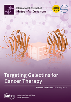
Galectins are soluble β-D-galactoside binding proteins overexpressed in cancerous cells.  They modulate tumor progression, influencing the disease outcome. Therefore, the development of potent inhibitors capable of selectively reducing the activity of galectins represents an important strategy for cancer therapy. In this frame, we have rationally designed a novel selenium-containing diglycosylated compound, featuring the presence of a lipophilic benzyl group at both saccharide residues. The relatively high binding affinity of this new compound to the carbohydrate recognition domain of galectin 3 and galectin 9, its good anti-proliferative and antimigration activity towards melanoma cells, as well as its anti-angiogenesis properties, stimulate further studies aimed at its development as an antitumor agent. View this paper
  • Issues are regarded as officially published after their release is announced to the table of contents alert mailing list.
  • You may sign up for e-mail alerts to receive table of contents of newly released issues.
  • PDF is the official format for papers published in both, html and pdf forms. To view the papers in pdf format, click on the "PDF Full-text" link, and use the free Adobe Reader to open them.
Order results
Result details
Section
Select all
Export citation of selected articles as:
14 pages, 2429 KiB  
Article
Bone Mass and Osteoblast Activity Are Sex-Dependent in Mice Lacking the Estrogen Receptor α in Chondrocytes and Osteoblast Progenitor Cells
by Lena Steppe, Jasmin Bülow, Jan Tuckermann, Anita Ignatius and Melanie Haffner-Luntzer
Int. J. Mol. Sci. 2022, 23(5), 2902; https://doi.org/10.3390/ijms23052902 - 07 Mar 2022
Cited by 7 | Viewed by 3661 | Correction
Abstract
While estrogen receptor alpha (ERα) is known to be important for bone development and homeostasis, its exact function during osteoblast differentiation remains unclear. Conditional deletion of ERα during specific stages of osteoblast differentiation revealed different bone phenotypes, which were also shown to be [...] Read more.
While estrogen receptor alpha (ERα) is known to be important for bone development and homeostasis, its exact function during osteoblast differentiation remains unclear. Conditional deletion of ERα during specific stages of osteoblast differentiation revealed different bone phenotypes, which were also shown to be sex-dependent. Since hypertrophic chondrocytes can transdifferentiate into osteoblasts and substantially contribute to long-bone development, we aimed to investigate the effects of ERα deletion in both osteoblast and chondrocytes on bone development and structure. Therefore, we generated mice in which the ERα gene was inactivated via a Runx2-driven cyclic recombinase (ERαfl/fl; Runx2Cre). We analyzed the bones of 3-month-old ERαfl/fl; Runx2Cre mice by biomechanical testing, micro-computed tomography, and cellular parameters by histology. Male ERαfl/fl; Runx2Cre mice displayed a significantly increased cortical bone mass and flexural rigidity of the femurs compared to age-matched controls with no active Cre-transgene (ERαfl/fl). By contrast, female ERαfl/fl; Runx2Cre mice exhibited significant trabecular bone loss, whereas in cortical bone periosteal and endosteal diameters were reduced. Our results indicate that the ERα in osteoblast progenitors and hypertrophic chondrocytes differentially contributes to bone mass regulation in male and female mice and improves our understanding of ERα signaling in bone cells in vivo. Full article
(This article belongs to the Special Issue Bone Development and Growth)
Show Figures

Figure 1

14 pages, 663 KiB  
Review
Targeting Myotonic Dystrophy Type 1 with Metformin
by Mikel García-Puga, Ander Saenz-Antoñanzas, Ander Matheu and Adolfo López de Munain
Int. J. Mol. Sci. 2022, 23(5), 2901; https://doi.org/10.3390/ijms23052901 - 07 Mar 2022
Cited by 11 | Viewed by 4447
Abstract
Myotonic dystrophy type 1 (DM1) is a multisystemic disorder of genetic origin. Progressive muscular weakness, atrophy and myotonia are its most prominent neuromuscular features, while additional clinical manifestations in multiple organs are also common. Overall, DM1 features resemble accelerated aging. There is currently [...] Read more.
Myotonic dystrophy type 1 (DM1) is a multisystemic disorder of genetic origin. Progressive muscular weakness, atrophy and myotonia are its most prominent neuromuscular features, while additional clinical manifestations in multiple organs are also common. Overall, DM1 features resemble accelerated aging. There is currently no cure or specific treatment for myotonic dystrophy patients. However, in recent years a great effort has been made to identify potential new therapeutic strategies for DM1 patients. Metformin is a biguanide antidiabetic drug, with potential to delay aging at cellular and organismal levels. In DM1, different studies revealed that metformin rescues multiple phenotypes of the disease. This review provides an overview of recent findings describing metformin as a novel therapy to combat DM1 and their link with aging. Full article
(This article belongs to the Special Issue Myotonic Dystrophy: From Molecular Pathogenesis to Therapeutics 2.0)
Show Figures

Figure 1

17 pages, 21791 KiB  
Article
Role of Nuclear-Receptor-Related 1 in the Synergistic Neuroprotective Effect of Umbilical Cord Blood and Erythropoietin Combination Therapy in Hypoxic Ischemic Encephalopathy
by Joo-Wan Choi, Su Jung Kang, Jee In Choi, KyuBum Kwack and MinYoung Kim
Int. J. Mol. Sci. 2022, 23(5), 2900; https://doi.org/10.3390/ijms23052900 - 07 Mar 2022
Cited by 3 | Viewed by 2257
Abstract
Neonatal hypoxic–ischemic encephalopathy (HIE) results in neurological impairments; cell-based therapy has been suggested as a therapeutic avenue. Previous research has demonstrated the synergistically potentiated therapeutic efficacy of human umbilical cord blood (UCB) by combining recombinant human erythropoietin (EPO) treatment for recovery from HIE. [...] Read more.
Neonatal hypoxic–ischemic encephalopathy (HIE) results in neurological impairments; cell-based therapy has been suggested as a therapeutic avenue. Previous research has demonstrated the synergistically potentiated therapeutic efficacy of human umbilical cord blood (UCB) by combining recombinant human erythropoietin (EPO) treatment for recovery from HIE. However, its molecular mechanism is not entirely understood. In the present study, we analyzed the mechanisms underlying the effect of combination treatment with EPO and UCB by transcriptomic analysis, followed by gene enrichment analysis. Mouse HIE model of the neonate was prepared and randomly divided into five groups: sham, HIE, and UCB, EPO, and UCB+EPO treatments after HIE. A total of 376 genes were differentially expressed when |log2FC| ≥ 1-fold change expression values were considered to be differentially expressed between UCB+EPO and HIE. Further assessment through qRT-PCR and gene enrichment analysis confirmed the expression and correlation of its potential target, Nurr1, as an essential gene involved in the synergistic effect of the UCB+EPO combination. The results indicated the remarkable activation of Wnt/β-catenin signaling by reducing the infarct size by UCB+EPO treatment, accompanied by Nurr1 activity. In conclusion, these findings suggest that the regulation of Nurr1 through the Wnt/β-catenin pathway exerts a synergistic neuroprotective effect in UCB and EPO combination treatment. Full article
(This article belongs to the Special Issue Stem Cells—from Bench to Bedside 2021)
Show Figures

Figure 1

17 pages, 2849 KiB  
Article
Development and Characterization of Gentamicin-Loaded Arabinoxylan-Sodium Alginate Films as Antibacterial Wound Dressing
by Abdulaziz I. Alzarea, Nabil K. Alruwaili, Muhammad Masood Ahmad, Muhammad Usman Munir, Adeel Masood Butt, Ziyad A. Alrowaili, Muhammad Syafiq Bin Shahari, Ziyad S. Almalki, Saad S. Alqahtani, Anton V. Dolzhenko and Naveed Ahmad
Int. J. Mol. Sci. 2022, 23(5), 2899; https://doi.org/10.3390/ijms23052899 - 07 Mar 2022
Cited by 16 | Viewed by 3272
Abstract
Biopolymer-based antibacterial films are attractive materials for wound dressing application because they possess chemical, mechanical, exudate absorption, drug delivery, antibacterial, and biocompatible properties required to support wound healing. Herein, we fabricated and characterized films composed of arabinoxylan (AX) and sodium alginate (SA) loaded [...] Read more.
Biopolymer-based antibacterial films are attractive materials for wound dressing application because they possess chemical, mechanical, exudate absorption, drug delivery, antibacterial, and biocompatible properties required to support wound healing. Herein, we fabricated and characterized films composed of arabinoxylan (AX) and sodium alginate (SA) loaded with gentamicin sulfate (GS) for application as a wound dressing. The FTIR, XRD, and thermal analyses show that AX, SA, and GS interacted through hydrogen bonding and were thermally stable. The AXSA film displays desirable wound dressing characteristics: transparency, uniform thickness, smooth surface morphology, tensile strength similar to human skin, mild water/exudate uptake capacity, water transmission rate suitable for wound dressing, and excellent cytocompatibility. In Franz diffusion release studies, >80% GS was released from AXSA films in two phases in 24 h following the Fickian diffusion mechanism. In disk diffusion assay, the AXSA films demonstrated excellent antibacterial effect against E.coli, S. aureus, and P. aeruginosa. Overall, the findings suggest that GS-loaded AXSA films hold potential for further development as antibacterial wound dressing material. Full article
(This article belongs to the Special Issue Biopolymers in Drug and Gene Delivery Systems)
Show Figures

Figure 1

11 pages, 2158 KiB  
Article
Generation of a Dystrophin Mutant in Dog by Nuclear Transfer Using CRISPR/Cas9-Mediated Somatic Cells: A Preliminary Study
by Hyun Ju Oh, Eugene Chung, Jaehwan Kim, Min Jung Kim, Geon A. Kim, Seok Hee Lee, Kihae Ra, Kidong Eom, Soojin Park, Jong-Hee Chae, Jin-Soo Kim and Byeong Chun Lee
Int. J. Mol. Sci. 2022, 23(5), 2898; https://doi.org/10.3390/ijms23052898 - 07 Mar 2022
Cited by 3 | Viewed by 2328
Abstract
Dystrophinopathy is caused by mutations in the dystrophin gene, which lead to progressive muscle degeneration, necrosis, and finally, death. Recently, golden retrievers have been suggested as a useful animal model for studying human dystrophinopathy, but the model has limitations due to difficulty in [...] Read more.
Dystrophinopathy is caused by mutations in the dystrophin gene, which lead to progressive muscle degeneration, necrosis, and finally, death. Recently, golden retrievers have been suggested as a useful animal model for studying human dystrophinopathy, but the model has limitations due to difficulty in maintaining the genetic background using conventional breeding. In this study, we successfully generated a dystrophin mutant dog using the CRISPR/Cas9 system and somatic cell nuclear transfer. The dystrophin mutant dog displayed phenotypes such as elevated serum creatine kinase, dystrophin deficiency, skeletal muscle defects, an abnormal electrocardiogram, and avoidance of ambulation. These results indicate that donor cells with CRISPR/Cas9 for a specific gene combined with the somatic cell nuclear transfer technique can efficiently produce a dystrophin mutant dog, which will help in the successful development of gene therapy drugs for dogs and humans. Full article
Show Figures

Figure 1

15 pages, 25553 KiB  
Article
Corrosion Resistance and Electrical Conductivity of Hybrid Coatings Obtained from Polysiloxane and Carbon Nanotubes by Electrophoretic Co-Deposition
by Patryk Bezkosty, Elżbieta Długoń, Maciej Sowa, Jacek Nizioł, Piotr Jeleń, Jakub Marchewka, Marta Błażewicz and Maciej Sitarz
Int. J. Mol. Sci. 2022, 23(5), 2897; https://doi.org/10.3390/ijms23052897 - 07 Mar 2022
Cited by 4 | Viewed by 2210
Abstract
Nanocomposites developed based on siloxanes modified with carbon nanoforms are materials with great application potential in the electronics industry, medicine and environmental protection. This follows from the fact that such nanocomposites can be endowed with biocompatibility characteristics, electric conductivity and a high mechanical [...] Read more.
Nanocomposites developed based on siloxanes modified with carbon nanoforms are materials with great application potential in the electronics industry, medicine and environmental protection. This follows from the fact that such nanocomposites can be endowed with biocompatibility characteristics, electric conductivity and a high mechanical durability. Moreover, their surface, depending on the type and the amount of carbon nanoparticles, may exhibit antifouling properties, as well as those that limit bacterial adhesion. The paper reports on the properties of polysiloxane (PS) and carbon nanotubes (CNT) nanocomposite coatings on metal surfaces produced by the electrophoretic deposition (EPD). A comparison with coatings made of pure PS or pure CNT on the same substrates using the same deposition method (EPD) is provided. The coatings were examined for morphology and elemental composition (SEM, EDS), structural characteristics (confocal Raman spectroscopy), electrical conductivity and were tested for corrosion (electrochemical impedance spectroscopy-EIS, potentiodynamic polarization-PDP). The results obtained in this study clearly evidenced that such hybrid coatings conduct electricity and protect the metal from corrosion. However, their corrosion resistance differs slightly from that of a pure polymeric coating. Full article
(This article belongs to the Section Materials Science)
Show Figures

Graphical abstract

32 pages, 6752 KiB  
Review
Overview of the Importance of Biotics in Gut Barrier Integrity
by Aleksandra Maria Kocot, Elżbieta Jarocka-Cyrta and Natalia Drabińska
Int. J. Mol. Sci. 2022, 23(5), 2896; https://doi.org/10.3390/ijms23052896 - 07 Mar 2022
Cited by 26 | Viewed by 7996
Abstract
Increased gut permeability is suggested to be involved in the pathogenesis of a growing number of disorders. The altered intestinal barrier and the subsequent translocation of bacteria or bacterial products into the internal milieu of the human body induce the inflammatory state. Gut [...] Read more.
Increased gut permeability is suggested to be involved in the pathogenesis of a growing number of disorders. The altered intestinal barrier and the subsequent translocation of bacteria or bacterial products into the internal milieu of the human body induce the inflammatory state. Gut microbiota maintains intestinal epithelium integrity. Since dysbiosis contributes to increased gut permeability, the interventions that change the gut microbiota and correct dysbiosis are suggested to also restore intestinal barrier function. In this review, the current knowledge on the role of biotics (probiotics, prebiotics, synbiotics and postbiotics) in maintaining the intestinal barrier function is summarized. The potential outcome of the results from in vitro and animal studies is presented, and the need for further well-designed randomized clinical trials is highlighted. Moreover, we indicate the need to understand the mechanisms by which biotics regulate the function of the intestinal barrier. This review is concluded with the future direction and requirement of studies involving biotics and gut barrier. Full article
Show Figures

Figure 1

17 pages, 3465 KiB  
Article
Insight into the Phylogeny and Binding Ability of WRKY Transcription Factors
by Kuan-Ting Hsin, Min-Che Hsieh, Yu-Hsuan Lee, Kai-Chun Lin and Yi-Sheng Cheng
Int. J. Mol. Sci. 2022, 23(5), 2895; https://doi.org/10.3390/ijms23052895 - 07 Mar 2022
Cited by 10 | Viewed by 2544
Abstract
WRKY transcription factors (TFs), which make up one of the largest families of TFs in the plant kingdom, are key players in modulating gene expression relating to embryogenesis, senescence, pathogen resistance, and abiotic stress responses. However, the phylogeny and grouping of WRKY TFs [...] Read more.
WRKY transcription factors (TFs), which make up one of the largest families of TFs in the plant kingdom, are key players in modulating gene expression relating to embryogenesis, senescence, pathogen resistance, and abiotic stress responses. However, the phylogeny and grouping of WRKY TFs and how their binding ability is affected by the flanking regions of W-box sequences remain unclear. In this study, we reconstructed the phylogeny of WRKY across the plant kingdom and characterized the DNA-binding profile of Arabidopsis thaliana WRKY (WRKY54) based on its W-box recognition sequence. We found that WRKY TFs could be separated into five clades, and that the functional zinc-finger motif at the C-terminal of WRKY appeared after several nucleotide substitutions had occurred at the 3′-end of the zinc-finger region in chlorophytes. In addition, we found that W-box flanking regions affect the binding ability of WRKY54 based on the results of a fluorescence-based electrophoretic mobility shift assay (fEMSA) and quartz crystal microbalance (QCM) analysis. The great abundance of WRKY TFs in plants implicates their involvement in diverse molecular regulatory networks, and the flanking regions of W-box sequences may contribute to their molecular recognition mechanism. This phylogeny and our findings on the molecular recognition mechanism of WRKY TFs should be helpful for further research in this area. Full article
Show Figures

Figure 1

19 pages, 379 KiB  
Review
Does Bisphenol A Confer Risk of Neurodevelopmental Disorders? What We Have Learned from Developmental Neurotoxicity Studies in Animal Models
by Chloe Welch and Kimberly Mulligan
Int. J. Mol. Sci. 2022, 23(5), 2894; https://doi.org/10.3390/ijms23052894 - 07 Mar 2022
Cited by 18 | Viewed by 4219
Abstract
Substantial evidence indicates that bisphenol A (BPA), a ubiquitous environmental chemical used in the synthesis of polycarbonate plastics and epoxy resins, can impair brain development. Clinical and epidemiological studies exploring potential connections between BPA and neurodevelopmental disorders in humans have repeatedly identified correlations [...] Read more.
Substantial evidence indicates that bisphenol A (BPA), a ubiquitous environmental chemical used in the synthesis of polycarbonate plastics and epoxy resins, can impair brain development. Clinical and epidemiological studies exploring potential connections between BPA and neurodevelopmental disorders in humans have repeatedly identified correlations between early BPA exposure and developmental disorders, such as attention deficit/hyperactivity disorder and autism spectrum disorder. Investigations using invertebrate and vertebrate animal models have revealed that developmental exposure to BPA can impair multiple aspects of neuronal development, including neural stem cell proliferation and differentiation, synapse formation, and synaptic plasticity—neuronal phenotypes that are thought to underpin the fundamental changes in behavior-associated neurodevelopmental disorders. Consistent with neuronal phenotypes caused by BPA, behavioral analyses of BPA-treated animals have shown significant impacts on behavioral endophenotypes related to neurodevelopmental disorders, including altered locomotor activity, learning and memory deficits, and anxiety-like behavior. To contextualize the correlations between BPA and neurodevelopmental disorders in humans, this review summarizes the current literature on the developmental neurotoxicity of BPA in laboratory animals with an emphasis on neuronal phenotypes, molecular mechanisms, and behavioral outcomes. The collective works described here predominantly support the notion that gestational exposure to BPA should be regarded as a risk factor for neurodevelopmental disorders. Full article
(This article belongs to the Special Issue Advances in Endocrine Disruptors)
21 pages, 4597 KiB  
Article
Key Soybean Seedlings Drought-Responsive Genes and Pathways Revealed by Comparative Transcriptome Analyses of Two Cultivars
by Huidong Xuan, Yanzhong Huang, Li Zhou, Sushuang Deng, Congcong Wang, Jianyu Xu, Haitang Wang, Jinming Zhao, Na Guo and Han Xing
Int. J. Mol. Sci. 2022, 23(5), 2893; https://doi.org/10.3390/ijms23052893 - 07 Mar 2022
Cited by 14 | Viewed by 3043
Abstract
Seedling drought stress is one of the most important constraints affecting soybean yield and quality. To unravel the molecular mechanisms under soybean drought tolerance, we conducted comprehensive comparative transcriptome analyses of drought-tolerant genotype Jindou 21 (JD) and drought-sensitive genotype Tianlong No.1 (N1) seedlings [...] Read more.
Seedling drought stress is one of the most important constraints affecting soybean yield and quality. To unravel the molecular mechanisms under soybean drought tolerance, we conducted comprehensive comparative transcriptome analyses of drought-tolerant genotype Jindou 21 (JD) and drought-sensitive genotype Tianlong No.1 (N1) seedlings that had been exposed to drought treatment. A total of 6038 and 4112 differentially expressed genes (DEGs) were identified in drought-tolerant JD and drought-sensitive N1, respectively. Subsequent KEGG pathway analyses showed that numerous DEGs in JD are predominately involved in signal transduction pathways, including plant hormone signaling pathway, calcium signaling pathway, and MAPK signaling pathway. Interestingly, JA and BR plant hormone signal transduction pathways were found specifically participating in drought-tolerant JD. Meanwhile, the differentially expressed CPKs, CIPKs, MAPKs, and MAP3Ks of calcium and MAPK signaling pathway were only identified in JD. The number of DEGs involved in transcription factors (TFs) is larger in JD than that of in N1. Moreover, some differently expressed transcriptional factor genes were only identified in drought-tolerant JD, including FAR1, RAV, LSD1, EIL, and HB-PHD. In addition, this study suggested that JD could respond to drought stress by regulating the cell wall remodeling and stress-related protein genes such as EXPs, CALSs, CBPs, BBXs, and RD22s. JD is more drought tolerant than N1 owing to more DEGs being involved in multiple signal transduction pathways (JA, BR, calcium, MAPK signaling pathway), stress-related TFs, and proteins. The above valuable genes and pathways will deepen the understanding of the molecular mechanisms under drought stress in soybean. Full article
(This article belongs to the Section Molecular Plant Sciences)
Show Figures

Figure 1

16 pages, 33274 KiB  
Article
A Novel CDK4/6 and PARP Dual Inhibitor ZC-22 Effectively Suppresses Tumor Growth and Improves the Response to Cisplatin Treatment in Breast and Ovarian Cancer
by Chenchen Tian, Yufan Wei, Jianjun Li, Zhi Huang, Qiong Wang, Yingxue Lin, Xingping Lv, Yanan Chen, Yan Fan, Peiqing Sun, Rong Xiang, Antao Chang and Shuang Yang
Int. J. Mol. Sci. 2022, 23(5), 2892; https://doi.org/10.3390/ijms23052892 - 07 Mar 2022
Cited by 6 | Viewed by 3988
Abstract
In recent years, three PARP inhibitors and three CDK4/6 inhibitors have been approved by the FDA for the treatment of recurrent ovarian cancer and advanced ER-positive breast cancer, respectively. However, the clinical benefits of the PARPi or CDK4/6i monotherapy are not as satisfied [...] Read more.
In recent years, three PARP inhibitors and three CDK4/6 inhibitors have been approved by the FDA for the treatment of recurrent ovarian cancer and advanced ER-positive breast cancer, respectively. However, the clinical benefits of the PARPi or CDK4/6i monotherapy are not as satisfied as expected and benefit only a fraction of patients. Current studies have shown therapeutic synergy for combinations of PARPi and CDK4/6i in breast and ovarian cancers with homologous recombination (HR) proficiency, which represents a new synthetic lethal strategy for treatment of these cancers regardless HR status. Thus, any compounds or strategies that can combine PARP and CDK4/6 inhibition will likely have great potential in improving clinic outcomes and in benefiting more patients. In this study, we developed a novel compound, ZC-22, that effectively inhibited both PARP and CDK4/6. This dual-targeting compound significantly inhibited breast and ovarian cancer cells by inducing cell cycle arrest and severe DNA damage both in vitro and in vivo. Interestingly, the efficacy of ZC-22 is even higher than the combination of PARPi Olaparib and CDK4/6i Abemaciclib in most breast and ovarian cancer cells, suggesting that it may be an effective alternative for the PARPi and CDK4/6i combination therapy. Moreover, ZC-22 sensitized breast and ovarian cancer cells to cisplatin treatment, a widely used chemotherapeutic agent. Altogether, our study has demonstrated the potency of a novel CDK4/6 and PARP dual inhibitor, which can potentially be developed into a monotherapy or combinatorial therapy with cisplatin for breast and ovarian cancer patients with HR proficiency. Full article
(This article belongs to the Special Issue Molecular Research and Treatment of Breast Cancer)
Show Figures

Figure 1

19 pages, 655 KiB  
Review
Effects of Antiarrhythmic Drugs on Antiepileptic Drug Action—A Critical Review of Experimental Findings
by Kinga K. Borowicz-Reutt
Int. J. Mol. Sci. 2022, 23(5), 2891; https://doi.org/10.3390/ijms23052891 - 07 Mar 2022
Cited by 4 | Viewed by 4526
Abstract
Severe cardiac arrhythmias developing in the course of seizures increase the risk of SUDEP (sudden unexpected death in epilepsy). Hence, epilepsy patients with pre-existing arrhythmias should receive appropriate pharmacotherapy. Concomitant treatment with antiarrhythmic and antiseizure medications creates, however, the possibility of drug–drug interactions. [...] Read more.
Severe cardiac arrhythmias developing in the course of seizures increase the risk of SUDEP (sudden unexpected death in epilepsy). Hence, epilepsy patients with pre-existing arrhythmias should receive appropriate pharmacotherapy. Concomitant treatment with antiarrhythmic and antiseizure medications creates, however, the possibility of drug–drug interactions. This is due, among other reasons, to a similar mechanism of action. Both groups of drugs inhibit the conduction of electrical impulses in excitable tissues. The aim of this review was the analysis of such interactions in animal seizure models, including the maximal electroshock (MES) test in mice, a widely accepted screening test for antiepileptic drugs. Full article
(This article belongs to the Collection Feature Papers in Molecular Pharmacology)
Show Figures

Figure 1

5 pages, 209 KiB  
Editorial
Natural and Synthetic Compounds for Management, Prevention and Treatment of Obesity
by Antonella D’Anneo and Marianna Lauricella
Int. J. Mol. Sci. 2022, 23(5), 2890; https://doi.org/10.3390/ijms23052890 - 07 Mar 2022
Cited by 3 | Viewed by 1672
Abstract
For a long time, adipose tissue has been considered an inert tissue involved in fat accumulation [...] Full article
14 pages, 5226 KiB  
Article
Expression of the AHPND Toxins PirAvp and PirBvp Is Regulated by Components of the Vibrio parahaemolyticus Quorum Sensing (QS) System
by Shin-Jen Lin, Jiun-Yan Huang, Phuoc-Thien Le, Chung-Te Lee, Che-Chang Chang, Yi-Yuan Yang, Emily Chia-Yu Su, Chu-Fang Lo and Hao-Ching Wang
Int. J. Mol. Sci. 2022, 23(5), 2889; https://doi.org/10.3390/ijms23052889 - 07 Mar 2022
Cited by 9 | Viewed by 3391
Abstract
Acute hepatopancreatic necrosis disease (AHPND) in shrimp is caused by Vibrio strains that harbor a pVA1-like plasmid containing the pirA and pirB genes. It is also known that the production of the PirA and PirB proteins, which are the key factors that drive [...] Read more.
Acute hepatopancreatic necrosis disease (AHPND) in shrimp is caused by Vibrio strains that harbor a pVA1-like plasmid containing the pirA and pirB genes. It is also known that the production of the PirA and PirB proteins, which are the key factors that drive the observed symptoms of AHPND, can be influenced by environmental conditions and that this leads to changes in the virulence of the bacteria. However, to our knowledge, the mechanisms involved in regulating the expression of the pirA/pirB genes have not previously been investigated. In this study, we show that in the AHPND-causing Vibrio parahaemolyticus 3HP strain, the pirAvp and pirBvp genes are highly expressed in the early log phase of the growth curve. Subsequently, the expression of the PirAvp and PirBvp proteins continues throughout the log phase. When we compared mutant strains with a deletion or substitution in two of the quorum sensing (QS) master regulators, luxO and/or opaR (luxOD47E, ΔopaR, ΔluxO, and ΔopaRΔluxO), our results suggested that expression of the pirAvp and pirBvp genes was related to the QS system, with luxO acting as a negative regulator of pirAvp and pirBvp without any mediation by opaRvp. In the promoter region of the pirAvp/pirBvp operon, we also identified a putative consensus binding site for the QS transcriptional regulator AphB. Real-time PCR further showed that aphBvp was negatively controlled by LuxOvp, and that its expression paralleled the expression patterns of pirAvp and pirBvp. An electrophoretic mobility shift assay (EMSA) showed that AphBvp could bind to this predicted region, even though another QS transcriptional regulator, AphAvp, could not. Taken together, these findings suggest that the QS system may regulate pirAvp/pirBvp expression through AphBvp. Full article
Show Figures

Figure 1

15 pages, 15058 KiB  
Article
Molecular Hydrogen Enhances Proliferation of Cancer Cells That Exhibit Potent Mitochondrial Unfolded Protein Response
by Tomoya Hasegawa, Mikako Ito, Satoru Hasegawa, Masaki Teranishi, Koki Takeda, Shuto Negishi, Hiroshi Nishiwaki, Jun-ichi Takeda, Tyler W. LeBaron and Kinji Ohno
Int. J. Mol. Sci. 2022, 23(5), 2888; https://doi.org/10.3390/ijms23052888 - 07 Mar 2022
Cited by 8 | Viewed by 15317
Abstract
Molecular hydrogen ameliorates pathological states in a variety of human diseases, animal models, and cell models, but the effects of hydrogen on cancer have been rarely reported. In addition, the molecular mechanisms underlying the effects of hydrogen remain mostly unelucidated. We found that [...] Read more.
Molecular hydrogen ameliorates pathological states in a variety of human diseases, animal models, and cell models, but the effects of hydrogen on cancer have been rarely reported. In addition, the molecular mechanisms underlying the effects of hydrogen remain mostly unelucidated. We found that hydrogen enhances proliferation of four out of seven human cancer cell lines (the responders). The proliferation-promoting effects were not correlated with basal levels of cellular reactive oxygen species. Expression profiling of the seven cells showed that the responders have higher gene expression of mitochondrial electron transport chain (ETC) molecules than the non-responders. In addition, the responders have higher mitochondrial mass, higher mitochondrial superoxide, higher mitochondrial membrane potential, and higher mitochondrial spare respiratory capacity than the non-responders. In the responders, hydrogen provoked mitochondrial unfolded protein response (mtUPR). Suppression of cell proliferation by rotenone, an inhibitor of mitochondrial ETC complex I, was rescued by hydrogen in the responders. Hydrogen triggers mtUPR and induces cell proliferation in cancer cells that have high basal and spare mitochondrial ETC activities. Full article
(This article belongs to the Topic Cellular Redox Homeostasis)
Show Figures

Figure 1

16 pages, 3460 KiB  
Article
Glyceollins Trigger Anti-Proliferative Effects in Hormone-Dependent Aromatase-Inhibitor-Resistant Breast Cancer Cells through the Induction of Apoptosis
by Rashidra R. Walker, Jankiben R. Patel, Akash Gupta, A. Michael Davidson, Christopher C. Williams, Florastina Payton-Stewart, Stephen M. Boué, Matthew E. Burow, Rahul Khupse and Syreeta L. Tilghman
Int. J. Mol. Sci. 2022, 23(5), 2887; https://doi.org/10.3390/ijms23052887 - 07 Mar 2022
Cited by 7 | Viewed by 2068
Abstract
Aromatase inhibitors (AIs) are standard treatment for estrogen-dependent postmenopausal breast tumors; however, resistance develops leading to tumor relapse and metastasis. We previously demonstrated that glyceollin inhibits proliferation, survival, and migration of hormone-independent letrozole-resistant breast cancer. Since many AI-resistant tumors remain hormone-dependent, identifying distinctions [...] Read more.
Aromatase inhibitors (AIs) are standard treatment for estrogen-dependent postmenopausal breast tumors; however, resistance develops leading to tumor relapse and metastasis. We previously demonstrated that glyceollin inhibits proliferation, survival, and migration of hormone-independent letrozole-resistant breast cancer. Since many AI-resistant tumors remain hormone-dependent, identifying distinctions between estrogen-receptor-positive (ER+) and ER-negative (ER-) AI-resistant tumor response to therapy is critical. We hypothesize that treating ER+ letrozole-resistant T47D breast cancer cells (T47DaromLR) with a combination of 10 μM glyceollin and 0.5 μM lapatinib (a dual EGFR/HER2 inhibitor) will decrease cell proliferation through induction of apoptosis. The T47DaromLR cells were found to overexpress HER2 and MAPK while maintaining aromatase and ER levels compared to their letrozole-sensitive (T47Darom) counterparts. In the absence of estrogen stimulation, glyceollin ± lapatinib had no effect on the proliferation of the T47Darom cells, while glyceollin treatment caused 46% reduction in the proliferation of T47DaromLR cells, which was further diminished when combined with lapatinib. While neither agent influenced cell migration, glyceollin and lapatinib reduced S and G2/M phase cell entry and exclusively induced apoptosis by 1.29-fold in the T47DaromLR cells. Taken together, these results suggest that glyceollins and lapatinib may have potential as a novel combination therapeutic approach for hormone-dependent, letrozole-resistant tumors. Full article
(This article belongs to the Special Issue Dietary Bioactive Compounds and Breast Cancer)
Show Figures

Figure 1

10 pages, 1557 KiB  
Review
Brain Mechanisms of Exercise-Induced Hypoalgesia: To Find a Way Out from “Fear-Avoidance Belief”
by Katsuya Kami, Fumihiro Tajima and Emiko Senba
Int. J. Mol. Sci. 2022, 23(5), 2886; https://doi.org/10.3390/ijms23052886 - 07 Mar 2022
Cited by 13 | Viewed by 6453
Abstract
It is well known that exercise produces analgesic effects (exercise-induced hypoalgesia (EIH)) in animal models and chronic pain patients, but the brain mechanisms underlying these EIH effects, especially concerning the emotional aspects of pain, are not yet fully understood. In this review, we [...] Read more.
It is well known that exercise produces analgesic effects (exercise-induced hypoalgesia (EIH)) in animal models and chronic pain patients, but the brain mechanisms underlying these EIH effects, especially concerning the emotional aspects of pain, are not yet fully understood. In this review, we describe drastic changes in the mesocorticolimbic system of the brain which permit the induction of EIH effects. The amygdala (Amyg) is a critical node for the regulation of emotions, such as fear and anxiety, which are closely associated with chronic pain. In our recent studies using neuropathic pain (NPP) model mice, we extensively examined the association between the Amyg and EIH effects. We found that voluntary exercise (VE) activated glutamate (Glu) neurons in the medial basal Amyg projecting to the nucleus accumbens (NAc) lateral shell, while it almost completely suppressed NPP-induced activation of GABA neurons in the central nucleus of the Amyg (CeA). Furthermore, VE significantly inhibited activation of pyramidal neurons in the ventral hippocampus-CA1 region, which play important roles in contextual fear conditioning and the retrieval of fear memory. This review describes novel information concerning the brain mechanisms underlying EIH effects as a result of overcoming the fear-avoidance belief of chronic pain. Full article
Show Figures

Figure 1

30 pages, 1123 KiB  
Review
Prenatal Hypoxia Affects Foetal Cardiovascular Regulatory Mechanisms in a Sex- and Circadian-Dependent Manner: A Review
by Hana Sutovska, Katarina Babarikova, Michal Zeman and Lubos Molcan
Int. J. Mol. Sci. 2022, 23(5), 2885; https://doi.org/10.3390/ijms23052885 - 07 Mar 2022
Cited by 11 | Viewed by 5741
Abstract
Prenatal hypoxia during the prenatal period can interfere with the developmental trajectory and lead to developing hypertension in adulthood. Prenatal hypoxia is often associated with intrauterine growth restriction that interferes with metabolism and can lead to multilevel changes. Therefore, we analysed the effects [...] Read more.
Prenatal hypoxia during the prenatal period can interfere with the developmental trajectory and lead to developing hypertension in adulthood. Prenatal hypoxia is often associated with intrauterine growth restriction that interferes with metabolism and can lead to multilevel changes. Therefore, we analysed the effects of prenatal hypoxia predominantly not associated with intrauterine growth restriction using publications up to September 2021. We focused on: (1) The response of cardiovascular regulatory mechanisms, such as the chemoreflex, adenosine, nitric oxide, and angiotensin II on prenatal hypoxia. (2) The role of the placenta in causing and attenuating the effects of hypoxia. (3) Environmental conditions and the mother’s health contribution to the development of prenatal hypoxia. (4) The sex-dependent effects of prenatal hypoxia on cardiovascular regulatory mechanisms and the connection between hypoxia-inducible factors and circadian variability. We identified that the possible relationship between the effects of prenatal hypoxia on the cardiovascular regulatory mechanism may vary depending on circadian variability and phase of the days. In summary, even short-term prenatal hypoxia significantly affects cardiovascular regulatory mechanisms and programs hypertension in adulthood, while prenatal programming effects are not only dependent on the critical period, and sensitivity can change within circadian oscillations. Full article
(This article belongs to the Special Issue Molecular Mechanisms of Adaptation to Hypoxia)
Show Figures

Graphical abstract

22 pages, 5167 KiB  
Review
Cosmeceutical Therapy: Engaging the Repercussions of UVR Photoaging on the Skin’s Circadian Rhythm
by Camille Keisha Mahendra, Hooi-Leng Ser, Priyia Pusparajah, Thet Thet Htar, Lay-Hong Chuah, Wei Hsum Yap, Yin-Quan Tang, Gokhan Zengin, Siah Ying Tang, Wai Leng Lee, Kai Bin Liew, Long Chiau Ming and Bey Hing Goh
Int. J. Mol. Sci. 2022, 23(5), 2884; https://doi.org/10.3390/ijms23052884 - 07 Mar 2022
Cited by 7 | Viewed by 4128
Abstract
Sunlight is an important factor in regulating the central circadian rhythm, including the modulation of our sleep/wake cycles. Sunlight had also been discovered to have a prominent influence on our skin’s circadian rhythm. Overexposure or prolonged exposure to the sun can cause skin [...] Read more.
Sunlight is an important factor in regulating the central circadian rhythm, including the modulation of our sleep/wake cycles. Sunlight had also been discovered to have a prominent influence on our skin’s circadian rhythm. Overexposure or prolonged exposure to the sun can cause skin photodamage, such as the formation of irregular pigmentation, collagen degradation, DNA damage, and even skin cancer. Hence, this review will be looking into the detrimental effects of sunlight on our skin, not only at the aspect of photoaging but also at its impact on the skin’s circadian rhythm. The growing market trend of natural-product-based cosmeceuticals as also caused us to question their potential to modulate the skin’s circadian rhythm. Questions about how the skin’s circadian rhythm could counteract photodamage and how best to maximize its biopotential will be discussed in this article. These discoveries regarding the skin’s circadian rhythm have opened up a completely new level of understanding of our skin’s molecular mechanism and may very well aid cosmeceutical companies, in the near future, to develop better products that not only suppress photoaging but remain effective and relevant throughout the day. Full article
Show Figures

Graphical abstract

19 pages, 3021 KiB  
Article
Effect of Empagliflozin on Sphingolipid Catabolism in Diabetic and Hypertensive Rats
by Roxana Pérez-Villavicencio, Javier Flores-Estrada, Martha Franco, Bruno Escalante, Oscar Pérez-Méndez, Adriana Mercado and Rocio Bautista-Pérez
Int. J. Mol. Sci. 2022, 23(5), 2883; https://doi.org/10.3390/ijms23052883 - 07 Mar 2022
Cited by 5 | Viewed by 2409
Abstract
The profile of sphingomyelin and its metabolites shows changes in the plasma, organs, and tissues of patients with cardiovascular, renal, and metabolic diseases. The objective of this study was to investigate the effect of empagliflozin on the levels of sphingomyelin and its metabolites, [...] Read more.
The profile of sphingomyelin and its metabolites shows changes in the plasma, organs, and tissues of patients with cardiovascular, renal, and metabolic diseases. The objective of this study was to investigate the effect of empagliflozin on the levels of sphingomyelin and its metabolites, as well as on the activity of acid and neutral sphingomyelinase (aSMase and nSMase) and neutral ceramidase (nCDase) in the plasma, kidney, heart, and liver of streptozotocin-induced diabetic and Angiotensin II (Ang II)-induced hypertension rats. Empagliflozin treatment decreased hyperglycemia in diabetic rats whereas blood pressure remained elevated in hypertensive rats. In diabetic rats, empagliflozin treatment decreased sphingomyelin in the plasma and liver, ceramide in the heart, sphingosine-1-phosphate (S1P) in the kidney, and nCDase activity in the plasma, heart, and liver. In hypertensive rats, empagliflozin treatment decreased sphingomyelin in the plasma, kidney, and liver; S1P in the plasma and kidney; aSMase in the heart, and nCDase activity in the plasma, kidney, and heart. Our results suggest that empagliflozin downregulates the interaction of the de novo pathway and the catabolic pathway of sphingolipid metabolism in the diabetes, whereas in Ang II-dependent hypertension, it only downregulates the sphingolipid catabolic pathway. Full article
Show Figures

Figure 1

4 pages, 185 KiB  
Editorial
Recent Developments in NAFLD
by Alessandro Mantovani and Andrea Dalbeni
Int. J. Mol. Sci. 2022, 23(5), 2882; https://doi.org/10.3390/ijms23052882 - 07 Mar 2022
Cited by 3 | Viewed by 2024
Abstract
The aim of our Special Edition, entitled “Nonalcoholic Fatty Liver Disease/Metabolic Associated Fatty Liver Disease: New Insights”, is to point out recent developments in the area of NAFLD pathogenesis and treatment [...] Full article
15 pages, 689 KiB  
Review
The Targeting of Nuclear Factor Kappa B by Drugs Adopted for the Prevention and Treatment of Preeclampsia
by Agata Sakowicz
Int. J. Mol. Sci. 2022, 23(5), 2881; https://doi.org/10.3390/ijms23052881 - 07 Mar 2022
Cited by 5 | Viewed by 2679
Abstract
Preeclampsia (PE) is characterised by high levels and activity of the transcription factor Nuclear Factor kappa B (NFĸB) in the maternal blood and placental cells. This factor is responsible for the regulation of over 400 genes known to influence processes related to inflammation, [...] Read more.
Preeclampsia (PE) is characterised by high levels and activity of the transcription factor Nuclear Factor kappa B (NFĸB) in the maternal blood and placental cells. This factor is responsible for the regulation of over 400 genes known to influence processes related to inflammation, apoptosis and angiogenesis, and cellular responses to oxidative stress and hypoxia. Although high NFĸB activity induces hypoxia and inflammation, which are beneficial for the process of implantation, NFĸB level should be reduced in the later stages of physiological pregnancy to favour maternal immunosuppression and maintain gestation. It is believed that the downregulation of NFĸB activity by pharmacotherapy might be a promising way to treat preeclampsia. Interestingly, many of the drugs adopted for the prevention and treatment of preeclampsia have been found to regulate NFĸB activity. Despite this, further innovation is urgently needed to ensure treatment safety and efficacy. The present article summarizes the current state of knowledge about the drugs recommended by cardiology, obstetrics, and gynaecology societies for the prevention and treatment of preeclampsia with regard to their impact on the cellular regulation of NFĸB pathways. Full article
(This article belongs to the Topic Pathogenesis of Pregnancy-Related Complications)
Show Figures

Figure 1

15 pages, 1562 KiB  
Review
Chromosome Segregation in the Oocyte: What Goes Wrong during Aging
by Marta Wasielak-Politowska and Paweł Kordowitzki
Int. J. Mol. Sci. 2022, 23(5), 2880; https://doi.org/10.3390/ijms23052880 - 07 Mar 2022
Cited by 20 | Viewed by 7922
Abstract
Human female fertility and reproductive lifespan decrease significantly with age, resulting in an extended post-reproductive period. The central dogma in human female reproduction contains two important aspects. One is the pool of oocytes in the human ovary (the ovarian reserve; approximately 106 [...] Read more.
Human female fertility and reproductive lifespan decrease significantly with age, resulting in an extended post-reproductive period. The central dogma in human female reproduction contains two important aspects. One is the pool of oocytes in the human ovary (the ovarian reserve; approximately 106 at birth), which diminishes throughout life until menopause around the age of 50 (approximately 103 oocytes) in women. The second is the quality of oocytes, including the correctness of meiotic divisions, among other factors. Notably, the increased rate of sub- and infertility, aneuploidy, miscarriages, and birth defects are associated with advanced maternal age, especially in women above 35 years of age. This postponement is also relevant for human evolution; decades ago, the female aging-related fertility drop was not as important as it is today because women were having their children at a younger age. Spindle assembly is crucial for chromosome segregation during each cell division and oocyte maturation, making it an important event for euploidy. Consequently, aberrations in this segregation process, especially during the first meiotic division in human eggs, can lead to implantation failure or spontaneous abortion. Today, human reproductive medicine is also facing a high prevalence of aneuploidy, even in young females. However, the shift in the reproductive phase of humans and the strong increase in errors make the problem much more dramatic at later stages of the female reproductive phase. Aneuploidy in human eggs could be the result of the non-disjunction of entire chromosomes or sister chromatids during oocyte meiosis, but partial or segmental aneuploidies are also relevant. In this review, we intend to describe the relevance of the spindle apparatus during oocyte maturation for proper chromosome segregation in the context of maternal aging and the female reproductive lifespan. Full article
(This article belongs to the Special Issue Mammalian Gametes: Molecular Traits Shaping Their Form and Fate 2.0)
Show Figures

Figure 1

13 pages, 2363 KiB  
Article
Hyperglycemia Promotes Endothelial Cell Senescence through AQR/PLAU Signaling Axis
by Yiqi Wan, Zhirui Liu, Andong Wu, Abdul Haseeb Khan, Ying Zhu, Shuangjin Ding, Xueer Li, Ya Zhao, Ximo Dai, Jin Zhou, Jiankun Liu, Yuanyuan Li, Xueting Gong, Man Liu and Xiao-Li Tian
Int. J. Mol. Sci. 2022, 23(5), 2879; https://doi.org/10.3390/ijms23052879 - 07 Mar 2022
Cited by 18 | Viewed by 3339
Abstract
Hyperglycemia is reported to accelerate endothelial cell senescence that contributes to diabetic complications. The underlying mechanism, however, remains elusive. We previously demonstrated AQR as a susceptibility gene for type 2 diabetes mellitus (T2DM) and showed that it was increased in multiple tissues in [...] Read more.
Hyperglycemia is reported to accelerate endothelial cell senescence that contributes to diabetic complications. The underlying mechanism, however, remains elusive. We previously demonstrated AQR as a susceptibility gene for type 2 diabetes mellitus (T2DM) and showed that it was increased in multiple tissues in models with T2DM or metabolic syndrome. This study aimed to investigate the role of AQR in hyperglycemia-induced senescence and its underlying mechanism. Here, we retrieved several datasets of the aging models and found the expression of AQR was increased by high glucose and by aging across species, including C. elegans (whole-body), rat (cardiac tissues), and monkey (blood). we validated the increased AQR expression in senescent human umbilical vein endothelial cells (HUVECs). When overexpressed, AQR promoted the endothelial cell senescence, confirmed by an increased number of cells stained with senescence-associated beta-galactosidase and upregulation of CDKN1A (P21) as well as the prohibited cellular colony formation and G2/M phase arrest. To explore the mechanism by which AQR regulated the cellular senescence, transcriptomic analyses of HUVECs with the overexpression and knockdown of the AQR were performed. We identified 52 co-expressed genes that were enriched, in the terms of plasminogen activation, innate immunity, immunity, and antiviral defense. Among co-expressed genes, PLAU was selected to evaluate its contribution to senescence for its highest strength in the enrichment of the biological process. We demonstrated that the knockdown of PLAU rescued senescence-related phenotypes, endothelial cell activation, and inflammation in models induced by AQR or TNF-α. These findings, for the first time, indicate that AQR/PLAU is a critical signaling axis in the modulation of endothelial cell senescence, revealing a novel link between hyperglycemia and vascular dysfunction. The study may have implications in the prevention of premature vascular aging associated with T2DM. Full article
(This article belongs to the Special Issue MicroRNA, Insulin Resistance, and Metabolic Disorders)
Show Figures

Figure 1

16 pages, 897 KiB  
Review
STAT3 Role in T-Cell Memory Formation
by Yaroslav Kaminskiy and Jan Joseph Melenhorst
Int. J. Mol. Sci. 2022, 23(5), 2878; https://doi.org/10.3390/ijms23052878 - 07 Mar 2022
Cited by 9 | Viewed by 4446
Abstract
Along with the clinical success of immuno-oncology drugs and cellular therapies, T-cell biology has attracted considerable attention in the immunology community. Long-term immunity, traditionally analyzed in the context of infection, is increasingly studied in cancer. Many signaling pathways, transcription factors, and metabolic regulators [...] Read more.
Along with the clinical success of immuno-oncology drugs and cellular therapies, T-cell biology has attracted considerable attention in the immunology community. Long-term immunity, traditionally analyzed in the context of infection, is increasingly studied in cancer. Many signaling pathways, transcription factors, and metabolic regulators have been shown to participate in the formation of memory T cells. There is increasing evidence that the signal transducer and activator of transcription-3 (STAT3) signaling pathway is crucial for the formation of long-term T-cell immunity capable of efficient recall responses. In this review, we summarize what is currently known about STAT3 role in the context of memory T-cell formation and antitumor immunity. Full article
(This article belongs to the Special Issue Advances in Immuno-Oncology)
Show Figures

Figure 1

16 pages, 3785 KiB  
Article
DRG2 Depletion Promotes Endothelial Cell Senescence and Vascular Endothelial Dysfunction
by Anh-Nhung Le, Seong-Soon Park, Minh-Xuan Le, Unn Hwa Lee, Byung Kyun Ko, Hye Ryeong Lim, Ri Yu, Seong Hee Choi, Byung Ju Lee, Soo-Youn Ham, Chang Man Ha and Jeong Woo Park
Int. J. Mol. Sci. 2022, 23(5), 2877; https://doi.org/10.3390/ijms23052877 - 06 Mar 2022
Cited by 5 | Viewed by 2913
Abstract
Endothelial cell senescence is involved in endothelial dysfunction and vascular diseases. However, the detailed mechanisms of endothelial senescence are not fully understood. Here, we demonstrated that deficiency of developmentally regulated GTP-binding protein 2 (DRG2) induces senescence and dysfunction of endothelial cells. [...] Read more.
Endothelial cell senescence is involved in endothelial dysfunction and vascular diseases. However, the detailed mechanisms of endothelial senescence are not fully understood. Here, we demonstrated that deficiency of developmentally regulated GTP-binding protein 2 (DRG2) induces senescence and dysfunction of endothelial cells. DRG2 knockout (KO) mice displayed reduced cerebral blood flow in the brain and lung blood vessel density. We also determined, by Matrigel plug assay, aorta ring assay, and in vitro tubule formation of primary lung endothelial cells, that deficiency in DRG2 reduced the angiogenic capability of endothelial cells. Endothelial cells from DRG2 KO mice showed a senescence phenotype with decreased cell growth and enhanced levels of p21 and phosphorylated p53, γH2AX, senescence-associated β-galactosidase (SA-β-gal) activity, and senescence-associated secretory phenotype (SASP) cytokines. DRG2 deficiency in endothelial cells upregulated arginase 2 (Arg2) and generation of reactive oxygen species. Induction of SA-β-gal activity was prevented by the antioxidant N-acetyl cysteine in endothelial cells from DRG2 KO mice. In conclusion, our results suggest that DRG2 is a key regulator of endothelial senescence, and its downregulation is probably involved in vascular dysfunction and diseases. Full article
(This article belongs to the Section Molecular Pathology, Diagnostics, and Therapeutics)
Show Figures

Figure 1

13 pages, 858 KiB  
Review
Toxicological Impact of Rare Earth Elements (REEs) on the Reproduction and Development of Aquatic Organisms Using Sea Urchins as Biological Models
by Chiara Martino, Teresa Chianese, Roberto Chiarelli, Maria Carmela Roccheri and Rosaria Scudiero
Int. J. Mol. Sci. 2022, 23(5), 2876; https://doi.org/10.3390/ijms23052876 - 06 Mar 2022
Cited by 8 | Viewed by 2555
Abstract
The growing presence of lanthanides in the environment has drawn the attention of the scientific community on their safety and toxicity. The sources of lanthanides in the environment include diagnostic medicine, electronic devices, permanent magnets, etc. Their exponential use and the poor management [...] Read more.
The growing presence of lanthanides in the environment has drawn the attention of the scientific community on their safety and toxicity. The sources of lanthanides in the environment include diagnostic medicine, electronic devices, permanent magnets, etc. Their exponential use and the poor management of waste disposal raise serious concerns about the quality and safety of the ecosystems at a global level. This review focused on the impact of lanthanides in marine organisms on reproductive fitness, fertilization and embryonic development, using the sea urchin as a biological model system. Scientific evidence shows that exposure to lanthanides triggers a wide variety of toxic insults, including reproductive performance, fertilization, redox metabolism, embryogenesis, and regulation of embryonic gene expression. This was thoroughly demonstrated for gadolinium, the most widely used lanthanide in diagnostic medicine, whose uptake in sea urchin embryos occurs in a time- and concentration-dependent manner, correlates with decreased calcium absorption and primarily affects skeletal growth, with incorrect regulation of the skeletal gene regulatory network. The results collected on sea urchin embryos demonstrate a variable sensitivity of the early life stages of different species, highlighting the importance of testing the effects of pollution in different species. The accumulation of lanthanides and their emerging negative effects make risk assessment and consequent legislative intervention on their disposal mandatory. Full article
(This article belongs to the Special Issue Molecular Research on Reproductive Toxicity)
Show Figures

Figure 1

12 pages, 3459 KiB  
Article
CPR Gene Contributes to Integument Function and Ovary Development in a Rice Planthopper
by Zhe-Chao Wang, Shuai Tao, Xu Cheng, Dan-Ting Li, Chuan-Xi Zhang and Yan-Yuan Bao
Int. J. Mol. Sci. 2022, 23(5), 2875; https://doi.org/10.3390/ijms23052875 - 06 Mar 2022
Cited by 2 | Viewed by 1883
Abstract
Nicotinamide adenine dinucleotide phosphate (NADPH)-cytochrome P450 reductase (CPR) is an essential enzyme that transfers electrons from NADPH to cytochrome P450 monooxygenases. CPR is involved in cuticular hydrocarbon (CHC) synthesis in insects and is vital for insect development and survival. Here, we clarify the [...] Read more.
Nicotinamide adenine dinucleotide phosphate (NADPH)-cytochrome P450 reductase (CPR) is an essential enzyme that transfers electrons from NADPH to cytochrome P450 monooxygenases. CPR is involved in cuticular hydrocarbon (CHC) synthesis in insects and is vital for insect development and survival. Here, we clarify the physiological function of a CPR gene in Nilaparvata lugens, an important rice pest, by using RNA interference. CPR gene knockdown leads to the functional loss of waterproofing and water retention in the integument of female adults, which causes significantly reduced body weight and a lethal phenotype. Scanning electron microscopy shows that the lipid layer on the outermost surface of the abdominal cuticle becomes thin in dsCPR-injected adults. Furthermore, CHC profile analysis reveals that CPR knockdown significantly decreases the contents of CHCs with a carbon chain length ≥ C27 in adult females. Moreover, we find that CPR knockdown generates a deficient phenotype in ovaries with deformed oocytes and a complete failure of egg-laying. These findings suggest that CPR plays multiple functional roles in CHC biosynthesis and embryo development in insects. Full article
(This article belongs to the Section Molecular Plant Sciences)
Show Figures

Graphical abstract

15 pages, 4761 KiB  
Article
Synthesis of MIL-Modified Fe3O4 Magnetic Nanoparticles for Enhancing Uptake and Efficiency of Temozolomide in Glioblastoma Treatment
by Luca Pulvirenti, Francesca Monforte, Francesca Lo Presti, Giovanni Li Volti, Giuseppe Carota, Fulvia Sinatra, Corrado Bongiorno, Giovanni Mannino, Maria Teresa Cambria and Guglielmo Guido Condorelli
Int. J. Mol. Sci. 2022, 23(5), 2874; https://doi.org/10.3390/ijms23052874 - 06 Mar 2022
Cited by 13 | Viewed by 2523
Abstract
A nanometric hybrid system consisting of a Fe3O4 magnetic nanoparticles modified through the growth of Fe-based Metal-organic frameworks of the MIL (Materials Institute Lavoiser) was developed. The obtained system retains both the nanometer dimensions and the magnetic properties of the [...] Read more.
A nanometric hybrid system consisting of a Fe3O4 magnetic nanoparticles modified through the growth of Fe-based Metal-organic frameworks of the MIL (Materials Institute Lavoiser) was developed. The obtained system retains both the nanometer dimensions and the magnetic properties of the Fe3O4 nanoparticles and possesses increased the loading capability due to the highly porous Fe-MIL. It was tested to load, carry and release temozolomide (TMZ) for the treatment of glioblastoma multiforme one of the most aggressive and deadly human cancers. The chemical characterization of the hybrid system was performed through various complementary techniques: X-ray-diffraction, thermogravimetric analysis, FT-IR and X-ray photoelectron spectroscopies. The nanomaterial showed low toxicity and an increased adsorption capacity compared to bare Fe3O4 magnetic nanoparticles (MNPs). It can load about 12 mg/g of TMZ and carry the drug into A172 cells without degradation. Our experimental data confirm that, after 48 h of treatment, the TMZ-loaded hybrid nanoparticles (15 and 20 μg/mL) suppressed human glioblastoma cell viability much more effectively than the free drug. Finally, we found that the internalization of the MIL-modified system is more evident than bare MNPs at all the used concentrations both in the cytoplasm and in the nucleus suggesting that it can be capable of overcoming the blood-brain barrier and targeting brain tumors. In conclusion, these results indicate that this combined nanoparticle represents a highly promising drug delivery system for TMZ targeting into cancer cells. Full article
(This article belongs to the Special Issue Magnetic Nanoparticles for Biomedical and Imaging Applications)
Show Figures

Figure 1

18 pages, 1203 KiB  
Review
An Overview of Systematic Reviews of the Role of Vitamin D on Inflammation in Patients with Diabetes and the Potentiality of Its Application on Diabetic Patients with COVID-19
by Christiano Argano, Raffaella Mallaci Bocchio, Marika Lo Monaco, Salvatore Scibetta, Giuseppe Natoli, Attilio Cavezzi, Emidio Troiani and Salvatore Corrao
Int. J. Mol. Sci. 2022, 23(5), 2873; https://doi.org/10.3390/ijms23052873 - 06 Mar 2022
Cited by 8 | Viewed by 3937
Abstract
Almost two years have passed since the outbreak reported for the first time in Wuhan of coronavirus disease 2019 (COVID-19), due to severe acute respiratory syndrome (SARS)-CoV-2 coronavirus, rapidly evolved into a pandemic. This infectious disease has stressed global health care systems. The [...] Read more.
Almost two years have passed since the outbreak reported for the first time in Wuhan of coronavirus disease 2019 (COVID-19), due to severe acute respiratory syndrome (SARS)-CoV-2 coronavirus, rapidly evolved into a pandemic. This infectious disease has stressed global health care systems. The mortality rate is higher, particularly in elderly population and in patients with comorbidities such as hypertension, diabetes mellitus, cardiovascular disease, chronic lung disease, chronic renal disease, and malignancy. Among them, subjects with diabetes have a high risk of developing severe form of COVID-19 and show increased mortality. How diabetes contributes to COVID-19 severity remains unclear. It has been hypothesized that it may be correlated with the effects of hyperglycemia on systemic inflammatory responses and immune system dysfunction. Vitamin D (VD) is a modulator of immune-response. Data from literature showed that vitamin D deficiency in COVID-19 patients increases COVID-19 severity, likely because of its negative impact on immune and inflammatory responses. Therefore, the use of vitamin D might play a role in some aspects of the infection, particularly the inflammatory state and the immune system function of patients. Moreover, a piece of evidence highlighted a link among vitamin D deficiency, obesity and diabetes, all factors associated with COVID-19 severity. Given this background, we performed an overview of the systematic reviews to assess the association between vitamin D supplementation and inflammatory markers in patients with diabetes; furthermore, vitamin D’s possible role in COVID-19 patients was assessed as well. Three databases, namely MEDLINE, PubMed Central and the Cochrane Library of Systematic Reviews, were reviewed to retrieve the pertinent data. The aim of this review is to provide insight into the recent advances about the molecular basis of the relationship between vitamin D, immune response, inflammation, diabetes and COVID-19. Full article
(This article belongs to the Special Issue The Role of Vitamin D in Human Health and Diseases)
Show Figures

Figure 1

Previous Issue
Next Issue
Back to TopTop