Article

Plasma Fibulin-5 Levels as an Independent Predictor of a Poor Outcome after an Aneurysmal Subarachnoid Hemorrhage

Department of Neurosurgery, Mie University Graduate School of Medicine, 2-174 Edobashi, Tsu 514-8507, Japan
*
Author to whom correspondence should be addressed.
Members along with their affiliations listed in Appendix A.
Int. J. Mol. Sci. 2022, 23(23), 15184; https://doi.org/10.3390/ijms232315184
Submission received: 24 October 2022 / Revised: 27 November 2022 / Accepted: 1 December 2022 / Published: 2 December 2022

Abstract

:
Aneurysmal subarachnoid hemorrhage (SAH) is a poor-outcome disease with a delayed neurological exacerbation. Fibulin-5 (FBLN5) is one of matricellular proteins, some of which have been involved in SAH pathologies. However, no study has investigated FBLN5’s roles in SAH. This study was aimed at examining the relationships between serially measured plasma FBLN5 levels and neurovascular events or outcomes in 204 consecutive aneurysmal SAH patients, including 77 patients (37.7%) with poor outcomes (90-day modified Rankin Scale 3–6). Plasma FBLN5 levels were not related to angiographic vasospasm, delayed cerebral ischemia, and delayed cerebral infarction, but elevated levels were associated with severe admission clinical grades, any neurological exacerbation and poor outcomes. Receiver-operating characteristic curves indicated that the most reasonable cut-off values of plasma FBLN5, in order to differentiate 90-day poor from good outcomes, were obtained from analyses at days 4–6 for all patients (487.2 ng/mL; specificity, 61.4%; and sensitivity, 62.3%) and from analyses at days 7–9 for only non-severe patient (476.8 ng/mL; specificity, 66.0%; and sensitivity, 77.8%). Multivariate analyses revealed that the plasma FBLN5 levels were independent determinants of the 90-day poor outcomes in both all patients’ and non-severe patients’ analyses. These findings suggest that the delayed elevation of plasma FBLN5 is related to poor outcomes, and that FBLN5 may be a new molecular target to reveal a post-SAH pathophysiology.

1. Introduction

About 85% of subarachnoid hemorrhage (SAH) cases are caused by ruptured intracranial aneurysms [1], and an aneurysmal SAH remains a life-threatening disease, due to a high risk of neurovascular complications, which usually occur within the first 2 weeks of onset [2]. Early-phase (from onset to day 3) complications include transient global cerebral ischemia, acute hydrocephalus, seizures, and stress-induced cardiomyopathy [3,4,5,6]. Late-phase (from day 4 to day 14 or later) complications include cerebral vasospasms and other types of delayed cerebral ischemia (DCI) [7]. In recent years, the concept of early brain injury (EBI) has become popular [8]. EBI develops within 3 days of the SAH onset, and the complex pathophysiology includes microthrombosis, early vasoconstriction, disturbance of the cerebral autoregulation, blood–brain barrier (BBB) disruption, venous drainage dysfunction, cortical spreading depolarization, and neuronal apoptosis [9], possibly followed by inflammatory reactions and a DCI [8].
Fibulin-5 (FBLN5) is a 66-kDa extracellular matrix protein (ECM) belonging to class II of the fibulin family [10], and is required for the construction of elastic fiber and angiogenesis [11,12,13]. It is reported that a lack of FBLN5 causes loose skin, aortic tortuosity, emphysema, and genital prolapse in vivo [11,12]. In contrast, FBLN5 was reported to prevent cortical cell apoptosis and the BBB disruption in a transient cerebral ischemia model of rats [14,15]. In a clinical setting, plasma FBLN5 levels were reported to increase in an acute phase of both ischemic and hemorrhagic strokes [16,17]. However, clinical significances of plasma FBLN5 levels have never been investigated in patients with an aneurysmal SAH. In this study, the authors measured the plasma FBLN5 levels serially, and examined whether plasma FBLN5 levels are related to the clinical course and outcomes in patients with an aneurysmal SAH.

2. Results

2.1. Clinical Variables Related to a Poor Outcome

In 305 consecutive patients registered in the period, the present study analyzed 204 patients, according to the inclusion and exclusion criteria (Figure 1). In five patients whose 90-day modified Rankin scales (mRSs) were missing, mRS at discharge was used instead: of those, four patients had poor outcomes (mRS 4 or 5), and one patient had a good outcome (mRS 2). In addition, the angiographic vasospasm was not evaluated in one patient due to their poor general condition, who was included in the study and analyzed, excluding the missing data.
The clinical variables of the SAH patients were shown in Table 1. The median age was 66.0 (interquartile range, 51.8–74.3) years old, and 51 patients (25.0%) were aged 75 years or older. The study population included 149 female patients (73.0%), 77 patients (37.7%) with World Federation of Neurological Surgeons (WFNS) grades IV-V at admission, 72 patients (35.3%) with modified a Fisher grade 4 at admission computed tomography (CT), and 67 patients (32.8%) with acute hydrocephalus. Most of ruptured aneurysms (186 patients, 91.2%) were located in the anterior circulation, and were treated with clipping (161 patients, 78.9%). Cerebrospinal fluid (CSF) drainage was performed in 69 patients (33.8%), and a Rho kinase inhibitor fasudil hydrochloride was prophylactically administered for a DCI in 201 patients (98.5%). As a result, an angiographic vasospasm, a DCI, and a delayed cerebral infarction occurred in 62 patients (30.4%), 29 patients (14.2%), and 45 patients (22.1%), respectively. Seventy-seven patients (37.7%) had poor outcomes (90-day mRS 3–6) and eight patients (3.9%) died.
The univariate analyses found that a 90-day poor outcome (mRS 3–6) was significantly associated with the following variables: increasing age, female, past history of cerebral infarction, non-pre-onset mRS 0, pre-onset mRS 1, WFNS grades IV-V at admission, non-modified Fisher grade 1, non-modified Fisher grade 3, modified Fisher grade 4, acute hydrocephalus, CSF drainage, DCI, and delayed cerebral infarction (Table 1).

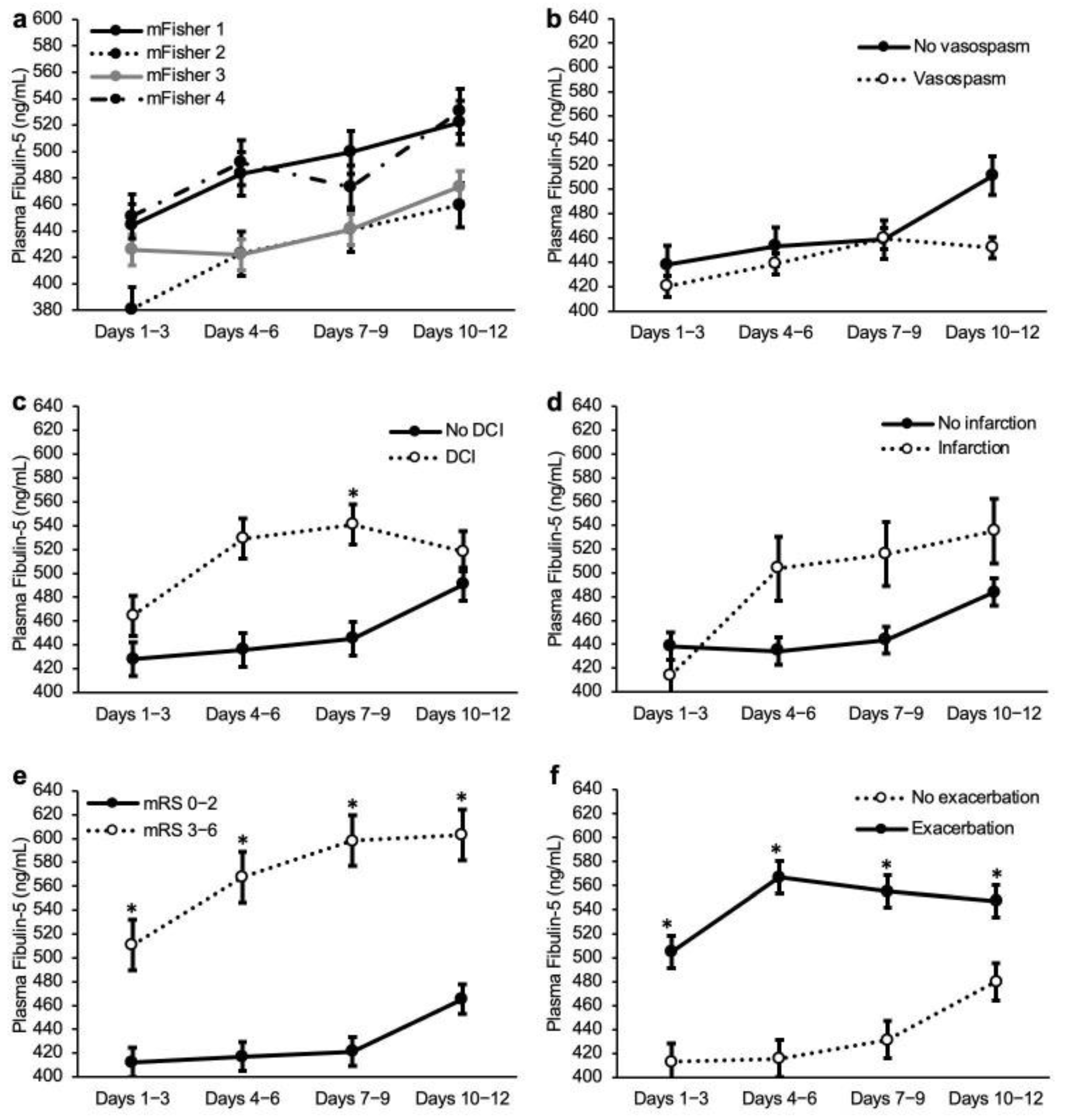
2.2. Plasma FBLN5 Levels

The control samples were obtained from 10 patients with unruptured cerebral aneurysms, but one patient’s data were excluded because active gingivitis was associated and thereby the plasma FBLN5 levels were extremely high (899.0 ng/mL; Smirnov–Grubbs’s test, p = 0.03). Age was similar between the control and SAH patients (67.9 ± 9.5 versus 64.2 ± 14.6 years; p = 0.454; Mann–Whitney U test), and plasma FBLN5 levels were not different between male and female patients in the control (428.2 ± 38.7 versus 473.0 ± 91.2 ng/mL; p = 0.348; unpaired t test) and SAH (459.2 ± 197.8 versus 489.7 ± 209.8 ng/mL; p = 0.063; Mann–Whitney U test). Plasma FBLN5 levels tended to increase from days 1–3 to 10–12 after a SAH, but were not different between nine control patients and SAH patients: 448.1 ± 66.5 versus 438.1 ± 66.5 at days 1–3, 490.9 ± 220.3 at days 4–6, 486.1 ± 212.8 at days 7–9, and 510.8 ± 195.5 ng/mL at days 10–12 post-SAH (p = 0.469, 0.701, 0.603, and 0.368, respectively; Mann–Whitney U test; Figure 2a).
As to the relationships between the plasma FBLN5 levels and the severity of the SAH or the outcome measures, patients with WFNS grades IV-V at admission had significantly higher FBLN5 levels than those with WFNS grades I-III at days 4–6 and 7–9 post-SAH (Figure 2b; Supplementary Table S1). Plasma FBLN5 levels were not different among the modified Fisher grades (Figure 2c), and between patients with and without an angiographic vasospasm, a DCI, and a delayed cerebral infarction, at any sampling point (Figure 2d–f). However, plasma FBLN5 levels were significantly higher in poor-outcome (90-day mRS 3–6) patients, compared with good-outcome (90-day mRS 0–2) patients at days 4–6, 7–9, and 10–12 post-SAH (Figure 2g).
When compared between patients with and without any neurological exacerbation, including a DCI by day 14, compared to at admission, the plasma FBLN5 levels were significantly higher in the exacerbation group at days 7–9 post-SAH (Figure 2h). In the exacerbation group, the plasma FBLN5 levels tended to increase preceding the neurological exacerbation (Figure 2i).

2.3. Receiver-Operating Characteristic (ROC) Curve Analyses for the Performance of Plasma FBLN5, to Differentiate the Poor Outcomes

To differentiate the poor from the good outcomes, the ROC curve analyses indicated that plasma FBLN5 with a cut-off value of 458.2 ng/mL, resulted in a specificity of 62.2% and a sensitivity of 46.8% at days 1–3 post-SAH (area under the curve (AUC), 0.517; 95% confidence interval (CI), 0.435–0.599). As well, according to the curve, the specificity was 61.4% and the sensitivity was 62.3%, when a cut-off value of 487.2 ng/mL was set at days 4–6 (AUC, 0.627; 95% CI, 0.549–0.704); the specificity was 66.1% and the sensitivity was 54.6%, when a cut-off value of 499.4 ng/mL was set at days 7–9 (AUC, 0.622; 95% CI, 0.543–0.701); and the specificity was 89.8% and the sensitivity was 32.5%, when a cut-off value of 707.8 ng/mL was set at days 10–12 (AUC, 0.593; 95% CI, 0.511–0.676) (Supplementary Figure S1). As the AUC was the highest at days 4–6, the plasma FBLN5 values at days 4–6 were used for the following multivariate analyses.

2.4. Independent Determinants for a 90-Day Poor Outcome

Among the significant variables on the univariate analyses (Table 1) and days 4–6 plasma FBLN5 cut-off values, the following variables had significant correlations: between the pre-onset mRSs 0 and 1 (r = −0.894), between WFNS grades I-III and IV-V (r = −1.000) at admission, between WFNS grades IV-V at admission, and the modified Fisher grade 4 (r = 0.335), between the modified Fisher grades 1 and 3 (r = −0.363), between the modified Fisher grades 3 and 4 (r = −0.717), between acute hydrocephalus and CSF drainage (r = 0.449), and between a DCI and a delayed cerebral infarction (r = 0.359). Among these similar clinical variables that were intercorrelated, only the variable with the smallest p value was used as a candidate variable for the multivariate analyses, in addition to other independent variables that were significantly related to a poor outcome on the univariate analyses. The multivariate analyses revealed that the plasma FBLN5 levels at days 4–6 post-SAH ≥ 487.2 ng/mL (odds ratio (OR), 2.55; 95% CI, 1.10-5.88; p = 0.029), were an independent determinant of a 90-day poor outcome, in addition to ≥75 years old, delayed cerebral infarction, non-pre-onset mRS 0, acute hydrocephalus, non-modified Fisher grade 3, and at admission, WFNS grades IV-V (Table 2).
In Supplementary Figure S2, the representative cases were shown: the 90-day outcomes were poor when the plasma FBLN5 levels at days 4–6 post-SAH were ≥487.2 ng/mL, and good (mRS 0–2), when those were <487.2 ng/mL, regardless of the admission WFNS grades, the modified Fisher grades, and the presence or absence of a delayed cerebral infarction.

2.5. Analyses in Non-Severe SAH Cases

We also performed an analysis limited to patients with, at admission, WFNS grades I-III in which the diagnosis of a DCI is easier. The total number of cases was 127, of which 27 cases had poor outcomes (Supplementary Table S2). The univariate analyses were limited to non-severe SAH cases, although most of the 90-day poor outcome-related variables were similar to the analyses in all patients, a pre-onset mRS 1, non-modified Fisher grades 1 and 3, and CSF drainage were no longer significant variables, and a past history of hypertension, a WFNS grade I not at admission, a WFNS grade III at admission, a ruptured aneurysm located at the anterior cerebral artery (ACA) and internal carotid artery (ICA), were added as 90-day poor outcome-related variables (Table 1 and Table S2). The relationships between the plasma FBLN5 levels and the outcome measures were also similar, but patients with a DCI had higher values at days 7–9 post-SAH in the analyses limited to the non-severe SAH cases (Figure 3 and Supplementary Table S3). The ROC curve analyses for the performance of the plasma FBLN5 levels, in order to differentiate the 90-day poor from the good outcomes, showed a better AUC, compared with the analyses of all patients, especially at days 7–9 post-SAH: a cut-off value of 476.8 ng/mL, a sensitivity of 77.8%, and a specificity of 66.0% (AUC, 0.749; 95% CI, 0.642–0.856; Supplementary Figure S3). All significant variables on the univariate analyses and a cut-off value of plasma FBLN5 at days 7–9 post-SAH were candidate variables for the multivariate analyses, but only the variable with the smallest p value was used among the following intercorrelated variables: between ACA and ICA (r = −0.644), between DCI and delayed cerebral infarction (r = 0.506), and between an at admission WFNS grade III and acute hydrocephalus (r = 0.328). The multivariate analyses revealed that the plasma FBLN5 levels at days 7–9 post-SAH ≥ 476.8 ng/mL (OR, 18.14; 95% CI, 3.25–101.39; p < 0.001) were an independent determinants of a 90-day poor outcome, although some independent variables were different from those in the analyses for all SAH patients (Supplementary Table S4).

3. Discussion

The novel findings in this study were as follows: (1) the plasma FBLN5 levels in patients with WFNS grades IV-V at admission, were significantly higher than those in WFNS grades I-III patients, at days 4–9 post-SAH; (2) the plasma FBLN5 levels were not related to an angiographic vasospasm, DCI, or delayed cerebral infarction, but any neurological exacerbation and 90-day poor outcome were associated with elevated FBLN5 levels at days 7–9 and days 4–12, respectively, in all SAH patients’ analyses; (3) in patients with WFNS grades I-III at admission, higher plasma FBLN5 levels were not related to an angiographic vasospasm and delayed cerebral infarction, but those at days 7–9 and days 1–12 were associated with a DCI and any neurological exacerbation or 90-day poor outcome, respectively; and (4) higher plasma FBLN5 levels were independent determinants of a 90-day poor outcome at days 4–6 in all patients’ analyses, and at days 7–9 in the non-severe patients’ analyses. These findings suggest that the plasma FBLN5 levels can be a predictor of the 90-day outcomes after a SAH, and that FBLN5 may be related to an EBI and a delayed neurological exacerbation, leading to poor outcomes.
FBLN5 is one of ECMs belonging to the FBLN family, and it is also a matricellular protein [10]. FBLNs have a repeated calcium binding epidermal growth factor (EGF)-like motifs and a C-terminal fibulin domain [18]. FBLN5 is a family of the short type FBLNs (named as class II type FBLNs) [10], and reported to play an important role in the development of elastic fibers in vivo [18]. FBLN5 is also known as an embryonic vascular EGF-like repeat-containing protein (EVEC) [19] or developing arteries and neural crest EGF-like (DANCE) [20,21], and is expressed mainly in the vascular smooth muscle cells of the developing arteries in embryos [19,22]. Its expression is downregulated in adult arteries, except for tissues with ongoing angiogenesis [19], but the re-expression is observed in balloon-injured vessels or atherosclerotic lesions [20]. Thus, FBLN5 has been involved in the vascular remodeling of injured or atherosclerotic vessels through the growth regulation, matrix maturation, or signaling between vascular smooth muscles and endothelial cells [19,20]. In SAH, FBLN5 may increase the reflecting of the rupture of intracranial aneurysms or vessel injuries, associated with an angiographic vasospasm, but the findings in this study did not support the hypothesis.
In the central nervous system, there are only several studies reported as to FBLN5. Guo et al. reported that overexpressed FBLN5 reduced the reactive oxygen species production, the apoptosis of cortical cells and capillary endothelial cells, and the BBB disruption, resulting in improved neurological functions, in a transient cerebral ischemia model of rats, although the infarct volume was not decreased [14]. Zang et al. reported that limb remote ischemic postconditioning reduced the infarct volume, improved the neurobehavior, and inhibited the BBB disruption by increasing FBLN5 in rat transient cerebral ischemia models [15]. In a clinical setting, Hu et al. reported that the plasma FBLN5 levels significantly increased within 3 days after an intracerebral hemorrhage, and were correlated with the disease severity and the 90-day outcomes [16]. According to a report by Elshony et al., the plasma FBLN5 levels were increased in all types of strokes, especially in hemorrhagic strokes, in the first few hours of onset, and the higher values were observed in more severe diseases [17]. Taken together, the experimental studies have shown that FBLN5 is neuroprotective for ischemic stroke, whereas clinical studies have found that higher plasma FBLN5 levels in an acute phase are associated with more severe ischemic or hemorrhagic strokes with worse outcomes. Although the present study is the first report to investigate the plasma FBLN5 levels serially from an acute phase to a subacute phase, the findings were similar to the previous clinical studies, in terms of that higher plasma FBLN5 levels at a certain time were associated with worse admission clinical grades and outcomes. However, it was unique that higher plasma FBLN5 levels in a delayed phase (FBLN5 levels at days 4–6 in all patients’ analyses and those at days 7–9 in non-severe patients’ analyses) were independent determinants for 90-day poor outcomes.
The matricellular proteins, such as tenascin-C, galectin-3, periostin, secreted protein acidic, and rich in cysteine, and osteopontin (OPN), have been implicated in diverse post-SAH pathologies [23,24,25,26,27,28,29,30,31]. Most of the matricellular proteins have been reported to exert harmful effects on post-SAH pathologies, such as neuroinflammation [24], BBB disruption [23,26,30,31], neuronal apoptosis [24], and cerebral vasospasms [29]. Moreover, only OPN has been repeatedly reported to be protective against an EBI, in terms of neuronal apoptosis [27], BBB disruption [27,32], and microcirculatory dysfunctions [33,34], as well as cerebral vasospasms [28] in experimental animal models. In a clinical setting, however, even OPN has been related to poor outcomes: that is, the plasma OPN levels increased after an aneurysmal SAH, and were significantly higher in patients with 90-day poor outcomes, compared with good outcome patients [35]. Although FBLN5 is one of the matricellular proteins, the functional significance has never been investigated in experimental SAH models. Considering the findings of the basic studies on FBLN5 in other fields, as described above, FBLN5 may act protectively similar to OPN. In the present clinical study, the higher plasma FBLN5 levels were associated with poor outcomes: the findings are also similar to those in clinical studies of OPN in patients with an aneurysmal SAH [35,36]. At the moment, thus, the authors hypothesize that the plasma FBLN5 levels may increase in a delayed fashion, reflecting the severity of an EBI that can be indicated by the WFNS grades, and that the increased FBLN5 may act protectively against an EBI but be insufficient to recover from the EBI, followed by a secondary neurological exacerbation and poor outcomes.
This study has some limitations. First, it is unknown if the sampling point in this study was the best, because the pathophysiology of SAH patients may change individually. Second, this study was a multicenter study, in which magnetic resonance imaging was not required in an acute phase of a SAH, and neurovascular events were diagnosed by CT. Third, it was not tested if FBLN5 is superior to the previously reported ones, as a biomarker. This is because there are no established or validated biomarkers known in a SAH, although many plausible biomarkers have been reported, including non-specific but central nervous system-derived cell death, or recovery-related ones, such as neurofilaments and S100β [37]. In addition, this study focused on discovering a new molecular target to elucidate the pathology of a SAH, rather than establishing a new biomarker: in the first place, we think that a single biomarker may not adequately capture the multiple complex and inter-related pathophysiological cascades associated with a SAH. Fourth, the authors cannot determine where FBLN5 was produced, because only the plasma FBLN5 levels were measured, and the CSF FBLN5 levels were not measured. However, this is the first study to show that FBLN5 may be involved in the post-SAH pathology. Future experimental and clinical studies would clarify the function, mechanisms and clinical significance of FBLN5.

4. Materials and Methods

The Institutional Ethics Committee approved the study (approval No. 2544), and the written informed consent was obtained from the relatives.

4.1. Study Population

The present study used the data of consecutive patients registered in the prospective registry for searching mediators of neurovascular events after an aneurysmal SAH (pSEED), which was conducted in nine tertiary referral centers in Mie prefecture, in Japan, from September 2013 to December 2016 [35,38,39,40,41,42]. In the registry, the clinical variables were recorded, and the plasma samples were serially collected. The inclusion criteria of the registry were as follows: ≥20 years old at onset, mRS 0–2 before onset, the SAH was diagnosed by CT scans, a saccular aneurysm as the cause of the SAH was confirmed on the CT angiography or digital subtraction angiography, and an aneurysmal obliteration by clipping or coil embolization occurred within 48 h of onset. The following was excluded from the present study (Figure 1): unknown etiology of the SAH, a SAH caused by a ruptured fusiform, dissecting, traumatic, mycotic, or arteriovenous malformation-related aneurysms, treatment by parent artery occlusion with or without bypass surgery, no prophylactic medication for a DCI (fasudil hydrochloride or cilostazol), any angiographic or treatment-related complications, concomitant inflammatory diseases that are known to upregulate FBLN5, missing blood samples, and missing data of the clinical variables. Timing, treatment modality (clipping or coil embolization) of the aneurysmal obliteration, and other medical management or treatment strategies, depended on the on-site investigators. In the present study, the plasma FBLN5 levels were measured using stocked plasma samples, and the relationship between the plasma FBLN5 levels and the clinical variables was retrospectively analyzed.

4.2. Clinical Variables

The following variables were analyzed: age, sex, past history, co-morbidities (hypertension, dyslipidemia, and diabetes mellitus; previously diagnosed or on medication), current smoking, family history of SAH, pre-onset mRS, location of the ruptured aneurysms, the WFNS grade at admission, the modified Fisher grade at admission, CT scans [43], and acute hydrocephalus. Age was divided into two groups, over and under 75 years old [42]. In the analyses of all SAH cases, the WFNS grades at admission were divided into non-severe cases (WFNS grades I-III) and severe cases (WFNS grades IV-V) [38], and in the analyses of the non-severe cases, the WFNS grades were analyzed individually [38]. Acute hydrocephalus was recorded when there was evidence of ventriculomegaly, proved by CT scans at admission, which was considered to cause neurological impairments, such as disturbance of consciousness. In the case of acute hydrocephalus, a catheter for CSF drainage was placed at the clipping or immediately before or after the coil embolization. As the treatment-related variables, the following factors were evaluated: treatment modality (clipping or coil embolization) performed for the aneurysmal obliteration, CSF drainage, including ventricular, cisternal, and spinal drainage, to manage the hydrocephalus and/or to promote the hematoma clearance, medication to prevent a DCI (intravenously administered fasudil hydrochloride, oral or enteral cilostazol, ethyl icosapentate, and statin). The outcomes were assessed as to the angiographic vasospasm, DCI, delayed cerebral infarction, and mRS, at 90 days post-SAH. A DCI was defined as otherwise an unexplained clinical deterioration (i.e., focal neurological impairments, a decrease of at least two points on the Glasgow coma scale, or both), which lasted for at least one hour [2]. Other potential causes of clinical deterioration were excluded, based on clinical assessments including CT scans, magnetic resonance images, electroencephalography, and laboratory studies. Any clinical deterioration including, DCI by day 14, compared with at admission, was also recorded as a neurological exacerbation, irrespective of the causes. An angiographic vasospasm was defined as a 50% or more reduction in the baseline vessel diameter of the major cerebral arteries on a CT angiography or digital subtraction angiography, irrespective of the symptoms, which was performed around 7 days post-SAH, and a neurological deterioration. A delayed cerebral infarction was defined as newly developed non-iatrogenic infarcts, which were not observed on CT scans or magnetic resonance images on the day after the aneurysmal obliteration, and developed within 1 month of the SAH. In cases where the 90-day mRS was missing, the mRS at discharge was used instead. The determination of these variables was made at each center, and the organizing committee qualified them.

4.3. Measurement of Plasma FBLN5

Following the aneurysmal obliteration, the blood samples were serially collected with minimal stasis, from a peripheral vein at days 1–3, 4–6, 7–9, and 10–12 after onset. All samples were centrifuged for 5 min at 3000× g, and the supernatants were stored at −78 °C, until assayed. The plasma FBLN5 levels were determined using a commercially available enzyme-linked immunosorbent assay kit for human FBLN5 (27121; IBL, Gunma, Japan). As a control, the blood samples were also obtained from 10 patients with unruptured cerebral aneurysms who provided written informed consent before any invasive procedure, and the plasma FBLN5 levels were determined, as described above.

4.4. Statistical Analysis

All statistical analyses were performed with SPSS software, version 28.0.1.0 (IBM, Armonk, New York, NY, USA). The continuous variables were expressed as a mean ± standard deviation (SD) or standard error of the mean (for graphs), or a median ±25–75 percentile, and were compared between two groups using unpaired t tests or Mann-Whitney U tests, respectively, and among more than three groups, by a one-way analysis of variance or Kruskal-Wallis test with Steel-Dwass’s post hoc test, as appropriate, after examining if each population compared, followed a normal distribution using Shapiro-Wilk tests. The categorical variables were reported as percentages and were compared using Pearson’s chi-squared tests or Fisher’s exact tests, as appropriate. The ROC curves and the AUC were analyzed to obtain the cut-off values of the plasma FBLN5 for the 90-day poor outcome. The correlation between the plasma FBLN5 levels and the clinical variables, or between the clinical variables was evaluated by Pearson’s correlation coefficient for continuous variables or Spearman’s correlation coefficient for categorical variables. The correlation coefficient |r| > 0.3 was considered a significant correlation. Any variable with a p value of <0.05 on the univariate analyses was entered into the multivariate logistic regression models using the 90-day dichotomous mRS outcome (good or poor), as the dependent variable, although only the variable with the smallest p value was used as a candidate variable, among the similar clinical variables that were intercorrelated. The multivariate logistic regression analyses were performed by the forward stepwise method. The analyses were performed in all patients, as well as in non-severe SAH (defined as WFNS grades I-III at admission) patients. A significant level was set at a p value < 0.05.

5. Conclusions

This study first revealed that the delayed elevation of the plasma FBLN5 levels was an independent determinant for 90-day poor outcomes, and that FBLN5 might be a new molecular target to clarify the post-SAH pathologies and to develop a new therapy against them.

Supplementary Materials

The following supporting information can be downloaded at: https://www.mdpi.com/article/10.3390/ijms232315184/s1.

Author Contributions

Conceptualization and design, Y.S. and H.S.; methodology, material preparation, data collection, Y.S., H.O., H.N., M.N., F.K., Y.M., R.Y., N.T., H.S. and pSEED Group; data analyses, Y.S. and H.S.; writing—original draft preparation, Y.S.; writing—review & editing, H.O., H.N., M.N., F.K., Y.M., R.Y., N.T. and H.S. All authors have read and agreed to the published version of the manuscript.

Funding

This work was funded by JSPS KAKENHI Grant Number JP20K17963 and Sanikai Foundation (Grant Number, N/A) to Dr. Kawakita, and the Taiju Life Social Welfare Foundation (Grant Number, N/A) to Dr. H. Suzuki.

Institutional Review Board Statement

The Institutional Ethics Committee approved the study (approval No. 2544).

Informed Consent Statement

Written informed consent was obtained from the relatives.

Data Availability Statement

Data from this study will be made available to qualified investigators upon reasonable inquiry.

Acknowledgments

We thank Chiduru Nakamura, Department of Neurosurgery, Mie University Graduate School of Medicine, for providing technical assistance.

Conflicts of Interest

H. Suzuki reported personal fees from Eisai and Kowa, and a research fund from Japan Blood Products Organization outside of the submitted work. The other authors declare that they have no conflict of interest.

Abbreviations

ACA: anterior cerebral artery; AN, aneurysm; AUC, area under curve; BBB, blood-brain barrier; CI, confidence interval; CSF, cerebrospinal fluid; CT, computed tomography; DANCE, developing arteries and neural crest EGF-like; DCI, delayed cerebral ischemia; EBI, early brain injury; ECM, extracellular matrix protein; EGF, epidermal growth factor; EVEC, embryonic vascular EGF-like repeat-containing protein; FBLN5, fibulin-5; ICA, internal carotid artery; IQR, interquartile range; MCA, middle cerebral artery; mRS, modified Rankin scale; OPN, osteopontin; OR, odds ratio; pSEED, prospective registry for searching mediators of neurovascular events after aneurysmal subarachnoid hemorrhage; ROC, receiver-operating characteristic; SAH, subarachnoid hemorrhage; SD, standard deviation; VA-BA, vertebral artery-basilar artery; WFNS, World Federation of Neurological Surgeons.

Appendix A

Prospective Registry for Searching Mediators of Neurovascular Events After Aneurysmal Subarachnoid Hemorrhage (pSEED) Group: Department of Neurosurgery, Kuwana City Medical Center, Kuwana (Hiroshi Sakaida, and Kazuhide Hamada), Mie Prefectural General Medical Center, Yokkaichi (Yusuke Kamei, and Yasuyuki Umeda), Suzuka Kaisei Hospital, Suzuka (Tomohiro Araki, Masato Shiba, Naoki Ichikawa, and Yoshinari Nakatsuka), Suzuka Chuo General Hospital, Suzuka (Shigetoshi Shimizu, Takuro Tsuchiya, and Reona Asada), Mie University Graduate School of Medicine, Tsu (Hidenori Suzuki, Naoki Toma, Ryuta Yasuda, Yoichi Miura, Masashi Fujimoto, Fumihiro Kawakita, Hirofumi Nishikawa, Hideki Kanamaru, and Yotaro Kitano), Mie Chuo Medical Center, Tsu (Fujimaro Ishida, Katsuhiro Tanaka, and Satoru Tanioka), Matsusaka Chuo General Hospital, Matsusaka (Kazuhiko Tsuda, and Yu Sato), Saiseikai Matsusaka General Hospital, Matsusaka (Hiroto Murata, and Keiji Fukazawa), and Ise Red Cross Hospital, Ise (Fumitaka Miya, Hiroshi Tanemura, and Takanori Sano).

References

  1. Van Gijn, J.; Rinkel, G.J. Subarachnoid haemorrhage: Diagnosis, causes and management. Brain 2001, 124 Pt 2, 249–278. [Google Scholar] [CrossRef] [PubMed]
  2. Vergouwen, M.D.; Vermeulen, M.; van Gijn, J.; Rinkel, G.J.; Wijdicks, E.F.; Muizelaar, J.P.; Mendelow, A.D.; Juvela, S.; Yonas, H.; Terbrugge, K.G.; et al. Definition of delayed cerebral ischemia after aneurysmal subarachnoid hemorrhage as an outcome event in clinical trials and observational studies: Proposal of a multidisciplinary research group. Stroke 2010, 41, 2391–2395. [Google Scholar] [CrossRef] [Green Version]
  3. Suzuki, H. What is early brain injury? Transl. Stroke Res. 2015, 6, 1–3. [Google Scholar] [CrossRef] [PubMed]
  4. Chen, S.; Luo, J.; Reis, C.; Manaenko, A.; Zhang, J. Hydrocephalus after Subarachnoid Hemorrhage: Pathophysiology, Diagnosis, and Treatment. Biomed. Res. Int. 2017, 2017, 8584753. [Google Scholar] [CrossRef] [Green Version]
  5. Rush, B.; Wiskar, K.; Fruhstorfer, C.; Hertz, P. Association between seizures and mortality in patients with aneurysmal subarachnoid hemorrhage: A nationwide retrospective cohort analysis. Seizure 2016, 41, 66–69. [Google Scholar] [CrossRef] [Green Version]
  6. Taki, W.; Sakai, N.; Suzuki, H. Determinants of poor outcome after aneurysmal subarachnoid hemorrhage when both clipping and coiling are available: Prospective Registry of Subarachnoid Aneurysms Treatment (PRESAT) in Japan. World Neurosurg. 2011, 76, 437–445. [Google Scholar] [CrossRef]
  7. Buce-Satoba, I.; Rozkalne, D.; Stepanovs, J.; Mamaja, B.; Krumina, G.; Ozolina, A. Literature Review of Aneurysmal Subarachnoid Hemorrhage from Incidence to Treatment Options. Acta Chir. Latv. 2020, 18, 70–78. [Google Scholar] [CrossRef]
  8. Suzuki, H.; Kanamaru, H.; Kawakita, F.; Asada, R.; Fujimoto, M.; Shiba, M. Cerebrovascular pathophysiology of delayed cerebral ischemia after aneurysmal subarachnoid hemorrhage. Histol. Histopathol. 2021, 36, 143–158. [Google Scholar] [CrossRef]
  9. Hasegawa, Y.; Suzuki, H.; Uekawa, K.; Kawano, T.; Kim-Mitsuyama, S. Characteristics of Cerebrovascular Injury in the Hyperacute Phase after Induced Severe Subarachnoid Hemorrhage. Transl. Stroke Res. 2015, 6, 458–466. [Google Scholar] [CrossRef]
  10. Yanagisawa, H.; Schluterman, M.K.; Brekken, R.A. Fibulin-5, an integrin-binding matricellular protein: Its function in development and disease. J. Cell Commun. Signal 2009, 3, 337–347. [Google Scholar] [CrossRef]
  11. Nakamura, T.; Lozano, P.R.; Ikeda, Y.; Iwanaga, Y.; Hinek, A.; Minamisawa, S.; Cheng, C.F.; Kobuke, K.; Dalton, N.; Takada, Y.; et al. Fibulin-5/DANCE is essential for elastogenesis in vivo. Nature 2002, 415, 171–175. [Google Scholar] [CrossRef] [PubMed]
  12. Yanagisawa, H.; Davis, E.C.; Starcher, B.C.; Ouchi, T.; Yanagisawa, M.; Richardson, J.A.; Olson, E.N. Fibulin-5 is an elastin-binding protein essential for elastic fibre development in vivo. Nature 2002, 415, 168–171. [Google Scholar] [CrossRef] [PubMed]
  13. Nonaka, R.; Onoue, S.; Wachi, H.; Sato, F.; Urban, Z.; Starcher, B.C.; Seyama, Y. DANCE/fibulin-5 promotes elastic fiber formation in a tropoelastin isoform-dependent manner. Clin. Biochem. 2009, 42, 713–721. [Google Scholar] [CrossRef]
  14. Guo, J.; Cheng, C.; Chen, C.S.; Xing, X.; Xu, G.; Feng, J.; Qin, X. Overexpression of Fibulin-5 Attenuates Ischemia/Reperfusion Injury after Middle Cerebral Artery Occlusion in Rats. Mol. Neurobiol. 2016, 53, 3154–3167. [Google Scholar] [CrossRef] [PubMed]
  15. Zhang, W.; Wang, Y.; Bi, G. Limb remote ischaemic postconditioning-induced elevation of fibulin-5 confers neuroprotection to rats with cerebral ischaemia/reperfusion injury: Activation of the AKT pathway. Clin. Exp. Pharmacol. Physiol. 2017, 44, 656–663. [Google Scholar] [CrossRef]
  16. Hu, L.; Dong, M.X.; Zhao, H.; Xu, G.H.; Qin, X.Y. Fibulin-5: A novel biomarker for evaluating severity and predicting prognosis in patients with acute intracerebral haemorrhage. Eur. J. Neurol. 2016, 23, 1195–1201. [Google Scholar] [CrossRef]
  17. Elshony, H.S.; Okda, M.A.; El-Kabany, R.A. Ischemia-modified albumin and fibulin-5 as diagnostic and prognostic markers for acute cerebrovascular disease. Egypt. J. Neurol. Psychiatr. Neurosurg. 2021, 57, 12. [Google Scholar] [CrossRef]
  18. Papke, C.L.; Yanagisawa, H. Fibulin-4 and fibulin-5 in elastogenesis and beyond: Insights from mouse and human studies. Matrix Biol. 2014, 37, 142–149. [Google Scholar] [CrossRef]
  19. Kowal, R.C.; Richardson, J.A.; Miano, J.M.; Olson, E.N. EVEC, a novel epidermal growth factor-like repeat-containing protein upregulated in embryonic and diseased adult vasculature. Circ. Res. 1999, 84, 1166–1176. [Google Scholar] [CrossRef] [Green Version]
  20. Nakamura, T.; Ruiz-Lozano, P.; Lindner, V.; Yabe, D.; Taniwaki, M.; Furukawa, Y.; Kobuke, K.; Tashiro, K.; Lu, Z.; Andon, N.L.; et al. DANCE, a novel secreted RGD protein expressed in developing, atherosclerotic, and balloon-injured arteries. J. Biol. Chem. 1999, 274, 22476–22483. [Google Scholar] [CrossRef]
  21. Hirai, M.; Ohbayashi, T.; Horiguchi, M.; Okawa, K.; Hagiwara, A.; Chien, K.R.; Kita, T.; Nakamura, T. Fibulin-5/DANCE has an elastogenic organizer activity that is abrogated by proteolytic cleavage in vivo. J. Cell Biol. 2007, 176, 1061–1071. [Google Scholar] [CrossRef] [Green Version]
  22. Chailley-Heu, B.; Boucherat, O.; Barlier-Mur, A.M.; Bourbon, J.R. FGF-18 is upregulated in the postnatal rat lung and enhances elastogenesis in myofibroblasts. Am. J. Physiol. Lung Cell Mol. Physiol. 2005, 288, L43–L51. [Google Scholar] [CrossRef]
  23. Fujimoto, M.; Shiba, M.; Kawakita, F.; Liu, L.; Shimojo, N.; Imanaka-Yoshida, K.; Yoshida, T.; Suzuki, H. Deficiency of tenascin-C and attenuation of blood-brain barrier disruption following experimental subarachnoid hemorrhage in mice. J. Neurosurg. 2016, 124, 1693–1702. [Google Scholar] [CrossRef] [PubMed] [Green Version]
  24. Liu, L.; Fujimoto, M.; Nakano, F.; Nishikawa, H.; Okada, T.; Kawakita, F.; Imanaka-Yoshida, K.; Yoshida, T.; Suzuki, H. Deficiency of Tenascin-C Alleviates Neuronal Apoptosis and Neuroinflammation after Experimental Subarachnoid Hemorrhage in Mice. Mol. Neurobiol. 2018, 55, 8346–8354. [Google Scholar] [CrossRef]
  25. Suzuki, H.; Fujimoto, M.; Kawakita, F.; Liu, L.; Nakatsuka, Y.; Nakano, F.; Nishikawa, H.; Okada, T.; Kanamaru, H.; Imanaka-Yoshida, K.; et al. Tenascin-C in brain injuries and edema after subarachnoid hemorrhage: Findings from basic and clinical studies. J. Neurosci. Res. 2020, 98, 42–56. [Google Scholar] [CrossRef] [PubMed] [Green Version]
  26. Okada, T.; Suzuki, H.; Travis, Z.D.; Altay, O.; Tang, J.; Zhang, J.H. SPARC Aggravates Blood-Brain Barrier Disruption via Integrin alphaVbeta3/MAPKs/MMP-9 Signaling Pathway after Subarachnoid Hemorrhage. Oxid. Med. Cell Longev. 2021, 2021, 9739977. [Google Scholar] [CrossRef] [PubMed]
  27. Kawakita, F.; Kanamaru, H.; Asada, R.; Suzuki, H. Potential roles of matricellular proteins in stroke. Exp. Neurol. 2019, 322, 113057. [Google Scholar] [CrossRef]
  28. Suzuki, H.; Hasegawa, Y.; Chen, W.; Kanamaru, K.; Zhang, J.H. Recombinant osteopontin in cerebral vasospasm after subarachnoid hemorrhage. Ann. Neurol. 2010, 68, 650–660. [Google Scholar] [CrossRef] [Green Version]
  29. Fujimoto, M.; Shiba, M.; Kawakita, F.; Liu, L.; Shimojo, N.; Imanaka-Yoshida, K.; Yoshida, T.; Suzuki, H. Effects of Tenascin-C Knockout on Cerebral Vasospasm after Experimental Subarachnoid Hemorrhage in Mice. Mol. Neurobiol. 2018, 55, 1951–1958. [Google Scholar] [CrossRef]
  30. Nishikawa, H.; Liu, L.; Nakano, F.; Kawakita, F.; Kanamaru, H.; Nakatsuka, Y.; Okada, T.; Suzuki, H. Modified Citrus Pectin Prevents Blood-Brain Barrier Disruption in Mouse Subarachnoid Hemorrhage by Inhibiting Galectin-3. Stroke 2018, 49, 2743–2751. [Google Scholar] [CrossRef]
  31. Liu, L.; Kawakita, F.; Fujimoto, M.; Nakano, F.; Imanaka-Yoshida, K.; Yoshida, T.; Suzuki, H. Role of Periostin in Early Brain Injury after Subarachnoid Hemorrhage in Mice. Stroke 2017, 48, 1108–1111. [Google Scholar] [CrossRef] [PubMed]
  32. Suzuki, H.; Hasegawa, Y.; Kanamaru, K.; Zhang, J.H. Mechanisms of osteopontin-induced stabilization of blood-brain barrier disruption after subarachnoid hemorrhage in rats. Stroke 2010, 41, 1783–1790. [Google Scholar] [CrossRef] [Green Version]
  33. Suzuki, H.; Ayer, R.; Sugawara, T.; Chen, W.; Sozen, T.; Hasegawa, Y.; Kanamaru, K.; Zhang, J.H. Protective effects of recombinant osteopontin on early brain injury after subarachnoid hemorrhage in rats. Crit. Care Med. 2010, 38, 612–618. [Google Scholar] [CrossRef] [PubMed]
  34. Topkoru, B.C.; Altay, O.; Duris, K.; Krafft, P.R.; Yan, J.; Zhang, J.H. Nasal administration of recombinant osteopontin attenuates early brain injury after subarachnoid hemorrhage. Stroke 2013, 44, 3189–3194. [Google Scholar] [CrossRef] [PubMed] [Green Version]
  35. Nakatsuka, Y.; Shiba, M.; Nishikawa, H.; Terashima, M.; Kawakita, F.; Fujimoto, M.; Suzuki, H.; pSEED Group. Acute-Phase Plasma Osteopontin as an Independent Predictor for Poor Outcome After Aneurysmal Subarachnoid Hemorrhage. Mol. Neurobiol. 2018, 55, 6841–6849. [Google Scholar] [CrossRef]
  36. Asada, R.; Suzuki, H. Osteopontin in post-subarachnoid hemorrhage pathologies. J. Integr. Neurosci. 2022, 21, 62. [Google Scholar] [CrossRef] [PubMed]
  37. Chou, S.H.; Macdonald, R.L.; Keller, E. Biospecimens and molecular and cellular biomarkers in aneurysmal subarachnoid hemorrhage studies: Common data elements and standard reporting recommendations. Neurocrit. Care 2019, 30, S46–S59. [Google Scholar] [CrossRef]
  38. Nakatsuka, Y.; Kawakita, F.; Yasuda, R.; Umeda, Y.; Toma, N.; Sakaida, H.; Suzuki, H. Preventive effects of cilostazol against the development of shunt-dependent hydrocephalus after subarachnoid hemorrhage. J. Neurosurg. 2017, 127, 319–326. [Google Scholar] [CrossRef] [Green Version]
  39. Nakatsuka, Y.; Terashima, M.; Nishikawa, H.; Kawakita, F.; Fujimoto, M.; Shiba, M.; Yasuda, R.; Toma, N.; Sakaida, H.; Suzuki, H. Current Status of Ruptured Cerebral Aneurysm Treatment in Regional Hospitals and Results of Coil Embolization. J. Neuroendovascular. Ther. 2018, 12, 109–116. [Google Scholar] [CrossRef] [Green Version]
  40. Nishikawa, H.; Nakatsuka, Y.; Shiba, M.; Kawakita, F.; Fujimoto, M.; Suzuki, H.; pSEED Group. Increased Plasma Galectin-3 Preceding the Development of Delayed Cerebral Infarction and Eventual Poor Outcome in Non-Severe Aneurysmal Subarachnoid Hemorrhage. Transl. Stroke Res. 2018, 9, 110–119. [Google Scholar] [CrossRef]
  41. Kanamaru, H.; Kawakita, F.; Nakano, F.; Miura, Y.; Shiba, M.; Yasuda, R.; Toma, N.; Suzuki, H.; pSEED Group. Plasma Periostin and Delayed Cerebral Ischemia after Aneurysmal Subarachnoid Hemorrhage. Neurotherapeutics 2019, 16, 480–490. [Google Scholar] [CrossRef] [PubMed] [Green Version]
  42. Kanamaru, H.; Kawakita, F.; Asada, R.; Miura, Y.; Shiba, M.; Toma, N.; Suzuki, H.; pSEED Group. Prognostic factors varying with age in patients with aneurysmal subarachnoid hemorrhage. J. Clin. Neurosci. 2020, 76, 118–125. [Google Scholar] [CrossRef] [PubMed]
  43. Claassen, J.; Bernardini, G.L.; Kreiter, K.; Bates, J.; Du, Y.E.; Copeland, D.; Connolly, E.S.; Mayer, S.A. Effect of cisternal and ventricular blood on risk of delayed cerebral ischemia after subarachnoid hemorrhage: The Fisher scale revisited. Stroke 2001, 32, 2012–2020. [Google Scholar] [CrossRef] [PubMed]
Figure 1. A flow chart showing the included and excluded patients in this study. mRS, modified Rankin scale; SAH, subarachnoid hemorrhage.
Ijms 23 15184 g001
Figure 2. Chronological changes in the plasma fibulin-5 levels after a subarachnoid hemorrhage (SAH). Comparison between the control and SAH patients (a), between SAH patients with World Federation of Neurological Surgeons (WFNS) grades I-III and IV-V at admission (b), among the modified Fisher grades (c), between patients with and without angiographic vasospasm (d), delayed cerebral ischemia (DCI); (e), or delayed cerebral infarction (f), between the 90-day modified Rankin scale (mRS) 0–2 and 3–6 (g), and between patients with and without neurological exacerbations (h). Relationships between the changes in the plasma fibulin-5 levels and the timing of the neurological exacerbation (i). Data, a mean ±standard error of the mean. * p < 0.05, Mann–Whitney U test; ** p < 0.05, unpaired t test.
Ijms 23 15184 g002
Figure 3. Chronological changes in the plasma fibulin-5 levels in patients with, at admission, World Federation of Neurological Surgeons grades I-III of a subarachnoid hemorrhage. Comparison among the modified Fisher grades (a), between the patients with and without an angiographic vasospasm (b), a delayed cerebral ischemia (DCI); (c), or a delayed cerebral infarction (d), between the 90-day modified Rankin scale (mRS) 0–2 and 3–6 (e), and between patients with and without a neurological exacerbation (f). Data, a mean ±standard error of the mean. * p < 0.05, Mann-Whitney U test.
Ijms 23 15184 g003
Table 1. Clinical variables related to the 90-day poor outcome or modified Rankin scale (mRS) 3–6.
Total (n = 204)mRS 0–2 (n = 127)mRS 3–6 (n = 77)p Value
Age
Median (IQR), years66.0 (51.8–74.3)60.0 (48.0–69.0)74.0 (66.0–82.0)<0.001 a
≥75 years old51 (25.0)14 (11.0)37 (48.1)<0.001 b
Female149 (73.0)86 (67.7)63 (81.8)0.028 b
Past history
SAH10 (4.9)5 (3.9)5 (6.5)0.308 c
Cerebral infarction9 (4.4)2 (1.6)7 (9.1)0.016 c
Hypertension90 (44.1)56 (44.1)34 (44.2)0.993 b
Dyslipidemia23 (11.3)12 (9.4)11 (14.3)0.290 b
Diabetes mellitus15 (7.4)8 (6.3)7 (9.1)0.459 b
Family history of SAH21 (10.3)16 (12.6)5 (6.5)0.164 b
Current smoking45 (22.1)33 (26.0)12 (15.6)0.082 b
Pre-onset mRS
0188 (92.2)123 (96.9)65 (84.4)0.002 c
113 (6.4)4 (3.1)9 (11.7)0.018 c
23 (1.5)0 (0.0)3 (3.9)0.052 c
Admission WFNS grade
I-III127 (62.3)100 (78.7)27 (35.1)<0.001 b
IV-V77 (37.7)27 (21.3)50 (64.9)<0.001 b
Modified Fisher grade
125 (12.3)22 (17.3)3 (3.9)0.003 c
28 (3.9)7 (5.5)1 (1.3)0.127 c
399 (48.5)74 (58.3)25 (32.5)<0.001 b
472 (35.3)24 (18.9)48 (62.3)<0.001 b
Acute hydrocephalus67 (32.8)25 (19.7)42 (54.5)<0.001 b
Ruptured AN location
ACA70 (34.3)48 (37.8)22 (28.6)0.179 b
ICA76 (37.3)45 (35.4)31 (40.3)0.489 b
MCA40 (19.6)23 (18.1)17 (22.1)0.489 b
VA-BA18 (8.8)11 (8.7)7 (9.1)0.917 b
Anterior circulation186 (91.2)116 (91.3)70 (90.9)0.917 b
Multiple Aneurysms56 (27.5)34 (26.8)22 (28.6)0.780 b
Treatment modality
Clipping161 (78.9)96 (75.6)65 (84.4)0.134 b
Coiling43 (21.1)31 (24.4)12 (15.6)0.134 b
CSF drainage69 (33.8)35 (27.6)34 (44.2)0.015 b
Prophylactic drug for DCI
Fasudil hydrochloride201 (98.5)126 (99.2)75 (97.4)0.319 c
Cilostazol166 (81.4)105 (82.7)61 (79.2)0.539 b
Ethyl icosapentate103 (50.5)68 (53.5)35 (45.5)0.263 b
Statin50 (24.5)35 (27.6)15 (19.5)0.193 b
Angiographic vasospasm62 (30.4)33 (26.0)29 (37.7)0.068 b
DCI29 (14.2)11 (8.7)18 (23.4)0.004 b
Delayed cerebral infarction45 (22.1)15 (11.8)30 (39.0)<0.001 b
Data are number of cases (%) unless otherwise specified and compared between mRS 0–2 and 3–6. p values are determined by a Mann–Whitney U test, b Pearson’s chi-squared test, or c Fisher’s exact test. ACA, anterior cerebral artery; AN, aneurysm; CSF, cerebrospinal fluid; DCI, delayed cerebral ischemia; ICA, internal carotid artery; IQR, interquartile range; MCA, middle cerebral artery; SAH, subarachnoid hemorrhage; VA-BA, vertebral artery-basilar artery; WFNS, World Federation of Neurological Surgeons.
Table 2. Multivariate logistic regression analyses for predicting a 90-day poor outcome after a subarachnoid hemorrhage.
VariablesOdds Ratio95% CIp Value
Age ≥ 75 years old3.311.38–7.930.007
Delayed cerebral infarction12.644.48–35.68<0.001
Fibulin-5 at days 4–6 ≥ 487.2 ng/mL2.551.10–5.880.029
Pre-onset mRS 00.100.02–0.620.013
Acute hydrocephalus4.842.03–11.54<0.001
Modified Fisher grade 30.300.13–0.710.006
Admission WFNS grades IV-V5.932.61–13.47<0.001
Female 0.887
Past history of a cerebral infarction 0.120
Variables are selected by the forward stepwise method. Plasma fibulin-5 levels at days 4–6 are categorized using the cut-off value that was determined in Supplementary Figure S1. CI, confidence interval; mRS, modified Rankin scale; WFNS, World Federation of Neurosurgical Surgeons.
Publisher’s Note: MDPI stays neutral with regard to jurisdictional claims in published maps and institutional affiliations.

