Article

Knockdown of YY1 Inhibits XIST Expression and Enhances Cloned Pig Embryo Development

1
National Engineering Research Center for Breeding Swine Industry, South China Agricultural University, Guangzhou 510030, China
2
Department of Animal Genetics, Breeding and Reproduction, College of Animal Science, Guangzhou 510030, China
3
Guangdong Provincial Key Laboratory of Agro-Animal Genomics and Molecular Breeding, South China Agricultural University, Guangzhou 510030, China
4
Guangdong Provincial Laboratory of Lingnan Modern Agricultural Science and Technology, Guangzhou 510642, China
*
Authors to whom correspondence should be addressed.
These authors contributed equally to this work.
Int. J. Mol. Sci. 2022, 23(23), 14572; https://doi.org/10.3390/ijms232314572
Submission received: 2 November 2022 / Revised: 18 November 2022 / Accepted: 21 November 2022 / Published: 23 November 2022

Abstract

:
The technique of cloning has wide applications in animal husbandry and human biomedicine. However, the very low developmental efficiency of cloned embryos limits the application of cloning. Ectopic XIST-expression-induced abnormal X chromosome inactivation (XCI) is a primary cause of the low developmental competence of cloned mouse and pig embryos. Knockout or knockdown of XIST improves cloning efficiency in both pigs and mice. The transcription factor Yin yang 1(YY1) plays a critical role in XCI by triggering the transcription of X-inactive specific transcript (XIST) and facilitating the localization of XIST RNA on the X chromosome. This study aimed to investigate whether RNA interference to suppress the expression of YY1 can inhibit erroneous XIST expression, rescue abnormal XCI, and improve the developmental ability of cloned pig embryos. The results showed that YY1 binds to the 5′ regulatory region of the porcine XIST gene in pig cells. The microinjection of YY1 siRNA into cloned pig embryos reduced the transcript abundance of XIST and upregulated the mRNA level of X-linked genes at the 4-cell and blastocyst stages. The siRNA-mediated knockdown of YY1 altered the transcriptome and enhanced the in vitro and in vivo developmental efficiency of cloned porcine embryos. These results suggested that YY1 participates in regulating XIST expression and XCI in cloned pig embryos and that the suppression of YY1 expression can increase the developmental rate of cloned pig embryos. The present study established a new method for improving the efficiency of pig cloning.

1. Introduction

The technique of cloning, also known as somatic cell nuclear transfer (SCNT), is used to propagate superior livestock [1,2], preserve endangered animals [3,4,5,6], generate cloned human embryos for isolation of therapeutic embryonic stem cells [7,8,9,10], and produce multi-purpose genetically engineered animals [11,12,13]. Thus, cloning is a powerful tool that has wide applications in animal production, life science, and biomedicine. Nevertheless, the efficiency of SCNT embryos in developing to term is very low, at about 2% [14,15] in mice and 1% in pigs [16]. Such a low developmental rate of full-term SCNT embryos limits the application and development of the SCNT technique.
Ectopic XIST expression induces abnormal silence of a part of X-linked genes, which resembles X chromosome inactivation (XCI); it is one of the primary causes of the low developmental competence of cloned mouse and pig embryos. Knockout of the XIST gene or knockdown of Xist expression not only rescued aberrant XCI but also resulted in an eight—ten-fold increase in the full-term developmental efficiency of cloned mouse embryos [17,18]. The injection of anti-XIST small interfering RNA (siRNA) or the short hairpin RNA (shRNA) plasmid and the inactivation of the XIST gene also increased the expression of X-linked genes and promoted the development of cloned pig embryos [19,20,21]. In addition, RNA interference (RNAi) of RLIM, a XIST transcription inducer, inhibited the expression of XIST and improved the developmental efficiency of cloned pig embryos [16].
Transcription factor YY1 plays key roles in regulating XIST functions. YY1 can activate XIST transcription and XCI in mouse and human cells by binding to the 5′ regulatory region of the XIST gene [22,23]. YY1 can also bind to XIST RNA to facilitate its localization on the X chromosome, thus promoting XCI in mouse and human cells [23,24,25].
We hypothesized that YY1 is involved in mediating the aberrant activation of XIST expression and XCI in cloned pig embryos. To test this hypothesis, we injected YY1 siRNA into cloned pig embryos to suppress YY1 expression and investigated its effects on XIST expression, XCI, and cloned pig embryo development. We demonstrated that siRNA-mediated knockdown of YY1 expression can suppress XIST expression, upregulate the expression of X-linked genes, and improve the developmental efficiency of cloned pig embryos.

2. Results

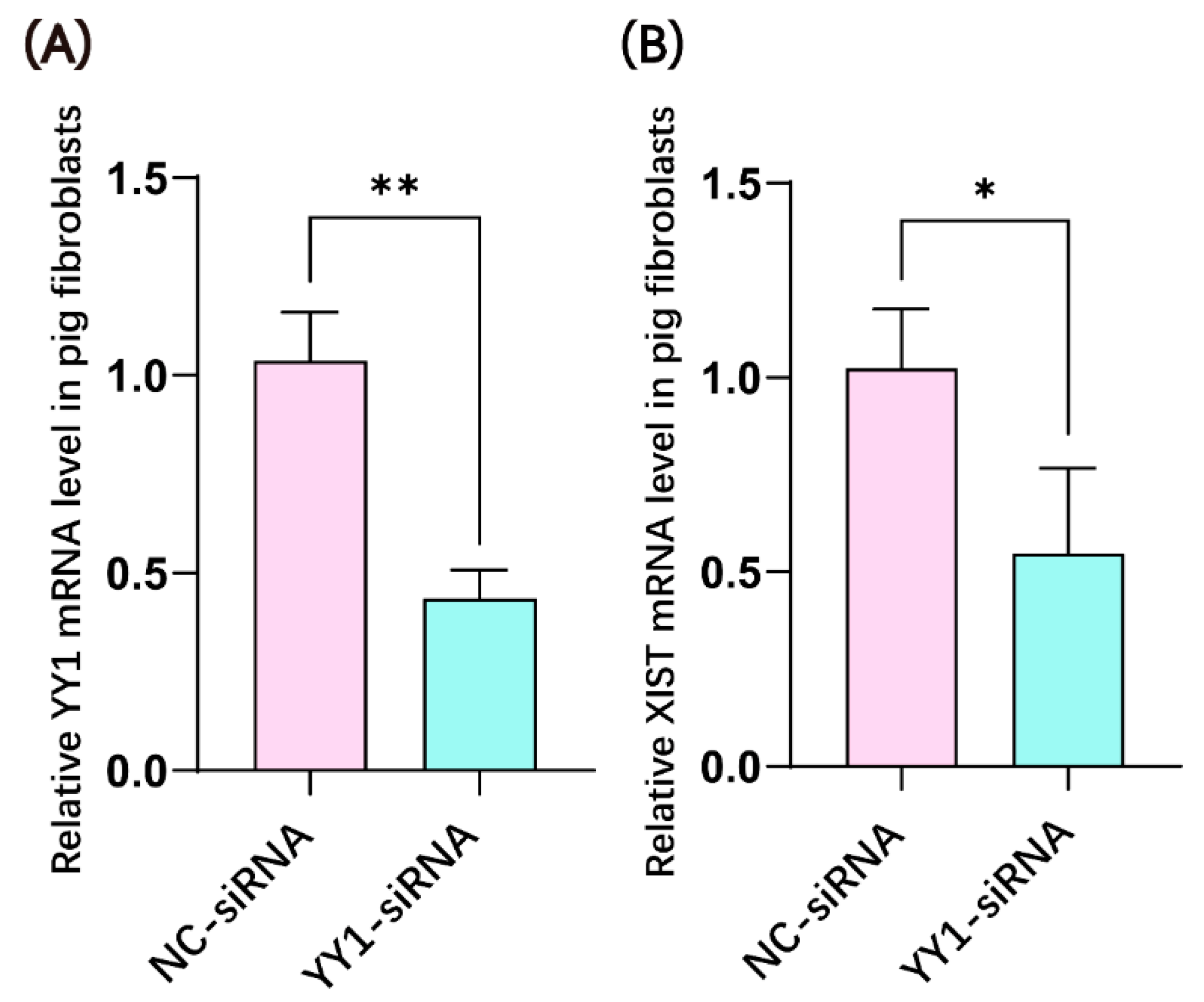
2.1. YY1 siRNA Transfection Inhibited the mRNA Expression of YY1 and XIST in Porcine Fibroblasts

YY1 siRNA was transfected into porcine female fibroblasts to examine its effects on YY1 and XIST mRNA expression. YY1 siRNA not only significantly decreased the level of YY1 transcript (Figure 1A) but also significantly down-regulated XIST mRNA expression (Figure 1B).

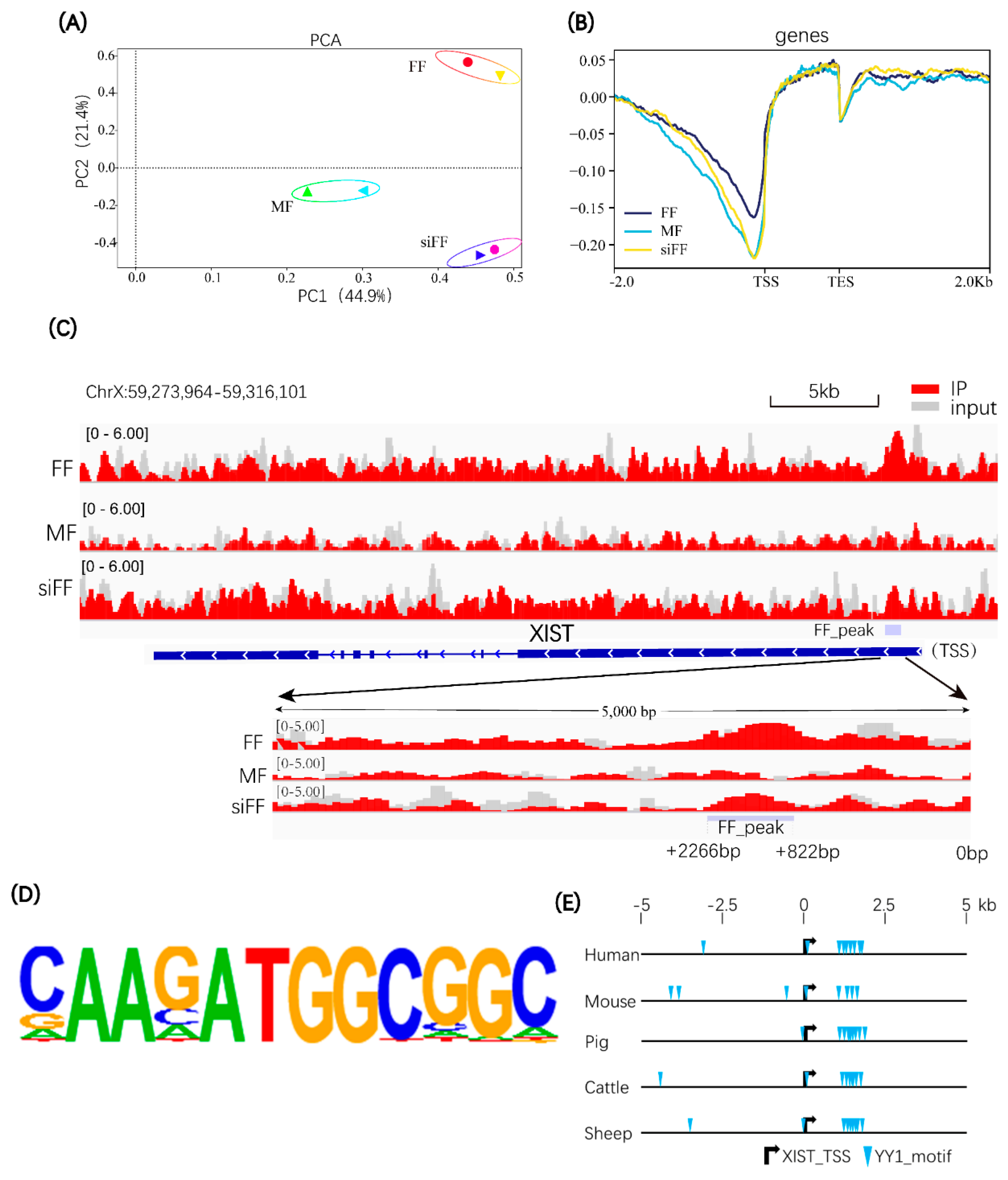
2.2. YY1 Can Bind to the 5′ Regulatory Region of the Porcine XIST Gene

To investigate whether YY1 activates porcine XIST transcription by binding to its regulatory region, we performed ChiP-seq analysis (for YY1) of three groups of pig cells, including male fibroblasts (MF), female fibroblasts (FF), and female fibroblasts transfected with YY1 siRNA (siFF). We hypothesized that in MF cells, the X-linked XIST gene should be silent to maintain the activation of the single X chromosome; in FF cells, one XIST allele should be expressed to maintain random XCI, and in siFF cells, XIST expression should be inhibited by transfected YY1 siRNA. The PCA results showed a distinct clustering of the samples from three groups (Figure 2A). The binding site analysis showed that YY1 had a central binding peak near the transcription start site (TSS) of genome-wide genes (Figure 2B). The analysis of the binding site of YY1 on the porcine XIST gene revealed the YY1 binding peak at the 5′ regulatory region (between +822 bp to +2266 bp) of the pig XIST gene in the FF cells, but this was not observed in the MF and siFF cells (Figure 2C). The DNA binding motif of YY1 was predicted by HOMER software analysis (Figure 2D). This motif was used to analyze the potential binding sites of YY1 on the XIST gene in human, mouse, pig, cattle, and sheep. In all the five analyzed mammalian species, the XIST gene contained similar clustered YY1 binding sites in the 5′ regulatory region (Figure 2E), implying that the binding of YY1 to the 5′ regulatory region of the XIST gene is conserved in mammals.

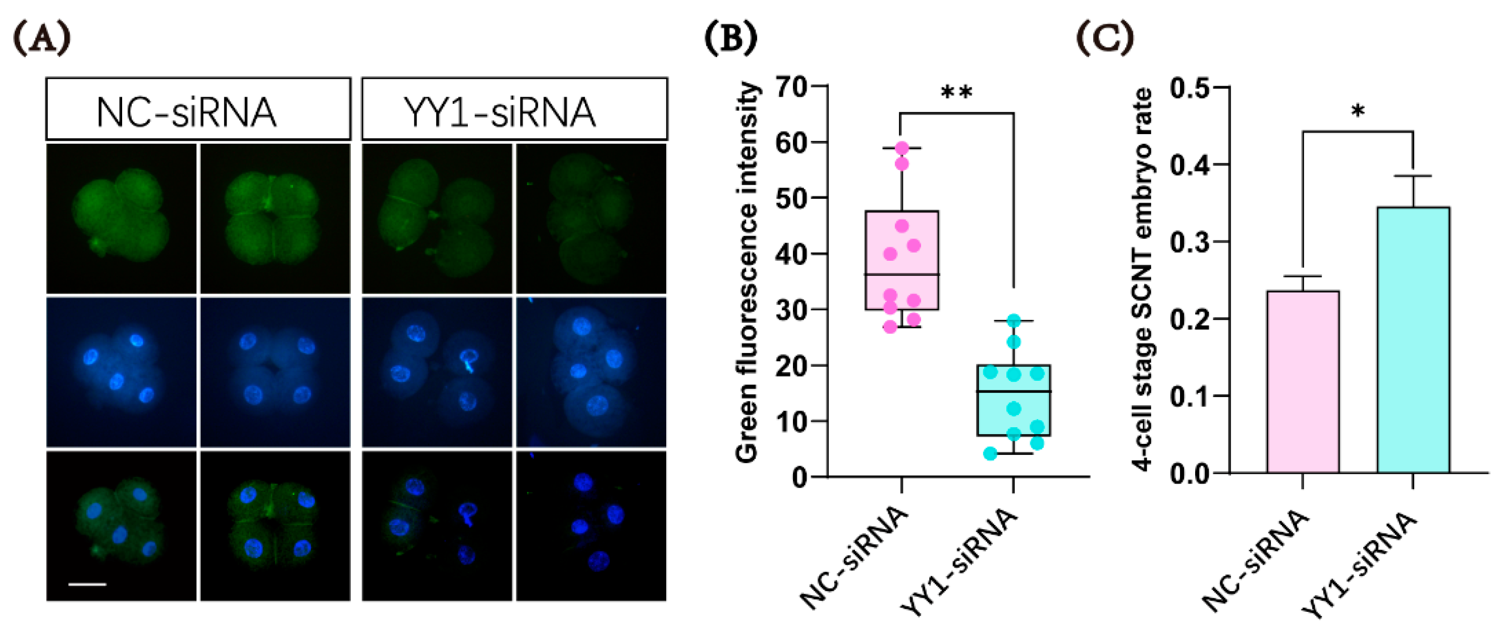
2.3. Microinjection of YY1 siRNA Inhibited the Expression of the YY1 Protein and Improved the Developmental Ability of Cloned Pig Embryos

YY1 siRNA was microinjected into cloned pig embryos to examine whether it could inhibit the expression of the YY1 protein. The immunofluorescence analysis showed that the level of the YY1 protein in the cloned pig embryos injected with YY1 siRNA was indeed significantly lower than that in the control group at the 4-cell stage (Figure 3A,B). When we collected cloned embryos at the 4-cell stage for immunofluorescence analysis, we also observed that the ratio of 4-cell-stage cloned embryos in the YY1 siRNA-injected group was significantly higher than that in the control group (Figure 3C)
To further study the effects of YY1 expression knockdown on the development of cloned pig embryos, we injected 10 µM or 50 µM of YY1 siRNA into cloned pig embryos. We found that the microinjection of 50 µM YY1 siRNA significantly increased the cleavage rate and blastocyst rate of the injected cloned embryos. In contrast, no significant effect was noticed on cloned pig embryo development after the injection of 10 µM YY1 siRNA (Table 1). This positive influence of 50 µM of YY1 siRNA on increasing the developmental competence of the cloned pig embryos was subsequently confirmed with more replicates and a larger number of injected embryos (Table 2). Moreover, 50 µM YY1 siRNA also improved the in vivo full-term developmental efficiency of the cloned pig embryos (Table 3).

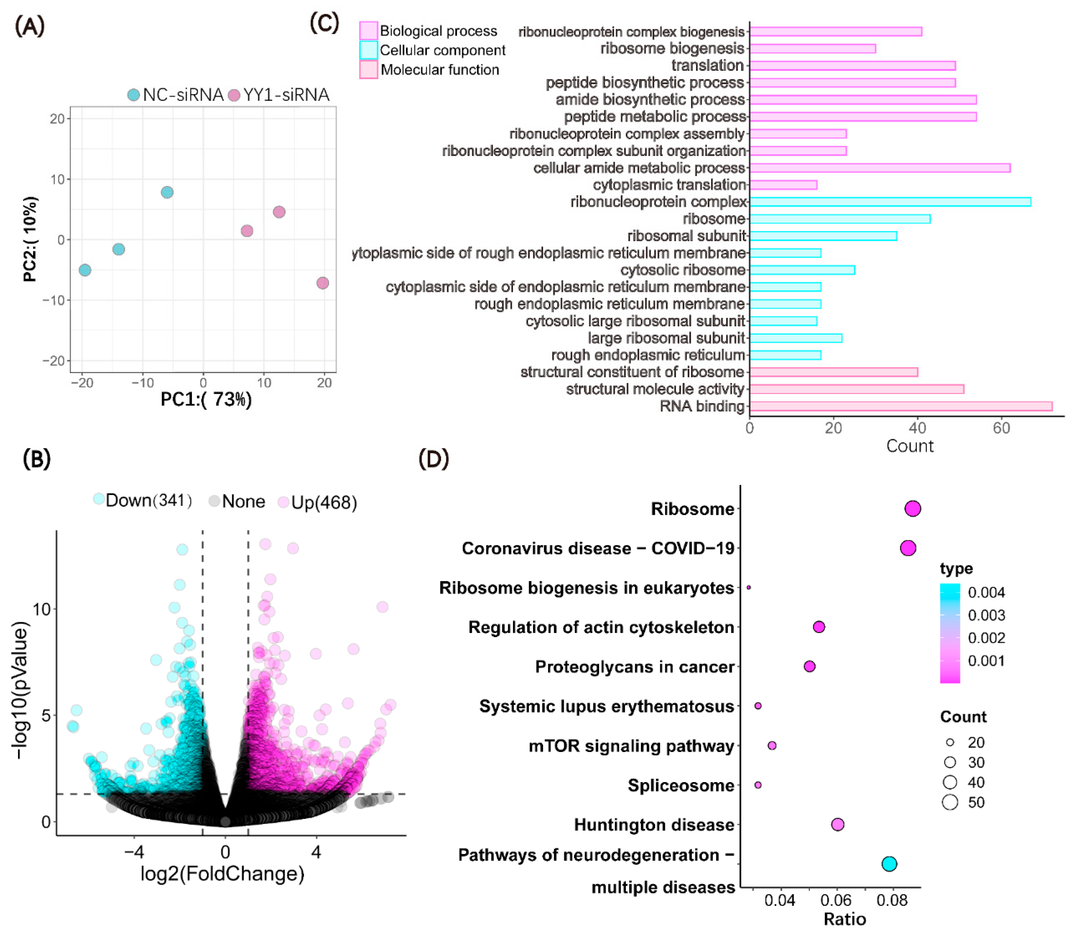
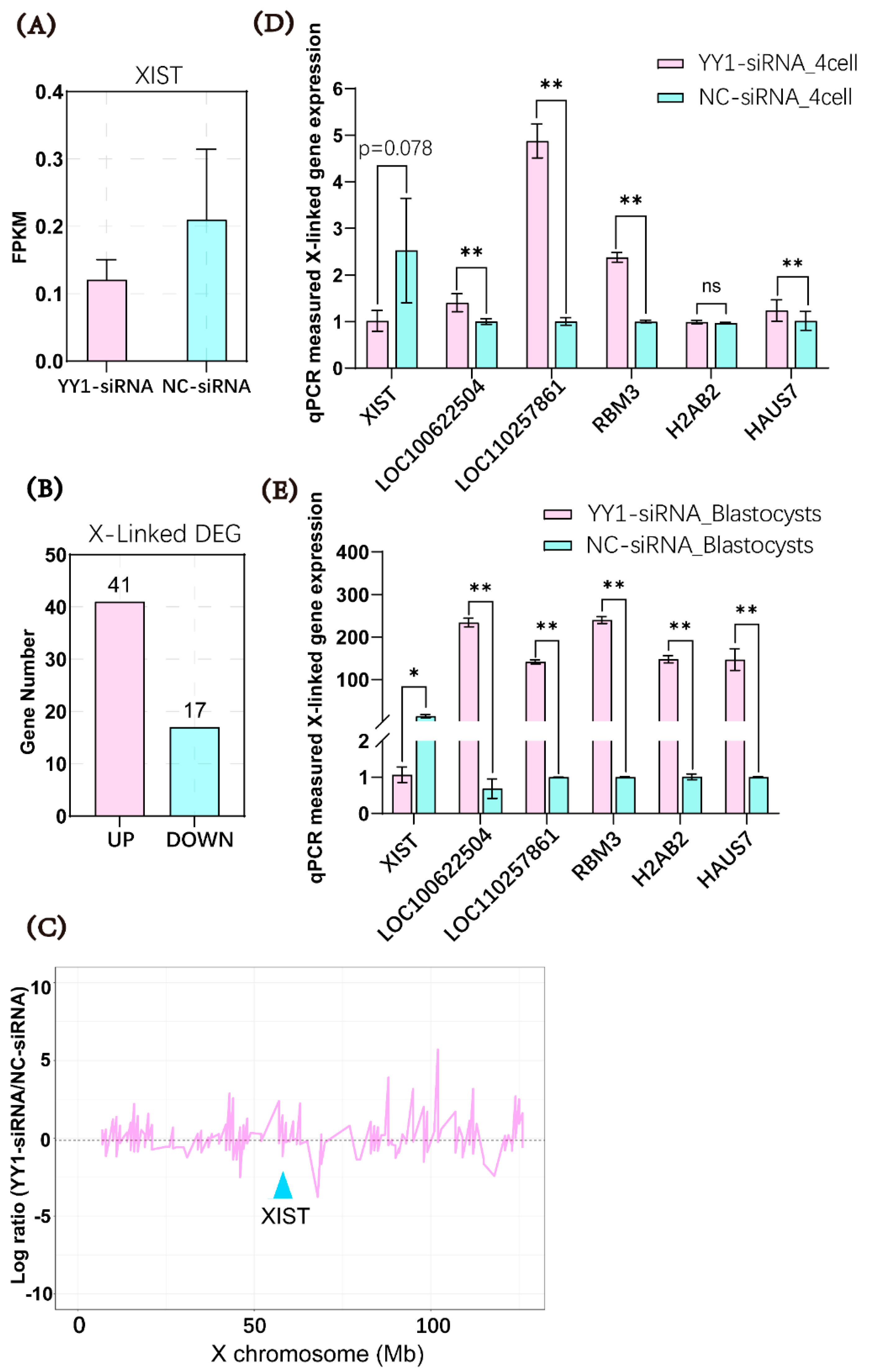
2.4. Microinjection of YY1 siRNA Altered the Transcriptome of Cloned Pig Embryos

To investigate the effects of the injection of YY1 siRNA on the gene expression patterns of pig cloned embryos, we collected 4-cell-stage control embryos and YY1-knockdown embryos for transcriptome sequencing. According to the PCA results, the samples of the YY1-siRNA-injection group were separated from those of the NC-siRNA-injection group (Figure 4A), suggesting the different gene expression patterns of the two groups of embryos. A total of 809 differentially expressed genes (DEGs) were detected in the two groups of SCNT embryos; of these, 341 were down-regulated and 468 were upregulated in the YY1-siRNA-injected group relative to the control group (Figure 4B).
The DEGs identified by RNA sequencing were subjected to GO and KEGG analysis. The analysis of the biological process (BP), cellular component (CC), and molecular function (MF) based on the GO database showed that the DEGs were primarily enriched in ribonucleoprotein complex biogenesis, ribosome biogenesis, translation, ribonucleoprotein complex, ribosome, ribosomal subunits, structural constituent of ribosome, structural molecular activity, and RNA binding, etc. (Figure 4C). The KEGG analysis revealed that the DEGs between two groups of embryos were mainly enriched in the ribosome, Coronavirus disease, Huntington’s disease, etc. (Figure 4D).

2.5. Microinjection of YY1 siRNA Inhibited XIST Expression and Upregulated the mRNA Levels of X-Linked Genes in Cloned Pig Embryos

The transcriptomic data analysis of the two groups of embryos showed that YY1 siRNA tended to inhibit the expression of XIST in the cloned embryos at the 4-cell stage (Figure 5A). The number of upregulated X-linked DEGs (41) in the YY1-siRNA-injection group was much higher than the number of downregulated X-linked DEGs (17) (Figure 5B). Furthermore, the analysis of the relative expression level of all the RNA-seq-detected X-linked genes of YY1-siRNA group indicated that expression of most of the detected X-linked genes were upregulated in the YY1-siRNA-injected embryos compared with control embryos (Figure 5C). These findings suggested that the RNAi of YY1 favored the activation of the X chromosome in the cloned pig embryos. The influence of the knockdown of YY1 on XIST and X-linked gene expression was further investigated by performing qPCR to examine the mRNA level of the XIST gene and five randomly selected X-linked genes in the two groups of cloned embryos at the 4-cell and blastocyst stages. The mRNA expression of XIST was almost significantly downregulated (p = 0.078), and the transcript levels of the four tested X-linked genes were significantly increased in the 4-cell stage of embryos with YY1 knockdown compared with the control embryos (Figure 5D). At the blastocyst stage, the mRNA abundance of XIST decreased, and that of the five randomly selected X-linked genes was significantly elevated in the YY1-siRNA-injected embryos compared with the control embryos (Figure 5E).

3. Discussion

YY1 can activate XIST transcription and thereby induce XCI via binding to the 5′ regulatory region of the XIST gene [22,23]. YY1 can also facilitate XIST RNA localization to the X chromosome, thereby promoting XCI by binding to XIST RNA [23,24,25]. Therefore, YY1 possibly regulates XCI by either modulating XIST transcription or controlling the anchoring of XIST RNA on the X chromosome, or by both processes in different tissues at different developmental stages, or under different circumstances. In the cloned pig embryos injected with YY1 siRNA, the expression of X-linked genes was significantly upregulated at both the 4-cell stage and blastocyst stage, while the XIST mRNA level was only significantly decreased at the blastocyst stage but not at the 4-cell stage. This implies that in cloned pig embryos, YY1 may modulate X-linked gene expression at the 4-cell stage mainly by regulating the localization of XIST RNA, but the modulation at the blastocyst stage occurs mainly by regulating the transcription of the XIST gene.
The genes coding for many ribosomal proteins contain multiple YY1 binding sites in their promoter regions [26,27]. This suggests that YY1 can regulate the expression of ribosomal proteins and thereby modulate ribosome-related pathways. The GO and KEGG pathway analysis of the DEGs between the control and YY1-siRNA-injected embryos in this study showed that the DEGs were mainly enriched in ribosome-related pathways. This indicated that in addition to inhibiting XIST expression and promoting X-linked gene expression, the knockdown of YY1 expression in the cloned pig embryos also affected ribosome-related pathways. However, whether the changes in the ribosome-related pathways induced by RNAi of YY1 had a positive impact, a negative impact, or no impact on the development of the cloned pig embryos remains to be further examined.
In human and mouse cells, YY1 regulates XCI by binding to XIST DNA and RNA [22,23,24,25]. This study also demonstrated that YY1 might regulate XCI in pig cells and cloned embryos by binding to XIST DNA and RNA. The present study also indicated that the binding region of YY1 on the porcine XIST gene was similar to that on the human and mouse XIST gene, which is located at about +1500 bp downstream of the XIST transcription start site [22,23,25]. These data suggest that the regulatory mechanism of YY1 on XIST functions may be conserved in mammals.
We previously demonstrated that the siRNA-mediated knockdown of RLIM, another XIST transcription activator, also suppressed XIST expression and improved the developmental rate of cloned pig embryos [16]. However, RNA interference of RLIM exhibited only a minor effect on increasing X-linked gene expression in the cloned pig embryos [16]. This was different from the result from knocking down YY1 in the cloned pig embryos. The differences in the ability to upregulate the expression of X-linked genes between the two methods may be related to the fact that knockdown of YY1 can inhibit both XIST transcription and XIST RNA localization on the X chromosome, whereas the knockdown of RLIM can only block XIST expression. The direct knockdown of XIST by siRNA also improved cloned embryo development, and this approach resulted in a higher cloning efficiency than the RNA interference of YY1 or RLIM [18]. The siRNA-mediated knockdown of YY1 or RLIM only inhibited the production of newly transcribed YY1 and RLIM mRNA but had no effect on the YY1 or RLIM protein that was already synthesized and maintained in the cloned embryos, which could still promote XIST expression. Thus, for improving the cloning efficiency, the direct knockdown of XIST was better than the indirect suppression of XIST expression by RNA interference of YY1 or RLIM.

4. Materials and Methods

4.1. Ethics Statement

This study was conducted following the “Guiding Opinions on the Care of Laboratory Animals” promulgated by the Ministry of Science and Technology of China. The protocol for animal experiments was approved by the Institutional Animal Care and Utilization Committee of South China Agricultural University. All efforts were made to minimize the suffering of the animals tested.

4.2. Design, Synthesis, and Transfection of siRNA

One siRNA that targeted the mRNA of the porcine YY1 gene (XM_021099699) was designed.
The sequences of the anti-YY1 siRNA and NC siRNA are as follows:
  • YY1-siRNA (sense): CCGAGUACAUGACAGGAAATT
  • YY1-siRNA (antisense): UUUCCUGUCAUGUACUCGGTT
  • NC-siRNA (sense): UUCUCCGAACGUGUCACGUTT
  • NC-siRNA (antisense): ACGUGACACGUUCGGAGAATT
Anti-YY1 siRNA and NC siRNA were synthesized by GenePharma (Shanghai, China).
Porcine fetal fibroblasts (PFF) were cultured in Dulbecco’s modified Eagle’s medium (DMEM) complete medium containing 12% fetal bovine serum (Gibco) in a 10 cm diameter petri dish at 37 °C and 5% CO2. The cells were digested with trypsin after they were 90% confluent. siRNA transfection was performed using the transfection reagent Lipofectamine™ RNAiMAX (Invitrogen) according to the instructions. Forty-eight hours post-transfection, cells were collected for total RNA extraction and were stored at −80 °C for later use.

4.3. Real-Time Quantitative Polymerase Chain Reaction (qPCR) Analysis

Total RNA was extracted from PFF and microinjected embryos using the E.Z.N.A.® Total RNA Kit II and Qiagen AllPrep DNA/RNA Micro Kit (Qiagen; Gaithersburg, MD, USA), respectively, according to the manufacturer’s instructions. The resulting total RNA was reverse transcribed using the PrimeScript™ RT reagent Kit with gDNA Eraser. The synthesized complementary DNA (cDNA) was detected by qPCR using the PowerUp™ SYBR™ Green Master Mix. GAPDH was used as the internal reference gene. The primers and their sequences are mentioned in Table 4. PCR reactions were performed using a QuantStudio 7 Flex system (Thermo Fisher Scientific Waltham, MA, USA) according to the manufacturer’s recommended parameters. The relative expression of genes was calculated using the 2−∆∆Ct method.

4.4. Chromatin Immunoprecipitation-Sequencing (ChIP-Seq) and Data Analysis

ChIP was performed using the Bersin Bio Chromatin immunoprecipitation Kit according to the instructions. After the digestion of PFF, cells were cross-linked using 1% formaldehyde for 10 min at room temperature. Cross-linked cells were treated with glycine at room temperature for 5 min, centrifuged at 1000 rpm for 5 min at 4 °C, and rinsed twice with PBS. Chromatin fragments were generated by shearing cell lysates through sonication in 1% SDS lysis buffer. The sonicated sample was centrifuged at 20,000 g at 4 °C for 10 min, and the supernatant was transferred to a fresh centrifuge tube. Magnetic beads were prepared in advance by overnight incubation with YY1 antibody (Proteintech, Rosemont, IL, USA) and IgG antibody (Proteintech, Rosemont, IL, USA). The ultrasonically lysed samples were mixed with the antibody-incubated magnetic beads and incubated for 2–4 h. The DNA on the magnetic beads was eluted, and RNase A and Proteinase K were added sequentially to remove RNase and protease from the elute. This DNA was purified using phenol–chloroform extraction and sequenced by Novogene Corporation (Beijing, China) using the Illumina high-throughput sequencing platform NovaSeq 6000.
FastQC and Bowtie2 were used for the quality control and sequence alignment of the sequencing data [28]. Peaks calling was analyzed by the Model-based Analysis of ChIP-seq 2 (MACS2) software with parameters set to p < 0.005 [29]. Peaks were identified by MACS2, and their regions were annotated using the R package ChIPseeke [30]. The ChIP-seq IP data with the ChIP-Seq input data as a control were normalized using the bamCompare tool in the deepTools software based on the log2 fold difference in RPKM as a pattern, and a bigwig file was generated for visualization [31]. The multiBigwigSummary tool of deepTools was used to summarize the genome coverage of each sample bigwig file. The plotPCA tool of deepTools was used to perform the principal component analysis (PCA) of the data. The plotHeatmap and plotProfile tools were used to draw the upstream and downstream 2000 bp ChIP-seq data distribution heatmap and profile plot of signal enrichment (with the transcriptional start site as the center). The HOMER software was used to detect significantly enriched known motifs in identified peaks [32].

4.5. Analysis of the Binding Site of YY1 on the XIST Gene

The XIST gene sequences of each species were downloaded from the National Center for Biotechnology Information website. The binding sites of YY1 on the XIST gene of each species were searched on the MEME Suite website (https://meme-suite.org/meme/ (accessed on 22 November 2021)).

4.6. Production of Cloned Embryos

Fresh gilt ovaries were collected from local slaughterhouses, placed in normal saline (containing 1% penicillin and streptomycin sulfate), preheated to 37 °C, and transported to the laboratory within 2–3 h. The follicular fluid containing cumulus–oocyte complexes (COCs) was aspirated from antral follicles using a 10 mL syringe equipped with a needle. COCs with homogeneous cytoplasm and at least three layers of cumulus cells were selected, transferred to the oocyte maturation medium, and then cultured at 38.5 °C in a cell culture incubator with 5% CO2 for 42–44 h. After in vitro maturation, COCs were transferred into 200 µL of DPBS-PVA containing 1.0 mg/mL hyaluronidase to remove the surrounding cumulus cells by gentle pipetting. For the subsequent SCNT, mature oocytes with intact cell membranes, clear perivitelline spaces, and clearly visible extruded polar bodies were selected. The selected oocytes were fixed using a fixation needle and enucleated by blind suction. A single donor cell (Duroc boar) was injected into the perivitelline space of the enucleated oocytes. The reconstituted embryos were cultured in porcine zygote medium 3 (PZM-3) for 1 h and then transferred to an electrofusion solution with a direct current pulse of 100 v/mm, 100 μs, and 2DC to induce fusion. The reconstituted embryos were transferred to fresh PZM-3 and further cultured. After activation, the cleavage embryos and blastocysts were counted at 48 h and 144 h, respectively.

4.7. Microinjection of siRNA into Cloned Embryos

Different concentrations of siRNA (10 pL) were microinjected into the activated cloned embryos using a micropipette driven by a Piezo (PiezoXpert). Following microinjection, the embryos were cultured in PZM-3 medium at 38.5 °C and 5% CO2 in a humidified atmosphere.

4.8. Immunofluorescence Assay

Embryos were collected, rinsed gently thrice in DPBS-PVB, and fixed with 4% paraformaldehyde (Biosharp Biotechnology Company) for 15 min at room temperature. The fixed embryos were then permeabilized with 0.2% Triton X-100 for 15 min and treated with a blocking solution for 1 h; both procedures were performed at room temperature. The embryos were then incubated overnight with YY1 antibody (1:500; Proteintech) at 4 °C. The next day, the embryos were transferred to a diluted fluorescent secondary antibody (1:500; Thermo Fisher Scientific) and incubated at 37 °C for 1 h. The genomic DNA in the embryos was stained with DAPI (1:1000; Thermo Fisher Scientific) for 5 min and observed by fluorescence microscopy.

4.9. Embryo Transfer

Embryos at the 2-cell stage were selected and loaded into transfer tubes 22–26 h after activation. The selected embryos were transported to a pig farm with a portable incubator (Minitube, Delavan, WI, USA). Within 40–46 h prior to embryo transfer, estrous-synchronized Yorkshire sows were used as recipients. Anesthesia was induced with ketamine (25 mg/kg body weight) and xylazine (1.1 mg/kg body weight), respectively, and maintained with 3% isoflurane. The ovaries and oviducts of sows were surgically exposed. A syringe was used to deliver the cloned embryos to the oviducts of sows. Ultrasound examination was performed to monitor the pregnancy status of the recipient sows at 1 mo after the embryo transfer. The recipients gave birth to cloned piglets through natural birth. If natural farrowing did not occur until gestation day 116, the recipient sows were injected with a prostaglandin analog (cloprostenol; 200 g/recipient) to induce farrowing, and the farrowing conditions of the sows were recorded.

4.10. RNA Sequencing and Bioinformatics Analysis

Cloned embryos were collected at the 4-cell stage, and five to six embryos were mixed as one sample for sequencing. The cDNA was generated and amplified for sequencing based on the smartseq2 method. Library quality was assessed using the Agilent Bioanalyzer 2100 system. The clustered library preparations were then sequenced on an Illumina Nova platform, and 150 bp paired-end reads were generated. Clean read data were mapped to the Sus scrofa 11.1 reference genome. Differentially expressed genes (DEGs) between the YY1-siRNA and NC-siRNA groups were analyzed using the R package DEseq2 [33]. Gene Ontology (GO) analysis and Kyoto Encyclopedia of Genes and Genomes (KEGG) analyses were performed with the R package clusterProfiler [34].

4.11. Statistical Analysis

Data were analyzed using SPSS software version 20. The differences in values from more than two groups were analyzed by one-way analysis of variance. The difference in values between two groups was analyzed by Student’s t-test.

5. Conclusions

The RNAi-mediated knockdown of YY1 expression suppressed XIST transcription, upregulated X-linked gene expression, and improved the developmental efficiency of cloned pig embryos. This study not only indicated the involvement of YY1 in the regulation of XIST expression and XCI in cloned pig embryos but also established a new approach to enhance the developmental competence of cloned pig embryos. These findings will help to promote the development and application of the pig cloning technique.

Author Contributions

Conceptualization, Y.D., Z.W. and Z.L.; methodology, X.W. and Y.D.; data curation, X.W. and X.P.; software, L.Y., B.T. and H.Z.; validation, E.Z. and G.C.; investigation, L.H.; writing—original draft preparation, Y.D.; writing—review and editing, Z.L.; project administration, Z.L., Z.W. and G.C.; funding acquisition, Z.W. and G.C. All authors have read and agreed to the published version of the manuscript.

Funding

This research was funded by two grants from the Department of Science and Technology of Guangdong Province, China (grant numbers: 2022B0202090003 and 2019BT02N630), and one grant from the Department of Science and Technology of Yunfu City, Guangdong Province, China (grant number: 2021020601).

Institutional Review Board Statement

The study was conducted according to the guidelines of the Declaration of Helsinki and approved by the Institutional Animal Care and Use Committee of South China Agricultural University (Approval number: 2020f005).

Informed Consent Statement

Not applicable.

Data Availability Statement

The data that support this study are available within the article and available from the authors upon request.

Conflicts of Interest

The authors declare no conflict of interest.

Abbreviation

bpBase pair
BPBiological process
CCCellular component
cDNAComplementary DNA
ChIP-seqChromatin immunoprecipitation sequence
COCsCumulus oocyte complexes
DEGsDifferentially expressed genes
DMEMDulbecco’s Modified Eagle Medium
DNADeoxyribonucleic acid
DPBSDulbecco’s Phosphate Buffered Saline
FBSFetal bovine serum
GAPDHGlyceraldehyde-3-phosphate dehydrogenase
GOGene Ontology
KEGGKyoto Encyclopedia of Genes and Genomes
mRNAMessenger RNA
MFMolecular function
PCAPrincipal component analysis
PEFPorcine embryonic fibroblast
PZM-3Porcine zygote medium 3
qPCRQuantitative Real-time PCR
RLIMRing finger protein, LIM domain interacting
RNARibonucleic acid
RPKMReads per kilobase per million mapped reads
SCNTSomatic cell nuclear transfer
sgRNASingle guide RNA
siRNASmall interfering RNA
TESTranscription termination site
TSSTranscriptional start site
XCIX-chromosome inactivation X
XISTX-inactive specific transcript
YY1Yin yang 1

References

  1. Lee, E.; Estrada, J.; Piedrahita, J.A. A comparative study on the efficiency of two enucleation methods in pig somatic cell nuclear transfer: Effects of the squeezing and the aspiration methods. Anim. Biotechnol. 2008, 19, 71–79. [Google Scholar] [CrossRef] [PubMed] [Green Version]
  2. Lagutina, I.; Fulka, H.; Lazzari, G.; Galli, C. Interspecies somatic cell nuclear transfer: Advancements and problems. Cell. Reprogram. 2013, 15, 374–384. [Google Scholar] [CrossRef] [PubMed] [Green Version]
  3. Wani, N.A.; Vettical, B.S.; Hong, S.B. First cloned Bactrian camel (Camelus bactrianus) calf produced by interspecies somatic cell nuclear transfer: A step towards preserving the critically endangered wild Bactrian camels. PLoS ONE 2017, 12, e177800. [Google Scholar] [CrossRef] [PubMed] [Green Version]
  4. Jeon, Y.; Nam, Y.H.; Cheong, S.A.; Kwak, S.S.; Lee, E.; Hyun, S.H. Absence of nucleolus formation in raccoon dog-porcine interspecies somatic cell nuclear transfer embryos results in embryonic developmental failure. J. Reprod. Dev. 2016, 62, 345–350. [Google Scholar] [CrossRef] [Green Version]
  5. Tunstall, T.; Kock, R.; Vahala, J.; Diekhans, M.; Fiddes, I.; Armstrong, J.; Paten, B.; Ryder, O.A.; Steiner, C.C. Evaluating recovery potential of the northern white rhinoceros from cryopreserved somatic cells. Genome Res. 2018, 28, 780–788. [Google Scholar] [CrossRef] [Green Version]
  6. Loi, P.; Modlinski, J.A.; Ptak, G. Interspecies somatic cell nuclear transfer: A salvage tool seeking first aid. Theriogenology 2011, 76, 217–228. [Google Scholar] [CrossRef]
  7. Matoba, S.; Zhang, Y. Somatic Cell Nuclear Transfer Reprogramming: Mechanisms and Applications. Cell Stem Cell 2018, 23, 471–485. [Google Scholar] [CrossRef] [Green Version]
  8. Loi, P.; Iuso, D.; Czernik, M.; Ogura, A. A New, Dynamic Era for Somatic Cell Nuclear Transfer? Trends Biotechnol. 2016, 34, 791–797. [Google Scholar] [CrossRef]
  9. Loi, P.; Beaujean, N.; Khochbin, S.; Fulka, J.J.; Ptak, G. Asymmetric nuclear reprogramming in somatic cell nuclear transfer? Bioessays 2008, 30, 66–74. [Google Scholar] [CrossRef]
  10. Lomax, G.P.; DeWitt, N.D. Somatic cell nuclear transfer in Oregon: Expanding the pluripotent space and informing research ethics. Stem Cells Dev. 2013, 22 (Suppl. 1), 25–28. [Google Scholar] [CrossRef] [PubMed]
  11. Zeng, F.; Liao, S.; Kuang, Z.; Zhu, Q.; Wei, H.; Shi, J.; Zheng, E.; Xu, Z.; Huang, S.; Hong, L.; et al. Genetically Engineered Pigs as Efficient Salivary Gland Bioreactors for Production of Therapeutically Valuable Human Nerve Growth Factor. Cells 2022, 11, 2378. [Google Scholar] [CrossRef] [PubMed]
  12. Skrzyszowska, M.; Samiec, M. Generating Cloned Goats by Somatic Cell Nuclear Transfer-Molecular Determinants and Application to Transgenics and Biomedicine. Int. J. Mol. Sci. 2021, 22, 7490. [Google Scholar] [CrossRef] [PubMed]
  13. Hodges, C.A.; Stice, S.L. Generation of bovine transgenics using somatic cell nuclear transfer. Reprod. Biol. Endocrinol. 2003, 1, 81. [Google Scholar] [CrossRef] [PubMed] [Green Version]
  14. Ogura, A.; Inoue, K.; Wakayama, T. Recent advancements in cloning by somatic cell nuclear transfer. Philos. Trans. R. Soc. Lond B Biol. Sci. 2013, 368, 20110329. [Google Scholar] [CrossRef] [Green Version]
  15. Thuan, N.V.; Kishigami, S.; Wakayama, T. How to improve the success rate of mouse cloning technology. J. Reprod. Dev. 2010, 56, 20–30. [Google Scholar] [CrossRef] [PubMed] [Green Version]
  16. Yang, L.; Wu, X.; Zhang, N.; Shi, J.; Zhou, R.; Su, Q.; Zheng, E.; Huang, S.; Xu, Z.; Hong, L.; et al. Knockdown of RLIM inhibits XIST expression and improves developmental competence of cloned male pig embryos. Mol. Reprod. Dev. 2021, 88, 228–237. [Google Scholar] [CrossRef] [PubMed]
  17. Inoue, K.; Kohda, T.; Sugimoto, M.; Sado, T.; Ogonuki, N.; Matoba, S.; Shiura, H.; Ikeda, R.; Mochida, K.; Fujii, T.; et al. Impeding Xist expression from the active X chromosome improves mouse somatic cell nuclear transfer. Science 2010, 330, 496–499. [Google Scholar] [CrossRef] [Green Version]
  18. Matoba, S.; Inoue, K.; Kohda, T.; Sugimoto, M.; Mizutani, E.; Ogonuki, N.; Nakamura, T.; Abe, K.; Nakano, T.; Ishino, F.; et al. RNAi-mediated knockdown of Xist can rescue the impaired postimplantation development of cloned mouse embryos. Proc. Natl. Acad. Sci. USA 2011, 108, 20621–20626. [Google Scholar] [CrossRef] [PubMed] [Green Version]
  19. Zeng, F.; Huang, Z.; Yuan, Y.; Shi, J.; Cai, G.; Liu, D.; Wu, Z.; Li, Z. Effects of RNAi-mediated knockdown of Xist on the developmental efficiency of cloned male porcine embryos. J. Reprod. Dev. 2016, 62, 591–597. [Google Scholar] [CrossRef] [Green Version]
  20. Yang, X.; Wu, X.; Yang, Y.; Gu, T.; Hong, L.; Zheng, E.; Xu, Z.; Zeng, F.; Shi, J.; Zhou, R.; et al. Improvement of developmental competence of cloned male pig embryos by short hairpin ribonucleic acid (shRNA) vector-based but not small interfering RNA (siRNA)-mediated RNA interference (RNAi) of Xist expression. J. Reprod. Dev. 2019, 65, 533–539. [Google Scholar] [CrossRef]
  21. Ruan, D.; Peng, J.; Wang, X.; Ouyang, Z.; Zou, Q.; Yang, Y.; Chen, F.; Ge, W.; Wu, H.; Liu, Z.; et al. XIST Derepression in Active X Chromosome Hinders Pig Somatic Cell Nuclear Transfer. Stem Cell Rep. 2018, 10, 494–508. [Google Scholar] [CrossRef] [PubMed] [Green Version]
  22. Makhlouf, M.; Ouimette, J.F.; Oldfield, A.; Navarro, P.; Neuillet, D.; Rougeulle, C. A prominent and conserved role for YY1 in Xist transcriptional activation. Nat. Commun. 2014, 5, 4878. [Google Scholar] [CrossRef] [Green Version]
  23. Chapman, A.G.; Cotton, A.M.; Kelsey, A.D.; Brown, C.J. Differentially methylated CpG island within human XIST mediates alternative P2 transcription and YY1 binding. BMC Genet. 2014, 15, 89. [Google Scholar] [CrossRef] [Green Version]
  24. Syrett, C.M.; Sindhava, V.; Hodawadekar, S.; Myles, A.; Liang, G.; Zhang, Y.; Nandi, S.; Cancro, M.; Atchison, M.; Anguera, M.C. Loss of Xist RNA from the inactive X during B cell development is restored in a dynamic YY1-dependent two-step process in activated B cells. PLoS Genet. 2017, 13, e1007050. [Google Scholar] [CrossRef] [PubMed] [Green Version]
  25. Jeon, Y.; Lee, J.T. YY1 tethers Xist RNA to the inactive X nucleation center. Cell 2011, 146, 119–133. [Google Scholar] [CrossRef] [Green Version]
  26. Perry, R.P. The architecture of mammalian ribosomal protein promoters. BMC Evol. Biol. 2005, 5, 15. [Google Scholar] [CrossRef] [PubMed] [Green Version]
  27. Chen, L.; Shioda, T.; Coser, K.R.; Lynch, M.C.; Yang, C.; Schmidt, E.V. Genome-wide analysis of YY2 versus YY1 target genes. Nucleic Acids Res. 2010, 38, 4011–4026. [Google Scholar] [CrossRef] [Green Version]
  28. Langmead, B.; Salzberg, S.L. Fast gapped-read alignment with Bowtie 2. Nat. Methods 2012, 9, 357–359. [Google Scholar] [CrossRef] [Green Version]
  29. Zhang, Y.; Liu, T.; Meyer, C.A.; Eeckhoute, J.; Johnson, D.S.; Bernstein, B.E.; Nusbaum, C.; Myers, R.M.; Brown, M.; Li, W.; et al. Model-based analysis of ChIP-Seq (MACS). Genome Biol. 2008, 9, R137. [Google Scholar] [CrossRef] [Green Version]
  30. Yu, G.; Wang, L.G.; He, Q.Y. ChIPseeker: An R/Bioconductor package for ChIP peak annotation, comparison and visualization. Bioinformatics 2015, 31, 2382–2383. [Google Scholar] [CrossRef]
  31. Ramirez, F.; Ryan, D.P.; Gruning, B.; Bhardwaj, V.; Kilpert, F.; Richter, A.S.; Heyne, S.; Dundar, F.; Manke, T. deepTools2: A next generation web server for deep-sequencing data analysis. Nucleic Acids Res. 2016, 44, W160–W165. [Google Scholar] [CrossRef] [PubMed]
  32. Heinz, S.; Benner, C.; Spann, N.; Bertolino, E.; Lin, Y.C.; Laslo, P.; Cheng, J.X.; Murre, C.; Singh, H.; Glass, C.K. Simple combinations of lineage-determining transcription factors prime cis-regulatory elements required for macrophage and B cell identities. Mol. Cell 2010, 38, 576–589. [Google Scholar] [CrossRef] [PubMed] [Green Version]
  33. Love, M.I.; Huber, W.; Anders, S. Moderated estimation of fold change and dispersion for RNA-seq data with DESeq2. Genome Biol. 2014, 15, 550. [Google Scholar] [CrossRef] [PubMed] [Green Version]
  34. Wu, T.; Hu, E.; Xu, S.; Chen, M.; Guo, P.; Dai, Z.; Feng, T.; Zhou, L.; Tang, W.; Zhan, L.; et al. clusterProfiler 4.0: A universal enrichment tool for interpreting omics data. Innovation 2021, 2, 100141. [Google Scholar] [CrossRef]
Figure 1. YY1 siRNA-mediated downregulation of YY1 and XIST mRNA expression in female pig fibroblasts. (A) Knockdown of YY1 expression by YY1 siRNA. (B) Inhibition of XIST expression via YY1 expression knockdown. * and ** represent a significant difference at p < 0.05 and p < 0.01, respectively, where n = 3 in each group.
Ijms 23 14572 g001
Figure 2. Analysis of the binding sites of YY1 on XIST Gene by Chip-seq. (A) Principal component analysis (PCA) plot. MF: male fibroblasts; FF: female fibroblasts; siFF: female fibroblasts transfected with YY1-siRNA. (B) Enrichment of YY1 binding sites within 2 kb upstream and 2 kb downstream of the transcription start site (TSS) and transcription termination site (TES) in the genome. The y-axis denotes the average log2 fold change relative to the control (C) Chip-seq revealed YY1 binding sites on the pig XIST gene. The white arrow in the blue box indicates the direction of transcription of the XIST gene. (D) Porcine YY1 DNA binding consensus motif. (E) Predicted YY1 binding sites on the XIST gene of different species.
Ijms 23 14572 g002
Figure 3. The effects of YY1-siRNA microinjection on YY1 protein expression and 4-cell-stage embryo rate of cloned pig embryos. (A) Immunofluorescence comparison of YY1 protein abundance between YY1-siRNA and NC-siRNA groups. Scale bar = 50 μm (B) YY1 protein quantification in YY1-siRNA and NC-siRNA groups (n = 10 in each group). (C) Comparison of 4-cell-stage embryo rate between YY1-siRNA and NC-siRNA groups (n = 4 in each group). * and ** represent a significant difference at p < 0.05 and p < 0.01, respectively.
Ijms 23 14572 g003
Figure 4. Transcriptome analysis of the 4-cell-stage control and YY1-knockdown SCNT embryos. (A) Principal component analysis (PCA) of two groups of cloned embryos. (B) Volcano plot of differentially expressed genes (DEGs) between two groups of cloned embryos. (C) The significantly enriched Gene Ontology (GO) terms of DEGs. The top 10 GO terms were shown in the biological process (BP) and cellular component (CC) categories. Only three GO terms were enriched in the category of molecular function (MF). (D) The Kyoto Encyclopedia of Genes and Genomes (KEGG) enrichment analysis of DEGs. The top 10 KEGG pathways are shown.
Ijms 23 14572 g004
Figure 5. Effects of YY1 siRNA on the expression of XIST and X-linked genes in cloned embryos. (A) Level of XIST in YY1-siRNA group and NC-siRNA group measured by RNA-seq. (B) The number of X-linked differentially expressed genes in the YY1-siRNA group (vs. NC-siRNA group) detected by RNA-seq (n = 3). (C) Relative expression level of all the RNA-seq-detected X-linked genes of YY1-siRNA group (relative to the NC-siRNA group) plotted on the positions of the X chromosome. (D) Quantitative polymerase chain reaction (qPCR) used to measure the expression of XIST mRNA and X-linked genes in 4-cell-stage SCNT embryos. (E) qPCR used to measure mRNA expression of XIST and X-inked genes in blastocyst-stage SCNT embryos, * and ** represent a significant difference at p < 0.05 and p < 0.01, respectively. n = 3 in each group in (AE).
Ijms 23 14572 g005
Table 1. Effects of the injection of YY1 siRNA on the in vitro developmental efficiency of porcine SCNT embryos.
GroupsNo. of RepetitionNo. of Cultured SCNT EmbryosNo. of Cleaved Embryos (%, Mean)No. of Blastocysts
(%, Mean)
YY1-siRNA
(10 μM)
28367 (80.75 ± 3.08) ab24 (28.92 ± 0.49) ab
YY1-siRNA
(50 μM)
26556 (86.08 ± 6.83) b21 (32.24 ± 5.83) b
NC-siRNA
(50 μM)
27549 (65.33 ± 0.65) a17 (22.65 ± 1.46) a
Values in the same column labeled with different superscripts differ at p < 0.05.
Table 2. Effects of injection of 50 µM YY1 siRNA on the in vitro developmental efficiency of porcine SCNT embryos.
GroupsNo. of RepetitionNo. of Cultured SCNT EmbryosNo. of Cleaved Embryos (%, Mean)No. of Blastocysts
(%, Mean)
YY1-siRNA
(50 μM)
8305257 (84.29 ± 5.05) b81 (26.85 ± 6.76) b
NC-siRNA
(50 μM)
8318229 (71.74 ± 5.21) a60 (18.58 ± 6.54) a
Values in the same column labeled with different superscripts differ at p < 0.05.
Table 3. Effects of injection of YY1 siRNA on the in vivo developmental rate of porcine sSCNT embryos.
GroupsNo. of Total/Farrowed RecipientsNo. of Transferred Cloned EmbryosNo. of Total Born Cloned PigletsNo. of Live-Born Cloned Piglets
YY1-siRNA
(50 μM)
6/2127183
NC-siRNA
(50 μM)
5/097100
Table 4. Primers used for the quantitative polymerase chain reaction.
GeneGenBank Accession No.Primer Sequence
YY1XM_021099699(forward): ATACCCGGCATCGACCTCTC
(reverse): AACATCTTTGTGCAGCCTTTGT
XISTKC753465(forward): CTTGCCGCAATCGAAAACAT
(reverse): ACCAATTCCACCACCCTTTC
LOC100622504XM_003360396(forward): GTGAGACAACGTGCTCTGAGA
(reverse): ATTCCATGGGTTCCTGAGCTG
LOC110257861XR_002340890(forward): GCACAGTACCTTCTCCCCTTC
(reverse): CCCTGTTGCTTTGGTTTCGAG
RBM3NM_001243419(forward): GCTTCAAATGCAATGAGAGCCA
(reverse): TCTGCCATTTCCATAGCCCTG
H2AB2XM_021080443(forward): AAAAGGAGCCGTCGAAAGTCA
(reverse): GTCAGGTACTCGATGGTGGC
HAUS7XM_021079955(forward): CTCTGACTTCCACCAGCTCATC
(reverse): CTCTTCCATTTTGCTGGTTGGC
GAPDHNM_001206359(forward): TGACCCCTTCATTGACCTCC
(reverse): CTCCGCCTTGACTGTGCC
Publisher’s Note: MDPI stays neutral with regard to jurisdictional claims in published maps and institutional affiliations.

