

Newly Designed Primers for the Sequencing of the inlA Gene of Lineage I and II Listeria monocytogenes Isolates


Abstract
:1. Introduction
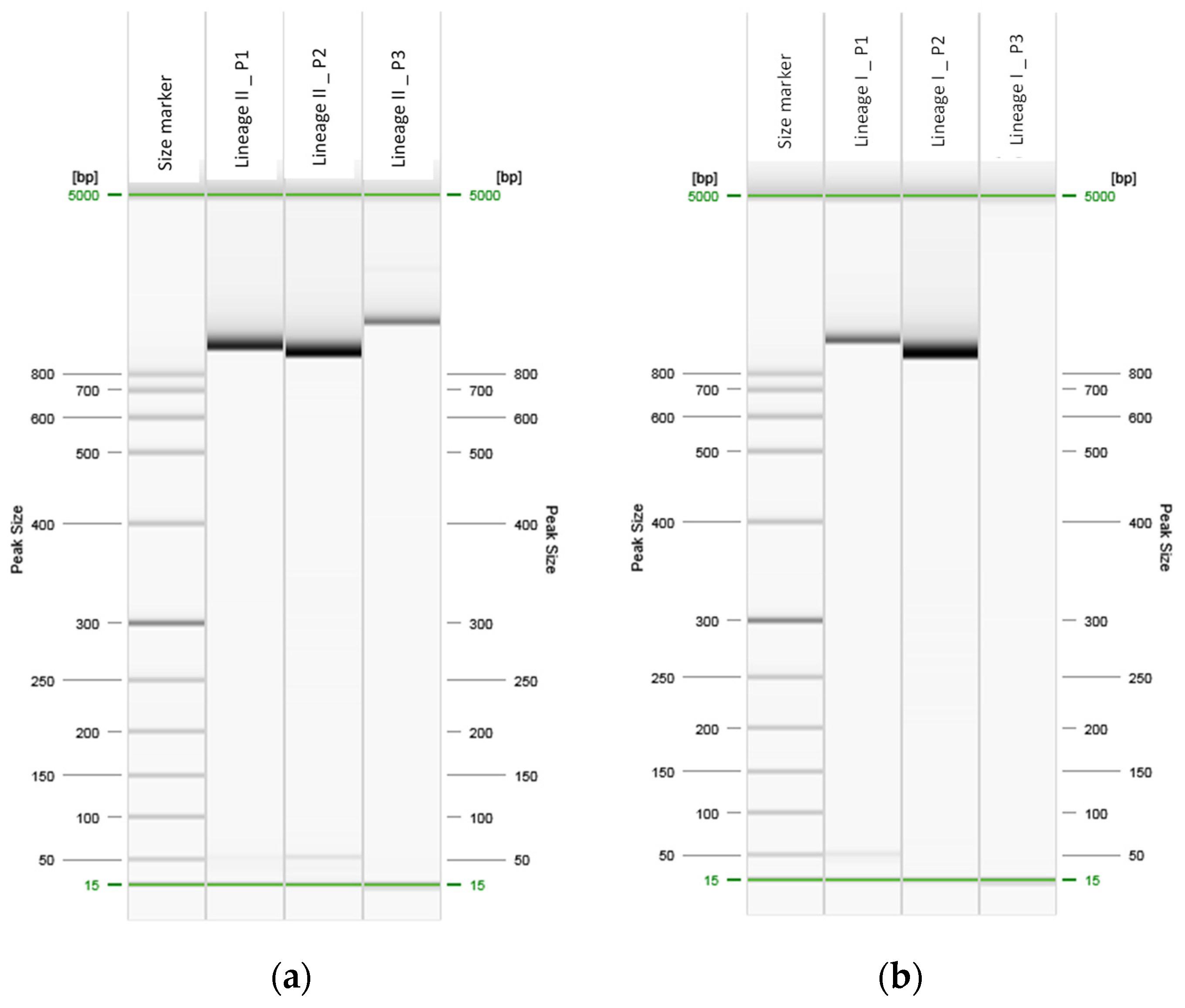
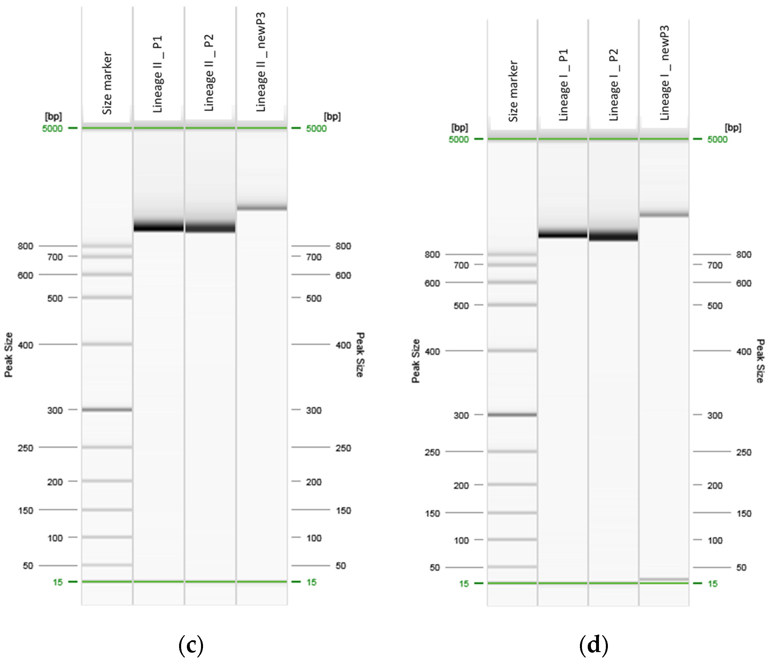
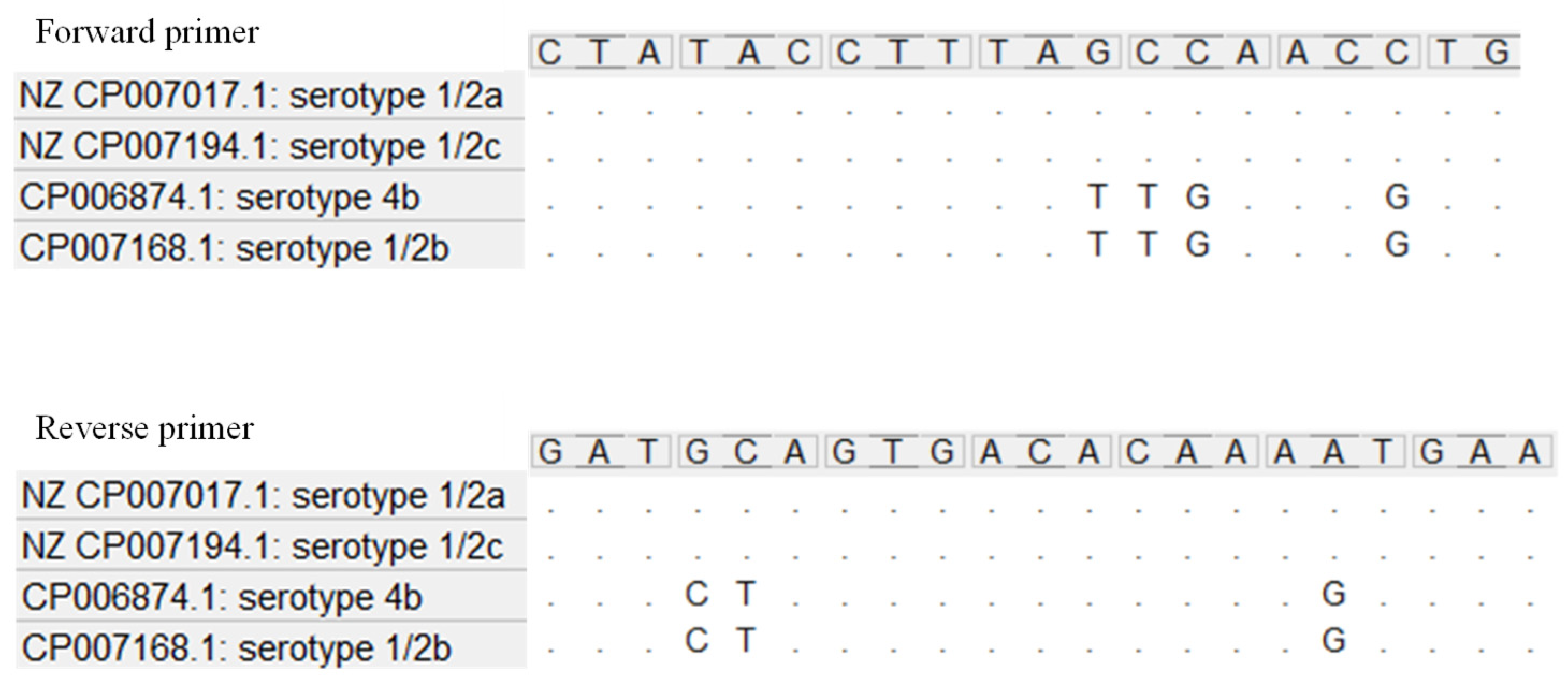
2. Results
3. Discussion
4. Materials and Methods
4.1. Isolates Collection
4.2. Primers
4.3. PCR Assay
5. Conclusions
Supplementary Materials
Author Contributions
Funding
Institutional Review Board Statement
Informed Consent Statement
Data Availability Statement
Conflicts of Interest
References
- Dreyer, M.; Aguilar-Bultet, L.; Rupp, S.; Guldimann, C.; Stephan, R.; Schock, A.; Otter, A.; Schüpbach, G.; Brisse, S.; Lecuit, M.; et al. Listeria Monocytogenes Sequence Type 1 Is Predominant in Ruminant Rhombencephalitis. Sci. Rep. 2016, 6, 36419. [Google Scholar] [CrossRef] [PubMed] [Green Version]
- Lecuit, M. Listeria monocytogenesa, Model in Infection Biology. Cell. Microbiol. 2020, 22, e13186. [Google Scholar] [CrossRef] [PubMed] [Green Version]
- Schlech III, W.F.; Acheson, D. Foodborne listeriosis. Clin. Infect. Dis. 2000, 31, 770–775. [Google Scholar] [CrossRef] [PubMed]
- Senay, T.E.; Ferrell, J.L.; Garrett, F.G.; Albrecht, T.M.; Cho, J.; Alexander, K.L.; Myers-Morales, T.; Grothaus, O.F.; D’Orazio, S.E.F. Neurotropic Lineage III Strains of Listeria Monocytogenes Disseminate to the Brain without Reaching High Titer in the Blood. mSphere 2020, 5, e00871-20. [Google Scholar] [CrossRef] [PubMed]
- Song, Y.; Peters, T.L.; Bryan, D.W.; Hudson, L.K.; Denes, T.G. Characterization of a Novel Group of Listeria Phages That Target Serotype 4b Listeria Monocytogenes. Viruses 2021, 13, 671. [Google Scholar] [CrossRef] [PubMed]
- Lee, B.-H.; Garmyn, D.; Gal, L.; Guérin, C.; Guillier, L.; Rico, A.; Rotter, B.; Nicolas, P.; Piveteau, P. Exploring Listeria Monocytogenes Transcriptomes in Correlation with Divergence of Lineages and Virulence as Measured in Galleria Mellonella. Appl. Environ. Microbiol. 2019, 85, e01370-19. [Google Scholar] [CrossRef] [Green Version]
- Phelps, C.C.; Vadia, S.; Arnett, E.; Tan, Y.; Zhang, X.; Pathak-Sharma, S.; Gavrilin, M.A.; Seveau, S. Relative Roles of Listeriolysin O, InlA, and InlB in Listeria Monocytogenes Uptake by Host Cells. Infect. Immun. 2018, 86, e00555-18. [Google Scholar] [CrossRef] [Green Version]
- Schiavano, G.F.; Ateba, C.N.; Petruzzelli, A.; Mele, V.; Amagliani, G.; Guidi, F.; De Santi, M.; Pomilio, F.; Blasi, G.; Gattuso, A.; et al. Whole-Genome Sequencing Characterization of Virulence Profiles of Listeria Monocytogenes Food and Human Isolates and In Vitro Adhesion/Invasion Assessment. Microorganisms 2021, 10, 62. [Google Scholar] [CrossRef]
- Su, X.; Cao, G.; Zhang, J.; Pan, H.; Zhang, D.; Kuang, D.; Yang, X.; Xu, X.; Shi, X.; Meng, J. Characterization of Internalin Genes in Listeria Monocytogenes from Food and Humans, and Their Association with the Invasion of Caco-2 Cells. Gut Pathog. 2019, 11, 30. [Google Scholar] [CrossRef]
- Da Silva, M.F.; Ferreira, V.; Magalhães, R.; Almeida, G.; Alves, A.; Teixeira, P. Detection of Premature Stop Codons Leading to Truncated Internalin A among Food and Clinical Strains of Listeria Monocytogenes. Food Microbiol. 2017, 63, 6–11. [Google Scholar] [CrossRef]
- Nightingale, K.K.; Ivy, R.A.; Ho, A.J.; Fortes, E.D.; Njaa, B.L.; Peters, R.M.; Wiedmann, M. InlA Premature Stop Codons Are Common among Listeria Monocytogenes Isolates from Foods and Yield Virulence-Attenuated Strains That Confer Protection against Fully Virulent Strains. Appl. Environ. Microbiol. 2008, 74, 6570–6583. [Google Scholar] [CrossRef] [Green Version]
- Medeiros, M.; Castro, V.H.L.D.; Mota, A.L.A.D.A.; Pereira, M.G.; De Martinis, E.C.P.; Perecmanis, S.; Santana, A.P. Assessment of Internalin A Gene Sequences and Cell Adhesion and Invasion Capacity of Listeria Monocytogenes Strains Isolated from Foods of Animal and Related Origins. Foodborne Pathog. Dis. 2021, 18, 243–252. [Google Scholar] [CrossRef]
- Fravalo, P.; Cherifi, T.; Neira Feliciano, K.D.; Letellier, A.; Fairbrother, J.; Bekal, S. Characterisation of InlA Truncation in Listeria Monocytogenes Isolates from Farm Animals and Human Cases in the Province of Quebec. Vet. Rec. Open 2017, 4, e000199. [Google Scholar] [CrossRef] [Green Version]
- Wagner, E.; Fagerlund, A.; Thalguter, S.; Jensen, M.R.; Heir, E.; Møretrø, T.; Moen, B.; Langsrud, S.; Rychli, K. Deciphering the Virulence Potential of Listeria Monocytogenes in the Norwegian Meat and Salmon Processing Industry by Combining Whole Genome Sequencing and in Vitro Data. Int. J. Food Microbiol. 2022, 383, 109962. [Google Scholar] [CrossRef]
- Fagerlund, A.; Wagner, E.; Møretrø, T.; Heir, E.; Moen, B.; Rychli, K.; Langsrud, S. Pervasive Listeria Monocytogenes Is Common in the Norwegian Food System and Is Associated with Increased Prevalence of Stress Survival and Resistance Determinants. Appl. Environ. Microbiol. 2022, 88, e00861-22. [Google Scholar] [CrossRef]
- Hingston, P.; Chen, J.; Dhillon, B.K.; Laing, C.; Bertelli, C.; Gannon, V.; Tasara, T.; Allen, K.; Brinkman, F.S.L.; Truelstrup Hansen, L.; et al. Genotypes Associated with Listeria Monocytogenes Isolates Displaying Impaired or Enhanced Tolerances to Cold, Salt, Acid, or Desiccation Stress. Front. Microbiol. 2017, 8, 369. [Google Scholar] [CrossRef] [Green Version]
- Orsi, R.H.; Ripoll, D.R.; Yeung, M.; Nightingale, K.K.; Wiedmann, M. Recombination and Positive Selection Contribute to Evolution of Listeria Monocytogenes InlA. Microbiology 2007, 153, 2666–2678. [Google Scholar] [CrossRef] [Green Version]
- Manuel, C.S.; Van Stelten, A.; Wiedmann, M.; Nightingale, K.K.; Orsi, R.H. Prevalence and Distribution of Listeria Monocytogenes InlA Alleles Prone to Phase Variation and InlA Alleles with Premature Stop Codon Mutations among Human, Food, Animal, and Environmental Isolates. Appl. Environ. Microbiol. 2015, 81, 8339–8345. [Google Scholar] [CrossRef] [Green Version]
- Gelbíčová, T.; Koláčková, I.; Pantu, R. A Novel Mutation Leading to a Premature Stop Codon in InlA of Listeria Monocytogenes Isolated from Neonatal Listeriosis. New Microbiol. 2015, 38, 293–296. [Google Scholar]
- Toledo, V.; den Bakker, H.C.; Hormazábal, J.C.; González-Rocha, G.; Bello-Toledo, H.; Toro, M.; Moreno-Switt, A.I. Genomic Diversity of Listeria Monocytogenes Isolated from Clinical and Non-Clinical Samples in Chile. Genes 2018, 9, 396. [Google Scholar] [CrossRef] [Green Version]
- Gorski, L.; Parker, C.T.; Liang, A.S.; Walker, S.; Romanolo, K.F. The Majority of Genotypes of the Virulence Gene InlA Are Intact among Natural Watershed Isolates of Listeria Monocytogenes from the Central California Coast. PLoS ONE 2016, 11, e0167566. [Google Scholar] [CrossRef] [PubMed]
- Pirone-Davies, C.; Chen, Y.; Pightling, A.; Ryan, G.; Wang, Y.; Yao, K.; Hoffmann, M.; Allard, M.W. Genes Significantly Associated with Lineage II Food Isolates of Listeria Monocytogenes. BMC Genom. 2018, 19, 708. [Google Scholar] [CrossRef] [PubMed] [Green Version]
- Zhang, L.; Wang, Y.; Liu, D.; Luo, L.; Wang, Y.; Ye, C. Identification and Characterization of Als Genes Involved in D-Allose Metabolism in Lineage II Strain of Listeria Monocytogenes. Front. Microbiol. 2018, 9, 621. [Google Scholar] [CrossRef] [PubMed]
- Jacquet, C.; Doumith, M.; Gordon, J.I.; Martin, P.M.V.; Cossart, P.; Lecuit, M. A Molecular Marker for Evaluating the Pathogenic Potential of Foodborne Listeria Monocytogenes. J. Infect. Dis. 2004, 189, 2094–2100. [Google Scholar] [CrossRef] [Green Version]
- Tamburro, M.; Ripabelli, G.; Fanelli, I.; Maria Grasso, G.; Lucia Sammarco, M. Typing of Listeria Monocytogenes Strains Isolated in Italy by Inl A Gene Characterization and Evaluation of a New Cost-Effective Approach to Antisera Selection for Serotyping. J. Appl. Microbiol. 2010, 108, 1602–1611. [Google Scholar] [CrossRef]
- Tamura, K.; Stecher, G.; Peterson, D.; Filipski, A.; Kumar, S. MEGA6: Molecular Evolutionary Genetics Analysis Version 6.0. Mol. Biol. Evol. 2013, 30, 2725–2729. [Google Scholar] [CrossRef]


| GenBank Accession Number | Isolation Year | Lineage | ST | Origin | Source | Clinical Syndrome |
|---|---|---|---|---|---|---|
| OP686908 | 2020 | I | 1 | Food | Meat | / |
| OP686909 | 2020 | I | 1 | Clinical | Blood | Sepsis |
| OP686910 | 2020 | I | 1 | Food | Cheese | / |
| OP686912 | 2020 | I | 1 | Clinical | Blood | Sepsis |
| OP686923 | 2021 | I | 1 | Food | Meat | / |
| OP686924 | 2021 | I | 1 | Clinical | Blood | Sepsis |
| OP686926 | 2021 | I | 1 | Clinical | Placenta | Maternal-neonatal |
| OP686918 | 2020 | I | 2 | Food | Other | / |
| OP686921 | 2021 | I | 2 | Food | Cheese | / |
| OP686928 | 2021 | I | 2 | Clinical | Cerebrospinal fluid | Meningitis |
| OP686913 | 2020 | I | 3 | Environmental | Meat | / |
| OP686920 | 2020 | I | 3 | Food | Fish | / |
| OP686914 | 2020 | I | 3 | Clinical | Blood | Sepsis |
| OP686911 | 2020 | I | 6 | Clinical | Blood | Sepsis |
| OP686929 | 2021 | I | 6 | Clinical | Blood | Sepsis |
| OP686917 | 2020 | II | 7 | Environmental | Meat | / |
| OP686906 | 2019 | II | 8 | Clinical | Blood | Sepsis |
| OP686907 | 2019 | II | 8 | Environmental | Grocery store | / |
| OP686919 | 2020 | II | 8 | Clinical | Blood | Sepsis |
| OP686925 | 2021 | II | 8 | Clinical | Blood | Sepsis |
| OP686944 | 2019 | II | 9 | Food | Meat | / |
| OP686936 | 2020 | II | 9 | Food | Other | / |
| OP686935 | 2020 | II | 9 | Environmental | Grocery store | / |
| OP686930 | 2021 | II | 26 | Clinical | Blood | Sepsis |
| OP686905 | 2020 | II | 37 | Food | Salami | / |
| OP686927 | 2021 | II | 37 | Clinical | Blood | Sepsis |
| OP686940 | 2020 | II | 121 | Environmental | Meat | / |
| OP686941 | 2020 | II | 121 | Food | Meat | / |
| OP686942 | 2020 | II | 121 | Food | Meat | / |
| OP686943 | 2020 | II | 121 | Food | Meat | / |
| OP686939 | 2020 | II | 121 | Food | Meat | / |
| OP686932 | 2020 | II | 155 | Environmental | Meat | / |
| OP686915 | 2020 | II | 204 | Environmental | Dairy | / |
| OP686922 | 2021 | I | 217 | Environmental | Meat | / |
| OP686916 | 2021 | I | 217 | Food | Milk | / |
| OP686933 | 2021 | I | 288 | Food | Cheese | / |
| OP686934 | 2021 | I | 288 | Environmental | Meat | / |
| OP686937 | 2013 | II | 325 | Environmental | Dairy | / |
| OP686938 | 2015 | II | 325 | Food | Cheese | / |
| OP686931 | 2020 | I | 330 | Food | Meat | / |
| Name | Forward 5′-3′ Sequence | Reverse 5′-3′ Sequence | Position (bp) |
|---|---|---|---|
| S_1 | GATATCACTAAACGGCTCC | TAGTTTTGTTAGACCCGACA | (−170)–872 |
| S_2 | TAAATCGGCTAGAACTATCCA | GTCAATAAATTCCCAGCTTC | 497–1540 |
| S_3 | CTATACCTTTAGCCAACCTG | TTCATTTTGTGTCACTGCATC | 1410–(+218) |
| Snew_3 | YTATACCTTTAVCCAAYCTG | TTCAYTTTGTGTCACTRSATC | 1410–(+218) |
Publisher’s Note: MDPI stays neutral with regard to jurisdictional claims in published maps and institutional affiliations. |
© 2022 by the authors. Licensee MDPI, Basel, Switzerland. This article is an open access article distributed under the terms and conditions of the Creative Commons Attribution (CC BY) license (https://creativecommons.org/licenses/by/4.0/).
Share and Cite
Magagna, G.; Finazzi, G.; Filipello, V. Newly Designed Primers for the Sequencing of the inlA Gene of Lineage I and II Listeria monocytogenes Isolates. Int. J. Mol. Sci. 2022, 23, 14106. https://doi.org/10.3390/ijms232214106
Magagna G, Finazzi G, Filipello V. Newly Designed Primers for the Sequencing of the inlA Gene of Lineage I and II Listeria monocytogenes Isolates. International Journal of Molecular Sciences. 2022; 23(22):14106. https://doi.org/10.3390/ijms232214106
Chicago/Turabian StyleMagagna, Giulia, Guido Finazzi, and Virginia Filipello. 2022. "Newly Designed Primers for the Sequencing of the inlA Gene of Lineage I and II Listeria monocytogenes Isolates" International Journal of Molecular Sciences 23, no. 22: 14106. https://doi.org/10.3390/ijms232214106