Article

Three Heat Shock Protein Genes and Antioxidant Enzymes Protect Pardosa pseudoannulata (Araneae: Lycosidae) from High Temperature Stress

Guizhou Provincial Key Laboratory for Agricultural Pest Management of Mountainous Regions, Institute of Entomology, Guizhou University, Guiyang 550025, China
*
Author to whom correspondence should be addressed.
Int. J. Mol. Sci. 2022, 23(21), 12821; https://doi.org/10.3390/ijms232112821
Submission received: 19 September 2022 / Revised: 19 October 2022 / Accepted: 20 October 2022 / Published: 24 October 2022
(This article belongs to the Collection Feature Papers in “Molecular Biology”)

Abstract

:
Pardosa pseudoannulata (P. pseudoannulata) is an essential natural predatory enemy in rice ecosystems. The fluctuating climate may cause them to experience heat stress, whereas heat shock proteins (HSPs) and antioxidant enzymes help resist heat damage. Herein, we cloned and characterized the full-length genes PpHSP27, PpHSP60, and PpHSC70 from P. pseudoannulata. Changes in gene expression levels and superoxide dismutase (SOD), catalase (CAT), and glutathione transferase (GST) activities in adult male and female P. pseudoannulata were measured at different stress exposure times and temperatures. We found that the abovementioned HSP genes belong to the sHSP, HSP60, and HSP70 families. The expression of the three HSP genes and the activities of SOD, CAT, and GST were significantly upregulated with the increasing stress temperature and time. The knockdown of the three HSP genes via RNA interference significantly decreased the survival rate of male and female P. pseudoannulata during high temperature stress. Thus, PpHSP27, PpHSP60, and PpHSC70 play an important role in the heat tolerance of P. pseudoannulata, and SOD, CAT, and GST enable recovery heat stress-induced oxidative damage. Their changes and regulation during high temperature stress can improve spiders’ adaptability in the field and enhance the biological control of environmental pests.

1. Introduction

Arthropods are relatively susceptible to high temperatures because they do not have mechanisms to regulate their body temperature. High temperatures can significantly affect their growth, development, reproduction, survival, and other processes [1,2]. The intensity, frequency, and duration of high temperatures are increasing because of global warming, and these trends are expected to deteriorate with increased global warming [3,4]. During their long evolution, arthropods have evolved various behavioral, morphological, physiological, and molecular strategies to avoid high-temperature damage. The production of heat shock proteins (HSPs) and the enhancement of antioxidant enzymes are considered important strategies for arthropods to cope with heat stress [5,6,7,8,9].
As with adverse environmental factors, such as hypoxia, heavy metal ion pollution, UV stress, and starvation, heat stress causes protein denaturation and loss of function in the organism [5,8,10]. As a chaperone protein, HSP cooperates with auxiliary molecular chaperones and auxiliary proteins to mediate activities such as protein folding, localization, and degradation, and prevents the protein denaturation caused by the abovementioned reasons [11,12,13]. Based on the relative molecular weight and amino acid (AA) sequence homology, HSPs may be divided into HSP110, HSP90, HSP70, HSP60, small heat shock proteins (sHSPs), and other families [13,14,15]. Of these, HSP70 is the most phylogenetically conserved and widely studied HSP in arthropods [9,16,17]. In arthropods, HSPs play a protective role in various tissues and organs. For example, HSPs can protect the normal neural circuit function of Drosophila under heat stress [18]. HSP genes in the midgut of Rhodnius prolixus and Aedes aegypti were significantly increased following the consumption of a hot blood meal, suggesting that HSP genes protect their midgut from the heat stress caused by hot blood [19,20]. Within the same species, there are also differences in the expression of HSP genes. In Nilaparvata lugens, there is a difference in heat tolerance between the two morphs: the macropters with migratory ability are more heat tolerant than the brachypters, and RNA interference (RNAi) experiments showed that this difference results from the overexpression of HSP genes [21]. Welte et al. [22] found that Drosophila melanogaster had the highest HSP70 genes expression in the first instar larvae, which were more thermotolerant than other instars, and the induction of HSP70 genes enhanced the heat tolerance of D. melanogaster in the first instar. The expression levels of the HSP70 and HSP23 genes were significantly higher in Bemisia tabaci females than in males. After knocking down these genes, heat tolerance was significantly decreased in females, whereas no change was observed in males. This means that the high expression of HSP genes in females results in a higher survival rate under conditions of heat stress [23]. These findings suggest that HSP genes play an important role in heat stress resistance in different tissues and at various developmental stages in arthropods.
Reactive oxygen species (ROS) are byproducts of metabolism in the mitochondria, endoplasmic reticulum, and lysosomes [24]. Low levels of ROS favor cell signaling and the induction of defense genes in insects, but if overproduced, it can result in oxidative damage to lipids, proteins, and nucleic acids within cells [9,25]. To cope with this damage, heat stressed arthropods reduce cell damage through antioxidant enzyme [7,26]. The main antioxidant enzymes in arthropods are superoxide dismutase (SOD), catalase (CAT), and glutathione transferase (GST) [26,27,28,29,30]. The function of SOD is to degrade superoxide anions (O2−) to hydrogen peroxide (H2O2) and oxygen (O2), following which CAT decomposes H2O2 to H2O and O2. GST not only catalyzes the reduction of H2O2 but also that of lipid H2O2 [31,32,33,34].
Rice field spiders are important natural enemies of rice field pests [35,36]. Pardosa pseudoannulata is one of the dominant species of rice field spiders, which is widely distributed across Asia [37]. It has the characteristics of high activity and strong predatory ability, fecundity, and resistance [35]. Environmental temperature is one of the most critical factors affecting the development, survival, reproduction, and predation of P. pseudoannulata [38]. The optimal temperature range for the survival of P. pseudoannulata is 20–32 °C. At a temperature of 25 °C, spawning amounts and egg hatching rates are the highest; however, when the temperature exceeds 35 °C, the lifespan of P. pseudoannulata is significantly shortened [35,39,40]. In paddy fields in southern China, the summer temperature often exceeds 40 °C [41]. Therefore, P. pseudoannulata in the paddy field system is often subjected to high temperature stress; thus, it is crucial to understand the molecular and physiological mechanisms of P. pseudoannulata in the context of high temperature resistance.
The functional studies of HSP genes and antioxidant enzymes in heat stress have made important progress in other arthropods, but the underlying mechanism of action in spiders requires further elucidation. In a previous study by our group, we performed a transcriptome sequencing study of P. pseudoannulata and found that the HSP27, HSP60 and HSC70 genes were significantly upregulated after high temperature stress, suggesting that they may have an essential role in the P. pseudoannulata’s resistance to high temperature stress [42]. Therefore, this study aimed to measure the changes in antioxidant enzyme activities and PpHSP27, PpHSP60, and PpHSC70 gene expression levels in male and female P. pseudoannulata after exposure to various temperatures (31 °C, 34 °C, 37 °C, 40 °C, and 43 °C) and for different durations (1, 2, 4, 8, and 12 h). In addition, changes in high temperature tolerance mechanisms of male and female adult P. pseudoannulata were observed after silencing the three abovementioned HSP genes. Our findings can provide insight into the molecular and physiological mechanisms of spider resistance to high temperatures, as well as a theoretical basis for spider protection and utilization.

2. Results

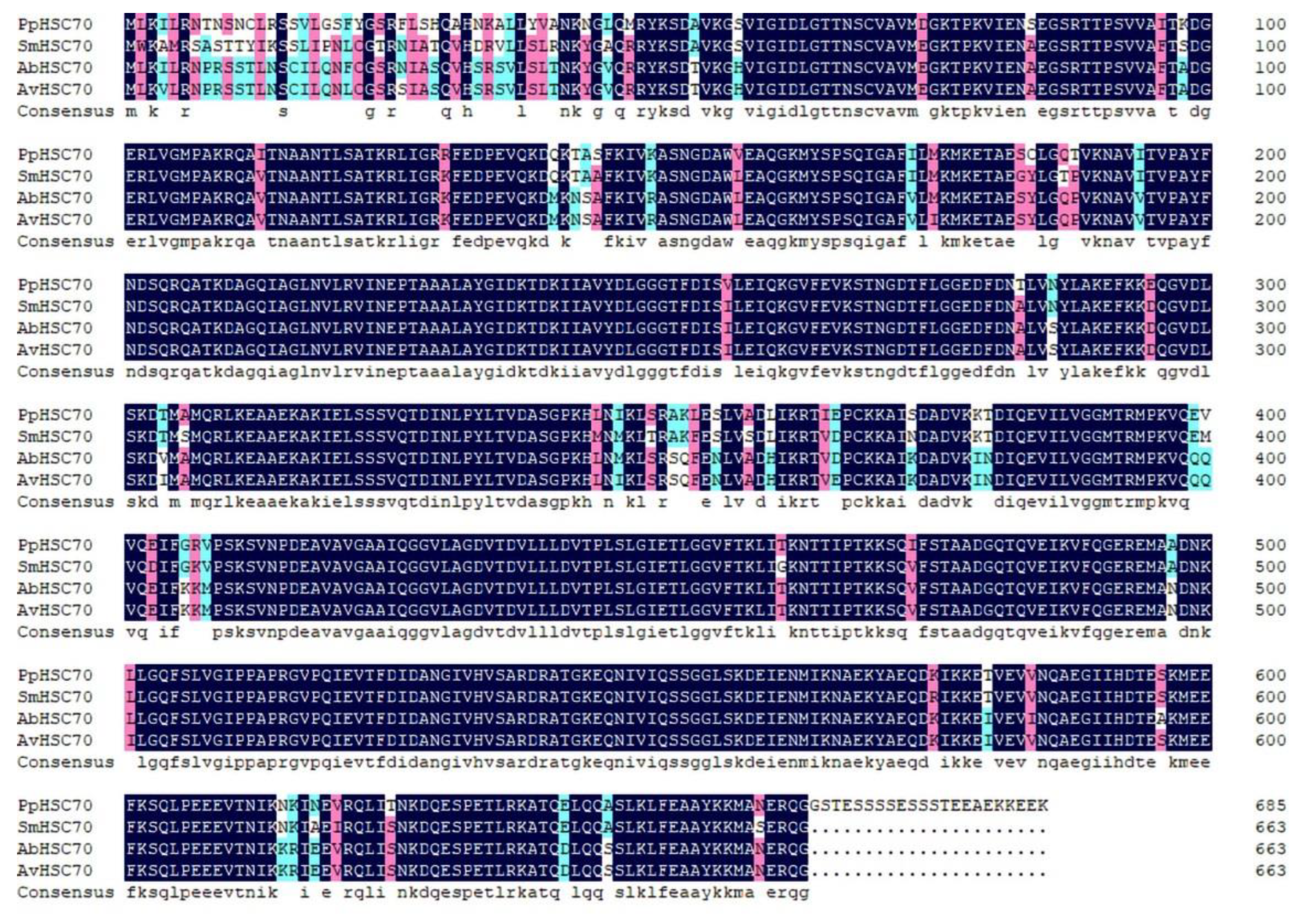
2.1. Sequence Analysis and Phylogenetic Tree

The full-length sequences of the three HSP genes PpHSP27, PpHSP60, and PpHSC70 were obtained using RT-PCR and RACE technology. The full length of PpHSP27 (GenBank Accession No. ON808409) was 997 base pairs (bp), included a 5′ untranslated region (UTR) of 108 bp, a 3′ UTR of 337 bp contained a typical polyA tail, and an open reading frame (ORF) of 552 bp. The ORF encoded 183 AAs and its predicted molecular weight was 21.18 kDa. The theoretical isoelectric point (pI) is 5.50. The instability index (II) was 55.25, indicating that it is an unstable protein. The grand average of hydropathicity (GRAVY) was −0.763, indicating that it is a hydrophilic protein. The deduced AA sequence of PpHSP27 contained an alpha crystallin domain (ACD), which is a structural feature of the sHSP family (Figure S1). In the phylogenetic tree, HSP27 was divided into two taxa, Arachnida and Insecta, and P. pseudoannulata was clustered with Arachnida (Figure 1). The deduced AA sequences were all greater than 88% similar to HSP27 from Parasteatoda tepidariorum (XP015921832.1), Stegodyphus dumicola (XP035231886.1), and Stegodyphus mimosarum (KFM65662.1) (Figure 2).
The full length of PpHSP60 (GenBank Accession No. ON808407) was 2065 bp, included a 5′ UTR of 158 bp, a 3′ UTR of 199 bp contained a typical polyA tail, and an ORF of 1710 bp. The ORF encodes 569 AAs and its predicted molecular weight was 61.17 kDa. The pI was 5.43. The II was 31.6, which indicated that it is a stable protein. The GRAVY was −0.184, which indicated that it is a hydrophilic protein. The deduced AA sequence of PpHSP60 contained a characteristic sequence of the HSP60 family, AAVEEGIVAGGG (Figure S2). In the phylogenetic tree, HSP60 was divided into two taxa, Arachnida and Insecta, and P. pseudoannulata was clustered with Arachnida (Figure 1). The deduced AA sequences were all greater than 92% similar to HSP60 from S. dumicola (XP035227740.1), Trichonephila clavata (GFR30939.1), and Trichonephila inaurata madagascariensis (GFS44331.1) (Figure 3).
The full length of PpHSC70 (GenBank Accession No. ON808408) was 2438 bp, including a 5′ UTR of 134 bp, a 3′ UTR of 247 bp containing a typical polyA tail, and an ORF of 2061 bp. Its ORF encodes 686 AAs and its predicted molecular weight was 74.56 kDa. The pI was 6.19. The II was 41.78, which indicated that it is a stable protein. The GRAVY was −0.369, which indicated that it is a hydrophilic protein. The deduced AA sequence of PpHSC70 contained three HSP70 family signature sequences: IDLGTTNS, VYDLGGGTFDISVL, and VILVGGMTRMPKVQE (Figure S3). In the phylogenetic tree, HSC60 was divided into two taxa, Arachnida and Insecta, and P. pseudoannulata was clustered with Arachnida (Figure 1). The deduced AA sequences were all more than 89% similar to HSC70 from S. mimosarum (KFM81131.1), Argiope bruennichi (KAF8774081.1), and Araneus ventricosus (GBM18724.1) (Figure 4).

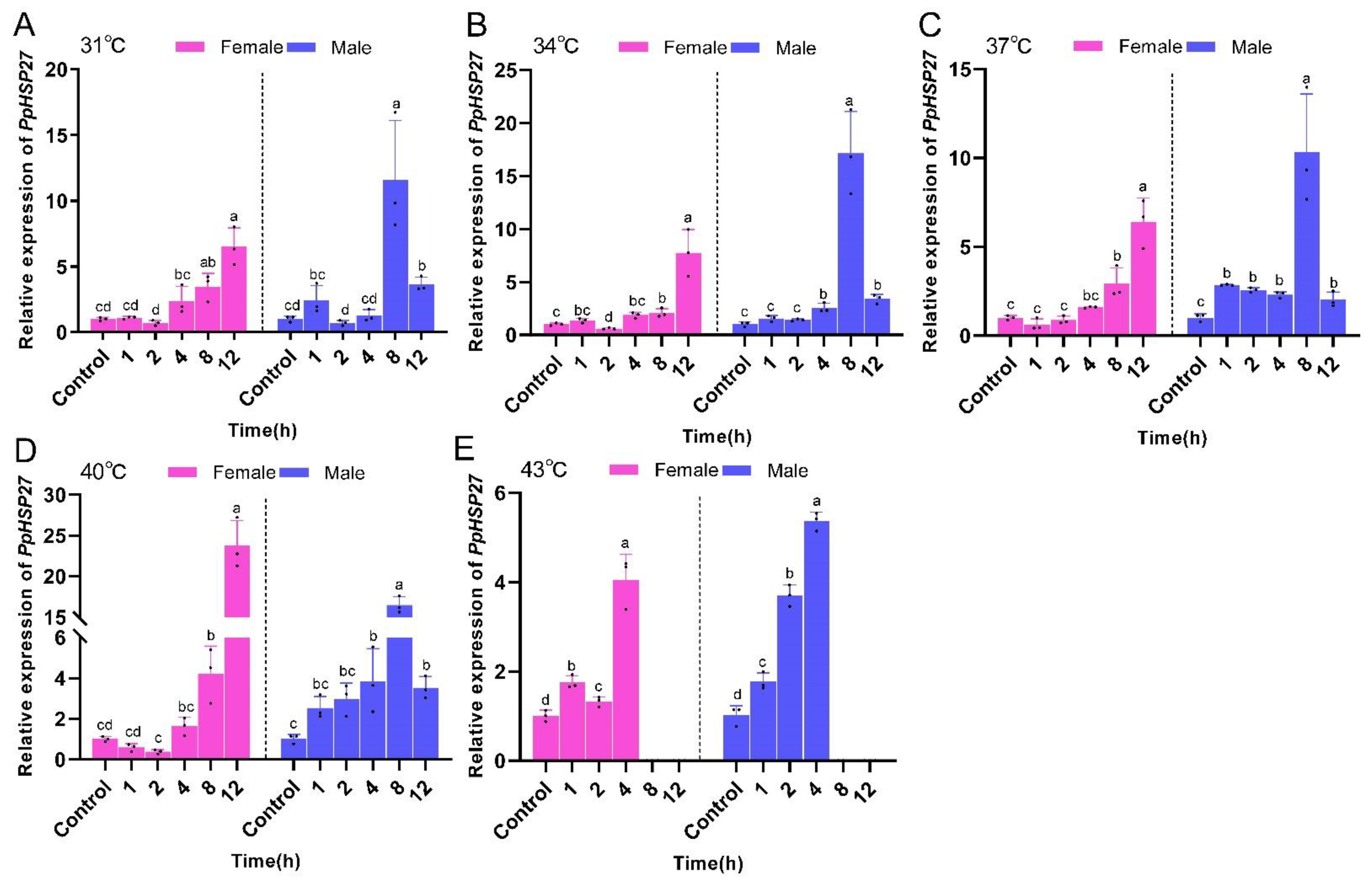
2.2. Effects of High Temperature Stress on PpHSP Genes Expression

The expression of PpHSP27, PpHSP60, and PpHSC70 in P. pseudoannulata were significantly affected by the temperature and time of heat stress as well as their interactions. At the same temperature, the expression of PpHSP27 in female P. pseudoannulata exhibited an overall increasing trend with increasing stress time. The highest expression was observed in the treatment group with the longest stress duration. The expression of PpHSP27 was not significantly different from that of the control after 1 or 2 h of stress at 31, 34, 37, and 40 °C. The trend of PpHSP27 expression in male P. pseudoannulata was consistent with that in females, and overall, its expression was highest after 8 h of stress. For the same duration of stress, the expression of the PpHSP27 gene in female P. pseudoannulata was highest at the highest stress temperature. The expression of PpHSP27 in female P. pseudoannulata was highest after 12 h of stress at 40 °C and was 25.75-fold (F = 93.615, df = 17, P < 0.001) higher than that of the control. PpHSP27 expression in male P. pseudoannulata was highest after 8 h of stress at 34 °C and 17.14-fold (F = 97.336, df = 17, P < 0.001) higher than that of the control, both significantly higher than their respective controls (Figure 5).
Under the same temperature stress, PpHSP60 gene expression in female P. pseudoannulata was highest in all groups, with the longest duration of stress treatment and overall expression increasing with time. The expression of the PpHSP60 gene in male P. pseudoannulata showed a general trend of increasing and then decreasing with time. The expression of PpHSP60 was not significantly different from the control group under the five time periods of stress at 31 °C and 34 °C. At the same duration of stress, PpHSP60 expression in both male and female P. pseudoannulata exhibited the highest expression in the highest temperature treatment group. PpHSP60 expression was highest in female P. pseudoannulata after 12 h of stress at 40 °C, which was 9.12-fold (F = 59.169, df = 17, P < 0.001) higher than the control. In males after 4 h of stress at 43 °C, the expression was 13.14-fold (F = 92.728, df = 11, P < 0.001) higher than the control, and both were significantly higher than their respective controls (Figure 6).
At the same temperature, the expression of the PpHSC70 gene in female P. pseudoannulata exhibited an overall increasing trend with time and the expression of the PpHSC70 gene in male P. pseudoannulata increased and then decreased with time. PpHSC70 expression was highest in female P. pseudoannulata after 8 h of stress at 40 °C, which was 21.85-fold (F = 146.728, df = 17, P < 0.001) higher than the control. In males after 1 h of stress at 37 °C, the expression was 3.97-fold (F = 24.665, df = 17, P < 0.001) higher than the control, and both were significantly higher than their respective controls (Figure 7).

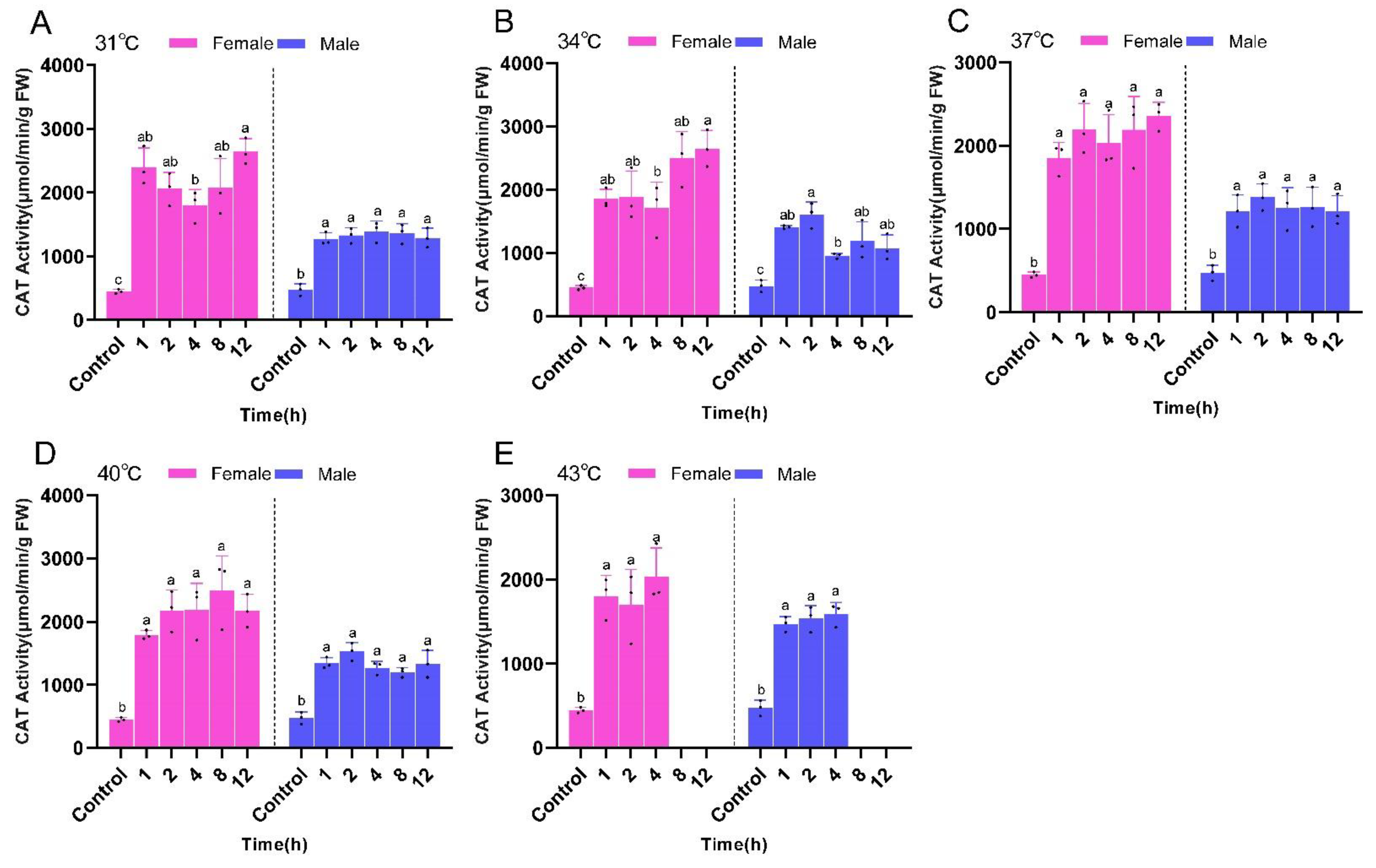
2.3. Changes in Antioxidant Enzyme Activity

The activities of SOD, CAT, and GST in P. pseudoannulata were significantly altered by the temperature and time of heat stress and their interactions. At the same temperature, SOD activities in both male and female P. pseudoannulata exhibited a trend of increasing and then decreasing with increased stress time. The SOD activity of female P. pseudoannulata was not significantly different from that of the control after 12 h of stress at different temperatures. SOD activity of the male P. pseudoannulata in all temperature treatment groups was highest after 2 h of stress and was significantly higher than that of the control group. The highest SOD activity occurred after 8 h of stress at 40 °C for female P. pseudoannulata (132.78 U/g FW, F =46.588, df = 17, P < 0.001) and 2 h of stress at 37 °C for males (133.01 U/g FW, F = 19.421, df = 17, P < 0.001). Both were significantly higher than their respective controls (Figure 8).
Overall, CAT activity in female P. pseudoannulata was significantly higher than that of the control group after different times of stress at the same temperature, and did not differ significantly from each other within the same temperature treatment group as did the male P. pseudoannulata. The highest CAT activity occurred after 12 h of stress at 34 °C for female P. pseudoannulata (2647.51 μmol/min/g FW, F = 17.338, df = 17, P < 0.001) and after 2 h of stress at 34 °C for the males (1603.44 μmol/min/g FW, F = 21.592, df = 17, P < 0.001). Both were significantly higher than their respective controls (Figure 9).
GST activities of both male and female P. pseudoannulata increased with increasing stress time at the same temperature and was significantly higher than that of the control group. However, there was no significant difference between the treatment groups at the same temperature. The highest GST activity appeared after 12 h of stress at 31 °C for female P. pseudoannulata (1.55 nmol/min/g FW, F = 19.180, df = 17, P < 0.001) and after 1 h of stress at 43 °C for the males (1.35 nmol/min/g FW, F = 37.571, df = 17, P < 0.001)). Both were significantly higher than their respective controls (Figure 10).

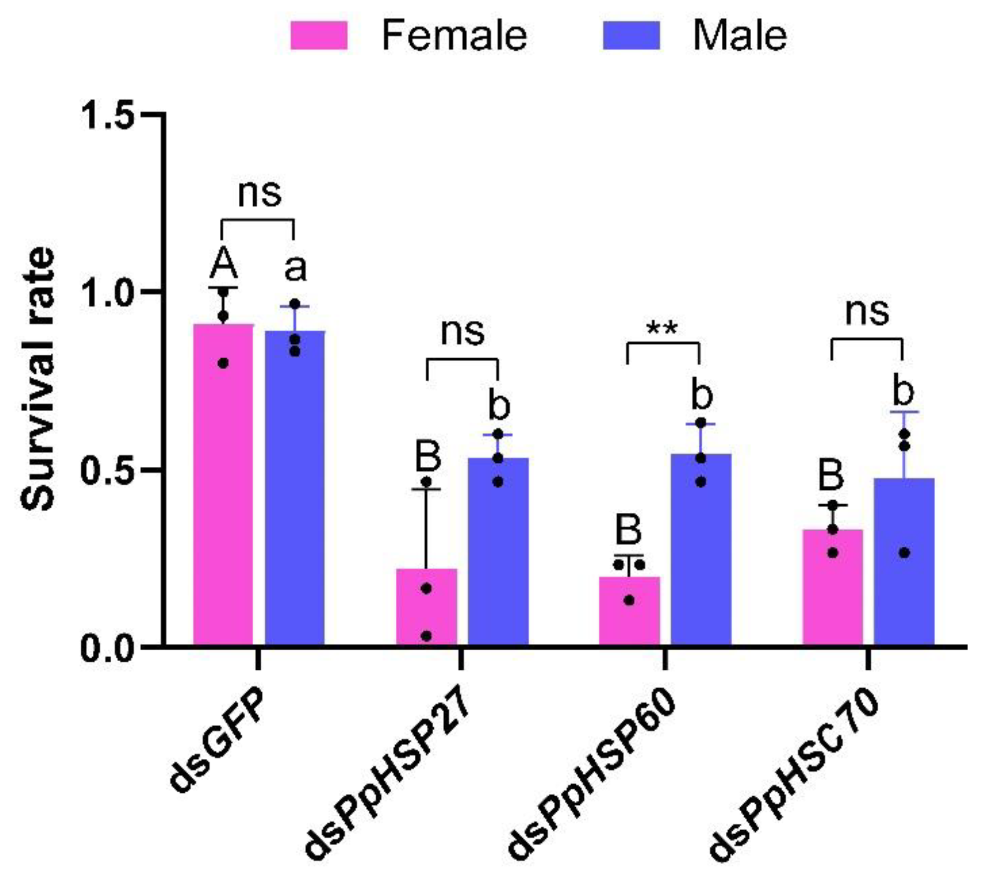
2.4. Effect of Silencing Three HSP Genes on High Temperature Tolerance in P. pseudoannulata

To further understand the role of the three HSP genes in heat resistance of P. pseudoannulata, we injected dsRNA into male and female P. pseudoannulata and examined changes in the survival rate of male and female P. pseudoannulata after high temperature stress. When compared with the control group injected with green fluorescent protein (GFP, GenBank accession number: L29345.1), the expression of the three HSP genes in female P. pseudoannulata injected with dsPpHSP27, dsPpHSP60, and dsPpHSC70 decreased by 41% (t = 3.775, df = 4, P = 0.020), 64% (t = 3.913, df = 4, P = 0.017), and 59% (t = 4.450, df = 4, P = 0.011), respectively. The expression levels of the three HSP genes in male P. pseudoannulata decreased by 55% (t = 4.864, df = 4, P = 0.008), 75% (t = 6.674, df = 4, P = 0.003), and 63% (t = 5.571, df = 4, P = 0.005), respectively. After interference, the expression levels of the three genes in male and female P. pseudoannulata were significantly lower than that in the control group (Figure 11).
After injection of dsPpHSP27, dsPpHSP60, and dsPpHSC70, the survival rates of male and female P. pseudoannulata were significantly lower than that of the control group injected with dsGFP after exposure to high temperature stress at 43 °C for 4 h. After 4 h of high temperature stress at 43 °C, the survival rates of female P. pseudoannulata injected with dsPpHSP27, dsPpHSP60, and dsPpHSC70 were 22%, 20%, and 33%, respectively, which was significantly lower than the 91% of the control group (F = 19.655, df = 11, P < 0.05). The survival rates of male P. pseudoannulata were 53%, 54%, and 48%, respectively, significantly higher than 89% in the control group (F = 8.575, df = 11, P < 0.05) (Figure 12). These results indicated that knockdown of the three HSP genes can significantly reduce the heat tolerance of P. pseudoannulata to high temperatures.

3. Discussion

High temperature is one of the main factors that affect P. pseudoannulata, which leads to decreased predation and a shortened lifespan [39,43]. This may indirectly lead to increased pests in rice fields and raise the cost of people to control pests. It is well-established that HSPs and antioxidant enzymes play an important role in arthropod resistance to high temperature stress. Previous studies suggested that HSPs and antioxidant enzymes may be key factors in the resistance of P. pseudoannulata to high temperature stress [44,45]. HSPs are highly conserved in arthropods [5]. In the present study, we cloned and characterized the full-length PpHSP27, PpHSP60, and PpHSC70 genes of P. pseudoannulata, which will improve the HSP gene data of P. pseudoannulata and provide a reference for future studies. The deduced AA sequences show that they contain characteristic sequences of HSP27, HSP60, and HSP70 families and the AA sequence alignment shows that they are highly similar to the HSP27, HSP60, and HSC70 of other Arachnids. Based on our analysis, the three HSP genes isolated from P. pseudoannulata belonged to the HSP27, HSP60, and HSP70 families.
There are sexual dimorphisms in P. pseudoannulata and there is a huge difference in body size between males and females, thus it is necessary to study male and female P. pseudoannulata separately. Our study showed that the expression of the PpHSP27, PpHSP60, and PpHSC70 genes increased significantly with increasing stress temperature and stress duration in all treatment groups. This is consistent with the results of Agasicles hygrophila, Neoseiulus barkeri, and Scylla paramamosain [46,47,48]. From this, we hypothesized that these three HSP genes may help P. pseudoannulata to resist heat stress. The HSC70 gene is considered to be constitutively expressed under non-stress conditions and heat stress does not readily affect its expression [49,50]. However, in recent studies on Tetranychus urticae, Sitodiplosis mosellana, Pteromalus puparum, Plutella xylostella, as well as others, the expression of the HSC70 gene increased significantly with increasing temperature [51,52,53,54]. In the present study, we also found that PpHSC70 gene expression in P. pseudoannulata was induced by high temperatures and significantly different from that of the control. This suggested that HSC70 and HSP70 have the same function in P. pseudoannulata. On average, the expression levels of the three HSP genes in P. pseudoannulata were not significantly different from those in the control group after stress at 31 °C and 34 °C for 1 and 2 h, which indicated that the results of short-term stress below 34 °C is not a harsh condition for P. pseudoannulata. This was consistent with the results of Zhao et al. [40], who found that 25–32 °C is the optimal temperature for P. pseudoannulata to survive. Although the synthesis of HSPs improves the body’s resistance, it also consumes the organism’s energy [55,56]. Thus, for P. pseudoannulata, energy may be used in more important places, such as spawning and reproduction, when the external conditions are suitable for survival. Multiple studies have shown that females are more tolerant than males to heat stress, for example, this phenomenon is associated with higher expression of the HSP genes in B. tabaci females [23,57,58,59]. In this study, we found that with the increase in stress time, the expression of three HSP genes in P. pseudoannulata continued to increase, and although the expression of three HSP genes in male spiders also increased, they decreased significantly after 8 h. Females P. pseudoannulata generally play a significant role in reproduction and are larger than males, and more energy is needed to store in their bodies for reproduction, which means they have more energy to regulate themselves against unfavorable conditions when they are exposed to unfavorable conditions [45]. Some studies showed that females were more resistant than males and survived longer than males without water or food [60]. In summary, we speculate that female P. pseudoannulata have a higher tolerance than male P. pseudoannulata over prolonged high temperature stress.
High temperature stress leads to excessive ROS production in arthropod cells, which in turn, causes damage to the arthropods themselves [9,25]. To avoid such adverse effects, arthropods have evolved the ability to maintain a balanced relationship between ROS and antioxidants [61]. In the present study, the SOD, CAT, and GST activities of P. pseudoannulata were significantly increased by stress temperature as well as stress time, suggesting that these enzymes are involved in the scavenging of excess ROS in P. pseudoannulata. Studies of Ophraella communa, Corythucha ciliate, Mononychellus mcgregori, and Hylyphantes graminicola also confirmed this result [62,63,64,65]. Overall, the SOD activities of male and female P. pseudoannulata were not significantly different from that of the control group after 12 h of heat stress at 37 °C and 40 °C. A similar phenomenon was reported for SOD activity in S. paramamosain [48]. Based on these results, we hypothesized that there is a limit to the regulatory capacity of SOD and when stress conditions exceed a certain level, the spider may scavenge the generated ROS through other mechanisms. The CAT enzyme is one of the main ways for arthropods to decompose H2O2 [31]. In the present study, the CAT activities of male and female P. pseudoannulata in all treatment groups were significantly higher than that of the control group. This was consistent with the changing trend of CAT for Myzus persicae and H. graminicola [29,65]. This indicated that CAT is the main pathway for the breakdown of H2O2 in P. pseudoannulata, so they need to maintain relatively high activity under heat stress at different times and temperatures. Unlike CAT and SOD, the GST activities of male and female P. pseudoannulata were not significantly different from the control group after 1 and 2 h of stress at 31 °C and 34 °C, respectively. In Mythimna separata and Liposcelis bostrychophila [28,66], GST activities significantly increased in a relatively short period of time. These changes in GST activities may result from interspecies differences. Overall, the activities of SOD, CAT, and GST all increased significantly after P. pseudoannulata was exposed to high temperature stress, suggested that antioxidant enzymes (SOD, CAT, and GST) play important roles in reducing oxidative damage caused by high temperature stress in P. pseudoannulata.
As a powerful tool for gene function studies, dsRNA-mediated silencing or inhibition of target genes has been widely used in various arthropods [67]. There were also studies using RNAi to silence in P. pseudoannulata [68,69]. Therefore, we performed RNAi experiments by injecting dsRNA into P. pseudoannulata to determine the effect of silencing HSP genes on the heat tolerance of P. pseudoannulata. The results showed that the expression levels of PpHSP27, PpHSP60, and PpHSC70 genes were significantly reduced in male and female P. pseudoannulata after the injection of dsRNA. After silencing the PpHSP27, PpHSP60, and PpHSC70 genes, the survival rates of female and male adult P. pseudoannulata were significantly decreased after high temperature stress. Therefore, our results indicated that changes in gene expression levels of PpHSP27, PpHSP60 and PpHSC70 could significantly affect the heat tolerance of spiders. Similar results have been observed for other arthropods. In Dermatophagoides farinae, the survival rates of D. farinae treated with the three dsHSP70 fragments were significantly reduced under heat stress at 41 °C for 1 h [70]. After knockdown of HSC70 and HSP90 in Plodia interpunctellalarvae, thermotolerance was significantly reduced [71]. The survival rate of male P. pseudoannulata after heat stress was higher than that of female P. pseudoannulata after knocking down all three HSP genes. A similar phenomenon was observed in B. tabaci, and D. melanogaster [22,23]; however, the exact cause of this phenomenon is not clear at this time and further studies are needed. The study on N. barkeri showed that the protein level of HSP70-1 was positively correlated with the expression of NbHSP70-1, and the overexpression of NbHSP70 protein in Escherichia coli (E. coli) enhanced the high temperature tolerance of E. coli [47]. Overexpression of HSP40 from Cydia pomonell in E. coli enhanced the high temperature tolerance of E. coli [72]. Therefore, we hypothesized that the HSC70, HSP60, HSP27 protein content of P. pseudoannulata correlates with the expression levels of PpHSP27, PpHSP60, and PpHSC70 genes, and that a greater accumulation of HSC70, HSP60, and HSP27 proteins could provide strong protection against heat tolerance in P. pseudoannulata.

4. Materials and Methods

4.1. Spider Feeding and Experimental Handling

Adult P. pseudoannulata used in this study were collected in Yanlou Town, Guiyang City, Guizhou Province (106.619227° E, 26.322845° N). Before the experiment, the P. pseudoannulata were acclimated in an artificial climate box (25 ± 0.5 °C, relative humidity 70–80%, and photoperiod of 14 L:10 D) for two weeks and fed five housefly pupae per week. To generate heat stress, male or female P. pseudoannulata were placed, respectively, in a climate chamber at 31 °C, 34 °C, 37 °C, 40 °C, and 43 °C for 1, 2, 4, 8, and 12 h, and then allowed to recover for 1 h in a climate chamber at 25 °C [45]. After treatment, surviving P. pseudoannulata were immediately cryopreserved in liquid nitrogen and placed in a −80 °C freezer for later use. Since P. pseudoannulata were housed at 25 °C, 25 °C was used as a control for all treatment groups. The processed samples were divided into two parts: one for measuring antioxidant enzyme activity and the other for extracting total RNA. Each treatment consisted of three replicates (n = 3); 3 spiders were included in each replicate to test HSP genes expression or antioxidant enzyme activity.

4.2. RNA Extraction and cDNA Synthesis

The total RNA from whole P. pseudoannulata was extracted using a Trizol Reagent Kit (Sangon Biotech, Shanghai, China). The concentration, purity, and integrity of the RNA were determined by NanoDrop 2000 spectrophotometer (Thermo Fisher Scientific, Waltham, MA, USA) and 1% agarose gel electrophoresis. First-strand cDNA was synthesized using StarScript II RT Mix with a gDNA Remover StarScript II kit (GenStar, Beijing, China). The agarose gel electropherograms of P. pseudoannulatais RNA were shown in Figure S4.

4.3. Cloning the Full-Length cDNA of HSP Genes

Three HSP gene DNA internal fragments were amplified using the first-strand cDNA as a template. The cloning primers (Table 1) were designed using Primer Premier 6.0 (PREMIER Biosoft International, Palo Alto, CA, USA) software based on the transcriptional master sequence of the P. pseudoannulata [42]. The PCR reaction was performed using Taq DNA Polymerase (GenStar, Beijing, China) under the following conditions: 94 °C initial denaturation for 3 min, 35 cycles of 94 °C denaturation for 30 s, 55–60 °C annealing for 30 s, 72 °C elongation for 1–2 min, and a final 5 min extension at 72 °C. The PCR products were purified with a FastPure® Gel DNA Extraction Mini Kit (Vazyme, Nanjing, China). Then, the purified products were cloned into the pGEM-T Easy vector (Promega, Madison, WI, USA) and finally transformed into E. coli DH5α-competent cells (Takara, Beijing, China) and sent to Sangon Biotech (Shanghai, China) for sequencing. The primers used for amplifying the 5′ and 3′ ends were designed according to the partial fragment sequences obtained by cloning and the instructions of the SMARTer® RACE 5′/3′ Kit (Takara, Beijing, China). The 5′ and 3′ ends were cloned according to the instructions of the SMARTer® RACE 5′/3′ Kit. The cloning steps of the 5′ and 3′ ends refer to the previous study of our task group [45]. The agarose gel electropherograms of PCR products of the clones PpHSP27, PpHSP60 and PpHSC70 are shown in Figures S5 and S6.

4.4. Sequence Analysis

The resulting gene fragments were assembled into the full length of each gene using the SeqMan program in DNAStar (Madison, WI, USA). AA sequences were deduced using ExPASy sequence analysis tools (https://web.expasy.org/translate/) (accessed on 18 August 2022) [73] and motifs were identified using ScanProsite software (http://www.expasy.org/tools/scanprosite) (accessed on 18 August 2022) [74]. Prediction of the relative molecular mass and theoretical isoelectric point of proteins was performed using ProtParam (http://web.expasy.org/protparam/) (accessed on 18 August 2022). The obtained AA sequences were subjected to a homology search using the NCBI BLAST program (https://blast.ncbi.nlm.nih.gov/Blast.cgi) (accessed on 18 August 2022). The MEGA-X (Mega Limited, AKL, NZ) software was used to construct a phylogenetic tree of the AA sequences of the HSPs of P. pseudoannulata and HSPs of other species by the neighbor-joining method, and the bootstrap value was calculated as 1000 repetitions [75].

4.5. Quantitative Real-Time PCR

Quantitative RT-PCR primers were designed based on the full length of the three cloned genes using primer6 software. Gene-specific primers were shown in Table 1. qRT-PCR was performed on all experimental samples using an iCycle iQ (Bio-Rad, Hercules, CA, USA). All mRNA expression levels were quantified using the β-actin gene as an internal reference [42]. The total volume of the qRT-PCR reaction was 20 μL and contained 10 μL of 2 × RealStar Fast SYBR qPCR Mix (GenStar, Beijing, China), 1 μL of each gene-specific primer (Table 1), 1 μL of cDNA template, and 8 μL of diethyl pyrocarbonate-treated H2O. The reaction conditions were as follows: 95 °C for 2 min, followed by 40 cycles of 95 °C for 15 s, 55 °C for 30 s, and 72 °C for 30 s. Melting curves were produced from 65 °C to 95 °C. Each experiment included three biological replicates and each biological replicate included three technical replicates [45]. The agarose gel electropherograms of PCR products of qPCR primers are shown in Figure S7.

4.6. RNA Interference

To further study the biological functions of the three HSP genes, we injected specific dsRNA into adult P. pseudoannulata to interfere with the expression of the three genes. The primers used for the RNAi experiments were shown in Table 1. The dsRNA of GFP was used as a negative control. Based on the manufacturer’s instructions, the dsRNA was synthesized using the Transcript Aid T7 High Yield Transcription Kit (Thermo Fisher Scientific, Waltham, MA, USA). The agarose gel electropherograms of dsRNA are shown in Figure S8.
A total of 99 male and 99 female P. pseudoannulata were used for each HSP gene interference experiment. dsRNA injections were performed using a microsyringe (HAMILTON, Shanghai, China). The spider was immobilized by hand (wearing gloves to prevent being bitten by the spider) and dsRNA was slowly injected from the back of the spider’s abdomen, with 1000 ng/500 nL of dsRNA injected into each spider. The injected P. pseudoannulata were reared in an artificial climate box (25 ± 0.5 °C, relative humidity 70–80%, and photoperiod of 14 L:10 D) and after 72 h post-injection of dsRNA, 9 female and 9 male P. pseudoannulata from each treatment group were randomly collected, frozen in liquid nitrogen, and stored in a −80 °C freezer for subsequent verification of RNAi efficiency. Each treatment consisted of three replicates (n = 3); 3 spiders were included in each replicate to determine silencing efficiency. The remaining 90 male and 90 female P. pseudoannulata were used to verify the effect of each HSP gene on spider thermotolerance. The injected male and female P. pseudoannulata were placed in a climate chamber at 43 °C for 4 h and survival rate was recorded. Each treatment consisted of three replicates (n = 3), 30 spiders were included in each replicate for the survival rate determination.

4.7. Antioxidant Enzyme Activity Detection

SOD, CAT, and GST activities were determined using commercial kits (Cat. No.: BC0175, BC0200, BC0350, Solarbio, Beijing, China). The three P. pseudoannulata were mixed with the 0.05 M pH 7.0 phosphate buffers and homogenized in an ice bath (adding 1 mL of buffer for every 0.1 g of spider), then 1 mL of the product after homogenization in ice bath was centrifuged at 12,000× g for 10 min, and the supernatant was put on ice for use. The absorbance was read in a Multiskan FC (Thermo Fisher Scientific, Waltham, MA, USA) with reference to the kit instructions and the antioxidant enzyme activity was calculated according to the fresh weight (FW) of the sample [76].

4.8. Data Analysis

According to the formula provided by the kit, SOD, CAT, and GST activities were calculated. The relative expression of the HSP gene for each treatment was calculated by the 2−ΔΔCt method [77]. All data analysis in this study was performed using SPSS 22 software (SPSS Inc., Chicago, IL, USA). In the data analysis of HSP gene expression and antioxidant enzyme activity of P. pseudoannulata under different conditions of high temperature stress, separate one-way Analysis of Variance (ANOVA) were used for each group of temperature or gender, followed by pairwise comparisons at different time points using Tukey’s Honest Significant Difference (HSD) method (P = 0.05). In the verification experiment of silence efficiency, the Student’s t-test was used to compare the difference of PpHSP gene expression in P. pseudoannulata after being injected with dsGFP and dsHSP (P = 0.05). In the function verification experiment of PpHSP genes, separate one-way ANOVAs were used for two genders, Tukey’s HSD method was used, followed by pairwise comparisons with different RNAi processing (P = 0.05), and the Student’s t-test was used to compare the difference in the survival rate of female and male P. pseudoannulata after the HSP genes were silenced (P = 0.05). All the data are expressed as the mean ± standard deviation (SD).

5. Conclusions

We cloned the full length of PpHSP27, PpHSP60, and PpHSC70 of P. pseudoannulata and analyzed their sequence features. Trends in the expression of these HSP genes and the enzymatic activities of SOD, CAT, and GST were explored at different high temperatures and stress durations, which revealed that they were significantly upregulated with increasing temperature and stress durations. We successfully knocked down HSP genes and verified their functions in P. pseudoannulata. The results indicated that knockdown of PpHSP27, PpHSP60, and PpHSC70 enhanced susceptibility to high temperatures in P. pseudoannulata. In conclusion, our study shows that SOD, CAT, and GST enable P. pseudoannulata to remove the metabolic byproducts produced by heat stress. PpHSP27, PpHSP60, and PpHSC70 are key factors that affect the heat tolerance of P. pseudoannulata. They all play an important role in the resistance of P. pseudoannulata to high temperature.

Supplementary Materials

The following supporting information can be downloaded at: https://www.mdpi.com/article/10.3390/ijms232112821/s1.

Author Contributions

Conceptualization, D.F. and R.X.; funding acquisition, R.X.; methodology, D.F., J.L. and Y.-N.P.; formal analysis, D.F., Y.-N.P., J.L., F.X., M.L. and J.-Y.Z.; investigation, D.F., F.X., M.L. and J.-Y.Z.; writing—original draft, D.F.; writing—review & editing, R.X. and D.F. All authors have read and agreed to the published version of the manuscript.

Funding

This research was supported by Guizhou Provincial Science and Technology Projects (Qian Ke He Support [2022] General 135, Qian Ke He Support [2018] 2354 and Qian Ke He Talent CXTD [2021] 004).

Institutional Review Board Statement

Not applicable.

Informed Consent Statement

Not applicable.

Data Availability Statement

The data and materials supporting the conclusions of this study are included within the article.

Acknowledgments

We wish to thank Tao Zhang (College of Agriculture, Guizhou University, China), Xibin Yang (College of Agriculture, Guizhou University, China) and Runa Zhao (College of Agriculture, Guizhou University, China) for assistance with improving the manuscript.

Conflicts of Interest

The authors declare no conflict of interest.

References

  1. Bale, J.S.; Masters, G.J.; Hodkinson, I.D.; Awmack, C.; Bezemer, T.M.; Brown, V.K.; Butterfield, J.; Buse, A.; Coulson, J.C.; Farrar, J.; et al. Herbivory in global climate change research: Direct effects of rising temperature on insect herbivores. Global Change Biol. 2002, 8, 1–16. [Google Scholar] [CrossRef]
  2. Horn, D.J. Temperature synergism in integrated pest management. In Temperature Sensitivity in Insects and Application in Integrated Pest Management; CRC Press: Boca Raton, FL, USA, 2019; pp. 125–140. [Google Scholar]
  3. Perkins-Kirkpatrick, S.E.; Lewis, S.C. Increasing trends in regional heatwaves. Nat. Commun. 2020, 11, 3357. [Google Scholar] [CrossRef]
  4. Wang, X.X.; Jiang, D.B.; Lang, X.M. Future extreme climate changes linked to global warming intensity. Sci. Bull. 2017, 62, 1673–1680. [Google Scholar] [CrossRef] [Green Version]
  5. Feder, M.E.; Hofmann, G.E. Heat-shock proteins, molecular chaperones, and the stress response: Evolutionary and ecological physiology. Annu. Rev. Physiol. 1999, 61, 243–282. [Google Scholar] [CrossRef] [PubMed] [Green Version]
  6. Srensen, J.G.; Kristensen, T.N.; Loeschcke, V. The evolutionary and ecological role of heat shock proteins. Ecol. Lett. 2010, 6, 1025–1037. [Google Scholar] [CrossRef]
  7. Felton, G.W.; Summers, C.B. Antioxidant systems in insects. Arch. Insect Biochem. Physiol. 1995, 29, 187–197. [Google Scholar] [CrossRef]
  8. Perez, R.; Aron, S. Adaptations to thermal stress in social insects: Recent advances and future directions. Biol. Rev. 2020, 95, 1535–1553. [Google Scholar] [CrossRef]
  9. Gonzalez-Tokman, D.; Cordoba-Aguilar, A.; Dattilo, W.; Lira-Noriega, A.; Sanchez-Guillen, R.A.; Villalobos, F. Insect responses to heat: Physiological mechanisms, evolution and ecological implications in a warming world. Biol. Rev. 2020, 95, 802–821. [Google Scholar] [CrossRef]
  10. Kregel Kevin, C. Invited Review: Heat shock proteins: Modifying factors in physiological stress responses and acquired thermotolerance. J. Appl. Physiol. 2002, 192, 2177–2186. [Google Scholar] [CrossRef] [Green Version]
  11. King, A.M.; MacRae, T.H. Insect heat shock proteins during stress and diapause. In Annual Review of Entomology; Berenbaum, M.R., Ed.; Palo Alto: Santa Clara, CA, USA, 2015; Volume 60, pp. 59–75. [Google Scholar]
  12. Ganea, E. Chaperone-like activity of alpha-crystallin and other small heat shock proteins. Curr. Protein Pept. Sci. 2001, 2, 205–225. [Google Scholar] [CrossRef]
  13. Basha, E.; O’Neill, H.; Vierling, E. Small heat shock proteins and alpha-crystallins: Dynamic proteins with flexible functions. Trends Biochem. Sci. 2012, 37, 106–117. [Google Scholar] [CrossRef] [PubMed] [Green Version]
  14. Fink, A.L. Chaperone-Mediated Protein Folding. Physiol. Rev. 1999, 79, 425–449. [Google Scholar] [CrossRef] [PubMed] [Green Version]
  15. Morimoto, R.I. Cells in stress: Transcriptional activation of heat shock genes. Science 1993, 259, 1409–1410. [Google Scholar] [CrossRef] [PubMed]
  16. Bukau, B.; Horwich, A.L. The Hsp70 and Hsp60 chaperone machines. Cell 1998, 92, 351–366. [Google Scholar] [CrossRef] [Green Version]
  17. Mayer, M.P.; Bukau, B. Hsp70 chaperones: Cellular functions and molecular mechanism. Cell. Mol. Life Sci. 2005, 62, 670–684. [Google Scholar] [CrossRef] [PubMed] [Green Version]
  18. Karunanithi, S.; Barclay, J.W.; Robertson, R.M.; Brown, I.R.; Atwood, H.L. Neuroprotection at Drosophila synapses conferred by prior heat shock. J. Neurosci. 1999, 19, 4360–4369. [Google Scholar] [CrossRef] [Green Version]
  19. Benoit, J.B.; Lopez-Martinez, G.; Patrick, K.R.; Phillips, Z.P.; Krause, T.B.; Denlinger, D.L. Drinking a hot blood meal elicits a protective heat shock response in mosquitoes. Proc. Natl. Acad. Sci. USA 2011, 108, 8026–8029. [Google Scholar] [CrossRef] [Green Version]
  20. Paim, R.M.; Araujo, R.N.; Leis, M.; Sant’Anna, M.R.; Gontijo, N.F.; Lazzari, C.R.; Pereira, M.H. Functional evaluation of Heat Shock Proteins 70 (HSP70/HSC70) on Rhodnius prolixus (Hemiptera, Reduviidae) physiological responses associated with feeding and starvation. Insect Biochem. Mol. Biol. 2016, 77, 10–20. [Google Scholar] [CrossRef]
  21. Lu, K.; Chen, X.; Liu, W.; Zhou, Q. Characterization of heat shock cognate protein 70 gene and its differential expression in response to thermal stress between two wing morphs of Nilaparvata lugens (Stål). Comp. Biochem. Physiol. Part A Mol. Integr. Physiol. 2016, 199, 47–53. [Google Scholar] [CrossRef]
  22. Krebs, R.A.; Feder, M.E.; Lee, J. Heritability of expression of the 70KD heat-shock protein in Drosophila melanogaster and its relevance to the evolution of thermotolerance. Evolution 1998, 52, 841–847. [Google Scholar] [CrossRef]
  23. Lü, Z.-C.; Wan, F.-H. Using double-stranded RNA to explore the role of heat shock protein genes in heat tolerance in Bemisia tabaci (Gennadius). J. Exp. Biol. 2011, 214, 764–769. [Google Scholar] [CrossRef] [PubMed] [Green Version]
  24. Zhou, D.; Shao, L.; Spitz, D.R. Reactive oxygen species in normal and tumor stem cells. Adv. Cancer Res. 2014, 122, 1–67. [Google Scholar] [PubMed] [Green Version]
  25. Jena, K.; Kar, P.K.; Kausar, Z.; Babu, C.S. Effects of temperature on modulation of oxidative stress and antioxidant defenses in testes of tropical tasar silkworm Antheraea mylitta. J. Therm. Biol. 2013, 38, 199–204. [Google Scholar] [CrossRef]
  26. Parsell, D.A.; Lindquist, S. The Function of Heat-Shock Proteins in Stress Tolerance: Degradation and Reactivation of Damaged Proteins. Annu. Rev. Genet. 1993, 27, 437–497. [Google Scholar] [CrossRef] [PubMed]
  27. Li, X.; Ma, W.; Shen, J.; Long, D.; Jiang, Y. Tolerance and response of two honeybee species Apis cerana and Apis mellifera to high temperature and relative humidity. PLoS ONE 2019, 14, e0217921. [Google Scholar] [CrossRef] [PubMed] [Green Version]
  28. Miao, Z.Q.; Tu, Y.Q.; Guo, P.Y.; He, W.; Wei, D.D. Antioxidant Enzymes and Heat Shock Protein Genes from Liposcelis bostrychophila Are Involved in Stress Defense upon Heat Shock. Insects 2020, 11, 839. [Google Scholar] [CrossRef]
  29. Khurshid, A.; Inayat, R.; Tamkeen, A.; Haq, I.U.; Li, C.; Boamah, S.; Zhou, J.-J.; Liu, C. Antioxidant Enzymes and Heat-Shock Protein Genes of Green Peach Aphid (Myzus persicae) Under Short-Time Heat Stress. Front. Physiol. 2021, 12, 805509. [Google Scholar] [CrossRef]
  30. Yang, L.H.; Huang, H.; Wang, J.J. Antioxidant responses of citrus red mite, Panonychus citri (McGregor) (Acari: Tetranychidae), exposed to thermal stress. J. Insect Physiol. 2010, 56, 1871–1876. [Google Scholar] [CrossRef]
  31. Schieber, M.; Chandel, N.S. ROS function in redox signaling and oxidative stress. Curr. Biol. 2014, 24, R453–R462. [Google Scholar] [CrossRef] [Green Version]
  32. Dong, L.; He, Y.; Wang, Y.; Dong, Z. Research progress on application of superoxide dismutase (SOD). J. Agric. Sci. Technol. 2013, 15, 53–58. [Google Scholar]
  33. Mhamdi, A.; Queval, G.; Chaouch, S.; Vanderauwera, S.; Van Breusegem, F.; Noctor, G. Catalase function in plants: A focus on Arabidopsis mutants as stress-mimic models. J. Exp. Bot. 2010, 61, 4197–4220. [Google Scholar] [CrossRef] [PubMed]
  34. Vaish, S.; Gupta, D.; Mehrotra, R.; Mehrotra, S.; Basantani, M.K. Glutathione S-transferase: A versatile protein family. 3 Biotech 2020, 10, 321. [Google Scholar] [CrossRef] [PubMed]
  35. Sigsgaard, L. Early season natural control of the brown planthopper, Nilaparvata lugens: The contribution and interaction of two spider species and a predatory bug. Bull. Entomol. Res. 2007, 97, 533–544. [Google Scholar] [CrossRef] [PubMed]
  36. Yang, H.; Peng, Y.; Tian, J.; Wang, J.; Wei, B.; Xie, C.; Wang, Z. Rice Field Spiders in China: A Review of the Literature. J. Econ. Entomol. 2018, 111, 53–64. [Google Scholar] [CrossRef] [PubMed]
  37. World Spider Catalog. In World Spider Catalog, Version 21.0; Natural History Museum Bern: London, UK, 2020.
  38. Wang, Z. Bionomics and behavior of the wolf spider, Pardosa pseudoannulata (Araneae: Lycosidae). Acta Entomol. Sin. 2007, 50, 927–932. [Google Scholar] [CrossRef]
  39. Wang, J.; Zhao, L.; Shi, Y.; Tang, M.; Wang, Z. Effect of temperature on growth and development of Pardosa pseudoannulata. Chin. J. Biol. Control 2017, 33, 597–603. [Google Scholar] [CrossRef]
  40. Zhao, J.; Yuan, A.; Yu, K. Effect of temperature on the development and fertility of Pardosa pseudoannulata. J. Hubei Univ. 1989, 1, 1–9. [Google Scholar]
  41. Song, Y.; Wang, C.; Linderholm, H.W.; Fu, Y.; Cai, W.; Xu, J.; Zhuang, L.; Wu, M.; Shi, Y.; Wang, G. The negative impact of increasing temperatures on rice yields in southern China. Sci. Total Environ. 2022, 820, 153262. [Google Scholar] [CrossRef]
  42. Xiao, R.; Wang, L.; Cao, Y.; Zhang, G. Transcriptome response to temperature stress in the wolf spider Pardosa pseudoannulata (Araneae: Lycosidae). Ecol. Evol. 2016, 6, 3540–3554. [Google Scholar] [CrossRef] [Green Version]
  43. Zeng, L.; Cheng, Y.; Yan, Z.; Ma, J.; Ren, S.; Wei, L.; Xue, Z. Predation of Pardosa pseudoannulata and Pardosa procurva Against Plutella xylostella. Agric. Sci. Technol. 2016, 17, 2866. [Google Scholar] [CrossRef]
  44. Li, C.; Wang, Y.; Yao, G.; Li, G. Cloning and analysis of Pardosa pseudoannulata heat shock protein 20 gene. J. South. Agric. 2018, 49, 1525–1530. [Google Scholar]
  45. Sun, L.-Y.; Liu, J.; Li, Q.; Fu, D.; Zhu, J.-Y.; Guo, J.-J.; Xiao, R.; Jin, D.-C. Cloning and differential expression of three heat shock protein genes associated with thermal stress from the wolf spider Pardosa pseudoannulata (Araneae: Lycosidae). J. Asia Pac. Entomol. 2021, 24, 158–166. [Google Scholar] [CrossRef]
  46. Jin, J.; Zhao, M.; Wang, Y.; Zhou, Z.; Guo, J. Induced Thermotolerance and Expression of Three Key Hsp Genes (Hsp70, Hsp21, and sHsp21) and Their Roles in the High Temperature Tolerance of Agasicles hygrophila. Front. Physiol. 2020, 10, 1593. [Google Scholar] [CrossRef] [PubMed]
  47. Tian, C.; Li, Y.; Wu, Y.; Chu, W.; Liu, H. Sustaining induced heat shock protein 70 confers biological thermotolerance in a high-temperature adapted predatory mite Neoseiulus barkeri (Hughes). Pest Manag. Sci. 2021, 77, 939–948. [Google Scholar] [CrossRef] [PubMed]
  48. Liu, Z.-M.; Zhu, X.-L.; Lu, J.; Cai, W.-J.; Ye, Y.-P.; Lv, Y.-P. Effect of high temperature stress on heat shock protein expression and antioxidant enzyme activity of two morphs of the mud crab Scylla paramamosain. Comp. Biochem. Physiol. Part A Mol. Integr. Physiol. 2018, 223, 10–17. [Google Scholar] [CrossRef] [PubMed]
  49. Flaherty, K.M.; DeLuca-Flaherty, C.; McKay, D.B. Three-dimensional structure of the ATPase fragment of a 70K heat-shock cognate protein. Nature 1990, 346, 623–628. [Google Scholar] [CrossRef] [PubMed]
  50. Snutch, T.P.; Heschl, M.F.; Baillie, D.L. The Caenorhabditis elegans hsp70 gene family: A molecular genetic characterization. Gene 1988, 64, 241–255. [Google Scholar] [CrossRef]
  51. Shim, J.-K.; Jung, D.-O.; Park, J.-W.; Kim, D.-W.; Ha, D.-M.; Lee, K.-Y. Molecular cloning of the heat-shock cognate 70 (Hsc70) gene from the two-spotted spider mite, Tetranychus urticae, and its expression in response to heat shock and starvation. Comp. Biochem. Physiol. Part B Biochem. Mol. Biol. 2006, 145, 288–295. [Google Scholar] [CrossRef]
  52. Cheng, W.; Li, D.; Wang, Y.; Liu, Y.; Zhu-Salzman, K. Cloning of heat shock protein genes (hsp70, hsc70 and hsp90) and their expression in response to larval diapause and thermal stress in the wheat blossom midge, Sitodiplosis mosellana. J. Insect Physiol. 2016, 95, 66–77. [Google Scholar] [CrossRef]
  53. Wang, H.; Dong, S.Z.; Kai, L.; Cui, H.; Ye, G.Y. A heat shock cognate 70 gene in the endoparasitoid, Pteromalus puparum, and its expression in relation to thermal stress. BMB Rep. 2008, 41, 388–393. [Google Scholar] [CrossRef] [Green Version]
  54. Sonoda, S.; Ashfaq, M.; Tsumuki, H. Cloning and nucleotide sequencing of three heat shock protein genes (hsp90, hsc70, and hsp19. 5) from the diamondback moth, Plutella xylostella (L.) and their expression in relation to developmental stage and temperature. Arch. Insect Biochem. Physiol. 2006, 62, 80–90. [Google Scholar] [CrossRef] [PubMed]
  55. Koehn, R.K.; Bayne, B.L. Towards a physiological and genetical understanding of the energetics of the stress response. Biol. J. Linn. Soc. 1989, 37, 157–171. [Google Scholar] [CrossRef]
  56. Ming, L.I.; Wen-Cai, L.U.; Feng, H.Z.; Yuan, L.; Wang, J.J.; Lin, H.E. Cloning and expression analysis of a heat shock protein 70 gene TCHSP70–4 from Tetranychus cinnabarinus (Acari:Tetranychidae). Acta Entomol. Sin. 2008, 14, 3036–3051. [Google Scholar]
  57. Andersen, J.P.; Schwartz, A.; Gramsbergen, J.B.; Loeschcke, V. Dopamine levels in the mosquito Aedes aegypti during adult development, following blood feeding and in response to heat stress. J. Insect Physiol. 2006, 52, 1163–1170. [Google Scholar] [CrossRef] [PubMed]
  58. Sarup, P.; Sørensen, J.; Dimitrov, K.; Barker, J.; Loeschcke, V. Climatic adaptation of Drosophila buzzatii populations in southeast Australia. Heredity 2006, 96, 479–486. [Google Scholar] [CrossRef]
  59. Wei, J.; Wang, R.; Wan, F. Comparative analysis of the copy numbers of hsp70 and hsp90 in Trialeurodes vaporariorum and three cryptic species of Bemisia tabaci complex (Hemiptera: Aleyrodidae). Acta Entomol. Sin. 2014, 57, 647–655. [Google Scholar] [CrossRef]
  60. Wen, L.-L.; Yang, F.; Chen, Y.-Y.; Guo, Y.-Y.; Wang, S.-Y.; Jiao, X.-G. Comparison of stress tolerance between the two wolf spiders Pardosa astrigera and Pardosa pseudoannulata. J. Hubei Univ. (Nat. Sci.) 2015, 37, 231–234. [Google Scholar]
  61. Krishnan, N.; Kodrík, D. Endocrine control of oxidative stress in insects. In Oxidative Stress in Vertebrates and Invertebrates: Molecular Aspects of Cell Signaling; John Wiley & Sons: Hoboken, NJ, USA, 2011; pp. 259–270. [Google Scholar]
  62. Chen, H.; Sarwar, S.G.; Guo, J.; Wan, F.; Zhou, Z. Antioxidant Responses of Ragweed Leaf Beetle Ophraella communa (Coleoptera: Chrysomelidae) Exposed to Thermal Stress. Front. Physiol. 2018, 9, e808. [Google Scholar] [CrossRef] [Green Version]
  63. Ju, R.-T.; Wei, H.-P.; Wang, F.; Zhou, X.-H.; Li, B. Anaerobic respiration and antioxidant responses of Corythucha ciliata (Say) adults to heat-induced oxidative stress under laboratory and field conditions. Cell Stress Chaperones 2014, 19, 255–262. [Google Scholar] [CrossRef] [Green Version]
  64. Lu, F.; Chen, Q.; Chen, Z.; Lu, H.; Xu, X.; Jing, F. Effects of heat stress on development, reproduction and activities of protective enzymes in Mononychellus mcgregori. Exp. Appl. Acarol. 2014, 63, 267–284. [Google Scholar] [CrossRef]
  65. Zhao, Y.; Li, Y.; He, M.; Yun, Y.; Peng, Y. Antioxidant responses of the pest natural enemy Hylyphantes graminicola (Araneae: Linyphiidae) exposed to short-term heat stress. J. Therm. Biol. 2020, 87, 102477. [Google Scholar] [CrossRef]
  66. Ali, A.; Rashid, M.; Huang, Q.; Wong, C.; Lei, C.-L. Response of antioxidant enzymes in Mythimna separata (Lepidoptera: Noctuidae) exposed to thermal stress. Bull. Entomol. Res. 2017, 107, 382–390. [Google Scholar] [CrossRef] [PubMed]
  67. Yu, N.; Christiaens, O.; Liu, J.; Niu, J.; Cappelle, K.; Caccia, S.; Huvenne, H.; Smagghe, G. Delivery of dsRNA for RNAi in insects: An overview and future directions. Insect Sci. 2013, 20, 4–14. [Google Scholar] [CrossRef] [PubMed]
  68. Meng, X.; Li, C.; Bao, H.; Fang, J.; Liu, Z.; Zhang, Y. Validating the importance of two acetylcholinesterases in insecticide sensitivities by RNAi in Pardosa pseudoannulata, an important predatory enemy against several insect pests. Pestic. Biochem. Physiol. 2015, 125, 26–30. [Google Scholar] [CrossRef] [PubMed]
  69. Yang, Z.M.; Lu, T.Y.; Wu, Y.; Yu, N.; Xu, G.M.; Han, Q.Q.; Liu, Z.W. The importance of vitellogenin receptors in the oviposition of the pond wolf spider, Pardosa pseudoannulata. Insect Sci. 2022, 29, 443–452. [Google Scholar] [CrossRef]
  70. Niu, D.; Zhao, Y.; Gong, X.; Yang, R.; Hu, L.; Zhang, W. Stress response and silencing verification of heat shock proteins in Dermatophagoides farinae under temperature stress. Int. J. Biol. Macromol. 2020, 144, 351–361. [Google Scholar] [CrossRef]
  71. Kim, M.; Lee, S.; Chun, Y.S.; Na, J.; Kwon, H.; Kim, W.; Kim, Y. Heat tolerance induction of the Indian meal moth (Lepidoptera: Pyralidae) is accompanied by upregulation of heat shock proteins and polyols. Environ. Entomol. 2017, 46, 1005–1011. [Google Scholar] [CrossRef]
  72. Yang, X.-Q.; Zhang, Y.-L.; Wang, X.-Q.; Dong, H.; Gao, P.; Jia, L.-Y. Characterization of multiple heat-shock protein transcripts from Cydia pomonella: Their response to extreme temperature and insecticide exposure. J. Agric. Food Chem. 2016, 64, 4288–4298. [Google Scholar] [CrossRef]
  73. Elisabeth, G.; Alexandre, G.; Christine, H.; Ivan, I.; Appel, R.D.; Amos, B. ExPASy: The proteomics server for in-depth protein knowledge and analysis. Nucleic Acids Res. 2003, 31, 3784–3788. [Google Scholar] [CrossRef] [Green Version]
  74. Edouard, D.C.; Sigrist, C.J.A.; Alexandre, G.; Virginie, B.; Langendijk-Genevaux, P.S.; Elisabeth, G.; Amos, B.; Nicolas, H. ScanProsite: Detection of PROSITE signature matches and ProRule-associated functional and structural residues in proteins. Nucleic Acids Res. 2006, 34, W362–W365. [Google Scholar] [CrossRef]
  75. Kumar, S.; Stecher, G.; Li, M.; Knyaz, C.; Tamura, K. MEGA X: Molecular evolutionary genetics analysis across computing platforms. Mol. Biol. Evol. 2018, 35, 1547–1549. [Google Scholar] [CrossRef] [PubMed]
  76. Hossain, M.M.; Huang, H.L.; Yuan, Y.; Wan, T.Y.; Jiang, C.F.; Dai, Z.H.; Xiong, S.L.; Cao, M.H.; Tu, S.X. Silicone stressed response of crayfish (Procambarus clarkii) in antioxidant enzyme activity and related gene expression. Environ. Pollut. 2021, 274, 115836. [Google Scholar] [CrossRef] [PubMed]
  77. Livak, K.J.; Schmittgen, T.D. Analysis of relative gene expression data using real-time quantitative PCR and the 2(-Delta Delta C(T)) Method. Methods 2001, 25, 402–408. [Google Scholar] [CrossRef] [PubMed]
Figure 1. The phylogenetic tree constructed from the deduced amino acid (AA) sequences of HSP27, HSP60, and HSP70 in P. pseudoannulata, Archinda and Insecta. Numbers at the nodes are bootstrap values (1000 replications). The organisms and accession numbers were as follows: DvvHSP27 = Diabrotica virgifera virgifera (XP028134524.1), PdHSP27 = Polistes dominula (XP015186657.1), PcHSP27 = Polistes canadensis (XP014612919.1), VcHSP27 = Venturia canescens (XP043268524.1), PpHSP27 = Pardosa pseudoannulata (ON808409), AbHSP27 = Argiope bruennichi (KAF8778562.1), PtHSP27 = Parasteatoda tepidariorum (XP015921832.1),PpHSP60 = P. pseudoannulata (ON808407), AbHSP60 = A. bruennichi (KAF8796449.1), AvHSP60 = Araneus ventricosus (GBN34335.1), CdHSP60 = Caerostris darwini (GIX94638.1), HiHSP60 = Hermetia illucens (XP037915503.1), ScHSP60 = Stomoxys calcitrans (XP013103765.1), PgHSP60 = Pseudomyrmex gracilis (XP020293187.1), MpHSP60 = Monomorium pharaonis (XP012536851.1),TpHSC70 = Trichogramma pretiosum (XP014221706.1), NvHSC70 = Nasonia vitripennis (XP001599525.2), SfHSC70 = Sogatella furcifera (QIQ19556.1), NlHSC70 = Nilaparvata lugens (AQP31367.1), DsHSC70 = Dermacentor silvarum (XP037555227.1), RmHSC70 = Rhipicephalus microplus (XP037283294.1), IsHSC70 = Ixodes scapularis (XP029836651.2), PtHSC70 = P. tepidariorum (XP042896938.1), PpHSC70 = P. pseudoannulata (ON808408).
Ijms 23 12821 g001
Figure 2. Multiple alignment of the deduced AA sequence of PpHSP27 with that of other species. Pt, P. tepidariorum (XP015921832.1); Sd, Stegodyphus dumicola (XP035231886.1); Sm, Stegodyphus mimosarum (KFM65662.1).
Ijms 23 12821 g002
Figure 3. Multiple alignment of the deduced AA sequence of PpHSP60 with that of other species. Sd, S. dumicola XP035231886.1; Tc, Trichonephila clavata GFR30939.1; Tim, Trichonephila inaurata madagascariensis GFS44331.1.
Ijms 23 12821 g003
Figure 4. Multiple alignment of the deduced AA sequence of PpHSC70 with that of other species. Sm, S. mimosarum KFM81131.1; Ab, A. bruennichi KAF8774081.1; Av, A. ventricosus GBM18724.1.
Ijms 23 12821 g004
Figure 5. Relative mRNA expression of PpHSP27 in P. pseudoannulata after high temperature stress. (A) 31 °C treatment group, (B) 34 °C treatment group, (C) 37 °C treatment group, (D) 40 °C treatment group, (E) 43 °C treatment group. Each value represents the mean ± standard deviation (SD) of three replications (n = 3). Separate one-way Analysis of Variances (ANOVAs) were used for each temperature and gender, followed by pairwise comparisons at different time points using Tukey’s Honest Significant Difference (HSD) method with different lowercase letters indicating significant differences in pairwise comparisons (P < 0.05). No data were available at 43 °C of 8 h and 12 h exposure due to the death of tested spiders.
Ijms 23 12821 g005
Figure 6. Relative mRNA expression of PpHSP60 in P. pseudoannulata after high temperature stress. (A) 31 °C treatment group, (B) 34 °C treatment group, (C) 37 °C treatment group, (D) 40 °C treatment group, (E) 43 °C treatment group. Each value represents the mean ± SD of three replications (n = 3). Separate one-way ANOVAs were used for each group of temperature or gender, followed by pairwise comparisons at different time points using Tukey’s HSD method with different lowercase letters indicating significant differences in pairwise comparisons (P < 0.05). No data were available at 43 °C of 8 h and 12 h exposure due to the death of tested spiders.
Ijms 23 12821 g006
Figure 7. Relative mRNA expression of PpHSC70 in P. pseudoannulata after high temperature stress. (A) 31 °C treatment group, (B) 34 °C treatment group, (C) 37 °C treatment group, (D) 40 °C treatment group, (E) 43 °C treatment group. Each value represents the mean ± SD of three replications (n = 3). Separate one-way ANOVAs were used for each group of temperature or gender, followed by pairwise comparisons at different time points using Tukey’s HSD method with different lowercase letters indicating significant differences in pairwise comparisons (P < 0.05). No data were available at 43 °C of 8 h and 12 h exposure due to the death of tested spiders.
Ijms 23 12821 g007
Figure 8. Superoxide dismutase (SOD) activity in P. pseudoannulata after high temperature stress. (A) 31 °C treatment group, (B) 34 °C treatment group, (C) 37 °C treatment group, (D) 40 °C treatment group, (E) 43 °C treatment group. Each value represents the mean ± SD of three replications (n = 3). Separate one-way ANOVAs were used for each group of temperature or gender, followed by pairwise comparisons at different time points using Tukey’s HSD method with different lowercase letters indicating significant differences in pairwise comparisons (P < 0.05). No data were available at 43 °C of 8 h and 12 h exposure due to the death of tested spiders.
Ijms 23 12821 g008
Figure 9. Catalase (CAT) activity in P. pseudoannulata after high temperature stress. (A) 31 °C treatment group, (B) 34 °C treatment group, (C) 37 °C treatment group, (D) 40 °C treatment group, (E) 43 °C treatment group. Each value represents the mean ± SD of three replications (n = 3). Separate one-way ANOVAs were used for each group of temperature or gender, followed by pairwise comparisons at different time points using Tukey’s HSD method with different lowercase letters indicating significant differences in pairwise comparisons (P < 0.05). No data were available at 43 °C of 8 h and 12 h exposure due to the death of tested spiders.
Ijms 23 12821 g009
Figure 10. Glutathione transferase (GST) activity in P. pseudoannulata after high temperature stress. (A) 31 °C treatment group, (B) 34 °C treatment group, (C) 37 °C treatment group, (D) 40 °C treatment group, (E) 43 °C treatment group. Each value represents the mean ± SD of three replications (n = 3). Separate one-way ANOVAs were used for each group of temperature or gender, followed by pairwise comparisons at different time points using Tukey’s HSD method with different lowercase letters indicating significant differences in pairwise comparisons (P < 0.05). No data were available at 43 °C of 8 h and 12 h exposure due to the death of tested spiders.
Ijms 23 12821 g010
Figure 11. Transcription levels of the PpHSP27, PpHSP60, and PpHSC70 genes after silencing. Each value represents the mean ± SD of three replications (n = 3). The * means significant differences between dsGFP and dsHSP according to a Student’s t-test. (* P < 0.05 and ** P < 0.01).
Ijms 23 12821 g011
Figure 12. Effects of silencing PpHSP27, PpHSP60, and PpHSC70 genes on P. pseudoannulata. Each value represents the mean ± SD of three replications (n = 3). Separate one-way ANOVAs were used for two genders, followed by pairwise comparisons with different dsRNA processing using Tukey’s HSD method, and different capital letters indicate significant differences among females and different lowercase letters indicate significant differences among males (P < 0.05). The * means significant differences between females and males based on a Student’s t-test. (* P < 0.05 and ** P < 0.01).
Ijms 23 12821 g012
Table 1. Primer names and sequences used in this study.
Primer NamesPrimer SequencesUse of Primers
PpHSP27-1FGGTTCCAGAAGCGTCTCTAmplification of internal HSP genes fragment
PpHSP27-1RCGTCCGAGTCCTGTATGAG
PpHSP27-2FCATTCAGAGCCACAGCACAG
PpHSP27-2RTTCAGAAGCGGTTACAGAGAAC
PpHSP60-FGGTGTAGATGTCTTGGCTGAT
PpHSP60-RCAGTTGGAATGGCATGAGTTC
PpHSC70-FATGTGTTGCTGTTATGGATGG
PpHSC70-RATGCTTGCTGTAGTTCTTGTG
3′-PpHSP27-FCAGATGGCGTCCTCACAGT3′RACE
3′-PpHSP60-FGATGCTGCTGTAGTCACTCA
3′-PpHSC70-FAGAGTCACCAGAAACATTGCGTAAAG
5′-PpHSP27-RTATGAGGGGCGAGCTGAGTCCGTCGAGC5′RACE
5′-PpHSP60-RGCCTCCAACCTTGAGAAGTGCAACACCA
5′-PpHSC70-RCCCTTGAGCTTCAACCCAAGCATCACC
qPpHSP27-FGGTTCAAGCCAAACACGAAGAqPCR
qPpHSP27-RGTGAGGACGCCATCTGTGA
qPpHSP60-FTGATGCTGCTGTAGTCACTCA
qPpHSP60-RACCCATACCGCCCATACCT
qPpHSC70-FGGCTATATCTGATGCTGATGTGA
qPpHSC70-RAAGAACACCACCCTGAATTGC
β-actin-FGACCCAATACTTCTAACG
β-actin-RACAGCAGGAAACACTTA
dsPpHSP27-FtaatacgactcactatagggTGAGGAGTGACTTCAGCGTGRNAi
dsPpHSP27-RtaatacgactcactatagggCAGGCTGATATTGGCTGACA
dsPpHSP60-FtaatacgactcactatagggCTCCAGAAGAAATAGCCCAGGTT
dsPpHSP60-RtaatacgactcactatagggAACGAGATCAGCATCATCACCAA
dsPpHSC70-FtaatacgactcactatagggGCTGGTGATGTCACAGATGTT
dsPpHSC70-RtaatacgactcactatagggCTTCTTCAGGTAACTGGCTCTT
dsGFP-FtaatacgactcactatagggGCCAACACTTGTCACTACTT
dsGFP-RtaatacgactcactatagggGGAGTATTTTGTTGATAATGGTC
The lowercase letters in the primer sequences represent the sequence of the T7 promoter.
Publisher’s Note: MDPI stays neutral with regard to jurisdictional claims in published maps and institutional affiliations.

