Article

Enhanced Photocatalytic Activity of Anatase/Rutile Heterojunctions by Lanthanum and Tin Co-Doping

1
School of Mechanical Engineering, Chengdu University, Chengdu 610106, China
2
School of Science, Xichang University, Xichang 615013, China
*
Authors to whom correspondence should be addressed.
Int. J. Mol. Sci. 2022, 23(19), 11339; https://doi.org/10.3390/ijms231911339
Submission received: 1 September 2022 / Revised: 11 September 2022 / Accepted: 20 September 2022 / Published: 26 September 2022
(This article belongs to the Special Issue Advances in Nanostructured Photovoltaics)

Abstract

:
Anatase/rutile heterojunctions were prepared using the sol–gel method and modified by La/Sn single doping and co-doping. Sn doping promoted the transformation from anatase to rutile, while La doping inhibited the phase transformation. La and Sn co-doping showed an inhibitory effect. The co-doping of La and Sn did not increase visible-light absorption, but exhibited a synergistic effect on inhibiting the recombination of photogenerated electrons and holes, which improved the photocatalytic activity on the basis of single-element modification. The first-order reaction rate constant of La/Sn co-doped sample was 0.027 min−1, which is 1.8 times higher than that of pure TiO2 (0.015 min−1). Meanwhile, the mechanism of photodegradation of methylene blue (MB) by La/Sn co-doped anatase/rutile heterojunctions was discussed through electrochemical measurements and free-radical trapping experiments.

1. Introduction

As a photocatalytic material, TiO2 is favored in the field of photocatalytic degradation of pollutants due to its advantages of stable chemical properties, nontoxicity, low cost, availability, and reusability [1,2,3]. The photogenerated electrons and holes of pure TiO2 are easily recombined, which limits its practical application. Modifying TiO2 by ion doping can form lattice defects, change the energy band structure, inhibit the recombination of photogenerated electrons and holes, and improve the photocatalytic efficiency [4,5,6,7]. Due to the synergistic effect of different elements, the co-doping modification is known to be more efficient than single doping in enhancing the photocatalytic performances of functional materials [8,9,10,11,12,13,14]. Chen et al. [15] loaded Ce/N co-doped TiO2 on diatomite, and the results showed that Ce/N co-doped sample had the smallest bandgap width because N doping made the valence band move up, and Ce doping formed an impurity level in the forbidden band, which were conducive to narrowing the bandgap width. Furthermore, the Ce/N co-doped sample showed the smallest grain size and the largest specific surface area. The activity of the co-doped sample was higher than that of the single-doped samples.
It has been reported that the photocatalytic activity of TiO2 is improved obviously after La doping [16,17,18,19]. Nesic et al. [16] and Peng et al. [17] found that the specific surface area of TiO2 increased after La doping, which was conducive to the photocatalytic activity. The study of Xin et al. [18] showed that La doping contributed to the increase in anatase content, the formation of the Ti–O–La bond was conducive to the improvement of adsorption performance, and the introduced O vacancies effectively inhibited the recombination of carriers, thus showing higher photocatalytic activity than pure TiO2. On the other hand, Sn-doped TiO2 exhibited better photocatalytic performance than pure TiO2 [20,21,22,23]. Mohamed et al. [20] found that, after Sn doping, the specific surface area increased, the visible-light absorption significantly improved, and the recombination of photogenerated electrons and holes was inhibited, which were advantageous to the photocatalytic activity. Therefore, it is reasonable to speculate that the La/Sn co-doping modification of TiO2 can give play to the advantages of each element, form a synergistic effect, and achieve a better modification effect.
In this work, pure TiO2 with anatase and rutile mixed crystal was prepared using the sol–gel method and then modified by single doping and co-doping of La and Sn elements, respectively. Pure TiO2, Sn-doped TiO2, La-doped TiO2, and La/Sn co-doped TiO2 were labeled as PT, ST, LT, and LST, respectively. The crystal structure, surface morphology, elemental composition, valence state, and optical properties of samples were analyzed, and the mechanism of improving the photocatalytic performance of co-doped TiO2 was systematically discussed.

2. Results and Discussion

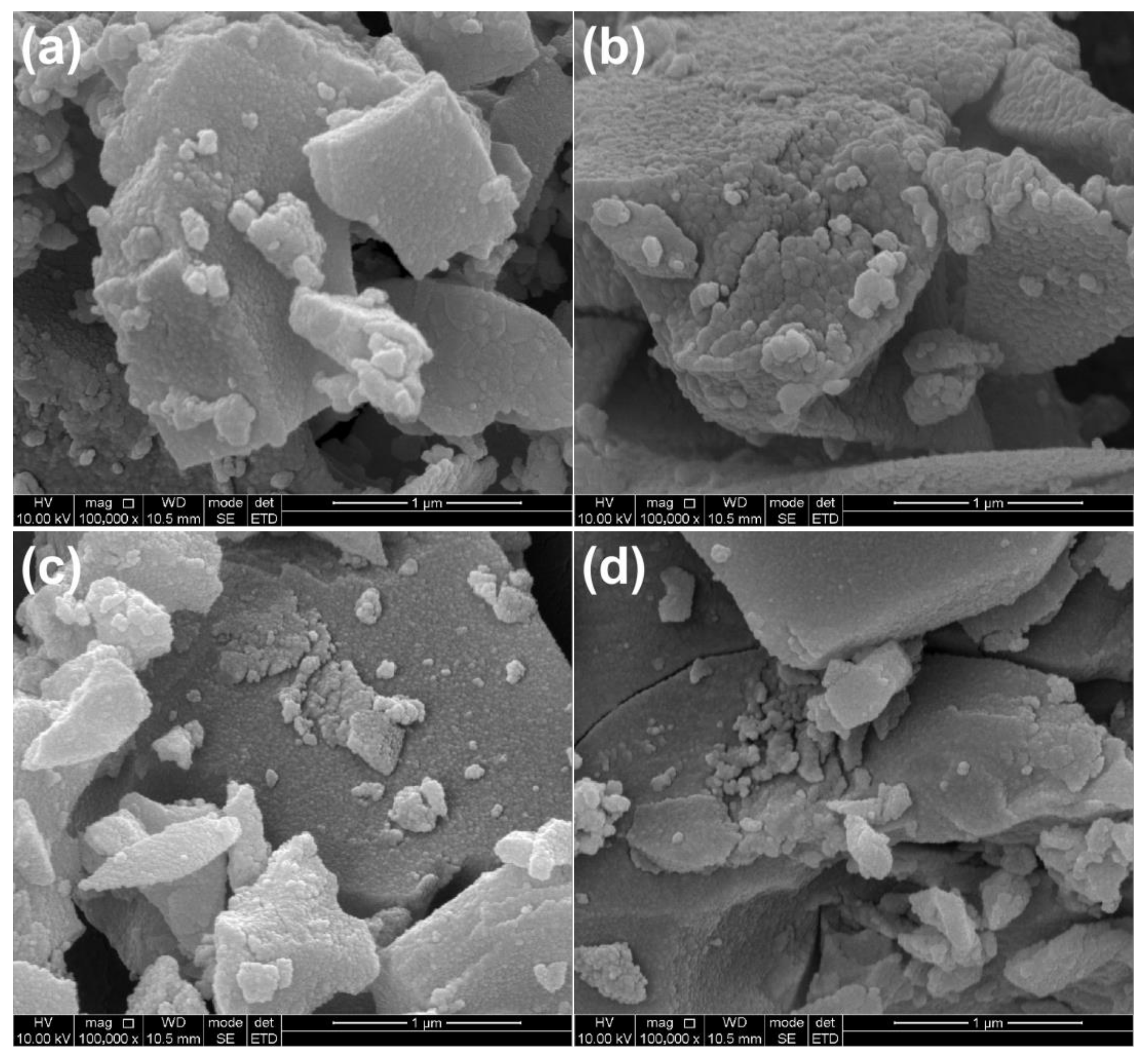
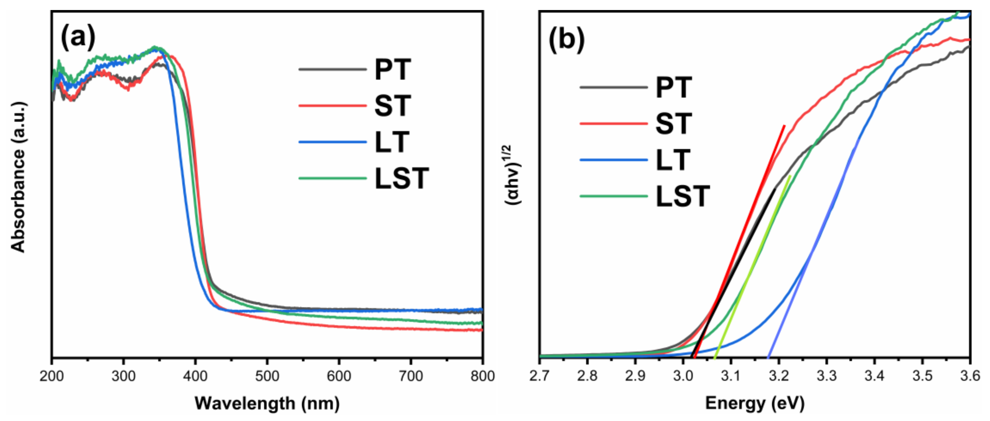
2.1. Photocatalyst Characterization

Figure 1 shows the XRD patterns of samples. The diffraction peaks of PT pattern appeared near 27.3°, 41.2°, and 44.3°, corresponding to the crystal planes of rutile (110), (111), and (210), respectively (JCPDS NO. 21-1276). The diffraction peaks of anatase (101), (004), and (200) crystal planes appeared at 25.3°, 37.8°, and 48.1° (JCPDS NO. 21-1272). The results show that PT had an anatase/rutile mixed crystalline structure. Most of the diffraction peaks corresponded to rutile phase in the ST pattern, indicating that Sn doping promoted the transformation from anatase to rutile. Correspondingly, anatase peaks could only be found in the pattern of LT, suggesting that the phase transformation was inhibited by La doping. Due to their similar ionic radius, Sn4+ ions (0.069 nm) could enter the lattice to replace Ti4+ ions (0.0605 nm), causing lattice defects, weakening the binding force between atoms, and making the Ti–O bonds easier to crack, which was conducive to phase transition [5]. Furthermore, since SnO2 and rutile have the same crystal structure, SnO2 would become the heterogeneous nucleation center of rutile, promoting the transformation [22]. The radius of La3+ ions (0.103 nm) is much larger than that of Ti4+ ions, hindering their entry into the TiO2 lattice. La elements should be dispersed on the surface of TiO2 particles in the form of oxide La2O3 [6]. The transformation requires the rearrangement of atoms and the breaking of bonds. La2O3 dispersed on TiO2 surface will hinder the migration of Ti and O atoms, and delay the nucleation and growth of rutile, inhibiting the phase transition [6,19]. It can be seen that the peaks in LST were indexed to anatase, and only a weak peak at 27.3° ascribed to rutile was detected, implying that LST was composed of mostly anatase and a small amount of rutile, revealing that the inhibition of phase transformation by La doping was stronger than the promotion of Sn doping.
Figure 2 presents the SEM images of samples. It can be observed that PT was composed of agglomerates with different shapes, and the size distribution of agglomerates ranged from tens to hundreds of nanometers. ST, LT, and LST samples were also composed of agglomerates of different shapes. No obvious change in particle morphology can be found in SEM images before and after doping.
Figure 3 shows the TEM images of PT (a) and LST (b). It can be observed that PT particles were seriously agglomerated, and it was difficult to distinguish the size of single particles. LST particles were also agglomerated, possibly due to the high temperature of 600 °C, and the particles grew larger during the process of heating. The HRTEM images of PT (c) and LST (d) are shown in Figure 3c,d. The crystal lattice stripes were clear, indicating that the samples had good crystallinity. The spacings of crystal planes marked in Figure 3c were 0.35 nm and 0.33 nm, corresponding to the anatase (101) crystal plane and the rutile (110) crystal plane, respectively [24,25,26,27], indicating that PT was a mixed crystal composed of anatase and rutile, consistent with the XRD results. The crystal plane spacing 0.36 nm marked in Figure 3d was indexed to the anatase (101) crystal plane of LST. The signals of Ti, O, La, and Sn elements could be found in the STEM mappings of LST (Figure 3e–k), indicating that La and Sn elements already existed in TiO2 due to doping, and the four elements were substantially uniformly distributed in the LST sample.
Figure 4 presents the XPS spectra of PT and LST. The appearance of the La 3d peak and Sn 3d peak in the LST spectra confirms the existence of La and Sn elements in the co-doped sample. Figure 4b shows the high-resolution spectra of Ti 2p. The Ti 2p of PT was decomposed into two peaks, located at 458.5 eV and 464.1 eV, corresponding to Ti 2p3/2 and Ti 2p1/2, indicating that the valence state of Ti was +4. The two peaks corresponding to Ti 2p3/2 and Ti 2p1/2 were located at 458.6 eV and 464.3 eV in LST, indicating that Ti in the co-doped sample also had a valence of +4 [15,28,29]. The high-resolution spectra of O 1s are shown in Figure 4c. The O 1s of PT was decomposed into two peaks corresponding to the lattice oxygen (OL) and surface hydroxyl (OH), located at 529.9 eV and 531.2 eV, respectively. The peaks of the lattice oxygen and surface hydroxyl of LST sample were at 530.0 eV and 531.1 eV respectively [30]. It can be observed that the surface hydroxyl peak area of the LST sample was significantly larger than that of PT, indicating that the co-doping advanced the surface adsorption performance and introduced more OH groups on the particle surface. The percentages of oxygen in the surface hydroxyl group of PT and LST were 12.1% and 22.6%, respectively, indicating that co-doping could enhance the surface hydroxyl content. This is beneficial to generate more hydroxyl radical and improve photocatalytic performance during photodegradation [31]. The Sn 3d high-resolution spectrum of LST is shown in Figure 4d. The two peaks at 486.3 eV and 494.9 eV corresponded to Sn 3d5/2 and Sn 3d3/2, respectively. The location of these two peaks indicates that the Sn element had a +4 valence [21,23,32]. The high-resolution spectrum of La 3d is shown in Figure 4e. The peaks at 834.8 eV and 838.1 eV corresponded to La 3d5/2, and the peaks at 852.2 eV and 855.1 eV corresponded to La 3d3/2, indicating that the La element existed in the form of La3+ [31,33].
Figure 5 shows the UV/Vis absorption spectra (a) and the bandgap (b) of the samples. In the ultraviolet region, all samples had high absorption, whereas, in the visible region, absorption decreased rapidly. The absorption edge of ST was almost the same as that of PT, and the absorption edges of LT and LST showed a weak blue shift. The bandgaps of PT, ST, LT, and LST were 3.02, 3.02, 3.17, and 3.06 eV, respectively. It has been documented that the absorption edge of TiO2 red-shifts and the bandgap decreases after Sn or La doping [31,34,35]. However, in this work, the bandgaps of LT and LST increased slightly. Combined with XRD, it can be seen that the transformation from anatase to rutile was inhibited by La doping. Due to the bandgap of anatase (3.2 eV) being larger than that of rutile (3.0 eV), the bandgaps of LT and LST samples increased compared to PT.
The PL spectra of samples are shown in Figure 6. PT and LST showed the same spectral shape but different intensity. The PL peak intensity decreased after Sn doping, indicating that the recombination of photogenerated electrons and holes was inhibited. The main PL peaks of PT and ST appeared at approximately 422 nm, due to the photons released when the photogenerated electrons returned directly from the conduction band to the valence band [32]. DRS results show that the bandgap of PT and ST was 3.02 eV, and the corresponding photon wavelength was 410 nm, which was about 12 nm less than the corresponding wavelength of 422 nm in the main peak of PL. This deviation could be attributed to the Stokes shift [36,37]. Compared with PT, the PL main peaks of LT and LST shifted to a lower wavelength, consistent with the bandgaps of LT and LST being larger than that of PT. The PL peak intensity of LT was lower than that of PT, indicating that La doping could also inhibit the recombination of photogenerated carriers. In particular, the PL peak intensity of LST was the lowest, implying that La/Sn co-doping had a synergistic effect on retarding the recombination of photogenerated charges, which was beneficial to the photocatalytic performance.

2.2. Photocatalytic Performance

Figure 7 shows the MB degradation degree curves (a) and kinetics curves (b) of samples. After 60 min, the degradation degree of PT was 59.3%, and the degradation degrees of ST, LT, and LST were 67.7%, 66.6%, and 80.6%, respectively. The degradation of MB on the surface of TiO2 photocatalyst conformed to a first-order reaction, and the reaction rate constant k was calculated using the formula kt = −ln(C/C0), where C0 and C are the initial concentration of MB solution and the concentration at time t, respectively. A larger k value denotes a faster reaction rate and a better photocatalytic activity. The first-order reaction rate constants of PT, ST, LT, and LST were 0.015, 0.019, 0.018, and 0.027 min−1, respectively. The first-order reaction rate constants of ST, LT, and LST are 1.3, 1.2, and 1.8 times higher than that of PT, respectively, indicating that the reaction rate was accelerated after doping. Table 1 summarizes the degradation degrees of reported photocatalysts.

2.3. Photocatalytic Degradation Mechanism

Figure 8 shows the results active species experiments of LST. When benzoquinone (BQ), isopropanol (IPA), and ammonium oxalate (AO) were added as the radical scavengers, the degradation degrees of LST decreased from 80.6% to 28.1%, 61.5%, and 78.0%, respectively. Since BQ, IPA, and AO are capture agents of •O2, •OH, and photogenerated holes (h+) [44,45], the results show that the •O2 radical was the main active group, whereas h+ and •OH played a subsidiary role in the degradation process toward MB.
Nitro-blue tetrazolium (NBT) and 2,3-dihydroxybenzoic acid (2,3-HBA) experiments were launched to further verify that the •O2 and •OH radicals were yielded in the degradation process, and the results are shown in Figure 9. The presence of •O2 radicals can be testified by the reaction of NBT with •O2 to form a purple precipitate. With the extension of illumination time, the •O2 radicals react with NBT to generate more and more purple precipitates, thus consuming NBT and gradually reducing its absorbance. On the other hand, salicylic acid (SA) reacts with •OH radicals to form 2,3-HBA which has a special absorption at 510 nm [44,45]. With the increase in time, the absorbance of NBT decreased and the absorbance of 2,3-HBA increased, indicating that •O2 radicals and •OH radicals were generated in the system.
Figure 10 shows the NBT and 2,3-HBA absorbance of LST and PT after illumination for 20 min. The NBT absorbance of PT was higher than that of LST, indicating that LST generated more •O2 radicals. The 2,3-HBA absorbance of PT was lower than that of LST, proving that LST produced more •OH radicals. During the photodegradation process, LST generated more free radicals, implying that it had a higher photogenerated charge separation rate compared to PT, consistent with the results of the PL spectra and photocatalytic activity experiments.
The separation and transfer of photogenerated charges of PT and LST were further studied through photoelectrochemical measurements. Figure 11a shows the photocurrent responses curves (PC) of PT and LST. Generally, a higher photocurrent density denotes a higher separation efficiency of photogenerated charges [46,47]. Both PT and LST generated photocurrent under light, and the photocurrent density of LST was higher than that of PT, indicating that Sn and La co-doping was beneficial to improve the separation efficiency of carriers. Figure 11b shows the electrochemical impedance spectra (EIS) of PT and LST. According to Nyquist’s theorem [46,48,49], the arc radius of LST was smaller than that of PT, indicating that LST had a lower charge motion resistance and a better electron mobility.
Figure 12 shows the Schottky curves of PT and LST. The electrode/electrolyte was is measured according to the Mott–Schottky equation (1) [48,50,51].
1 C 2 = 2 N D e ε 0 ε E E F B k T e ,
where C is the space charge capacitance in the semiconductor, ND is the electron carrier density, e is the elemental charge, ε0 is the permittivity of a vacuum, ε is the relative permittivity of the semiconductor, E is the applied potential, EFB is the flat band potential, T is the temperature, and k is the Boltzmann constant. The slope of the linear part of the curve was positive, indicating that LST is an n-type semiconductor [48,49,51]. In Equation (1), if 1/C2 = 0, the EFB value of the LST flat band potential could be estimated to be −0.55 V vs. Ag/AgCl. According to the formula ENHE = EAg/AgCl + 0.197 [52,53,54], the EFB of LST could be calculated to be about −0.35 V vs. NHE. It is generally believed that the CB bottom (ECB) is about 0.1 V more negative than the potential of EFB for n-type semiconductors [55,56]. Therefore, the ECB of LST could be determined to be −0.45 V vs. NHE. According to the formula EVB = Eg + ECB [49,51], the potential of VB (EVB) of LST could be calculated to be +2.61 V vs. NHE.
Some studies reported that ion doping brings impurity energy levels below the conduction band of TiO2, reduces the bandgap width, and increases the absorption of light [20,23]. Conversely, several studies also showed that there is a blue shift after doping [57,58]. In this work, DRS results show that the bandgap width of PT was 3.02 eV and that of LST was 3.06 eV, without a red shift, which could be ascribed to the fact that co-doping of Sn and La inhibited the transformation from anatase to rutile, and the forbidden band width of rutile was lower than that of anatase. Consequently, a weak blue shift occurred after La/Sn co-doping, implying that the utilization of visible light did not improve via co-doping. It can be seen from PL spectra that single doping of Sn and La or co-doping could inhibit the recombination of photogenerated electrons and holes, and LST exhibited the best inhibition ability. According to the DRS and Mott–Schottky results, the ECB of LST was −0.45 V, which is higher than E0(O2/•O2) (−0.046 V vs. NHE) [59,60]. The potential position of CB ensured the generation of •O2 radicals, as also confirmed by NBT experiments. On the basis of the above results, the mechanism diagram of photodegradation of MB by LST is shown in Figure 13.
On one hand, as the radius of Sn4+ is close to that of Ti4+, the replacement of Ti4+ by Sn4+ ions would form crystal defects, capture photogenerated charges, and improve quantum efficiency [61,62,63]. On the other hand, no peak related to La element was detected in the XRD pattern. The radius of La3+ ions is much larger than that of Ti4+ ions; thus, the possibility that La3+ ions entered the TiO2 lattice can be excluded. Instead they were dispersed on the surface of TiO2 particles in the form of oxide La2O3, which captured photogenerated charges, improving the separation of carriers [16,33,64]. Therefore, the co-doping of Sn and La produced a synergistic effect on improving the carrier separation; as a result, its photocatalytic activity was higher than that of single-element doping in PT.

3. Materials and Methods

Butyl titanate (Analytical Reagent, AR, ≥98.0%), anhydrous ethanol (AR, ≥99.7%), glacial acetic acid (AR, ≥99.5%), tin tetrachloride pentahydrate (AR, ≥99.0%), lanthanum nitrate hexahydrate (AR, ≥99.0%), benzoquinone (AR, ≥98.5%), ammonium oxalate (AR, ≥99.5%), and isopropanol (AR, ≥99.7%) were purchased from Chengdu Chron Chemicals Co., Ltd., (Chengdu, China).

3.1. Sample Preparation

Butyl titanate and anhydrous ethanol were added into the beaker at a volume ratio of 1:2 to form solution A. Deionized water, glacial acetic acid and anhydrous ethanol were mixed in a volume ratio of 2:3:7.5 to form solution B. After being evenly stirred, solution B was dropped into solution A, which was continuously stirred to form sol and then aged. After the aging, the gel was dried in the oven, and finally heat-treated at 600 °C. After grinding, pure TiO2 powder was obtained, labeled as PT. Certain amounts of SnCl4·5H2O and La(NO3)3·6H2O were added to solution B, and the other steps were the same to prepare the doped sample with a Sn/Ti molar ratio of 3% and a La/Ti molar ratio of 0.5%. Sn/La co-doped TiO2 could be prepared by adding SnCl4·5H2O and La(NO3)3·6H2O into solution B, where the molar ratio of Sn/Ti was 3% and the molar ratio of La/Ti was 0.5%. Sn-doped TiO2, La-doped TiO2, and La/Sn co-doped TiO2 were labeled as ST, LT, and LST, respectively.

3.2. Sample Characterization

The crystal structure of the samples was analyzed using a DX-2700 X-ray diffractometer (Dandong Haoyuan Instrument Co. Ltd., Dandong, China, XRD). An FEI-Inspect F50 scanning electron microscope (FEI Company, Hillsboro, OR, USA, SEM) and an FEI-Tecnai G2 F20 transmission electron microscope (FEI Company, Hillsboro, OR, USA, TEM and HRTEM) were used to observe the morphology. An XSAM800 multifunctional surface analysis system was used to analyze the element composition and valence state of samples (Kratos Ltd., Manchester, UK, XPS). A UV-3600 ultraviolet/visible-light spectrophotometer was used to study the optical absorption performance (Shimadzu Group Company, Kyoto, Japan, DRS). Detection of the recombination of photogenerated electrons and holes was investigated using an F-4600 fluorescence spectrometer (Shimadzu Group Company, Kyoto, Japan, PL). The photocurrent response curves, electrochemical impedance spectroscopy, and Mott–Schottky plots were measured using a DH-7000 electrochemical workstation (Jiangsu Donghua Analytical Instrument Co., Ltd., China, PC, EIS and MS).

3.3. Photocatalysis Experiment

The photocatalytic activity of the sample was evaluated with MB solution as the target pollutant. Briefly, 100 mL of MB (10 mg/L) solution and 0.05 g of sample powder were mixed at room temperature (25 °C) and kept neutral. After stirring for 30 min in the dark, the mixture was irradiated using a 250 W xenon lamp (300–800 nm). Samples were taken every 20 min. The absorbance of the supernatant was tested at 664 nm. The MB degradation degree was calculated using the formula (A0 − At)/A0 × 100%, where A0 and At are the initial and time t absorbances of MB.
On the basis of the MB degradation system, 2 mL (0.1 mol/L) of benzoquinone (BQ, •O2 trapping agent), isopropanol (IPA, •OH trapping agent), and ammonium oxalate (AO, h+ trapping agent) were added to investigate the active species.

4. Conclusions

Pure TiO2 was prepared using the sol–gel method and modified by La/Sn single doping and co-doping. PT had an anatase/rutile mixed crystalline structure, Sn promoted the transformation from anatase to rutile, and La inhibited the transformation. The inhibition of La was stronger than that of Sn, and the phase transformation from anatase to rutile was inhibited by co-doping. La/Sn co-doping did not produce an obvious red shift; however, the content of hydroxyl on the surface of TiO2 particles increased, and a synergistic effect was produced on inhibiting the recombination of photogenerated electrons and holes. The results of electrochemical experiments also showed that the separation and transfer of photogenerated charges were faster after La/Sn co-doping, and the quantum efficiency was improved. Therefore, the photocatalytic activity of LST was superior to that of ST, LT, and PT. The first-order reaction rate constant of PT was 0.015 min1, and the first-order reaction rate constants of ST, LT, and LST were 1.3, 1.2, and 1.8 times higher than that of PT, respectively. The active species experiments of LST showed that •O2 radicals were the main active groups during the photodegradation process.

Author Contributions

Methodology, X.Z., Y.J. and W.F.; investigation, F.Q. and L.H.; writing—original draft preparation, F.Q.; writing—review and editing, L.H.; supervision, W.F.; project administration, X.Z.; formal analysis, Y.J. and X.Z.; funding acquisition, X.Z. All authors have read and agreed to the published version of the manuscript.

Funding

This study was supported by the Key Research and Development Projects of Liangshan Prefecture Science and Technology Bureau of Sichuan Province (21ZDYF0202), the Higher-Education Talent Quality and Teaching Reform Project of Sichuan Province (JG2021-1104), and the Talent Training Quality and Teaching Reform Project of Chengdu University (cdjgb2022033).

Institutional Review Board Statement

Not applicable.

Informed Consent Statement

Not applicable.

Data Availability Statement

Not applicable.

Conflicts of Interest

The authors declare no conflict of interest.

References

  1. Sun, Y.; Liu, E.D.; Zhu, L.; Wen, Y.; Tan, Q.W.; Feng, W. Influence of annealing temperature of TiO2 nanotubes via hydrothermal method on Ti foil for photocatalytic degradation. Dig. J. Nanomater. Bios. 2019, 14, 463–470. [Google Scholar]
  2. Zhu, X.D.; Xu, H.Y.; Yao, Y.; Liu, H.; Wang, J.; Pu, Y.; Feng, W.; Chen, S.H. Effects of Ag0-modification and Fe3+-doping on the structural, optical and photocatalytic properties of TiO2. RSC Adv. 2019, 9, 40003–40012. [Google Scholar] [CrossRef] [PubMed]
  3. Sun, Y.; Xu, S.; Zeng, J.Y.; Yang, S.S.; Zhao, Q.R.; Yang, Y.; Zhao, Q.; Wang, G.X. Fabrication and photocatalytic activity of TiO2 nanotubes by hydrothermal treatment. Dig. J. Nanomater. Bios. 2021, 16, 239–246. [Google Scholar]
  4. Wu, D.; Li, C.; Zhang, D.S.; Wang, L.L.; Zhang, X.P.; Shi, Z.F.; Lin, Q. Photocatalytic improvement of Y3+ modified TiO2 prepared by a ball milling method and application in shrimp wastewater treatment. RSC Adv. 2019, 9, 14609–14620. [Google Scholar] [CrossRef]
  5. Solís-Casados, D.A.; Escobar-Alarcón, L.; Gómez-Oliván, L.M.; Haro-Poniatowski, E.; Klimova, T. Photodegradation of pharmaceutical drugs using Sn-modified TiO2 powders under visible light irradiation. Fuel 2017, 198, 3–10. [Google Scholar] [CrossRef]
  6. Yan, J.K.; Rong, X.Q.; Gu, X.; Du, J.H.; Gan, G.Y. Phase composition and photocatalytic properties of La3+-doped TiO2 nanopowders. Rare Metal Mat. Eng. 2020, 49, 0465–0475. [Google Scholar]
  7. Umar, K.; Ibrahim, M.N.M.; Ahmad, A.; Rafatullah, M. Synthesis of Mn-doped TiO2 by novel route and photocatalytic mineralization/intermediate studies of organic pollutants. Res. Chem. Intermediat. 2019, 45, 2927–2945. [Google Scholar] [CrossRef]
  8. Zhang, W.; Li, X.J.; Jia, G.; Gao, Y.F.; Wang, H.; Cao, Z.Z.; Li, C.H.; Liu, J.R. Preparation, characterization, and photocatalytic activity of boron and lanthanum co-doped TiO2. Catal. Commun. 2014, 45, 144–147. [Google Scholar] [CrossRef]
  9. Huang, T.Z.; Mao, S.; Yu, J.M.; Wen, Z.H.; Lu, G.H.; Chen, J.H. Effects of N and F doping on structure and photocatalytic properties of anatase TiO2 nanoparticles. RSC Adv. 2013, 3, 16657–16664. [Google Scholar] [CrossRef]
  10. Lin, X.X.; Rong, F.; Fu, D.G.; Yuan, C.W. Enhanced photocatalytic activity of fluorine doped TiO2 by loaded with Ag for degradation of organic pollutants. Powder Technol. 2012, 219, 173–178. [Google Scholar] [CrossRef]
  11. Wang, Y.Z.; Wu, Y.S.; Yang, H.; Xue, X.X.; Liu, Z.H. Doping TiO2 with boron or/and cerium elements: Effects on photocatalytic antimicrobial activity. Vacuum 2016, 131, 58–64. [Google Scholar] [CrossRef]
  12. Adyani, S.M.; Ghorbani, M. A comparative study of physicochemical and photocatalytic properties of visible light responsive Fe, Gd and P single and tri-doped TiO2 nanomaterials. J. Rare Earths 2018, 36, 72–85. [Google Scholar] [CrossRef]
  13. Kalantari, K.; Kalbasi, M.; Sohrabi, M.; Royaee, S.J. Enhancing the photocatalytic oxidation of dibenzothiophene using visible light responsive Fe and N co-doped TiO2 nanoparticles. Ceram. Int. 2017, 43, 973–981. [Google Scholar] [CrossRef]
  14. Shaban, M.; Ahmed, A.M.; Shehata, N.; Betiha, M.A.; Rabie, A.M. Ni-doped and Ni/Cr co-doped TiO2 nanotubes for enhancement of photocatalytic degradation of methylene blue. J. Colloid Interface Sci. 2019, 555, 31–41. [Google Scholar] [CrossRef]
  15. Chen, Y.; Liu, K.R. Fabrication of Ce/N co-doped TiO2/diatomite granule catalyst and its improved visible-light-driven photoactivity. J. Hazard. Mater. 2017, 324, 139–150. [Google Scholar] [CrossRef]
  16. Nešić, J.; Manojlović, D.D.; Andelković, I.; Dojcinović, B.P.; Vulić, P.J.; Krstić, J.; Roglić, G.M. Preparation, characterization and photocatalytic activity of lanthanum and vanadium co-doped mesoporous TiO2 for azo-dye degradation. J. Mol. Catal. A Chem. 2013, 378, 67–75. [Google Scholar] [CrossRef]
  17. Peng, H.; Cui, J.; Zhan, H.J.; Zhang, X. Improved photodegradation and detoxification of 2,4,6-trichlorophenol by lanthanum doped magnetic TiO2. Chem. Eng. J. 2015, 264, 316–321. [Google Scholar] [CrossRef]
  18. Xin, Y.J.; Liu, H.L. Study on mechanism of photocatalytic performance of La-doped TiO2/Ti photoelectrodes by theoretical and experimental methods. J. Solid State Chem. 2011, 184, 3240–3246. [Google Scholar] [CrossRef]
  19. Cruz, D.D.L.; Arévalo, J.C.; Torres, G.; Margulis, B.R.G.; Ornelas, C.; Aguilar-Elguézabal, A. TiO2 doped with Sm3+ by sol-gel: Synthesis, characterization and photocatalytic activity of diuron under solar light. Catal. Today 2011, 166, 152–158. [Google Scholar] [CrossRef]
  20. Mohamed, R.M.; Aazam, E.S. Effect of Sn loading on the photocatalytic aniline synthesis activity of TiO2 nanospheres. J. Alloys Compd. 2014, 595, 8–13. [Google Scholar] [CrossRef]
  21. Li, J.L.; Xu, X.T.; Liu, X.J.; Yu, C.Y.; Yan, D.; Sun, Z.; Pan, L.K. Sn doped TiO2 nanotube with oxygen vacancy for highly efficient visible light photocatalysis. J. Alloys Compd. 2016, 679, 454–462. [Google Scholar] [CrossRef]
  22. Alves, A.K.; Berutti, F.A.; Bergmann, C.P. Visible and UV photocatalytic characterization of Sn-TiO2 electrospun fibers. Catal. Today 2013, 208, 7–10. [Google Scholar] [CrossRef]
  23. Li, J.; Shi, J.; Li, Y.B.; Ding, Z.L.; Huang, J.G. A biotemplate synthesized hierarchical Sn-doped TiO2 with superior photocatalytic capacity under simulated solar light. Ceram. Int. 2021, 47, 8218–8227. [Google Scholar] [CrossRef]
  24. Dong, P.M.; Cheng, X.D.; Huang, Z.F.; Chen, Y.; Zhang, Y.Z.; Nie, X.X.; Zhang, X.W. In-situ and phase controllable synthesis of nanocrystalline TiO2 on flexible cellulose fabrics via a simple hydrothermal method. Mater. Res. Bull. 2018, 97, 89–95. [Google Scholar] [CrossRef]
  25. Nguyen-Phan, T.D.; Pham, V.H.; Chung, J.S.; Chhowalla, M.; Asefa, T.; Kim, W.J.; Shin, E.W. Photocatalytic performance of Sn-doped TiO2/reduced graphene oxide composite materials. Appl. Catal. A Gen. 2014, 473, 21–30. [Google Scholar] [CrossRef]
  26. Wu, M.C.; Wu, P.Y.; Lin, T.H.; Lin, T.F. Photocatalytic performance of Cu-doped TiO2 nanofibers treated by the hydrothermal synthesis and air-thermal treatment. Appl. Surf. Sci. 2018, 430, 390–398. [Google Scholar] [CrossRef]
  27. Tang, M.; Xia, Y.W.; Yang, D.X.; Lu, S.J.; Zhu, X.D.; Tang, R.Y.; Zhang, W.M. Ag decoration and SnO2 coupling modified anatase/rutile mixed crystal TiO2 composite photocatalyst for enhancement of photocatalytic degradation towards tetracycline hydrochloride. Nanomaterials 2022, 12, 873. [Google Scholar] [CrossRef]
  28. Zhu, X.D.; Wen, G.L.; Liu, H.; Han, S.H.; Chen, S.H.; Kong, Q.Q.; Feng, W. One-step hydrothermal synthesis and characterization of Cu-doped TiO2 nanoparticles/nanobucks/nanorods with enhanced photocatalytic performance under simulated solar light. J. Mater. Sci. Mater Electron. 2019, 30, 13826–13834. [Google Scholar] [CrossRef]
  29. Mathews, N.R.; Cortes Jacome, M.A.; Angeles-Chavez, C.; Toledo Antonio, J.A. Fe doped TiO2 powder synthesized by sol gel method: Structural and photocatalytic characterization. J. Mater. Sci. Mater. Electron. 2015, 26, 5574–5584. [Google Scholar] [CrossRef]
  30. Zhu, X.D.; Zhou, Q.; Xia, Y.W.; Wang, J.; Chen, H.J.; Xu, Q.; Liu, J.W.; Feng, W.; Chen, S.H. Preparation and characterization of Cu-doped TiO2 nanomaterials with anatase/rutile/brookite triphasic structure and their photocatalytic activity. J. Mater. Sci. Mater. Electron. 2021, 32, 21511–21524. [Google Scholar] [CrossRef]
  31. Chen, Y.; Wu, Q.; Zhou, C.; Jin, Q.T. Enhanced photocatalytic activity of La and N co-doped TiO2/diatomite composite. Powder Technol. 2017, 322, 296–300. [Google Scholar] [CrossRef]
  32. Zhu, X.D.; Zhu, R.R.; Pei, L.X.; Liu, H.; Xu, L.; Wang, J.; Feng, W.; Jiao, Y.; Zhang, W.M. Fabrication, characterization, and photocatalytic activity of anatase/rutile/SnO2 nanocomposites. J. Mater. Sci. Mater. Electron. 2019, 30, 21210–21218. [Google Scholar] [CrossRef]
  33. Yu, Y.M.; Piao, L.J.; Xia, J.X.; Wang, W.Z.; Geng, J.F.; Chen, H.Y.; Xing, X.; Li, H. A facile one-pot synthesis of N-La codoped TiO2 porous materials with bio-hierarchical architectures and enhanced photocatalytic activity. Mater. Chem. Phys. 2016, 182, 77–85. [Google Scholar] [CrossRef]
  34. Jiang, H.Q.; Liu, Y.D.; Li, J.S.; Wang, H.Y. Synergetic effects of lanthanum, nitrogen and phosphorus tri-doping on visible-light photoactivity of TiO2 fabricated by microwave-hydrothermal process. J. Rare Earth 2016, 34, 604–613. [Google Scholar] [CrossRef]
  35. Tripathi, A.K.; Mathpal, M.C.; Kumar, P.; Singh, M.K.; Soler, M.A.G.; Agarwal, A. Structural, optical and photoconductivity of Sn and Mn doped TiO2 nanoparticles. J. Alloys Compd. 2015, 622, 37–47. [Google Scholar] [CrossRef]
  36. Wang, S.X.; Song, Z.; Kong, Y.W.; Liu, Q.L. Relationship of Stokes shift with composition and structure in Ce3+/Eu2+-doped inorganic compounds. J. Lumin. 2019, 212, 250–263. [Google Scholar] [CrossRef]
  37. Khaidukov, N.M.; Makhov, V.N.; Zhang, Q.H.; Shi, R.; Liang, H.B. Extended broadband luminescence of dodecahedral multisite Ce3+ ions in garnets {Y3}[MgA](BAlSi)O12 (A = Sc, Ga, Al; B = Ga, Al). Dyes Pigments 2017, 142, 524–529. [Google Scholar] [CrossRef]
  38. Kamali, A.R.; Zhu, W.H.; Shi, Z.N.; Wang, D.X. Combustion synthesis-aqueous hybridization of nanostructured graphene-coated silicon and its dye removal performance. Mater. Chem. Phys. 2022, 277, 125565. [Google Scholar] [CrossRef]
  39. Wei, S.H.; Kamali, A.R. Waste plastic derived Co3Fe7/CoFe2O4@carbon magnetic nanostructures for efficient dye adsorption. J. Alloys Compd. 2021, 886, 161201. [Google Scholar] [CrossRef]
  40. Zhao, Z.Y.; Kamali, A.R. One-step conversion of Mg2Si into hydrogen-terminated porous silicon nanostructures. Mater. Today Chem. 2021, 22, 100621. [Google Scholar] [CrossRef]
  41. Si, Y.J.; Liu, H.H.; Li, N.T.; Zhong, J.B.; Li, J.Z.; Ma, D.M. SDBS-assisted hydrothermal treatment of TiO2 with improved photocatalytic activity. Mater. Lett. 2018, 212, 147–150. [Google Scholar] [CrossRef]
  42. Huang, J.; Ding, L.; Xi, Y.N.; Shi, L.; Su, G.; Gao, R.J.; Wang, W.; Dong, B.H.; Cao, L.X. Efficient silver modification of TiO2 nanotubes with enhanced photocatalytic activity. Solid State Sci. 2018, 80, 116–122. [Google Scholar] [CrossRef]
  43. Liu, L.; Liu, Z.W.; Yang, Y.X.; Geng, M.Q.; Zou, Y.M.; Shahzad, M.B.; Dai, Y.X.; Qi, Y. Photocatalytic properties of Fe-doped ZnO electrospun nanofibers. Ceram. Int. 2018, 44, 19998–20005. [Google Scholar] [CrossRef]
  44. Dou, L.; Zhong, J.B.; Li, J.Z.; Pandian, R.; Burda, C. In-situ construction of 3D nanoflower-like BiOI/Bi2SiO5 heterojunctions with enhanced photocatalytic performance for removal of decontaminants originated from a step-scheme mechanism. Appl. Surf. Sci. 2021, 544, 148883. [Google Scholar] [CrossRef]
  45. Zhu, X.D.; Wang, J.; Yang, D.X.; Liu, J.W.; He, L.L.; Tang, M.; Feng, W.; Wu, X.Q. Fabrication, characterization and high photocatalytic activity of Ag-ZnO heterojunctions under UV-visible light. RSC Adv. 2021, 11, 27257–27266. [Google Scholar] [CrossRef]
  46. Dou, L.; Jin, X.Y.; Chen, J.F.; Zhong, J.B.; Li, J.Z.; Zeng, Y.; Duan, R. One-pot solvothermal fabrication of S-scheme OVs-Bi2O3/Bi2SiO5 microsphere heterojunctions with enhanced photocatalytic performance toward decontamination of organic pollutants. Appl. Surf. Sci. 2020, 527, 146775. [Google Scholar] [CrossRef]
  47. Dou, L.; Li, J.J.; Long, N.; Lai, C.X.; Zhong, J.B.; Li, J.Z.; Huang, S.T. Fabrication of 3D flower-like OVs-Bi2SiO5 hierarchical microstructures for visible light-driven removal of tetracycline. Surf. Interfaces 2022, 29, 101787. [Google Scholar] [CrossRef]
  48. Ye, M.D.; Gong, J.J.; Lai, Y.K.; Lin, C.J.; Lin, Z.Q. High-efficiency photoelectrocatalytic hydrogen generation enabled by palladium quantum dots-sensitized TiO2 nanotube arrays. J. Am. Chem. Soc. 2012, 134, 15720–15723. [Google Scholar] [CrossRef]
  49. Chen, P.F.; Chen, L.; Ge, S.F.; Zhang, W.Q.; Wu, M.F.; Xing, P.X.; Rotamond, T.B.; Lin, H.J.; Wu, Y.; He, Y.M. Microwave heating preparation of phosphorus doped g-C3N4 and its enhanced performance for photocatalytic H2 evolution in the help of Ag3PO4 nanoparticles. Int. J. Hydrogen Energ. 2020, 45, 14354–14367. [Google Scholar] [CrossRef]
  50. Zhang, Z.H.; Yu, Y.J.; Wang, P. Hierarchical top-porous/bottom-tubular TiO2 nanostructures decorated with Pd nanoparticles for efficient photoelectrocatalytic decomposition of synergistic pollutants. ACS Appl. Mater. Inter. 2012, 4, 990–996. [Google Scholar] [CrossRef]
  51. Ding, Y.Y.; Zhang, J.Y.; Yang, Y.; Long, L.Z.; Yang, L.; Yan, L.J.; Kong, W.J.; Liu, F.C.; Lv, F.Z.; Liu, J. Fully-depleted dual p-n heterojunction with type-II band alignment and matched build-in electric field for high-efficient photocatalytic hydrogen production. Int. J. Hydrogen Energ. 2021, 46, 36069–36079. [Google Scholar] [CrossRef]
  52. Wang, J.M.; Kuo, M.T.; Zeng, P.; Xu, L.; Chen, S.T.; Peng, T.Y. Few-layer BiVO4 nanosheets decorated with SrTiO3: Rh nanoparticles for highly efficient visible-light-driven overall water splitting. Appl. Catal. B Environ. 2020, 279, 119377. [Google Scholar] [CrossRef]
  53. Zhou, W.J.; Jia, J.; Lu, J.; Yang, L.J.; Hou, D.M.; Li, G.Q.; Chen, S.W. Recent developments of carbon-based electrocatalysts for hydrogen evolution reaction. Nano Energy 2016, 28, 29–43. [Google Scholar] [CrossRef]
  54. Wang, Y.; Gao, P.; Li, B.H.; Yin, Z.; Feng, L.; Liu, Y.Z.; Du, Z.W.; Zhang, L.Q. Enhanced photocatalytic performance of visible-light-driven CuOx/TiO2-x for degradation of gaseous formaldehyde: Roles of oxygen vacancies and nano copper oxides. Chemosphere 2022, 291, 133007. [Google Scholar] [CrossRef]
  55. Sun, S.M.; Watanabe, M.; Wu, J.; An, Q.; Ishihara, T. Ultrathin WO3·0.33H2O nanotubes for CO2 photoreduction to acetate with high selectivity. J. Am. Chem. Soc. 2018, 140, 6474–6482. [Google Scholar] [CrossRef]
  56. He, W.; Liu, L.; Ma, T.T.; Han, H.M.; Zhu, J.J.; Liu, Y.P.; Fang, Z.; Yang, Z.; Guo, K. Controllable morphology CoFe2O4/g-C3N4 p-n heterojunction photocatalysts with built-in electric field enhance photocatalytic performance. Appl. Catal. B-Environ. 2022, 306, 121107. [Google Scholar] [CrossRef]
  57. Tahir, M. La-modified TiO2/carbon nanotubes assembly nanocomposite for efficient photocatalytic hydrogen evolution from glycerol-water mixture. Int. J. Hydrogen Energy 2019, 44, 3711–3725. [Google Scholar] [CrossRef]
  58. Dubnová, L.; Zvolská, M.; Edelmannová, M.; Matějová, L.; Reli, M.; Drobná, H.; Kuśtrowski, P.; Kočí, K.; Čapek, L. Photocatalytic decomposition of methanol-water solution over N-La/TiO2 photocatalysts. Appl. Surf. Sci. 2019, 469, 879–886. [Google Scholar] [CrossRef]
  59. Ye, L.Q.; Liu, J.Y.; Gong, C.Q.; Tian, L.H.; Peng, T.Y.; Zan, L. Two different roles of metallic Ag on Ag/AgX/BiOX (X = Cl, Br) visible light photocatalysts: Surface plasmon resonance and Z-scheme bridge. ACS Catal. 2012, 2, 1677–1683. [Google Scholar] [CrossRef]
  60. He, Y.M.; Zhang, L.H.; Teng, B.T.; Fan, M.H. A new application of Z-scheme Ag3PO4/g-C3N4 composite in converting CO2 to fuel. Environ. Sci. Technol. 2015, 49, 649–656. [Google Scholar] [CrossRef]
  61. Mohammadi, R.; Massoumi, B. Sn/Cu-TiO2 nanoparticles produced via sol-gel method: Synthesis, characterization, and photocatalytic activity. Russ. J. Phys. Chem. A 2014, 88, 1184–1190. [Google Scholar] [CrossRef]
  62. Du, J.M.; Zhao, G.Y.; Pang, H.; Qian, Y.T.; Liu, H.Q.; Kang, D.J. A template method for synthesis of porous Sn-doped TiO2 monolith and its enhanced photocatalytic activity. Mater. Lett. 2013, 93, 419–422. [Google Scholar] [CrossRef]
  63. Du, J.M.; Chen, H.J.; Yang, H.; Sang, R.R.; Qian, Y.T.; Li, Y.X.; Zhu, G.G.; Mao, Y.J.; He, W.; Kang, D.J. A facile sol–gel method for synthesis of porous Nd-doped TiO2 monolith with enhanced photocatalytic activity under UV–vis irradiation. Micropor. Mesopor. Mat. 2013, 182, 87–94. [Google Scholar] [CrossRef]
  64. Bokare, A.; Pai, M.; Athawale, A.A. Surface modified Nd doped TiO2 nanoparticles as photocatalysts in UV and solar light irradiation. Sol. Energy 2013, 91, 111–119. [Google Scholar] [CrossRef]
Figure 1. XRD patterns of samples.
Ijms 23 11339 g001
Figure 2. SEM images of PT (a), ST (b), LT (c), and LST (d).
Ijms 23 11339 g002
Figure 3. TEM and HRTEM images of PT (a,c) and LST (b,d), and the STEM mappings of LST (ek).
Ijms 23 11339 g003aIjms 23 11339 g003bIjms 23 11339 g003c
Figure 4. XPS survey of PT and LST (a); high resolution spectra of Ti 2p (b), O 1s (c), Sn 3d (d), and La 3d (e).
Ijms 23 11339 g004aIjms 23 11339 g004b
Figure 5. UV/Vis absorption spectra (a) and bandgap energy (b) of samples.
Ijms 23 11339 g005
Figure 6. PL spectra of samples.
Ijms 23 11339 g006
Figure 7. Degradation curves (100 mL of MB (10 mg/L) degraded by 0.05 g of sample at pH = 7 and room temperature of 25 °C) (a) and kinetics curves (b) of samples.
Ijms 23 11339 g007
Figure 8. Degradation degrees of LST in the presence of different scavengers.
Ijms 23 11339 g008
Figure 9. Absorbance curves of NBT (a) and 2,3-HBA (b) of LST.
Ijms 23 11339 g009
Figure 10. NBT absorbance (a) and 2,3-HBA (b) absorbance of LST and PT after 20 min.
Ijms 23 11339 g010
Figure 11. Photocurrent responses curves (a) and electrochemical impedance spectroscopy (b) of PT and LST.
Ijms 23 11339 g011
Figure 12. Mott–Schottky plot of LST.
Ijms 23 11339 g012
Figure 13. Schematic diagram of MB photodegradation by LST.
Ijms 23 11339 g013
Table 1. The summarization of degradation degrees by various photocatalysts.
Ref.MethodPhotocatalystLight SourceTarget PollutantDecolorization Degree
[29]Sol–gel methodFe–TiO2UV light (15 W)RB 69 (100 mg/L)98.0% in 120 min
[31]Sol–gel methodLa–N–TiO2/diatomiteXenon lamp (150 W)RhB (10 mg/L)93.0% in 240 min
[38]Combustion synthesis methodologyMg2Si(Si)/MgOLED lamp (100 W)MB (50 mg/L)90.0% in 120 min
[39]Ball-milling/molten salt processing approachCo3Fe7/CoFe2O4@
carbon
LED lamp (100 W)MB (100 mg/L)100% in 12 min
[40]Acid-treatment methodMg2SiLED lamp (100 W)MO (50 ppm)100% in 30 min
[41]Hydrothermal methodSDBS–TiO2Xenon lamp (500 W)RhB (10 mg/L)90.0% in 120 min
[42]Hydrothermal methodAg–TiO2Xenon lamp (500 W)RhB (20 mg/L)80.0% in 240 min
[43]Electrospinning
method
Fe–ZnOMercury lampMB (10 mg/L)88.0% in 360 min
[44]Solvothermal methodBiOI/Bi2SiO5Xenon lamp (500 W)MO (10 ppm)70.0% in 360 min
This workSol–gel methodLa–Sn–TiO2Xenon lamp (250 W)MB (10 mg/L)80.6% in 60 min
Publisher’s Note: MDPI stays neutral with regard to jurisdictional claims in published maps and institutional affiliations.

