Review

Unraveling Mitochondrial Determinants of Tumor Response to Radiation Therapy

by
Mattia Zaffaroni
1,†,
Maria Giulia Vincini
1,†,
Giulia Corrao
1,
Giulia Marvaso
1,2,*,
Matteo Pepa
1,
Giuseppe Viglietto
3,
Nicola Amodio
3,* and
Barbara Alicja Jereczek-Fossa
1,2
1
Division of Radiation Oncology, IEO—European Institute of Oncology, IRCCS, 20141 Milan, Italy
2
Department of Oncology and Hemato-Oncology, University of Milan, 20122 Milan, Italy
3
Department of Experimental and Clinical Medicine, Magna Graecia University of Catanzaro, 88100 Catanzaro, Italy
*
Authors to whom correspondence should be addressed.
These authors contributed equally to this work.
Int. J. Mol. Sci. 2022, 23(19), 11343; https://doi.org/10.3390/ijms231911343
Submission received: 29 August 2022 / Revised: 20 September 2022 / Accepted: 21 September 2022 / Published: 26 September 2022
(This article belongs to the Special Issue State-of-the-Art Molecular Oncology in Italy)

Abstract

:
Radiotherapy represents a highly targeted and efficient treatment choice in many cancer types, both with curative and palliative intents. Nevertheless, radioresistance, consisting in the adaptive response of the tumor to radiation-induced damage, represents a major clinical problem. A growing body of the literature suggests that mechanisms related to mitochondrial changes and metabolic remodeling might play a major role in radioresistance development. In this work, the main contributors to the acquired cellular radioresistance and their relation with mitochondrial changes in terms of reactive oxygen species, hypoxia, and epigenetic alterations have been discussed. We focused on recent findings pointing to a major role of mitochondria in response to radiotherapy, along with their implication in the mechanisms underlying radioresistance and radiosensitivity, and briefly summarized some of the recently proposed mitochondria-targeting strategies to overcome the radioresistant phenotype in cancer.

1. Introduction

It is historically known that mitochondria developed from an endosymbiotic association between an ancestral bacteria and a proto-eukaryotic host cell, inaugurating a two billion-year symbiotic partnership [1]. Since then, it has been recognized that the mitochondrion is a highly evolved system coordinating energy production and distribution for cellular maintenance and reproduction. Specifically, mitochondria are small, double-membrane organelles whose main task is to ensure general cellular metabolism and the supply of energy by means of ATP through the tricarboxylic acid cycle, electron transport chain (ETC), and oxidative phosphorylation (OXPHOS). While their main function is to serve as metabolic units, mitochondria have also co-evolved with their hosts to function as central signaling nodes in multiple pathways, involved in modulation of the intracellular redox status, reactive oxygen species (ROS) production, and modulation of inflammation and apoptosis. Over the years, mitochondrial DNA (mtDNA) has evolved alongside its nuclear counterpart (nDNA); however, given the relatively small size of the mitochondrial genome, consisting of 37 genes, it has often been ignored in the pioneering sequencing analyses and functional studies. Nevertheless, an increasing number of studies is reporting the influence of mutations in the mtDNA sequence in a large variety of diseases, from metabolic and musculoskeletal ones to cancer [2,3,4].
Overall, mitochondria may contribute to malignant transformation by two major mechanisms, also discussed in more specialized reviews: (i) ROS production, which potentially contributes to the accumulation of DNA mutations and to the activation of oncogenic pathways [5]; and (ii) mitochondria outer membrane permeability transition which is a crucial step required for malignant clones to escape programmed cell death [6,7]. In addition, a third mechanism has recently emerged, consisting in the abnormal accumulation of mitochondrial metabolites (i.e., fumarate and succinate), resulting in transforming effects from normal to malignant clones [8].
In many tumors, radiotherapy (RT) represents the first line of treatment, both with curative and palliative intent, and it is estimated that as many as half of all cancer patients will receive RT at some point throughout the course of disease [9]. Growing evidence suggests that specific mitochondrial changes and metabolic remodeling play a role in the onset of resistance to RT [10,11,12,13]. Radioresistance, defined as the adaptation of tumor cells to ionizing radiation (IR)-induced damages, represents a major clinical issue in various cancer types [14,15]. Its etiology is complex and includes interactions among various cellular mechanisms, such as DNA damage repair mechanisms, cell cycle arrest, oncogenes and/or tumor suppressor genes, tumor microenvironment (TME), microbiome changes, and altered regulation of ROS [12,16].
The present review highlights recent findings on the role of mitochondria in RT, focusing on their implication in the mechanisms underlying radioresistance and radiosensitivity and on the available mitochondria-targeting strategies in the radioresistant setting. A brief focus will be also given to the cross-talk among hypoxia-induced radioresistance, ROS regulation, metabolic reprogramming, and epigenetics.

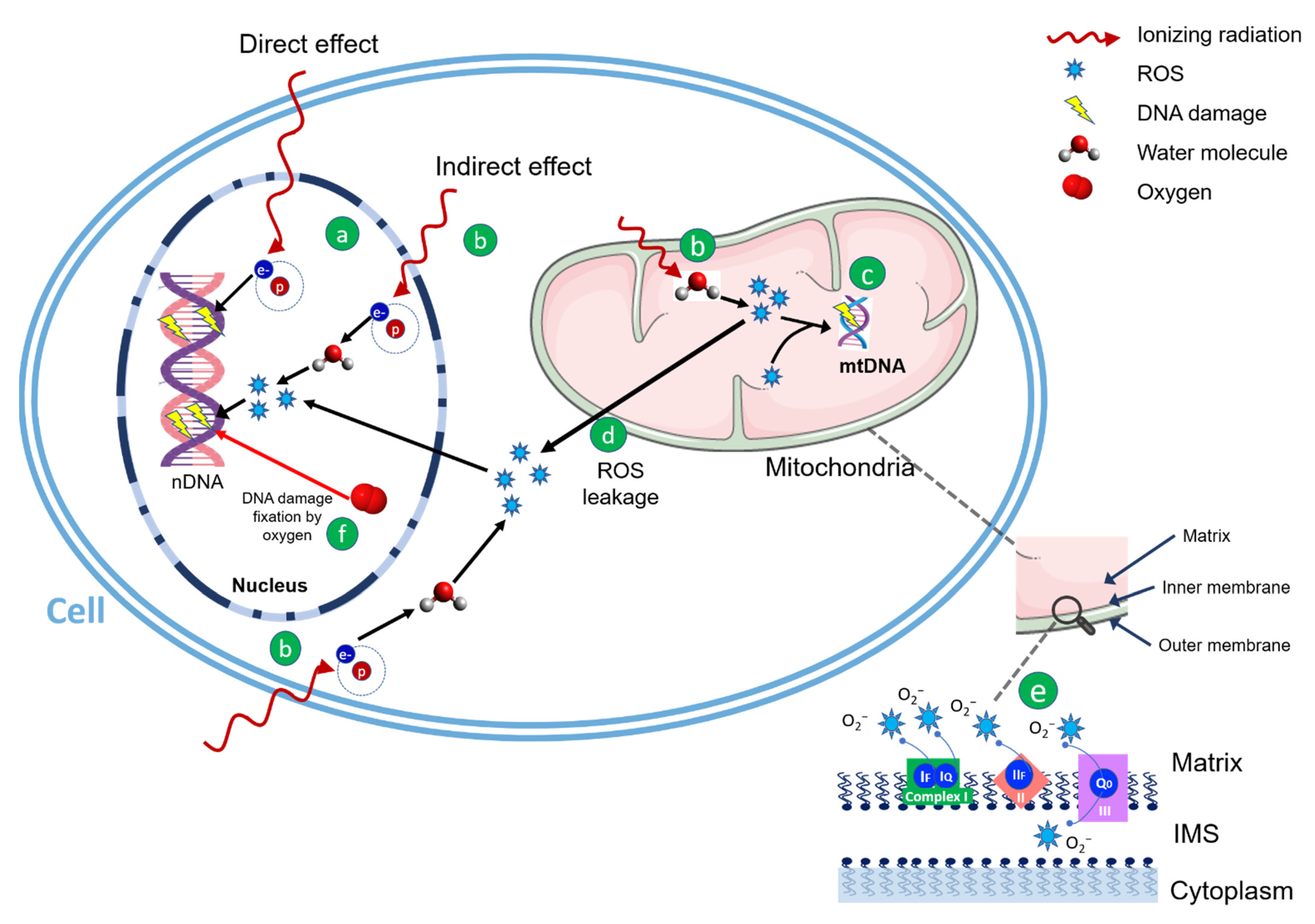
2. Radiation Therapy and Mitochondria

The long-held concept of radiation biology assumed that the effects of IRs were a direct result of targeted DNA damage in the nuclei of impacted cells. On the other hand, non-targeted effects and acquired genomic instability suggested that such a model could be flawed [17,18]. A significant body of the literature reported that IR exposure results in a long-term increase in oxidative stress [19]. In particular, cumulative targeting of mitochondrial metabolism and that of several redox-sensitive pathways by radiation were proven fundamental in elevating oxidative injury and altering cellular physiology within the intracellular microenvironment. The current hypothesis is that oxidative stress might promote an unstable phenotype functioning as a hypothetically unifying biochemical framework that might link multiple seemingly disparate cellular responses to past radiation assaults [20,21,22].
RT is a critical component of many cancer treatments, and generally, its mechanism of action relies on the direct induction of DNA damage or the indirect production of ROS (Figure 1).
In the direct action, electrons directly interact with DNA, causing double- and single-strand breaks (DSBs and SSBs, respectively), ultimately leading to cell death. This process is predominant with dense IR, such as charged particles with enough kinetic energy and high-linear energy transfer (LET) radiations [23], as protons in the end region of their range. In the indirect action, predominantly within sparse IR (photons), water molecules, representing the major constituent of the cell (about 80%), are hit-producing free radicals (such as the hydrogen radical –H•– and hydroxyl radical –OH•–), which are very reactive molecules because of the unpaired orbital electron in their structures. In turn, this event triggers a chain reaction: free radicals are able to react with oxygen, fixating the radiation-induced modification and resulting in irreversible DNA damage [24,25]. This phenomenon is largely explained by the oxygen enhancement ratio (OER) or oxygen enhancement effect, which in radiobiology refers to the boosting of therapeutic or detrimental effects of IR due to the presence of oxygen. Since sparse ionizing radiation induces about 70% of the damage by indirect effect, oxygen is necessary for the fixation of DNA damage by ROS and the main consequence is that tumor hypoxia represents one of the most important factors in the development of radioresistance.
Mitochondria, being the hub of energy generation, are a key supplier of reactive species, especially when metabolic stress disrupts oxidative phosphorylation processes [26]. Depending on the cell type, they may constitute about 4% to 25% of the cell volume, therefore representing a sizeable target for IR [27].
In mammalian cells, under physiological conditions, mitochondria represent the most important source of ROS [28], with ATP synthesis producing them during normal oxygen metabolism and accounting for about the great majority (more than 90%) of the total cellular ROS generation [29,30]. As summarized in Figure 1, ROS are produced, when oxygen is reduced during aerobic respiration, spotted from complexes I, II, and III [31], within the ETC in the inner mitochondrial membrane (IMM), or by oxidoreductase enzymes and metal-catalyzed oxidation throughout the lifetime of the cell cycle [32]. As a result, about 5% of the oxygen consumed by mitochondria gives rise to ROS, ultimately leading to oxidative stress affecting both mt- and nDNA alongside with all the other cellular constituents [33,34].
In different malignancies, mitochondria enable a higher proliferation rate of cancer clones by increasing their energy metabolism through multiple mechanisms, including the switch to glycolysis (instead of OXPHOS) for ATP production [35,36] as well as other metabolic changes, conferring both the ability to evade apoptosis as well as protection from chemical- or radiation-induced damages [37].
Notably, mitochondria not only produce most of a cell’s ROS, but they are also more susceptible than the nucleus to their own deleterious effects, due to the short half-lives of ROS in cells (≤1 μs) which limit their diffusion [38]. Since the respiratory chain located in IMM is the main site for generating ROS, high levels of oxidative stress in cells may be caused by mitochondria targeting drugs, causing oxidative damage to cellular components and leading to cell death [39].
Given the increased metabolic activity of cancer cells, mitochondrial hyperactivity in malignant transformation makes them preferential targets in anticancer therapy. Because of the increased production of ROS and the absence of robust protective mechanisms, cancer cells’ mitochondria result particularly susceptible to oxidative stress from other external factors such as IR [40]. In addition, leakage of the exceeding ROS from mitochondria induces oxidative stress affecting both n- and mtDNA and other cellular constituents [34]. As a result, therapeutic IR doses are able to increase mitochondrial oxidative stress, affecting their bioenergetic and biosynthetic metabolism and, in turn, inducing programmed cell death.

3. Effect of Radiation Quality on ROS Generation

It should be noted that the amount of ROS produced by a cell depends on the radiation type, and compared with low-LET and high-LET particles, e.g., carbon ions or protons toward the distal edge of the spread-out Bragg peak (SOBP), are much more effective in triggering diverse biological effects in mammalian cells, including genomic instability and malignant transformation [41]. This phenomenon is well-described by the relative biological effectiveness (RBE) parameter, which for instance for protons increases along with LET at the end of the SOBP [42]. A possible explanation for the higher efficiency of high LET radiations in increasing cellular ROS levels could be a stronger impairment of the antioxidative capacities of the exposed cells, as recently observed in human fibroblasts exposed to photons and carbon ions [43].
Regarding high-LET exposures, their specificity appears to rely on two main factors, namely the precise targeting of tumors associated with a high local energy deposition and the ability to induce mitochondrial dysfunction associated with non-irreversible apoptosis. Both factors contribute to the higher RBE typical for high-LET radiation. It is thought that the destructive power of highly dense ionization tracks causes mitochondrial dysfunction, metabolic distress, and widespread ROS that can overwhelm cancer cell defenses. Such an efficiency could be explained by the fact that particles could trigger an enhanced bystander effect since mitochondria are the main contributors to the regulation of innate and adaptive immunity, playing a crucial role in immunogenic antitumor response [44,45,46]. Indeed, the activation of neighboring cells and of circulating active immune cells enables more efficiently recognizing and eliminating aberrant cancer clones, in turn affecting tumor growth and metastasis formation. To support this hypothesis, several studies on patients with radioresistant solid tumors reported that particle therapy (in particular with carbon ions) is more beneficial in long-term outcomes with fewer side effects or fewer metastases and secondary cancers [47,48]. Interestingly, very recent data reveal that somewhat similar results may be obtained with conventional photon therapy in combination with DNA repair and immune checkpoint inhibitors [49,50,51]. On this basis, it could result in great interest to further explore the effect of radiation-induced mitochondrial dysfunction by employing low- and high-LET radiations.

4. Radioresistance and Mitochondria: Roles of Hypoxia and Metabolic Alterations

Radioresistance represents the main cause of RT treatment failure, ultimately leading to recurrence and metastatic progression. Although the mechanism underlying the development of radioresistance is fogged by a number of cellular signaling pathways and factors that contribute to such a complex process [52], an increasing number of studies demonstrates its close relation to alterations in tumor metabolism [53,54]. The development of tumor hypoxia and the associated metabolic pathways are one of the most important contributors [55], and, from a clinical standpoint, the main cause of cellular radioresistance is conferred by glycolytic/mitochondrial metabolic changes [56]. The decrease in oxygen availability means that cells must adapt their metabolic program to maintain the catabolic and anabolic reactions that rely on the availability of ATP normally supplied by OXPHOS. In this context, the metabolic reprogramming under hypoxia is mainly dependent on hypoxia-inducible factor 1-alpha (HIF-1α) transcription factor activity [57,58]. In general, HIF-1α signaling supports anaerobic ATP production and downregulates OXPHOS, thus reducing the cell’s reliance on oxygen-dependent energy production [59]. Since mitochondria are fundamental for oxygen-dependent metabolism, HIF-1α-dependent adaptation to hypoxia affects mitochondrial functions at many levels [60].
As a consequence of oxidative metabolism, which occurs in tumor cells, elevated amounts of ROS are produced from the mitochondrial ETC. High levels of mitochondrial ROS in turn activate signaling pathways proximal to the mitochondria to promote tumor cell proliferation and tumorigenesis [61]. However, if ROS are allowed to accumulate, cells undergo apoptosis [62]. Therefore, cancer cells generate an abundance of nicotinamide adenine dinucleotide phosphate (NADPH) in the mitochondria and cytosol to support high antioxidant activity and prevent the accumulation of potentially harmful ROS [63,64]. Thus, both glucose-dependent metabolic pathways and mitochondrial metabolism are essential for cancer cell proliferation.
The high rate of glycolysis in tumor cells is a consequence of deregulated signaling pathways, such as the phosphatidylinositol 3-kinase (PI3K) pathway and activation of oncogenes such as MYC and KRAS. In turn, this allows the generation of glycolytic intermediates that can funnel into multiple subsidiary biosynthetic pathways necessary for cell proliferation, such as the pentose phosphate pathway (PPP) for NADPH and nucleotide production [65]. The majority of cancer cells are known to produce energy primarily through accelerated glycolysis, followed by lactic acid fermentation even under normoxic conditions. This metabolic phenomenon, known as the Warburg effect, results less efficient, in terms of the amount of ATP for molecules of glucose produced, when compared with mitochondrial OXPHOS. However, the PPP-accompanying pathway can favor cancer cells by producing numerous substrates required for malignant proliferation and radioresistance [66]. Because many malignant cells are found themselves under hypoxic conditions during tumor growth, the metabolic reprogramming from OXPHOS to accelerated glycolysis is a key aspect of cancer cells’ adaptive response to hypoxia. In addition, under hypoxic circumstances, DNA free radicals can be reduced to their original form, decreasing ROS production and weakening radiation-induced DNA damage [67]. Hence, damage to cancer-cells DNA is greatly reduced at low oxygen levels, especially with low-LET sparse IR (e.g., photons) [68], since the influence of oxygen pressure increases as LET decreases, resulting in a condition called hypoxia-induced radioresistance, a common feature of solid tumors [69]. The influence of oxygen is well-defined by the OER, which compares the ratio of doses in hypoxic and normoxic conditions to obtain the same endpoint from a biological point of view, with values of ~2.5–3 for photons and ~1 (no oxygen effect) for high-LET radiations [70].
Hypoxia generally presents as a consequence of the rapid proliferation of malignant cells that exceeds their blood supply, therefore diminishing nutrients and oxygen available for the cells [71]. Hypoxic tumors have been reported to be highly aggressive, resistant to common strategies such as chemotherapy and RT, and associated with poor prognosis [72]. In fact, as stated above, the hypoxic microenvironment represents a significant barrier and affects the clinical outcome of RT requiring a higher radiation dose (up to three times the normal radiation) to achieve the desired apoptotic effect with respect to normoxic malignancies [73]. In this context, several efforts to improve clinical response by targeting cellular glucose and mitochondrial metabolism have been attempted [54,74]. In 2014, Shimura et al. reported how radioresistance is influenced by serine/threonine kinase (AKT)-mediated enhanced aerobic glycolysis acquired by tumor cells [54], demonstrating that radioresistant cells have higher lactate production rates and enhanced aerobic glycolysis compared with parental cells, thus suggesting that tumor cell metabolic pathway, in which mitochondria play a key role, is an attractive target to eliminate radioresistant clones and improve RT efficacy. On the other hand, Bol and colleagues investigated the impact of inhibition of the mitochondrial oxygen uptake on the tumor sensitivity to RT [74]. Their results underlined that even a subtle change in oxygen availability, due to cellular oxygen consumption, could modulate the response of malignant clones to radiation. These results provide a relevant rationale for combining therapeutic interventions aimed at decreasing the oxygen consumption rate of tumor cells during RT, resulting in anti-metabolic approaches.

5. Mitochondria and Epigenetics

In radiation oncology, past research has mainly focused on the direct damaging effects of IR on the DNA. However, chronic disruptions in mitochondrial metabolism and other redox-sensitive pathways, initiated during irradiation, also provide fundamental changes in the intracellular signaling environment to elevate oxidative injury and alter cellular physiology, prompting genomic instability [22,75].
In this regard, mitochondria play a major role in the radiation-induced genomic instability through epigenetic mechanisms.
As widely known and reported elsewhere, epigenetics is the study of heritable phenotype alterations not related to a change in DNA sequence [76]. Its regulation is largely reported as an important biological process involved in cancer development and spread, enabling adaptation to the microenvironment and growth advantage for tumor clones over normal cells [77]. Epigenetic modifications in nDNA have been well described and characterized and comprise different layers of regulation, including covalent modifications of DNA bases, post-translational histones modifications, and RNA and non-coding RNA (ncRNA) modulation [78,79,80,81]. Similar to its nuclear counterpart, mtDNA is under epigenetic regulation, the so-called mitoepigenetics [82,83]; mtDNA is mostly hypomethylated with respect to the nDNA and shows a slightly different epigenetic regulation, mainly related to the methylation activity [84] of the mitochondria-specific DNA methyltransferase (mtDNMT1) [85]. In particular, RT-derived oxidative stress deeply affects mtDNA methylation, by impairing methylation sites availability (by oxidizing CpG islets), as well as inhibiting mtDNMT1 activity, in turn altering mtDNA transcription and mitochondrial functions.
While the above-mentioned relationship among persistent oxidative stress, epigenetics, and mitochondrial function describes how RT acts on mtDNA, arguably it is also important to consider a further perspective, in which the RT-induced redox perturbations of mitochondrial functions represent the driving force affecting the whole epigenetic machinery, and in turn the regulation of gene expression, and finally genome integrity [22,86,87]. Accordingly, the physiologic mitochondrial activity involves the production of several cofactors crucial for epigenetic marks (e.g., ATP and acetyl-CoA) [82,88] and for the functioning of the whole epigenetic machinery [10,86,89]. Overall, as reported in a recent review by Baulch and colleagues, metabolic activities in the mitochondria are essential to provide the ATP required for phosphorylation and the acetyl coenzyme A (acetyl-CoA) needed for acetylation of histone tails [88]. Therefore, RT-derived redox perturbation of normal mitochondria functioning can, thus, affect the whole gene expression through different and yet-tobe unraveled layers of regulation [78,79,80,81].

6. Mitochondria-Targeting Strategies to Improve RT Effects

Due to their crucial role, mitochondria-targeting strategies in tumor cells have gained increasing attention. For example, the anticancer agent Lonidamine (LND) has been shown to selectively inhibit aerobic glycolysis in cancer cells and succinate:ubiquinone reductase activity of complex II, resulting in increased ROS [90,91]. The effectiveness of LND in combination with RT for the treatment of breast, brain, melanoma, prostate, and ovarian tumors has already been tested [92,93,94]. Modifying chemotherapy/RT drugs with mitochondria targeting units, such as LND, showed promising results also in radio-resistant malignant melanoma cells [95], although additional information is needed to clarify LND/RT therapeutic effectiveness before reaching the clinical side. Moreover, increasing oxygen delivery to counteract hypoxic radioresistance (e.g., hyperbaric oxygen) has been intensively explored; on the other hand, reduction in oxygen demand has attracted considerable attention, particularly with clinically relevant agents that are reported to overcome hypoxic radioresistance [96,97]. For example, nonsteroidal anti-inflammatory drugs (NSAIDs), such as piroxicam, indomethacin, and diclofenac, were demonstrated to increase tumor oxygenation when tested in murine transplantable liver tumor (TLT) and fibrosarcomas by influencing mitochondrial respiration [98]. When irradiation was applied at the time of maximal reoxygenation, the tumor radiosensitivity was enhanced (regrowth delay increased by a factor of 1.7), equivalent to the radiosensitization effect generated by hyperoxic gas breathing [55]. These results showed the potential utility of an acute administration of NSAIDs for radiosensitizing tumors, providing a new potential rationale for the treatment schedule when combining NSAIDs and radiotherapy. Such an effect triggered by anti-inflammatory agents has been also confirmed with steroid agents such as glucocorticoids, shown to promote tumor oxygenation by lowering oxygen consumption, thus resulting in an increase in tumor radiosensitivity [99].
Glucocorticoids such as hydrocortisone, dexamethasone, and prednisolone were tested, and when irradiation was applied, the tumor radiosensitivity was enhanced as observed with NSAIDs.
Metformin, a commonly used anti-diabetes drug, improves tumor oxygenation by inhibiting mitochondrial complex I [96], and the combination of radiation and metformin is being studied in various clinical trials [100]. In a recent review, Rao et al. [101] analyzed 17 studies on metformin-enhanced RT in patients with diabetes and different sites of tumor, showing that metformin correlated with improved tumor response to treatment, thus suggesting that it might represent an effective and inexpensive means to improve RT outcome with an optimal therapeutic ratio. Similar findings on the beneficial effect of metformin in RT were reported by Zannella and colleagues [96]. In this retrospective analysis, the authors found that metformin use was associated with a significant decrease in early biochemical relapse rates in 504 patients with localized prostate cancer under RT. In addition, Atovaquone, an anti-malarian drug and mitochondrial complex III-inhibitor, showed promising activity, reducing oxygen consumption by more than 80% in a variety of cancer cell lines and causing a delay in tumor growth [102]. The potential of this drug in combination with RT is under investigation in the ARCADIAN trial at Oxford University, with the aim to assess its safety and treatment improvements in the survival of patients with non-small cell lung cancer. Finally, Auranofin, an anti-arthritis drug considered for combined chemotherapy due to its ability to impair the redox homeostasis in tumor cells, has shown to significantly improve tumor radioresponse, when combined with buthionine sulfoximine, by arresting oxygen consumption in mitochondria [103]. A summary of the above-mentioned mitochondria-targeting strategies is provided in Table 1.
Multiple other modalities involving the modification of already available strategies with mitochondria targeting units to induce ROS formation, including photodynamic therapy (PDT) and photothermal therapy (PTT), have been also investigated, resulting promising in certain settings.
More in depth, PDT represents a clinically approved therapeutic procedure which uses three essential components—a special photosensitizer drug, light, and oxygen—to kill cancer and other abnormal cells [104]. The therapy consists in the administration of a photosensitizing agent followed by irradiation of a specific wavelength: the absorption of light by the photosensitizing drug results in the transfer of energy to molecular oxygen. This leads to the formation of ROS, O2⋅ (type I reaction), or O2 (type II reaction) only in the light-exposed region [105], which destroys the cells in which they have developed, leading to direct tumor cell death, damage to the microvasculature, and induction of a local inflammatory reaction.
Since ROS have a short half-life (40 ns) and diffusion ratio (<20 nm), the best effect is achieved when PS is transported to mitochondria for in situ ROS generation, enhancing the efficacy of PDT [106]. In particular, to increase the mitochondrial uptake in PDT, several lipophilic and cationic groups are used to penetrate the negatively charged mitochondrial membrane, such as organic phosphine/sulfur salt (e.g., triphenylphosphonium (TPP)), QA salts (rhodamine and rhodamine derivatives and pyridinium) transition metal complexes, guanidinium, and bisguanidinium [39].
TPP is also used in PTT, where it is the most common mitochondria-targeting unit connected to photothermal agents. PTT, an efficient complement to standard cancer treatments as RT and chemotherapy, relies on activation of PS by pulsed laser irradiation to generate heat for thermal ablation of cancer by inducing apoptosis in tumor tissues, and its advantages include deep penetration and minimal effects on the surrounding healthy tissues [107].
In a study published in 2015, Jung and colleagues [108] designed and synthesized a specific mitochondrion-targeting compound that was then tested according to its ability to induce significant cell hyperthermia upon near-infrared (NIR) irradiation. The compound was able to enhance the temperature of NIR-irradiated cells by more than 2 °C, boosting the therapeutic efficacy of hyperthermia treatment. A subsequent publication by the same group [109] described another TPP-based mitochondrion-targeting compound which showed the capability to further increase the temperature up to 13.5 °C, therefore confirming the potential of mitochondria-targeting modification and the subsequent ROS production and representing a new generation of PTT system.
Overall, the above-described techniques hold the promise to overcome hypoxia-induced radioresistance, and their combination with RT may open new avenues for novel therapeutic approaches [39,110,111].
Table 1. Summary of the combined principal radiotherapy−mitochondria targeting strategies to enhance radiosensitivity.
Compound NameMitochondria-Targeted UnitTesting ModelsReferences
Lonidaminecomplex IIhuman glioma cell linesPrabhakara et al. [92], 2018
human glioma cell linesKalia, V et al. [93], 2009
non-small cell lung cancer cell linesMeijer T W H et al. [94], 2018
Non-steroidal anti-inflammatory drugscomplex Imurine TLT liver tumors and FSaII fibrosarcomasCrokart et al. [98], 2005
Glucocorticoidscomplex I and complex IIImurine TLT liver tumors and FSaII fibrosarcomasCrokart et al. [99], 2007
Metformincomplex Iprostate cancerZannella VE et al. [96], 2013
prostate cancerTaira AV et al. [112], 2014
rectal cancerSkinner HD et al. [113], 2013
esophageal cancerSpierings et al. [114], 2015;
liver cancerJang et al. [115], 2015
head and neck cancerSpratt et al. [116], 2016
Atovaquonecomplex IIIFaDU and HCT116 xenografts in nude miceAshton et al. [102], 2016
non-small cell lung cancerARCADIAN TRIAL (currently in recruiting phase)
Auranofinmitochondrial thioredoxin reductaseH1299 tumor cell lines xenografted in murine modelsWang et al. [103], 2017

7. Conclusions and Future Perspectives

It is still a matter of debate how mitochondrial dysfunction contributes to radiation-derived genetic instability through epigenetic changes, metabolic reprogramming, and/or ROS generation. Unfortunately, partly due to differences in experimental design (e.g., dosage or quality of radiation and types of the tissue or model system), there is a lack of clarity about the precise role of the above-mentioned points, and the present literature review might provide many worthwhile observations to be further explored.
As of today, mitochondria have attracted considerable attention as targets for the development of novel anticancer agents, essentially for their central role in the pathways regulating cell death and chemio- and radio-resistance of cancer cells. Overall, a better understanding of mitochondria-dependent mechanisms of cancer cell resistance, also expanding the current knowledge on mitochondrial epigenetics, would lead to the development of more effective therapeutic strategies, allowing the design of combination therapies using mitochondria-targeting agents in association with current therapeutic regimens. Notably, the role of mitochondria in radiation-induced DNA instability via epigenetic mechanisms is a new subject with potentially significant and far-reaching implications. Arguably, studies supporting a direct link among IR exposure, genomic instability, and mitochondrial dysfunction are still lacking, and future efforts are warranted for unifying such three components.
Finally, further insights into the context of hypoxia and ROS generation could be derived from the use of protons and hadrons, which bring a smaller OER and a greater RBE as compared with photons, and from the analysis of their interaction with mitochondria. Refined strategies for modulating mitochondrial functions in selected tumor types are warranted to fully exploit the therapeutic potential of mitochondria-targeting drugs, with the final goal of enhancing RT effects or tackling radioresistance.

Author Contributions

Conceptualization, N.A. and G.M.; data gathering, M.Z., M.G.V., G.C., G.M. and N.A.; writing—original draft preparation, M.Z., G.C. and M.G.V.; writing—review and editing, N.A., G.M., M.P., B.A.J.-F. and G.V.; visualization, M.G.V.; supervision, B.A.J.-F., G.M., N.A. and G.V. All authors have read and agreed to the published version of the manuscript.

Funding

This research received no external funding.

Informed Consent Statement

Not applicable.

Acknowledgments

IEO received an institutional research grant from Accuray Inc. and was also partially supported by the Italian Ministry of Health with Ricerca Corrente and 5 × 1000 funds. G.C. received a research fellowship from Accuray Inc. N.A. was supported by a grant from the Italian Association for Cancer Research (AIRC-IG24449). M.Z. and M.G.V. received a research fellowship from the Associazione Italiana per la Ricerca sul Cancro (AIRC) entitled “Radioablation ± hormonotherapy for prostate cancer oligorecurrences (RADIOSA trial): potential of imaging and biology” registered at Clinical Trials.gov NCT03940235, approved by the Ethics Committee of IEO and Centro Cardiologico Monzino (IEO-997). The sponsors did not play any role in the study design, collection, analysis, and interpretation of data, nor in the writing of the manuscript, nor in the decision to submit the manuscript for publication.

Conflicts of Interest

B.A.J.-F. received speaker fees from Roche, Bayer, Janssen, Carl Zeiss, Ipsen, Accuray, Astellas, Ferring, Elekta, and IBA and consultation fees from Bayer, Janssen, Ipsen, and Astra Zeneca, all outside the current study. The remaining authors declare no conflicts of interest that are relevant to the content of this article.

References

  1. Wallace, D.C. Mitochondria and cancer. Nat. Rev. Cancer 2012, 12, 685–698. [Google Scholar] [CrossRef] [PubMed]
  2. Ishikawa, K.; Hayashi, J.-I. A novel function of mtDNA: Its involvement in metastasis. Ann. N. Y. Acad. Sci. 2010, 1201, 40–43. [Google Scholar] [CrossRef] [PubMed]
  3. Jackson, C.B.; Turnbull, D.M.; Minczuk, M.; Gammage, P.A. Therapeutic Manipulation of mtDNA Heteroplasmy: A Shifting Perspective. Trends Mol. Med. 2020, 26, 698–709. [Google Scholar] [CrossRef] [PubMed]
  4. Scheid, A.D.; Beadnell, T.C.; Welch, D.R. Roles of mitochondria in the hallmarks of metastasis. Br. J. Cancer 2021, 124, 124–135. [Google Scholar] [CrossRef] [PubMed]
  5. Sabharwal, S.S.; Schumacker, P.T. Mitochondrial ROS in cancer: Initiators, amplifiers or an Achilles’ heel? Nat. Rev. Cancer 2014, 14, 709–721. [Google Scholar] [CrossRef]
  6. Czabotar, P.E.; Lessene, G.; Strasser, A.; Adams, J.M. Control of apoptosis by the BCL-2 protein family: Implications for physiology and therapy. Nat. Rev. Mol. Cell Biol. 2014, 15, 49–63. [Google Scholar] [CrossRef]
  7. Izzo, V.; Bravo San Pedro, J.M.; Sica, V.; Kroemer, G.; Galluzzi, L. Mitochondrial Permeability Transition: New Findings and Persisting Uncertainties. Trends Cell Biol. 2016, 26, 655–667. [Google Scholar] [CrossRef]
  8. Gaude, E.; Frezza, C. Defects in mitochondrial metabolism and cancer. Cancer Metab. 2014, 2, 10. [Google Scholar] [CrossRef]
  9. Delaney, G.; Jacob, S.; Featherstone, C.; Barton, M. The role of radiotherapy in cancer treatment: Estimating optimal utilization from a review of evidence-based clinical guidelines. Cancer 2005, 104, 1129–1137. [Google Scholar] [CrossRef]
  10. Hanahan, D.; Weinberg, R.A. Hallmarks of cancer: The next generation. Cell 2011, 144, 646–674. [Google Scholar] [CrossRef] [Green Version]
  11. Lynam-Lennon, N.; Maher, S.; Maguire, A.; Phelan, J.; Muldoon, C.; Reynolds, J.V.; O’Sullivan, J. Altered Mitochondrial Function and Energy Metabolism Is Associated with a Radioresistant Phenotype in Oesophageal Adenocarcinoma. PLoS ONE 2014, 9, e100738. [Google Scholar] [CrossRef] [PubMed]
  12. Tang, L.; Wei, F.; Wu, Y.; He, Y.; Shi, L.; Xiong, F.; Gong, Z.; Guo, C.; Li, X.; Deng, H.; et al. Role of metabolism in cancer cell radioresistance and radiosensitization methods. J. Exp. Clin. Cancer Res. 2018, 37, 87. [Google Scholar] [CrossRef] [PubMed]
  13. McCann, E.; O’Sullivan, J.; Marcone, S. Targeting cancer-cell mitochondria and metabolism to improve radiotherapy response. Transl. Oncol. 2021, 14, 100905. [Google Scholar] [CrossRef] [PubMed]
  14. Jameel, J.; Rao, V.S.; Cawkwell, L.; Drew, P. Radioresistance in carcinoma of the breast. Breast 2004, 13, 452–460. [Google Scholar] [CrossRef]
  15. Ahmed, K.A.; Chinnaiyan, P.; Fulp, W.J.; Eschrich, S.; Torres-Roca, J.F.; Caudell, J.J. The radiosensitivity index predicts for overall survival in glioblastoma. Oncotarget 2015, 6, 34414–34422. [Google Scholar] [CrossRef]
  16. Diehn, M.; Cho, R.W.; Lobo, N.A.; Kalisky, T.; Dorie, M.J.; Kulp, A.N.; Qian, D.; Lam, J.S.; Ailles, L.E.; Wong, M.; et al. Association of reactive oxygen species levels and radioresistance in cancer stem cells. Nature 2009, 458, 780–783. [Google Scholar] [CrossRef]
  17. Morgan, W.F. Non-targeted and Delayed Effects of Exposure to Ionizing Radiation: I. Radiation-Induced Genomic Instability and Bystander Effects In Vitro. Radiat. Res. 2003, 159, 567–580. [Google Scholar] [CrossRef]
  18. Morgan, W.F. Non-targeted and Delayed Effects of Exposure to Ionizing Radiation: II. Radiation-Induced Genomic Instability and Bystander Effects In Vivo, Clastogenic Factors and Transgenerational Effects. Radiat. Res. 2003, 159, 581–596. [Google Scholar] [CrossRef]
  19. Spitz, D.R.; Hauer-Jensen, M. Ionizing Radiation-Induced Responses: Where Free Radical Chemistry Meets Redox Biology and Medicine. Antioxidants Redox Signal. 2014, 20, 1407–1409. [Google Scholar] [CrossRef]
  20. Clutton, S.; Townsend, K.; Walker, C.; Ansell, J.; Wright, E. Radiation-induced genomic instability and persisting oxidative stress in primary bone marrow cultures. Carcinogenesis 1996, 17, 1633–1639. [Google Scholar] [CrossRef]
  21. Leach, J.K.; Van Tuyle, G.; Lin, P.S.; Schmidt-Ullrich, R.; Mikkelsen, R.B. Ionizing radiation-induced, mitochondria-dependent generation of reactive oxygen/nitrogen. Cancer Res. 2001, 61, 3894–3901. [Google Scholar] [PubMed]
  22. Szumiel, I. Ionizing radiation-induced oxidative stress, epigenetic changes and genomic instability: The pivotal role of mitochondria. Int. J. Radiat. Biol. 2015, 91, 934929. [Google Scholar] [CrossRef] [PubMed]
  23. Chatzipapas, K.P.; Papadimitroulas, P.; Emfietzoglou, D.; Kalospyros, S.A.; Hada, M.; Georgakilas, A.G.; Kagadis, G.C. Ionizing Radiation and Complex DNA Damage: Quantifying the Radiobiological Damage Using Monte Carlo Simulations. Cancers 2020, 12, 799. [Google Scholar] [CrossRef] [PubMed]
  24. Ewing, D. The oxygen fixation hypothesis: A reevaluation. Am. J. Clin. Oncol. 1998, 21, 355–361. [Google Scholar] [CrossRef]
  25. Tharmalingham, H.; Hoskin, P. Clinical trials targeting hypoxia. Br. J. Radiol. 2019, 92, 20170966. [Google Scholar] [CrossRef] [PubMed]
  26. Spitz, D.R.; Azzam, E.I.; Li, J.J.; Gius, D. Metabolic oxidation/reduction reactions and cellular responses to ionizing radiation: A unifying concept in stress response biology. Cancer Metastasis Rev. 2004, 23, 311–322. [Google Scholar] [CrossRef] [PubMed]
  27. Averbeck, D.; Rodriguez-Lafrasse, C. Role of Mitochondria in Radiation Responses: Epigenetic, Metabolic, and Signaling Impacts. Int. J. Mol. Sci. 2021, 22, 11047. [Google Scholar] [CrossRef]
  28. Zhou, Z.; Song, J.; Nie, L.; Chen, X. Reactive oxygen species generating systems meeting challenges of photodynamic cancer therapy. Chem. Soc. Rev. 2016, 45, 6597–6626. [Google Scholar] [CrossRef]
  29. Gao, L.; Laude, K.; Cai, H. Mitochondrial Pathophysiology, Reactive Oxygen Species, and Cardiovascular Diseases. Vet. Clin. N. Am. Small Anim. Pract. 2008, 38, 137–155. [Google Scholar] [CrossRef]
  30. Jiang, H.; Wang, H.; De Ridder, M. Targeting antioxidant enzymes as a radiosensitizing strategy. Cancer Lett. 2018, 438, 154–164. [Google Scholar] [CrossRef]
  31. Brand, M.D. The sites and topology of mitochondrial superoxide production. Exp. Gerontol. 2010, 45, 466–472. [Google Scholar] [CrossRef] [PubMed]
  32. Ray, P.D.; Huang, B.-W.; Tsuji, Y. Reactive oxygen species (ROS) homeostasis and redox regulation in cellular signaling. Cell. Signal. 2012, 24, 981–990. [Google Scholar] [CrossRef] [PubMed]
  33. Srinivasan, S.; Guha, M.; Kashina, A.; Avadhani, N.G. Mitochondrial dysfunction and mitochondrial dynamics-The cancer connection. Biochim. Biophys. Acta 2017, 1858, 602–614. [Google Scholar] [CrossRef] [PubMed]
  34. Wong, H.-S.; Dighe, P.A.; Mezera, V.; Monternier, P.-A.; Brand, M.D. Production of superoxide and hydrogen peroxide from specific mitochondrial sites under different bioenergetic conditions. J. Biol. Chem. 2017, 292, 16804–16809. [Google Scholar] [CrossRef]
  35. Lu, J.; Tan, M.; Cai, Q. The Warburg effect in tumor progression: Mitochondrial oxidative metabolism as an anti-metastasis mechanism. Cancer Lett. 2014, 356, 156–164. [Google Scholar] [CrossRef]
  36. Vaupel, P.; Schmidberger, H.; Mayer, A. The Warburg effect: Essential part of metabolic reprogramming and central contributor to cancer progression. Int. J. Radiat. Biol. 2019, 95, 912–919. [Google Scholar] [CrossRef]
  37. Giampazolias, E.; Tait, S.W. Mitochondria and the hallmarks of cancer. FEBS J. 2016, 283, 803–814. [Google Scholar] [CrossRef]
  38. Richardson, R.B.; Harper, M.-E. Mitochondrial stress controls the radiosensitivity of the oxygen effect: Implications for radiotherapy. Oncotarget 2016, 7, 21469–21483. [Google Scholar] [CrossRef]
  39. Guo, X.; Yang, N.; Ji, W.; Zhang, H.; Dong, X.; Zhou, Z.; Li, L.; Shen, H.; Yao, S.Q.; Huang, W. Mito-Bomb: Targeting Mitochondria for Cancer Therapy. Adv. Mater. 2021, 33, 2007778. [Google Scholar] [CrossRef]
  40. O’Malley, J.; Kumar, R.; Inigo, J.; Yadava, N.; Chandra, D. Mitochondrial Stress Response and Cancer. Trends Cancer 2020, 6, 688–701. [Google Scholar] [CrossRef]
  41. Hada, M.; Wu, H.; Cucinotta, F.A. mBAND analysis for high- and low-LET radiation-induced chromosome aberrations: A review. Mutat. Res. Mol. Mech. Mutagen. 2011, 711, 187–192. [Google Scholar] [CrossRef] [PubMed]
  42. Paganetti, H. Relative biological effectiveness (RBE) values for proton beam therapy. Variations as a function of biological endpoint, dose, and linear energy transfer. Phys. Med. Biol. 2014, 59, R419–R472. [Google Scholar] [CrossRef] [PubMed]
  43. Laurent, C.; LeDuc, A.; Pottier, I.; Prevost, V.; Sichel, F.; Lefaix, J.-L. Dramatic Increase in Oxidative Stress in Carbon-Irradiated Normal Human Skin Fibroblasts. PLoS ONE 2013, 8, e85158. [Google Scholar] [CrossRef] [PubMed]
  44. Weinberg, S.E.; Sena, L.A.; Chandel, N.S. Mitochondria in the Regulation of Innate and Adaptive Immunity. Immunity 2015, 42, 406–417. [Google Scholar] [CrossRef]
  45. Breda, C.N.D.S.; Davanzo, G.G.; Basso, P.J.; Câmara, N.O.S.; Moraes-Vieira, P.M.M. Mitochondria as central hub of the immune system. Redox Biol. 2019, 26, 101255. [Google Scholar] [CrossRef] [PubMed]
  46. Yamazaki, T.; Galluzzi, L. Mitochondrial control of innate immune signaling by irradiated cancer cells. OncoImmunology 2020, 9, 1797292. [Google Scholar] [CrossRef]
  47. Durante, M.; Orecchia, R.; Loeffler, J.S. Charged-particle therapy in cancer: Clinical uses and future perspectives. Nat. Rev. Clin. Oncol. 2017, 14, 483–495. [Google Scholar] [CrossRef] [PubMed]
  48. Kong, L.; Gao, J.; Hu, J.; Lu, R.; Yang, J.; Qiu, X.; Hu, W.; Lu, J.J. Carbon ion radiotherapy boost in the treatment of glioblastoma: A randomized phase I/III clinical trial. Cancer Commun. 2019, 39, 5. [Google Scholar] [CrossRef]
  49. Keisari, Y.; Kelson, I. The Potentiation of Anti-Tumor Immunity by Tumor Abolition with Alpha Particles, Protons, or Carbon Ion Radiation and Its Enforcement by Combination with Immunoadjuvants or Inhibitors of Immune Suppressor Cells and Checkpoint Molecules. Cells 2021, 10, 228. [Google Scholar] [CrossRef]
  50. Marcus, D.; Lieverse, R.; Klein, C.; Abdollahi, A.; Lambin, P.; Dubois, L.; Yaromina, A. Charged Particle and Conventional Radiotherapy: Current Implications as Partner for Immunotherapy. Cancers 2021, 13, 1468. [Google Scholar] [CrossRef]
  51. Sato, H.; Demaria, S.; Ohno, T. The role of radiotherapy in the age of immunotherapy. Jpn. J. Clin. Oncol. 2021, 51, 513–522. [Google Scholar] [CrossRef] [PubMed]
  52. Szumiel, I. Intrinsic Radiation Sensitivity: Cellular Signaling is the Key. Radiat. Res. 2008, 169, 249–258. [Google Scholar] [CrossRef] [PubMed]
  53. Pitroda, S.P.; Wakim, B.T.; Sood, R.F.; Beveridge, M.G.; Beckett, M.A.; MacDermed, D.M.; Weichselbaum, R.R.; Khodarev, N.N. STAT1-dependent expression of energy metabolic pathways links tumour growth and radioresistance to the Warburg effect. BMC Med. 2009, 7, 68. [Google Scholar] [CrossRef]
  54. Shimura, T.; Noma, N.; Sano, Y.; Ochiai, Y.; Oikawa, T.; Fukumoto, M.; Kunugita, N. AKT-mediated enhanced aerobic glycolysis causes acquired radioresistance by human tumor cells. Radiother. Oncol. 2014, 112, 302–307. [Google Scholar] [CrossRef]
  55. Wang, H.; Jiang, H.; Van De Gucht, M.; De Ridder, M. Hypoxic Radioresistance: Can ROS Be the Key to Overcome It? Cancers 2019, 11, 112. [Google Scholar] [CrossRef] [PubMed]
  56. Yu, L.; Sun, Y.; Li, J.; Wang, Y.; Zhu, Y.; Shi, Y.; Fan, X.; Zhou, J.; Bao, Y.; Xiao, J.; et al. Silencing the Girdin gene enhances radio-sensitivity of hepatocellular carcinoma via suppression of glycolytic metabolism. J. Exp. Clin. Cancer Res. 2017, 36, 110. [Google Scholar] [CrossRef]
  57. Semenza, G.; Roth, P.; Fang, H.; Wang, G. Transcriptional regulation of genes encoding glycolytic enzymes by hypoxia-inducible factor 1. J. Biol. Chem. 1994, 269, 23757–23763. [Google Scholar] [CrossRef]
  58. Maxwell, P.H.; Dachs, G.U.; Gleadle, J.M.; Nicholls, L.G.; Harris, A.L.; Stratford, I.J.; Hankinson, O.; Pugh, C.W.; Ratcliffe, P.J. Hypoxia-inducible factor-1 modulates gene expression in solid tumors and influences both angiogenesis and tumor growth. Proc. Natl. Acad. Sci. USA 1997, 94, 8104–8109. [Google Scholar] [CrossRef]
  59. Papandreou, I.; Cairns, R.A.; Fontana, L.; Lim, A.L.; Denko, N.C. HIF-1 mediates adaptation to hypoxia by actively downregulating mitochondrial oxygen consumption. Cell Metab. 2006, 3, 187–197. [Google Scholar] [CrossRef]
  60. Briston, T.; Yang, J.; Ashcroft, M. HIF-1α localization with mitochondria: A new role for an old favorite? Cell Cycle Georget. Tex. 2011, 10, 4170–4171. [Google Scholar] [CrossRef]
  61. Schieber, M.; Chandel, N.S. ROS Function in Redox Signaling and Oxidative Stress. Curr. Biol. 2014, 24, R453–R462. [Google Scholar] [CrossRef]
  62. Gorrini, C.; Harris, I.S.; Mak, T.W. Modulation of oxidative stress as an anticancer strategy. Nat. Rev. Drug Discov. 2013, 12, 931–947. [Google Scholar] [CrossRef] [PubMed]
  63. Fan, J.; Ye, J.; Kamphorst, J.J.; Shlomi, T.; Thompson, C.B.; Rabinowitz, J.D. Quantitative flux analysis reveals folate-dependent NADPH production. Nature 2014, 510, 298–302. [Google Scholar] [CrossRef] [PubMed]
  64. Lewis, C.A.; Parker, S.J.; Fiske, B.P.; McCloskey, D.; Gui, D.Y.; Green, C.R.; Vokes, N.I.; Feist, A.M.; Heiden, M.G.V.; Metallo, C.M. Tracing Compartmentalized NADPH Metabolism in the Cytosol and Mitochondria of Mammalian Cells. Mol. Cell 2014, 55, 253–263. [Google Scholar] [CrossRef] [PubMed]
  65. Lunt, S.Y.; Vander Heiden, M.G. Aerobic Glycolysis: Meeting the Metabolic Requirements of Cell Proliferation. Annu. Rev. Cell Dev. Biol. 2011, 27, 441–464. [Google Scholar] [CrossRef] [PubMed]
  66. Nagao, A.; Kobayashi, M.; Koyasu, S.; Chow, C.C.T.; Harada, H. HIF-1-Dependent Reprogramming of Glucose Metabolic Pathway of Cancer Cells and Its Therapeutic Significance. Int. J. Mol. Sci. 2019, 20, 238. [Google Scholar] [CrossRef]
  67. Mudassar, F.; Shen, H.; O’Neill, G.; Hau, E. Targeting tumor hypoxia and mitochondrial metabolism with anti-parasitic drugs to improve radiation response in high-grade gliomas. J. Exp. Clin. Cancer Res. 2020, 39, 208. [Google Scholar] [CrossRef]
  68. Muz, B.; de la Puente, P.; Azab, F.; Azab, A.K. The role of hypoxia in cancer progression, angiogenesis, metastasis, and resistance to therapy. Hypoxia 2015, 3, 83–92. [Google Scholar] [CrossRef]
  69. Nejad, A.E.; Najafgholian, S.; Rostami, A.; Sistani, A.; Shojaeifar, S.; Esparvarinha, M.; Nedaeinia, R.; Javanmard, S.H.; Taherian, M.; Ahmadlou, M.; et al. The role of hypoxia in the tumor microenvironment and development of cancer stem cell: A novel approach to developing treatment. Cancer Cell Int. 2021, 21, 62. [Google Scholar] [CrossRef]
  70. Hirayama, R. Mechanism of oxygen effect for photon and heavy-ion beams. Igaku Butsuri Nihon Igaku Butsuri Gakkai kikanshi Jpn. J. Med. Phys. Off. J. Jpn. Soc. Med. Phys. 2014, 34, 65–69. [Google Scholar]
  71. Brahimi-Horn, M.C.; Chiche, J.; Pouysségur, J. Hypoxia and cancer. J. Mol. Med. Berl. Ger. 2007, 85, 1301–1307. [Google Scholar] [CrossRef] [PubMed] [Green Version]
  72. Baumann, R.; Depping, R.; Delaperriere, M.; Dunst, J. Targeting hypoxia to overcome radiation resistance in head & neck cancers: Real challenge or clinical fairytale? Expert Rev. Anticancer Ther. 2016, 16, 751–758. [Google Scholar] [PubMed]
  73. Wilson, W.R.; Hay, M.P. Targeting hypoxia in cancer therapy. Nat. Rev. Cancer 2011, 11, 393–410. [Google Scholar] [CrossRef] [PubMed]
  74. Bol, V.; Bol, A.; Bouzin, C.; Labar, D.; Lee, J.A.; Janssens, G.; Porporato, P.E.; Sonveaux, P.; Feron, O.; Grégoire, V. Reprogramming of tumor metabolism by targeting mitochondria improves tumor response to irradiation. Acta Oncol. 2015, 54, 266–274. [Google Scholar] [CrossRef] [PubMed]
  75. Azzam, E.I.; Jay-Gerin, J.-P.; Pain, D. Ionizing radiation-induced metabolic oxidative stress and prolonged cell injury. Cancer Lett. 2012, 327, 48–60. [Google Scholar] [CrossRef]
  76. Wu, C.T.; Morris, J.R. Genes, genetics, and epigenetics: A correspondence. Science 2001, 293, 1103–1105. [Google Scholar] [CrossRef]
  77. Timp, W.; Feinberg, A.P. Cancer as a dysregulated epigenome allowing cellular growth advantage at the expense of the host. Nat. Cancer 2013, 13, 497–510. [Google Scholar] [CrossRef]
  78. Goldberg, A.D.; Allis, C.D.; Bernstein, E. Epigenetics: A Landscape Takes Shape. Cell 2007, 128, 635–638. [Google Scholar] [CrossRef]
  79. Dupont, C.; Armant, D.R.; Brenner, C.A. Epigenetics: Definition, Mechanisms and Clinical Perspective. Semin. Reprod. Med. 2009, 27, 351–357. [Google Scholar] [CrossRef]
  80. Peschansky, V.; Wahlestedt, C. Non-coding RNAs as direct and indirect modulators of epigenetic regulation. Epigenetics 2014, 9, 3–12. [Google Scholar] [CrossRef]
  81. Belli, M.; Indovina, L. The Response of Living Organisms to Low Radiation Environment and Its Implications in Radiation Protection. Front. Public Health 2020, 8, 601711. [Google Scholar] [CrossRef] [PubMed]
  82. Iacobazzi, V.; Castegna, A.; Infantino, V.; Andria, G. Mitochondrial DNA methylation as a next-generation biomarker and diagnostic tool. Mol. Genet. Metab. 2013, 110, 25–34. [Google Scholar] [CrossRef] [PubMed]
  83. Cavalcante, G.C.; Schaan, A.P.; Cabral, G.F.; Santana-Da-Silva, M.N.; Pinto, P.; Vidal, A.F.; Ribeiro-Dos-Santos, Â. A Cell’s Fate: An Overview of the Molecular Biology and Genetics of Apoptosis. Int. J. Mol. Sci. 2019, 20, 4133. [Google Scholar] [CrossRef] [PubMed]
  84. Mechta, M.; Ingerslev, L.R.; Fabre, O.; Picard, M.; Barrès, R. Evidence Suggesting Absence of Mitochondrial DNA Methylation. Front. Genet. 2017, 8, 166. [Google Scholar] [CrossRef]
  85. Shock, L.S.; Thakkar, P.V.; Peterson, E.J.; Moran, R.G.; Taylor, S.M. DNA methyltransferase 1, cytosine methylation, and cytosine hydroxymethylation in mammalian mitochondria. Proc. Natl. Acad. Sci. USA 2011, 108, 3630–3635. [Google Scholar] [CrossRef]
  86. Kam, W.W.-Y.; Banati, R.B. Effects of ionizing radiation on mitochondria. Free Radic. Biol. Med. 2013, 65, 607–619. [Google Scholar] [CrossRef]
  87. Miousse, I.R.; Tobacyk, J.; Melnyk, S.; James, S.J.; Cheema, A.K.; Boerma, M.; Hauer-Jensen, M.; Koturbash, I. One-carbon metabolism and ionizing radiation: A multifaceted interaction. Biomol. Concepts 2017, 8, 83–92. [Google Scholar] [CrossRef]
  88. Baulch, J.E. Radiation-induced genomic instability, epigenetic mechanisms and the mitochondria: A dysfunctional ménage a trois? Int. J. Radiat. Biol. 2019, 95, 516–525. [Google Scholar] [CrossRef]
  89. Miousse, I.R.; Chang, J.; Shao, L.; Pathak, R.; Nzabarushimana, É.; Kutanzi, K.R.; Landes, R.D.; Tackett, A.J.; Hauer-Jensen, M.; Zhou, D.; et al. Inter-Strain Differences in LINE-1 DNA Methylation in the Mouse Hematopoietic System in Response to Exposure to Ionizing Radiation. Int. J. Mol. Sci. 2017, 18, 1430. [Google Scholar] [CrossRef]
  90. Guo, L.; Shestov, A.A.; Worth, A.J.; Nath, K.; Nelson, D.S.; Leeper, D.B.; Glickson, J.D.; Blair, I.A. Inhibition of Mitochondrial Complex II by the Anticancer Agent Lonidamine. J. Biol. Chem. 2016, 291, 42–57. [Google Scholar] [CrossRef]
  91. Nath, K.; Guo, L.; Nancolas, B.; Nelson, D.S.; Shestov, A.A.; Lee, S.-C.; Roman, J.; Zhou, R.; Leeper, D.B.; Halestrap, A.; et al. Mechanism of antineoplastic activity of lonidamine. Biochim. Biophys. Acta 2016, 1866, 151–162. [Google Scholar] [CrossRef] [PubMed] [Green Version]
  92. Prabhakara, S.; Kalia, V.K. Optimizing radiotherapy of brain tumours by a combination of temozolomide & lonidamine. Indian J. Med. Res. 2008, 128, 140. [Google Scholar] [PubMed]
  93. Kalia, V.; Prabhakara, S.; Narayanan, V. Modulation of cellular radiation responses by 2-deoxy-D-glucose and other glycolytic inhibitors: Implications for cancer therapy. J. Cancer Res. Ther. 2009, 5, 57–60. [Google Scholar] [CrossRef] [PubMed]
  94. Meijer, T.W.; Peeters, W.J.; Dubois, L.J.; van Gisbergen, M.W.; Biemans, R.; Venhuizen, J.-H.; Span, P.N.; Bussink, J. Targeting glucose and glutamine metabolism combined with radiation therapy in non-small cell lung cancer. Lung Cancer 2018, 126, 32–40. [Google Scholar] [CrossRef]
  95. Miyato, Y.; Ando, K. Apoptosis of Human Melanoma Cells by a Combination of Lonidamine and Radiation. J. Radiat. Res. 2004, 45, 189–194. [Google Scholar] [CrossRef]
  96. Zannella, V.E.; Dal Pra, A.; Muaddi, H.; McKee, T.D.; Stapleton, S.; Sykes, J.; Glicksman, R.; Chaib, S.; Zamiara, P.; Milosevic, M.; et al. Reprogramming Metabolism with Metformin Improves Tumor Oxygenation and Radiotherapy Response. Clin. Cancer Res. 2013, 19, 6741–6750. [Google Scholar] [CrossRef]
  97. Lin, A.; Maity, A. Molecular Pathways: A Novel Approach to Targeting Hypoxia and Improving Radiotherapy Efficacy via Reduction in Oxygen Demand. Clin. Cancer Res. 2015, 21, 1995–2000. [Google Scholar] [CrossRef]
  98. Crokart, N.; Radermacher, K.; Jordan, B.F.; Baudelet, C.; Cron, G.O.; Grégoire, V.; Beghein, N.; Bouzin, C.; Feron, O.; Gallez, B. Tumor Radiosensitization by Antiinflammatory Drugs: Evidence for a New Mechanism Involving the Oxygen Effect. Cancer Res. 2005, 65, 7911–7916. [Google Scholar] [CrossRef]
  99. Crokart, N.; Jordan, B.F.; Baudelet, C.; Cron, G.O.; Hotton, J.; Radermacher, K.; Grégoire, V.; Beghein, N.; Martinive, P.; Bouzin, C.; et al. Glucocorticoids Modulate Tumor Radiation Response through a Decrease in Tumor Oxygen Consumption. Clin. Cancer Res. 2007, 13, 630–635. [Google Scholar] [CrossRef]
  100. Coyle, C.; Cafferty, F.H.; Vale, C.; Langley, R.E. Metformin as an adjuvant treatment for cancer: A systematic review and meta-analysis. Ann. Oncol. 2016, 27, 2184–2195. [Google Scholar] [CrossRef]
  101. Rao, M.; Gao, C.; Guo, M.; Law, B.Y.K.; Xu, Y. Effects of metformin treatment on radiotherapy efficacy in patients with cancer and diabetes: A systematic review and meta-analysis. Cancer Manag. Res. 2018, 10, 4881–4890. [Google Scholar] [CrossRef] [PubMed] [Green Version]
  102. Ashton, T.M.; Fokas, E.; Kunz-Schughart, L.A.; Folkes, L.K.; Anbalagan, S.; Huether, M.; Kelly, C.J.; Pirovano, G.; Buffa, F.M.; Hammond, E.M.; et al. The anti-malarial atovaquone increases radiosensitivity by alleviating tumour hypoxia. Nat. Commun. 2016, 7, 12308. [Google Scholar] [CrossRef]
  103. Wang, H.; Bouzakoura, S.; de Mey, S.; Jiang, H.; Law, K.; Dufait, I.; Corbet, C.; Verovski, V.; Gevaert, T.; Feron, O.; et al. Auranofin radiosensitizes tumor cells through targeting thioredoxin reductase and resulting overproduction of reactive oxygen species. Oncotarget 2017, 8, 35728–35742. [Google Scholar] [CrossRef] [PubMed]
  104. dos Santos, A.F.; de Almeida, D.R.Q.; Terra, L.F.; Baptista, M.S.; Labriola, L. Photodynamic therapy in cancer treatment-an update review. J. Cancer Metastasis Treat. 2019, 5, 25. [Google Scholar] [CrossRef]
  105. Felsher, D.W. Cancer revoked: Oncogenes as therapeutic targets. Nat. Rev. Cancer 2003, 3, 375–380. [Google Scholar] [CrossRef] [PubMed]
  106. Han, K.; Lei, Q.; Wang, S.-B.; Hu, J.-J.; Qiu, W.-X.; Zhu, J.-Y.; Yin, W.-N.; Luo, X.; Zhang, X.-Z. Dual-Stage-Light-Guided Tumor Inhibition by Mitochondria-Targeted Photodynamic Therapy. Adv. Funct. Mater. 2015, 25, 2961–2971. [Google Scholar] [CrossRef]
  107. Jaque, D.; Maestro, L.M.; del Rosal, B.; Haro-Gonzalez, P.; Benayas, A.; Plaza, J.L.; Rodríguez, E.M.; Solé, J.G. Nanoparticles for photothermal therapies. Nanoscale 2014, 6, 9494–9530. [Google Scholar] [CrossRef]
  108. Jung, H.S.; Han, J.; Lee, J.-H.; Lee, J.H.; Choi, J.-M.; Kweon, H.-S.; Han, J.H.; Kim, J.-H.; Byun, K.M.; Jung, J.H.; et al. Enhanced NIR Radiation-Triggered Hyperthermia by Mitochondrial Targeting. J. Am. Chem. Soc. 2015, 137, 3017–3023. [Google Scholar] [CrossRef]
  109. Jung, H.S.; Lee, J.-H.; Kim, K.; Koo, S.; Verwilst, P.; Sessler, J.L.; Kang, C.; Kim, J.S. A Mitochondria-Targeted Cryptocyanine-Based Photothermogenic Photosensitizer. J. Am. Chem. Soc. 2017, 139, 9972–9978. [Google Scholar] [CrossRef]
  110. Wang, G.D.; Nguyen, H.T.; Chen, H.; Cox, P.B.; Wang, L.; Nagata, K.; Hao, Z.; Wang, A.; Li, Z.; Xie, J. X-Ray Induced Photodynamic Therapy: A Combination of Radiotherapy and Photodynamic Therapy. Theranostics 2016, 6, 2295–2305. [Google Scholar] [CrossRef]
  111. Cho, W.; Kessel, D.; Rakowski, J.; Loughery, B.; Najy, A.; Pham, T.; Kim, S.; Kwon, Y.; Kato, I.; Kim, H.; et al. Photodynamic Therapy as a Potent Radiosensitizer in Head and Neck Squamous Cell Carcinoma. Cancers 2021, 13, 1193. [Google Scholar] [CrossRef] [PubMed]
  112. Taira, A.V.; Merrick, G.S.; Galbreath, R.W.; Morris, M.; Butler, W.M.; Adamovich, E. Metformin is not associated with improved biochemical free survival or cause-specific survival in men with prostate cancer treated with permanent interstitial brachytherapy. J. Contemp. Brachytherapy 2014, 6, 254–261. [Google Scholar] [CrossRef] [PubMed]
  113. Skinner, H.D.; McCurdy, M.R.; Echeverria, A.E.; Lin, S.H.; Welsh, J.W.; O’Reilly, M.S.; Hofstetter, W.L.; Ajani, J.A.; Komaki, R.; Cox, J.D.; et al. Metformin use and improved response to therapy in esophageal adenocarcinoma. Acta Oncol. 2013, 52, 1002–1009. [Google Scholar] [CrossRef] [PubMed]
  114. Spierings, L.E.; Lagarde, S.M.; van Oijen, M.G.; Gisbertz, S.S.; Wilmink, J.W.; Hulshof, M.C.; Meijer, S.L.; Anderegg, M.C.; van Berge Henegouwen, M.I.; van Laarhoven, H.W. Metformin Use During Treatment of Potentially Curable Esophageal Cancer Patients is not Associated with Better Outcomes. Ann. Surg. Oncol. 2015, 22 (Suppl. 3), S766–S771. [Google Scholar] [CrossRef]
  115. Jang, W.I.; Kim, M.S.; Lim, J.S.; Yoo, H.J.; Seo, Y.S.; Han, C.J.; Han, C.J.; Park, S.C.; Kay, C.S.; Kim, M.; et al. Survival advantage associated with metformin usage in hepatocellular carcinoma patients receiving radiotherapy: A propensity score matching analysis. Anticancer Res. 2015, 35, 5047–5054. [Google Scholar]
  116. Spratt, D.E.; Zhang, C.; Zumsteg, Z.S.; Pei, X.; Zhang, Z.; Zelefsky, M.J. Metformin and prostate cancer: Reduced development of castration-resistant disease and prostate cancer mortality. Eur. Urol. 2013, 63, 709–716. [Google Scholar] [CrossRef] [Green Version]
Figure 1. DNA damage by IR can be direct or ROS-mediated. In the direct effect (a), DNA molecules are hit directly by the secondary electrons produced by the incident radiation, resulting in cleavage of the chemical bonds and lesions such as single- and double-strand breaks. In the indirect effect (b), secondary electrons interact with water to produce ROS which attack DNA molecules in the cell, in the nucleus and in the mitochondria. When mitochondria are exposed to IR, the generation of ROS rises and can harm mtDNA in the matrix (c) and nDNA by leakage in the cell (d). Some of the recognized locations for ROS formation during oxidative phosphorylation in ETC are shown in the bottom right (e) and include complexes I and III, which are the primary sources of ROS in mitochondria, as well as complex II. The most prevalent ROS in mitochondria are superoxide anions, extremely reactive free radicals that are easily changed into other ROS such as hydrogen peroxide (H2O2) and hydroxyl ions (OH). The indirect ROS-mediated effect of IR is enhanced in the presence of oxygen: under aerobic conditions, oxygen reacts extremely rapidly with DNA radicals, fixating the damage and ensuring an unrepairable strand break (f); in the absence of oxygen, DNA radicals can be reduced, and DNA repairs to its original form, preventing strand damage. Abbreviations: ETC, electron transport chain; IMS, intermembrane space; IR, ionizing radiation; mtDNA, mitochondrial DNA; nDNA, nuclear DNA; ROS, reactive oxygen species.
Ijms 23 11343 g001
Publisher’s Note: MDPI stays neutral with regard to jurisdictional claims in published maps and institutional affiliations.

