
Current Advances in Specialised Niosomal Drug Delivery: Manufacture, Characterization and Drug Delivery Applications


Abstract
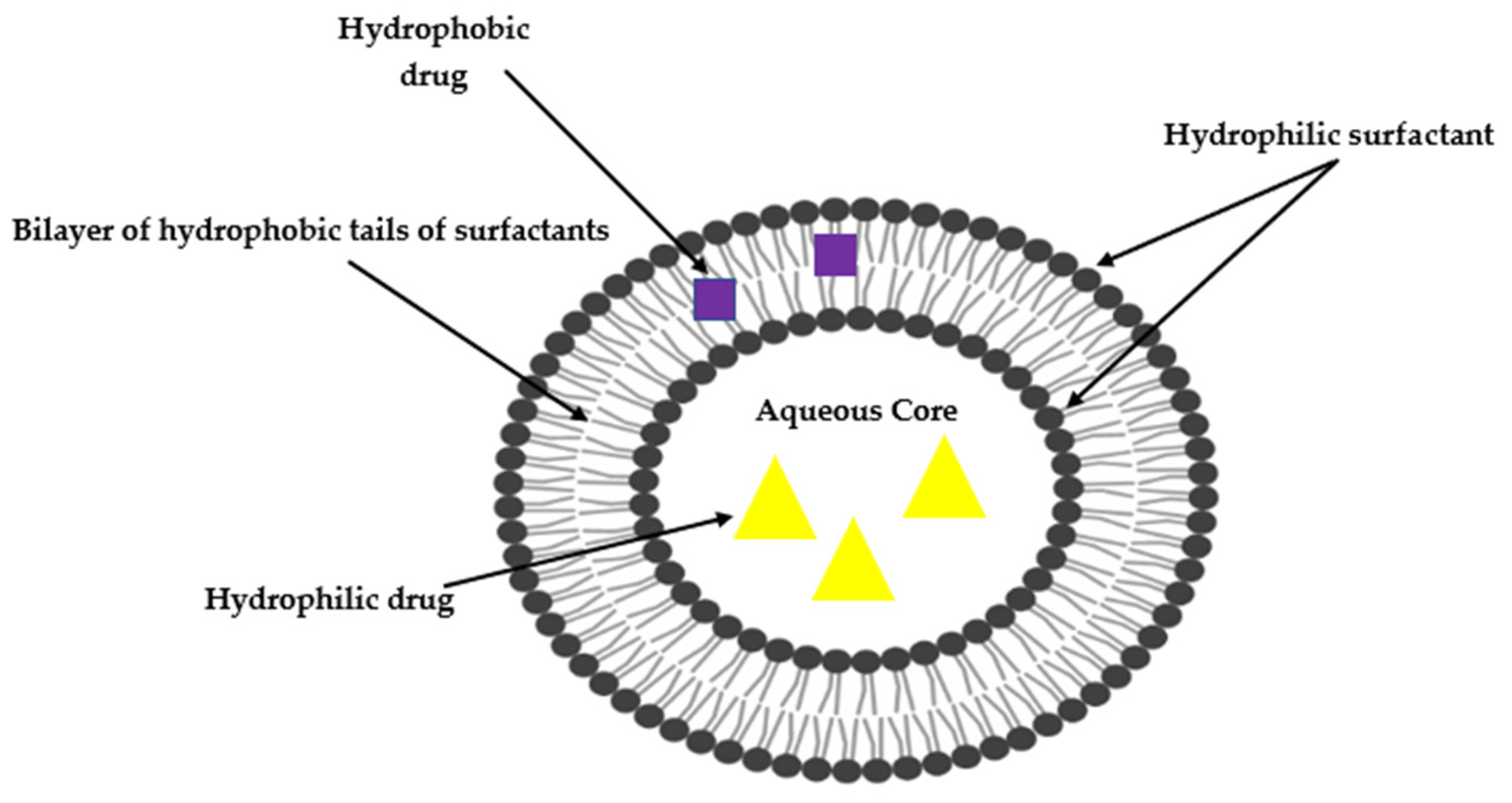
:1. Introduction
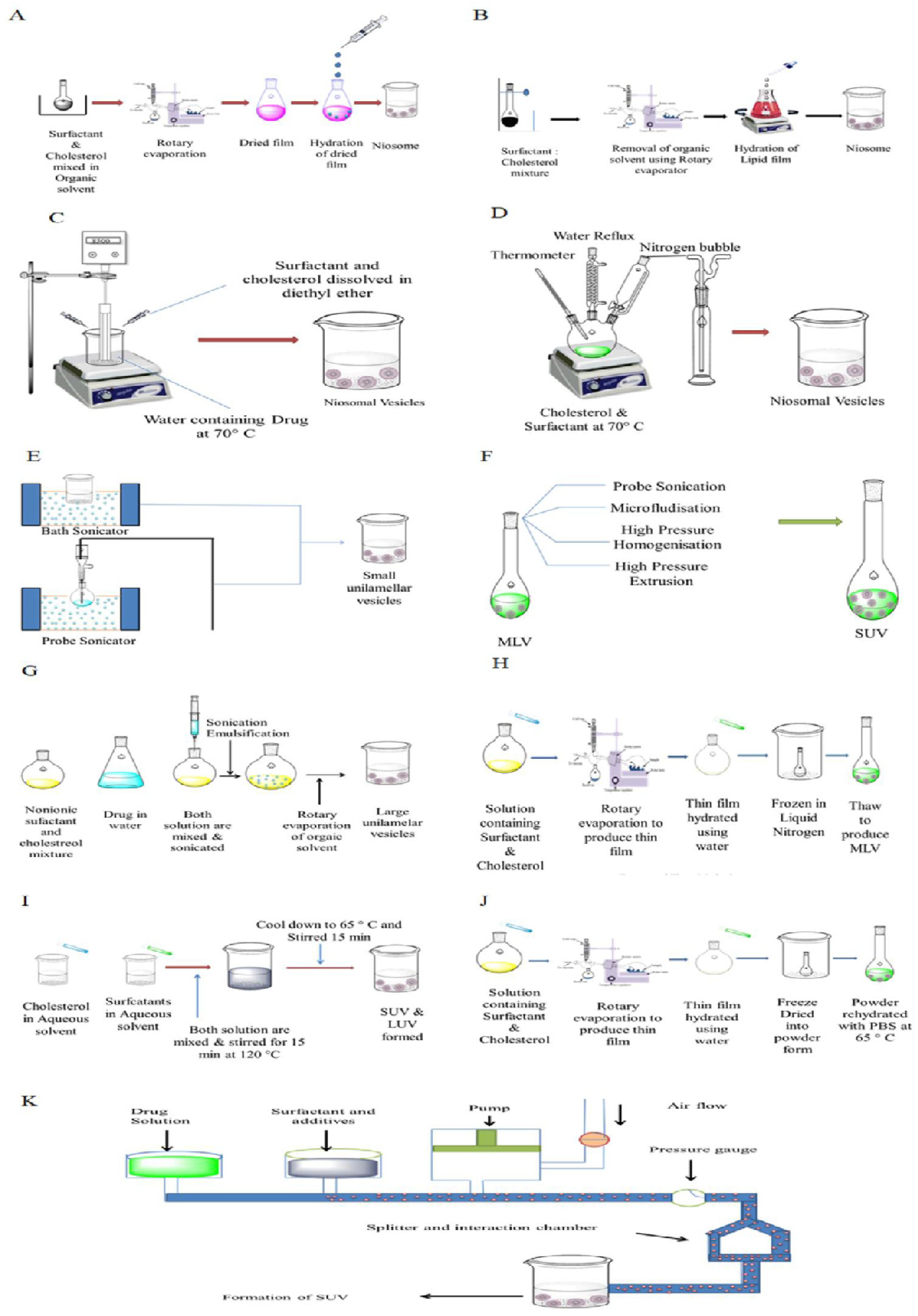
2. Preparation of Niosomes
2.1. Preparation of Multilamellar Vesicles (MLV)
2.1.1. Thin-Film Hydration
2.1.2. Transmembrane pH Gradient Drug Uptake
2.2. Preparation of Small Unilamellar Vesicle (SUV)
2.2.1. Microfluidics
2.2.2. Sonication
2.3. Preparation of Large Unilamellar Vesicles
2.3.1. Reversed-Phase Evaporation (REV)
2.3.2. Ether Injection
2.4. Miscellaneous Techniques
2.4.1. Emulsion Formation
2.4.2. Niosome Manufacture Using Micelles
2.4.3. Lipid Injection
2.4.4. Niosome Manufacture Using Polyoxyethylene Alkyl Ether
3. Characterisations of Niosomes
3.1. Particle Size (PS) and Polydispersity Index (PDI)
3.2. Zeta Potential (ZP)
3.3. Encapsulation Efficiency (EE)
3.4. Phase Behaviour
3.5. In Vitro Drug Release
3.6. Surface Elemental Composition
3.7. Bilayer Formation
3.8. Number of Lamellae
3.9. Membrane Rigidity
4. Factors Affecting the Critical Quality Attributes (CQA) of Niosomes
4.1. Choice of Surfactant
4.2. Critical Packing Parameters (CPP)
4.3. Nature of Payload
4.4. Cholesterol Content
4.5. Charge Inducers
4.6. Phase Transition Temperature (Tc)
4.7. Temperature of Hydration (Th)
5. Specialised Niosomes
5.1. pH-Responsive Niosomes
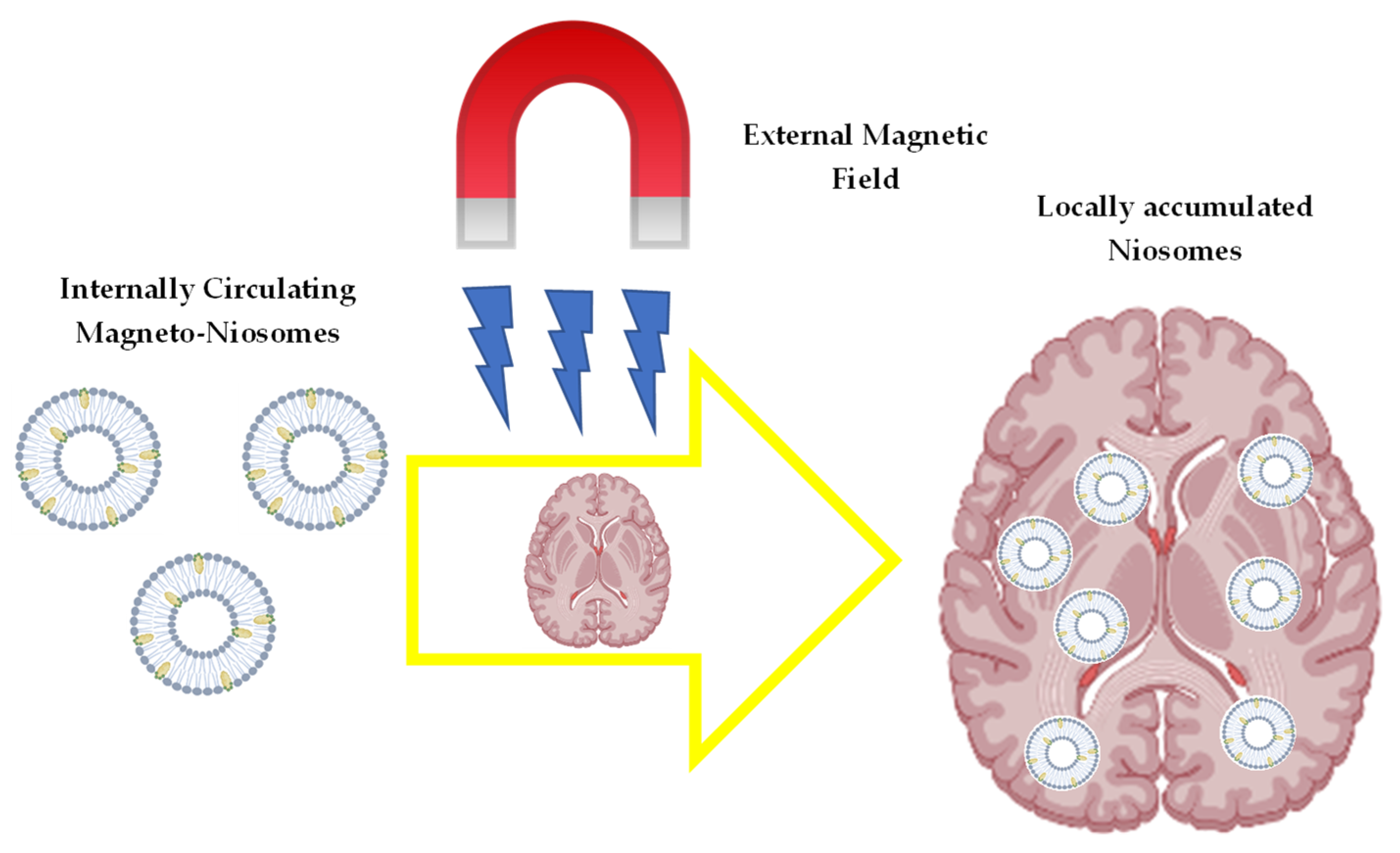
5.2. Magnetic Niosomes
5.3. Immuno-Niosomes
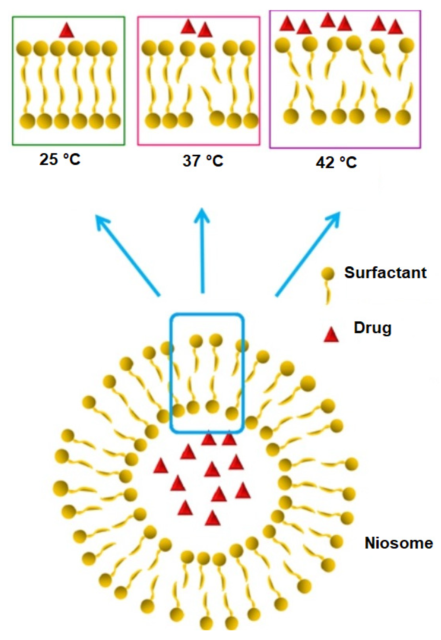
5.4. Thermoresponsive Niosomes
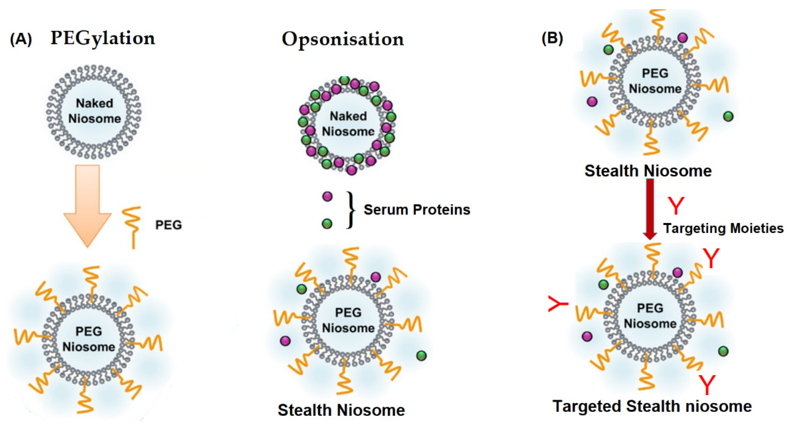
5.5. Stealth Niosomes
5.6. Deformable Vesicles (Transfersomes and Ethosomes)
5.7. Radio Niosomes
6. Limitations
7. Prospects and Conclusions
Author Contributions
Funding
Institutional Review Board Statement
Informed Consent Statement
Data Availability Statement
Acknowledgments
Conflicts of Interest
References
- Talegaonkar, S.; Mishra, P.; Khar, R.; Biju, S. Vesicular systems: An overview. Indian J. Pharm. Sci. 2006, 68, 141. [Google Scholar] [CrossRef]
- Alyami, H.; Abdelaziz, K.; Dahmash, E.Z.; Iyire, A. Nonionic surfactant vesicles (niosomes) for ocular drug delivery: Development, evaluation and toxicological profiling. J. Drug Deliv. Sci. Technol. 2020, 60, 102069. [Google Scholar] [CrossRef]
- Barani, M.; Mirzaei, M.; Torkzadeh-Mahani, M.; Lohrasbi-Nejad, A.; Nematollahi, M.H. A new formulation of hydrophobin-coated niosome as a drug carrier to cancer cells. Mater. Sci. Eng. C 2020, 113, 110975. [Google Scholar] [CrossRef]
- Paolino, D.; Cosco, D.; Muzzalupo, R.; Trapasso, E.; Picci, N.; Fresta, M. Innovative bola-surfactant niosomes as topical delivery systems of 5-fluorouracil for the treatment of skin cancer. Int. J. Pharm. 2008, 353, 233–242. [Google Scholar] [CrossRef] [PubMed]
- Moghassemi, S.; Hadjizadeh, A. Nano-niosomes as nanoscale drug delivery systems: An illustrated review. J. Control. Release 2014, 185, 22–36. [Google Scholar] [CrossRef]
- Kumar, K.K.; Sasikanth, K.; Sabareesh, M.; Dorababu, N. Formulation and evaluation of diacerein cream. Asian J. Pharm. Clin. Res. 2011, 4, 93–98. [Google Scholar] [CrossRef]
- Agarwal, R.; Katare, O.P.; Vyas, S.P. Preparation and in vitro evaluation of liposomal/niosomal delivery systems for antipsoriatic drug dithranol. Int. J. Pharm. 2001, 228, 43–52. [Google Scholar] [CrossRef]
- Jain, C.P.; Vyas, S.P. Preparation and characterization of niosomes containing rifampicin for lung targeting. J. Microencapsul. 1995, 12, 401–407. [Google Scholar] [CrossRef]
- Witika, B.A.; Walker, R.B. Development, manufacture and characterization of niosomes for the delivery for nevirapine. Pharmazie 2019, 74, 91–96. [Google Scholar] [CrossRef]
- Witika, B.A. The Development, Manufacture and Characterisation of Niosomes Intended To Deliver Nevirapine To the Brain. Master’s Thesis, Rhodes University, Makhanda, South Africa, 2017. [Google Scholar]
- Witika, B.A.; Walker, R.B. Preformulation characterization and identification of excipients for nevirapine loaded niosomes. Pharmazie 2021, 76, 77–83. [Google Scholar] [CrossRef]
- Sreya, M.; Krishna Sailaja, A. Preparation and evaluation of diclofenac sodium niosomal formulations. J. Bionanosci. 2017, 11, 489–496. [Google Scholar] [CrossRef]
- Srinivas, S.; Anand Kumar, Y.; Hemanth, A.; Anitha, M. Preparation and evaluation of niosomes containing aceclofenac. Dig. J. Nanomater. Biostruct. 2010, 5, 249–254. [Google Scholar]
- Shakya, V.; Bansal, B.K. Niosomes: A Novel Trend In Drug Delivery. Int. J. Res. Dev. Pharm. Life Sci. 2014, 3, 1036–1041. [Google Scholar]
- Gangwar, M.; Singh, R.; Goel, R.K.; Nath, G. Recent advances in various emerging vescicular systems: An overview. Asian Pac. J. Trop. Biomed. 2012, 2, S1176–S1188. [Google Scholar] [CrossRef]
- Rinaldi, F.; Hanieh, P.N.; Imbriano, A.; Passeri, D.; Del Favero, E.; Rossi, M.; Marianecci, C.; De Panfilis, S.; Carafa, M. Different instrumental approaches to understand the chitosan coated niosomes/mucin interaction. J. Drug Deliv. Sci. Technol. 2020, 55, 101339. [Google Scholar] [CrossRef]
- Bangham, A.D. Surrogate Cells or Trojan Horses: The Discovery of Liposomes. BioEssays 1995, 17, 1081–1088. [Google Scholar] [CrossRef]
- Singh, D.; Pradhan, M.; Nag, M.; Singh, M.R. Vesicular system: Versatile carrier for transdermal delivery of bioactives. Artif. Cells Nanomed. Biotechnol. 2015, 43, 282–290. [Google Scholar] [CrossRef]
- Kumar, G.P.; Rajeshwarrao, P. Nonionic surfactant vesicular systems for effective drug delivery—An overview. Acta Pharm. Sin. B 2011, 1, 208–219. [Google Scholar] [CrossRef]
- Uchegbu, I.F.; Vyas, S.P. Non-ionic surfactant based vesicles (niosomes) in drug delivery. Int. J. Pharm. 1998, 172, 33–70. [Google Scholar] [CrossRef]
- Chen, S.; Hanning, S.; Falconer, J.; Locke, M.; Wen, J. Recent advances in non-ionic surfactant vesicles (niosomes): Fabrication, characterization, pharmaceutical and cosmetic applications. Eur. J. Pharm. Biopharm. 2019, 144, 18–39. [Google Scholar] [CrossRef]
- El-Ridy, M.S.; Badawi, A.A.; Safar, M.M.; Mohsen, A.M. Niosomes as a novel pharmaceutical formulation encapsulating the hepatoprotective drug silymarin. Int. J. Pharm. Pharm. Sci. 2012, 4, 549–559. [Google Scholar]
- Daniela Stan, C.; Tǎtǎrîngǎ, G.; Gafiţanu, C.; Drǎgan, M.; Braha, S.; Popescu, M.C.; Lisǎ, G.; Ştefanache, A. Preparation and characterization of niosomes containing metronidazole. Farmacia 2013, 61, 1178–1185. [Google Scholar] [CrossRef]
- Balasubramaniam, A.; Kumar, V.A.; Pillai, K.S. Formulation and in vivo evaluation of niosome-encapsulated daunorubicin hydrochloride. Drug Dev. Ind. Pharm. 2002, 28, 1181–1193. [Google Scholar] [CrossRef] [PubMed]
- Biswal, S.; Murthy, P.N.; Sahu, J.; Sahoo, P.; Amir, F. Vesicles of non-ionic surfactants (niosomes) and drug delivery potential. Int. J. Pharm. Sci. Nanotechnol. 2008, 1, 1–8. [Google Scholar] [CrossRef]
- Marianecci, C.; Di Marzio, L.; Rinaldi, F.; Celia, C.; Paolino, D.; Alhaique, F.; Esposito, S.; Carafa, M. Niosomes from 80s to present: The state of the art. Adv. Colloid Interface Sci. 2014, 205, 187–206. [Google Scholar] [CrossRef]
- Bagheri, A.; Chu, B.S.; Yaakob, H. Niosomal drug delivery systems: Formulation, preparation and applications. World Appl. Sci. J. 2014, 32, 1671–1685. [Google Scholar] [CrossRef]
- Javani, R.; Hashemi, F.S.; Ghanbarzadeh, B.; Hamishehkar, H. Quercetin-loaded niosomal nanoparticles prepared by the thin-layer hydration method: Formulation development, colloidal stability, and structural properties. LWT Food Sci. Technol. 2021, 184, 107229. [Google Scholar] [CrossRef]
- Baillie, A.J.; Florence, A.T.; Hume, L.R.; Muirhead, G.T.; Rogerson, A. The preparation and properties of niosomes—non-ionic surfactant vesicles. J. Pharm. Pharmacol. 1985, 37, 863–868. [Google Scholar] [CrossRef]
- Ag Seleci, D.; Seleci, M.; Walter, J.G.; Stahl, F.; Scheper, T. Niosomes as nanoparticular drug carriers: Fundamentals and recent applications. J. Nanomater. 2016, 2016, 7372306. [Google Scholar] [CrossRef]
- Karim, K.; Mandal, A.; Biswas, N.; Guha, A.; Chatterjee, S.; Behera, M.; Kuotsu, K. Niosome: A future of targeted drug delivery systems. J. Adv. Pharm. Technol. Res. 2010, 1, 374–380. [Google Scholar] [CrossRef]
- Hunter, C.A.; Dolan, T.F.; Coombs, G.H.; Baillie, A.J. Vesicular Systems (Niosomes and Liposomes) for Delivery of Sodium Stibogluconate in Experimental Murine Visceral Leishmaniasis. J. Pharm. Pharmacol. 1988, 40, 161–165. [Google Scholar] [CrossRef] [PubMed]
- Hao, Y.; Zhao, F.; Li, N.; Yang, Y.; Li, K. Studies on a high encapsulation of colchicine by a niosome system. Int. J. Pharm. 2002, 244, 73–80. [Google Scholar] [CrossRef]
- Pardakhty, A.; Varshosaz, J.; Rouholamini, A. In vitro study of polyoxyethylene alkyl ether niosomes for delivery of insulin. Int. J. Pharm. 2007, 328, 130–141. [Google Scholar] [CrossRef]
- Mehta, S.K.; Jindal, N. Tyloxapol Niosomes as Prospective Drug Delivery Module for Antiretroviral Drug Nevirapine. AAPS PharmSciTech 2015, 16, 67–75. [Google Scholar] [CrossRef] [PubMed]
- Kamboj, S.; Saini, V.; Bala, S. Formulation and characterization of drug loaded nonionic surfactant vesicles (Niosomes) for oral bioavailability enhancement. Sci. World J. 2014, 2014, 959741. [Google Scholar] [CrossRef]
- Bragagni, M.; Mennini, N.; Ghelardini, C.; Mura, P. Development and characterization of niosomal formulations of doxorubicin aimed at brain targeting. J. Pharm. Pharm. Sci. 2012, 15, 184–196. [Google Scholar] [CrossRef]
- Mayer, L.D.; Bally, M.B.; Hope, M.J.; Cullis, P.R. Uptake of antineoplastic agents into large unilamellar vesicles in response to a membrane potential. BBA Biomembr. 1985, 816, 294–302. [Google Scholar] [CrossRef]
- Rajera, R.; Nagpal, K.; Singh, S.K.; Mishra, D.N. Niosomes: A Controlled and Novel Drug Delivery System. Biol. Pharm. Bull. 2011, 34, 945–953. [Google Scholar] [CrossRef]
- Mujoriya, R.; Bodla, R.B.; Dhamande, K.; Singh, D.; Patle, L. Niosomal drug delivery system: The magic bullet. J. Appl. Pharm. Sci. 2011, 1, 20–23. [Google Scholar]
- Mujoriya, R.Z.; Bodla, R.B. Niosomes—Challenge in preparation for pharmaceutical scientist. Int. J. Appl. Pharm. 2011, 3, 11–15. [Google Scholar]
- Szoka, F.; Papahadjopoulos, D. Procedure for preparation of liposomes with large internal aqueous space and high capture by reverse-phase evaporation. Proc. Natl. Acad. Sci. USA 1978, 75, 4194–4198. [Google Scholar] [CrossRef] [PubMed]
- Rogerson, A.; Cummings, J.; Willmott, N.; Florence, A.T. The Distribution of Doxorubicin in Mice Following Administration in Niosomes. J. Pharm. Pharmacol. 1988, 40, 337–342. [Google Scholar] [CrossRef]
- Batzri, S.; Korn, E.D. Single bilayer liposomes prepared without sonication. BBA Biomembr. 1973, 298, 1015–1019. [Google Scholar] [CrossRef]
- Sezgin-Bayindir, Z.; Yuksel, N. Investigation of Formulation Variables and Excipient Interaction on the Production of Niosomes. AAPS PharmSciTech 2012, 13, 826–835. [Google Scholar] [CrossRef] [PubMed]
- Lawrence, M.J. Surfactant systems: Their use in drug delivery. Chem. Soc. Rev. 1994, 23, 417. [Google Scholar] [CrossRef]
- Jadhav, S.M.; Morey, P.; Karpe, M.M.; Kadam, V. Novel vesicular system: An overview. J. Appl. Pharm. Sci. 2012, 2, 193–202. [Google Scholar]
- Aparajay, P.; Dev, A. Functionalized niosomes as a smart delivery device in cancer and fungal infection. Eur. J. Pharm. Sci. 2022, 168, 106052. [Google Scholar] [CrossRef]
- Blazek-Welsh, A.I.; Rhodes, D.G. SEM imaging predicts quality of niosomes from maltodextrin-based proniosomes. Pharm. Res. 2001, 18, 656–661. [Google Scholar] [CrossRef]
- Chudasama, A.; Patel, V.; Nivsarkar, M.; Vasu, K.; Shishoo, C. A novel lipid-based oral drug delivery system of nevirapine. Int. J. PharmTech Res. 2011, 3, 1159–1168. [Google Scholar]
- Semalty, A.; Semalty, M.; Singh, D.; Rawat, M.S.M. Development and physicochemical evaluation of pharmacosomes of diclofenac. Acta Pharm. 2009, 59, 335–344. [Google Scholar] [CrossRef]
- Le Maire, M.; Champeil, P.; Møller, J.V. Interaction of membrane proteins and lipids with solubilizing detergents. Biochim. Biophys. Acta Biomembr. 2000, 1508, 86–111. [Google Scholar] [CrossRef]
- Kuo, Y.C.; Chung, J.F. Physicochemical properties of nevirapine-loaded solid lipid nanoparticles and nanostructured lipid carriers. Colloids Surf. B Biointerfaces 2011, 83, 299–306. [Google Scholar] [CrossRef] [PubMed]
- Lu, C.T.; Zhao, Y.Z.; Wong, H.L.; Cai, J.; Peng, L.; Tian, X.Q. Current approaches to enhance CNS delivery of drugs across the brain barriers. Int. J. Nanomed. 2014, 9, 2241–2257. [Google Scholar] [CrossRef]
- Priyanka, K.; Singh, S. A review on skin targeted delivery of bioactives as ultradeformable vesicles: Overcoming the penetration problem. Curr. Drug Targets 2014, 15, 184–198. [Google Scholar] [CrossRef]
- Sergeev, G.B.; Klabunde, K.J. Experimental Techniques. In Nanochemistry, 2nd ed.; Sergeev, G.B., Klabunde, K.J.B.T.-N., Eds.; Elsevier: Oxford, UK, 2013; Volume 249, pp. 31–43. ISBN 978-0-444-59397-9. [Google Scholar]
- Winter, W.T. Measurement of suspended particles by quasi-elastic light scattering. J. Polym. Sci. Polym. Lett. Ed. 1983, 21, 1020. [Google Scholar] [CrossRef]
- Ruckmani, K.; Sankar, V. Formulation and Optimization of Zidovudine Niosomes. AAPS PharmSciTech 2010, 11, 1119–1127. [Google Scholar] [CrossRef] [PubMed]
- Begum, M.Y.; Dasari, S.; Sudhakar, M.; Lakshmi, B.V.S.; Manga, K.; Begum, M.Y. Development and Evaluation of Co-encapsulated Stavudine and Lamivudine niosomes for the Controlled Delivery. Der Pharm. Sin. 2014, 5, 1–10. [Google Scholar]
- Wagner, A.; Vorauer-Uhl, K. Liposome Technology for Industrial Purposes, 3rd ed.; Informa Healthcare Inc.: New York, NY, USA, 2011; Volume 2011, ISBN 9780849388217. [Google Scholar]
- Kumbhar, D.; Wavikar, P.; Vavia, P. Niosomal Gel of Lornoxicam for Topical Delivery: In vitro Assessment and Pharmacodynamic Activity. AAPS PharmSciTech 2013, 14, 1072–1082. [Google Scholar] [CrossRef] [Green Version]
- Wang, M.; Yuan, Y.; Gao, Y.; Ma, H.M.; Xu, H.T.; Zhang, X.N.; Pan, W.S. Preparation and characterization of 5-fluorouracil pH-sensitive niosome and its tumor-targeted evaluation: In vitro and in vivo. Drug Dev. Ind. Pharm. 2012, 38, 1134–1141. [Google Scholar] [CrossRef]
- Nkanga, C.I.; Krause, R.W.M. Encapsulation of Isoniazid-conjugated Phthalocyanine-In-Cyclodextrin-In-Liposomes Using Heating Method. Sci. Rep. 2019, 9, 11485. [Google Scholar] [CrossRef]
- Okafor, N.I.; Nkanga, C.I.; Walker, R.B.; Noundou, X.S.; Krause, R.W.M. Encapsulation and physicochemical evaluation of efavirenz in liposomes. J. Pharm. Investig. 2020, 50, 201–208. [Google Scholar] [CrossRef]
- Chen, H.; Torchilin, V.; Langer, R. Lectin-bearing polymerized liposomes as potential oral vaccine carriers. Pharm. Res. 1996, 13, 1378–1383. [Google Scholar] [CrossRef] [PubMed]
- Manosroi, A.; Wongtrakul, P.; Manosroi, J.; Sakai, H.; Sugawara, F.; Yuasa, M.; Abe, M. Characterization of vesicles prepared with various non-ionic surfactants mixed with cholesterol. Colloids Surf. B Biointerfaces 2003, 30, 129–138. [Google Scholar] [CrossRef]
- Lokamatha, K.M.; Bharathi, A.; Shanta Kumar, S.M.; Rama Rao, N. Effect of PVP-K30 on complexation and dissolution rate of nevirapine-β-cyclodextrin complexes. Int. J. Pharm. Pharm. Sci. 2010, 2, 169–176. [Google Scholar]
- Yoshioka, T.; Sternberg, B.; Florence, A.T. Preparation and properties of vesicles (niosomes) of sorbitan monoesters (Span 20, 40, 60 and 80) and a sorbitan triester (Span 85). Int. J. Pharm. 1994, 105, 1–6. [Google Scholar] [CrossRef]
- Griffin, W.C. Classification of surface-active agents by “HLB”. J. Soc. Cosmet. Chem. 1949, 1, 311–326. [Google Scholar]
- Shahiwala, A.; Misra, A. Studies in topical application of niosomally entrapped Nimesulide. J. Pharm. Pharm. Sci. 2002, 5, 220–225. [Google Scholar]
- Gregoriadis, G. Engineering liposomes for drug delivery: Progress and problems. Trends Biotechnol. 1995, 13, 527–537. [Google Scholar] [CrossRef]
- Israelachvili, J.N.; Mitchell, D.J.; Ninham, B.W. Theory of self-assembly of hydrocarbon amphiphiles into micelles and bilayers. J. Chem. Soc. Faraday Trans. 2 Mol. Chem. Phys. 1976, 72, 1525–1568. [Google Scholar] [CrossRef]
- Bouwstra, J.A.; Hoflan, H.E.J. Niosomes. In Colloidal Drug Delivery Systems; Kreuter, J., Ed.; Marcel Dekker: New York, NY, USA, 1996; Volume 66, pp. 191–217. [Google Scholar]
- Uchegbu, I.F.; Duncan, R. Niosomes containing N-(2-hydroxypropyl)methacrylamide copolymer-doxorubicin (PK1): Effect of method of preparation and choice of surfactant on niosome characteristics and a preliminary study of body distribution. Int. J. Pharm. 1997, 155, 7–17. [Google Scholar] [CrossRef]
- Sahin, N.O. Niosomes as nanocarrier systems. Nanomater. Nanosyst. Biomadical Appl. 2007, 67–81. [Google Scholar] [CrossRef]
- Mahale, N.B.; Thakkar, P.D.; Mali, R.G.; Walunj, D.R.; Chaudhari, S.R. Niosomes: Novel sustained release nonionic stable vesicular systems—An overview. Adv. Colloid Interface Sci. 2012, 183–184, 46–54. [Google Scholar] [CrossRef]
- Junyaprasert, V.B.; Teeranachaideekul, V.; Supaperm, T. Effect of charged and non-ionic membrane additives on physicochemical properties and stability of niosomes. AAPS PharmSciTech 2008, 9, 851–859. [Google Scholar] [CrossRef] [PubMed]
- Alsarra, I.A.; Bosela, A.A.; Ahmed, S.M.; Mahrous, G.M. Proniosomes as a drug carrier for transdermal delivery of ketorolac. Eur. J. Pharm. Biopharm. 2005, 59, 485–490. [Google Scholar] [CrossRef] [PubMed]
- Mokhtar, M.; Sammour, O.A.; Hammad, M.A.; Megrab, N.A. Effect of some formulation parameters on flurbiprofen encapsulation and release rates of niosomes prepared from proniosomes. Int. J. Pharm. 2008, 361, 104–111. [Google Scholar] [CrossRef]
- Edlow, D.W.; Sheldon, W.H. The pH of Inflammatory Exudates. Proc. Soc. Exp. Biol. Med. 1971, 137, 1328–1332. [Google Scholar] [CrossRef] [PubMed]
- Das, S.S.; Bharadwaj, P.; Bilal, M.; Barani, M.; Rahdar, A.; Taboada, P.; Bungau, S.; Kyzas, G. Stimuli-Responsive Polymeric Nanocarriers for Drug Delivery, Imaging, and Theragnosis. Polymers 2020, 12, 1397. [Google Scholar] [CrossRef] [PubMed]
- Johnson, R.P.; Preman, N.K. Dual and multistimuli-responsive block copolymers for drug delivery applications. In Stimuli Responsive Polymeric Nanocarriers for Drug Delivery Applications; Elsevier: Amsterdam, The Netherlands, 2019; pp. 249–267. [Google Scholar] [CrossRef]
- Pereira, M.C.; Pianella, M.; Wei, D.; Moshnikova, A.; Marianecci, C.; Carafa, M.; Andreev, O.A.; Reshetnyak, Y.K. pH-sensitive pHLIP® coated niosomes. Mol. Membr. Biol. 2016, 33, 51–63. [Google Scholar] [CrossRef]
- Hu, X.; Zhang, J.; Deng, L.; Hu, H.; Hu, J.; Zheng, G. Galactose-Modified PH-Sensitive Niosomes for Controlled Release and Hepatocellular Carcinoma Target Delivery of Tanshinone IIA. AAPS PharmSciTech 2021, 22, 96. [Google Scholar] [CrossRef]
- Sargazi, S.; Hosseinikhah, M.; Zargari, F.; Pal, N.; Chauhana, S.; Hassanisaadi, M.; Amani, S. pH-responsive cisplatin-loaded niosomes: Synthesis, characterization, cytotoxicity study and interaction analyses by simulation methodology. Nanofabrication 2021, 6, 1–15. [Google Scholar] [CrossRef]
- Marzoli, F.; Marianecci, C.; Rinaldi, F.; Passeri, D.; Rossi, M.; Minosi, P.; Carafa, M.; Pieretti, S. Long-lasting, antinociceptive effects of pH-sensitive niosomes loaded with ibuprofen in acute and chronic models of pain. Pharmaceutics 2019, 11, 62. [Google Scholar] [CrossRef] [PubMed]
- Barani, M.; Hajinezhad, M.R.; Sargazi, S.; Rahdar, A.; Shahraki, S.; Lohrasbi-Nejad, A.; Baino, F. In vitro and in vivo anticancer effect of pH-responsive paclitaxel-loaded niosomes. J. Mater. Sci. Mater. Med. 2021, 32, 147. [Google Scholar] [CrossRef] [PubMed]
- Allam, A.; El-Mokhtar, M.A.; Elsabahy, M. Vancomycin-loaded niosomes integrated within pH-sensitive in-situ forming gel for treatment of ocular infections while minimizing drug irritation. J. Pharm. Pharmacol. 2019, 71, 1209–1221. [Google Scholar] [CrossRef] [PubMed]
- Hafez, I.M.; Cullis, P.R. Cholesteryl hemisuccinate exhibits pH sensitive polymorphic phase behavior. Biochim. Biophys. Acta Biomembr. 2000, 1463, 107–114. [Google Scholar] [CrossRef]
- Marianecci, C.; Rinaldi, F.; Di Marzio, L.; Mastriota, M.; Pieretti, S.; Celia, C.; Paolino, D.; Iannone, M.; Fresta, M.; Carafa, M. Ammonium glycyrrhizinate-loaded niosomes as a potential nanotherapeutic system for anti-inflammatory activity in murine models. Int. J. Nanomed. 2014, 9, 635–651. [Google Scholar] [CrossRef]
- Rinaldi, F.; Forte, J.; Pontecorvi, G.; Hanieh, P.N.; Carè, A.; Bellenghi, M.; Tirelli, V.; Ammendolia, M.G.; Mattia, G.; Marianecci, C.; et al. pH-responsive oleic acid based nanocarriers: Melanoma treatment strategies. Int. J. Pharm. 2022, 613, 121391. [Google Scholar] [CrossRef]
- Carafa, M.; Marianecci, C.; Rinaldi, F.; Santucci, E.; Tampucci, S.; Monti, D. Span® and Tween® neutral and pH-sensitive vesicles: Characterization and in vitro skin permeation. J. Liposome Res. 2009, 19, 332–340. [Google Scholar] [CrossRef]
- Masotti, A.; Vicennati, P.; Alisi, A.; Marianecci, C.; Rinaldi, F.; Carafa, M.; Ortaggi, G. Novel Tween® 20 derivatives enable the formation of efficient pH-sensitive drug delivery vehicles for human hepatoblastoma. Bioorg. Med. Chem. Lett. 2010, 20, 3021–3025. [Google Scholar] [CrossRef]
- Widder, K.; Flouret, G.; Senyei, A. Magnetic microspheres: Synthesis of a novel parenteral drug carrier. J. Pharm. Sci. 1979, 68, 79–82. [Google Scholar] [CrossRef]
- Kong, M.; Park, H.; Feng, C.; Hou, L.; Cheng, X.; Chen, X. Construction of hyaluronic acid noisome as functional transdermal nanocarrier for tumor therapy. Carbohydr. Polym. 2013, 94, 634–641. [Google Scholar] [CrossRef]
- Tavano, L.; Vivacqua, M.; Carito, V.; Muzzalupo, R.; Caroleo, M.C.; Nicoletta, F. Doxorubicin loaded magneto-niosomes for targeted drug delivery. Colloids Surf. B Biointerfaces 2013, 102, 803–807. [Google Scholar] [CrossRef] [PubMed]
- Davarpanah, F.; Khalili Yazdi, A.; Barani, M.; Mirzaei, M.; Torkzadeh-Mahani, M. Magnetic delivery of antitumor carboplatin by using PEGylated-Niosomes. DARU J. Pharm. Sci. 2018, 26, 57–64. [Google Scholar] [CrossRef] [PubMed]
- Barani, M.; Nematollahi, M.H.; Zaboli, M.; Mirzaei, M.; Torkzadeh-Mahani, M.; Pardakhty, A.; Karam, G.A. In silico and in vitro study of magnetic niosomes for gene delivery: The effect of ergosterol and cholesterol. Mater. Sci. Eng. C 2019, 94, 234–246. [Google Scholar] [CrossRef] [PubMed]
- Maurer, V.; Altin, S.; Seleci, D.A.; Zarinwall, A.; Temel, B.; Vogt, P.M.; Strauß, S.; Stahl, F.; Scheper, T.; Bucan, V.; et al. In-vitro application of magnetic hybrid niosomes: Targeted sirna-delivery for enhanced breast cancer therapy. Pharmaceutics 2021, 13, 394. [Google Scholar] [CrossRef]
- Jamshidifar, E.; Eshrati Yeganeh, F.; Shayan, M.; Tavakkoli Yaraki, M.; Bourbour, M.; Moammeri, A.; Akbarzadeh, I.; Noorbazargan, H.; Hossein-Khannazer, N. Super magnetic niosomal nanocarrier as a new approach for treatment of breast cancer: A case study on sk-br-3 and mda-mb-231 cell lines. Int. J. Mol. Sci. 2021, 22, 7948. [Google Scholar] [CrossRef]
- Hood, E.; Gonzalez, M.; Plaas, A.; Strom, J.; VanAuker, M. Immuno-targeting of nonionic surfactant vesicles to inflammation. Int. J. Pharm. 2007, 339, 222–230. [Google Scholar] [CrossRef]
- Elliott, J.A.; Flam, B.; Muffly, K.; Strom, J.A.; Hood, E.; Vanauker, M.D. Targeted drug delivery with PEGylated immuno-niosomes. IFMBE Proc. 2009, 24, 363–366. [Google Scholar] [CrossRef]
- Gogoi, H.; Mani, R.; Bhatnagar, R. A niosome formulation modulates the Th1/Th2 bias immune response in mice and also provides protection against anthrax spore challenge. Int. J. Nanomed. 2018, 13, 7427–7440. [Google Scholar] [CrossRef]
- Mazzotta, E.; Tavano, L.; Muzzalupo, R. Thermo-sensitive vesicles in controlled drug delivery for chemotherapy. Pharmaceutics 2018, 10, 150. [Google Scholar] [CrossRef]
- Tavano, L.; Oliviero Rossi, C.; Picci, N.; Muzzalupo, R. Spontaneous temperature-sensitive Pluronic® based niosomes: Triggered drug release using mild hyperthermia. Int. J. Pharm. 2016, 511, 703–708. [Google Scholar] [CrossRef]
- Damera, D.P.; Nag, A. Tuning the phase transition temperature of hybrid Span60-L64 thermoresponsive niosomes: Insights from fluorescence and Raman spectroscopy. J. Mol. Liq. 2021, 340, 117110. [Google Scholar] [CrossRef]
- Dreher, M.R.; Raucher, D.; Balu, N.; Colvin, M.; Ludeman, S.M.; Chilkoti, A. Evaluation of an elastin-like polypeptide–doxorubicin conjugate for cancer therapy. J. Control Release 2002, 91, 31–43. [Google Scholar] [CrossRef]
- Shehata, T.; Kimura, T.; Higaki, K.; Ogawara, K. ichi In-vivo disposition characteristics of PEG niosome and its interaction with serum proteins. Int. J. Pharm. 2016, 512, 322–328. [Google Scholar] [CrossRef] [PubMed]
- Shi, B.; Fang, C.; Pei, Y. Stealth PEG-PHDCA niosomes: Effects of chain length of PEG and particle size on niosomes surface properties, in vitro drug release, phagocytic uptake, in vivo pharmacokinetics and antitumor activity. J. Pharm. Sci. 2006, 95, 1873–1887. [Google Scholar] [CrossRef]
- Haroun, M.; Elsewedy, H.S.; Shehata, T.M.; Tratrat, C.; Al Dhubiab, B.E.; Venugopala, K.N.; Almostafa, M.M.; Kochkar, H.; Elnahas, H.M. Significant of injectable brucine PEGylated niosomes in treatment of MDA cancer cells. J. Drug Deliv. Sci. Technol. 2022, 71, 103322. [Google Scholar] [CrossRef]
- Immordino, M.L.; Dosio, F.; Cattel, L. Stealth liposomes: Review of the basic science, rationale, and clinical applications, existing and potential. Int. J. Nanomed. 2006, 1, 297–315. [Google Scholar] [CrossRef]
- Karmali, P.P.; Simberg, D. Interactions of nanoparticles with plasma proteins: Implication on clearance and toxicity of drug delivery systems. Expert Opin. Drug Deliv. 2011, 8, 343–357. [Google Scholar] [CrossRef]
- Pengnam, S.; Patrojanasophon, P.; Rojanarata, T.; Ngawhirunpat, T.; Yingyongnarongkul, B.E.; Radchatawedchakoon, W.; Opanasopit, P. A novel plier-like gemini cationic niosome for nucleic acid delivery. J. Drug Deliv. Sci. Technol. 2019, 52, 325–333. [Google Scholar] [CrossRef]
- Rajan, R.; Vasudevan, D.; Biju Mukund, V.; Jose, S. Transferosomes—A vesicular transdermal delivery system for enhanced drug permeation. J. Adv. Pharm. Technol. Res. 2011, 2, 138–143. [Google Scholar] [CrossRef]
- Opatha, S.A.T.; Titapiwatanakun, V.; Chutoprapat, R. Transfersomes: A promising nanoencapsulation technique for transdermal drug delivery. Pharmaceutics 2020, 12, 855. [Google Scholar] [CrossRef]
- Jeong, W.Y.; Kwon, M.; Choi, H.E.; Kim, K.S. Recent advances in transdermal drug delivery systems: A review. Biomater. Res. 2021, 25, 24. [Google Scholar] [CrossRef] [PubMed]
- Tan, A.; Saul, J.M.; Hsin-I, C.; Hua, S. Lipid-based nano-delivery systems for skin delivery of drugs and bioactives. Front. Pharmacol. 2015, 6, 219. [Google Scholar] [CrossRef]
- El Zaafarany, G.M.; Awad, G.A.S.; Holayel, S.M.; Mortada, N.D. Role of edge activators and surface charge in developing ultradeformable vesicles with enhanced skin delivery. Int. J. Pharm. 2010, 397, 164–172. [Google Scholar] [CrossRef] [PubMed]
- Prasanthi, D.; Lakshmi, P.K. Vesicles - mechanism of transdermal permeation: A review. Asian J. Pharm. Clin. Res. 2012, 5, 18–25. [Google Scholar]
- Khamkat, P.; Ghosh, A.; Mukherjee, S. Transfersomes: An Innovative Vesicular Carrier for Boosted Transdermal Delivery System. Res. J. Pharm. Technol. 2022, 15, 2793–2800. [Google Scholar] [CrossRef]
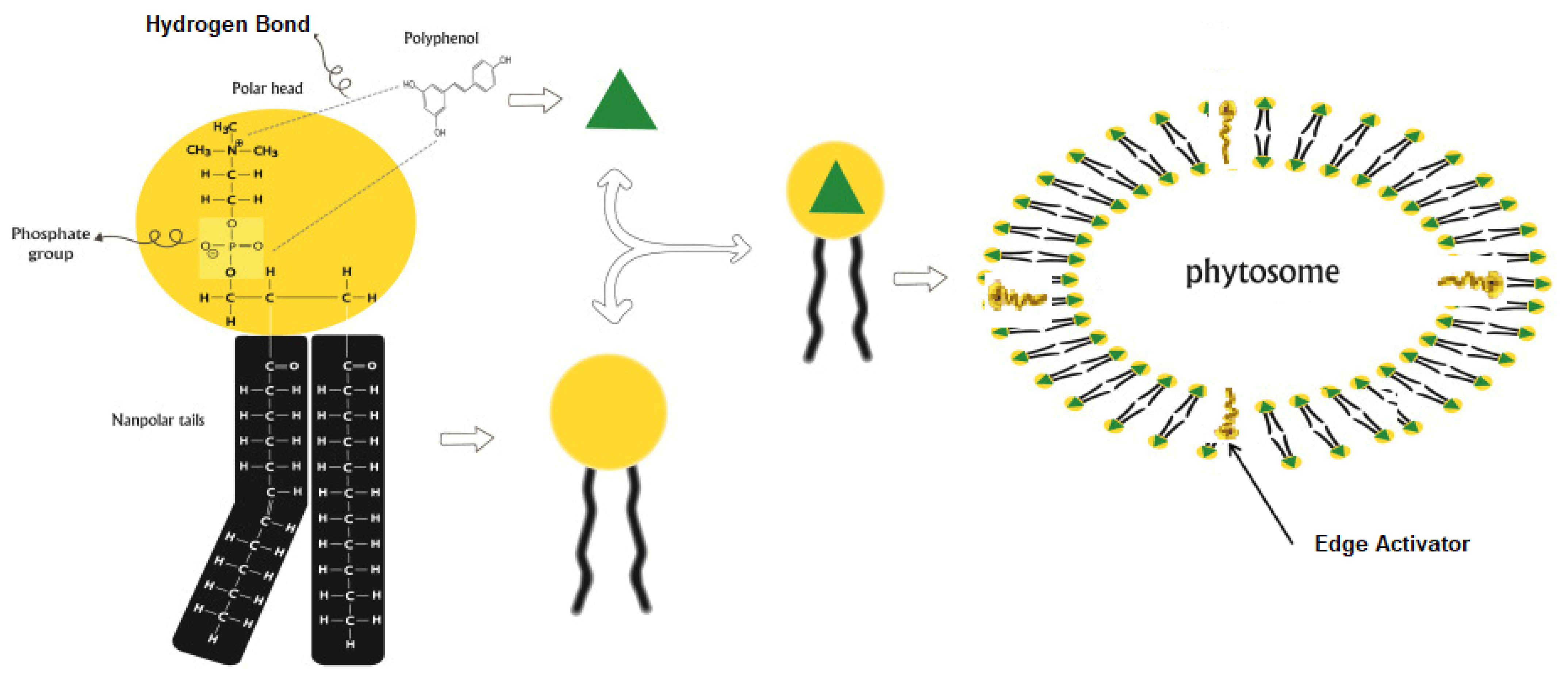
- Chen, R.P.; Chavda, V.P.; Patel, A.B.; Chen, Z.S. Phytochemical Delivery Through Transferosome (Phytosome): An Advanced Transdermal Drug Delivery for Complementary Medicines. Front. Pharmacol. 2022, 13, 850862. [Google Scholar] [CrossRef] [PubMed]
- Ali, M.F.M.; Salem, H.F.; Abdelmohsen, H.F.; Attia, S.K. Preparation and clinical evaluation of nano-transferosomes for treatment of erectile dysfunction. Drug Des. Devel. Ther. 2015, 9, 2431–2447. [Google Scholar] [CrossRef]
- Shang, H.; Younas, A.; Zhang, N. Recent advances on transdermal delivery systems for the treatment of arthritic injuries: From classical treatment to nanomedicines. Wiley Interdiscip. Rev. Nanomed. Nanobiotechnology 2022, 14, e1778. [Google Scholar] [CrossRef]
- Barani, M.; Sangiovanni, E.; Angarano, M.; Rajizadeh, M.A.; Mehrabani, M.; Piazza, S.; Gangadharappa, H.V.; Pardakhty, A.; Mehrbani, M.; Dell’agli, M.; et al. Phytosomes as innovative delivery systems for phytochemicals: A comprehensive review of literature. Int. J. Nanomed. 2021, 16, 6983–7022. [Google Scholar] [CrossRef]
- Lu, M.; Qiu, Q.; Luo, X.; Liu, X.; Sun, J.; Wang, C.; Lin, X.; Deng, Y.; Song, Y. Phyto-phospholipid complexes (phytosomes): A novel strategy to improve the bioavailability of active constituents. Asian J. Pharm. Sci. 2019, 14, 265–274. [Google Scholar] [CrossRef]
- Ramsden, J.J. Applied Nanoechnology; Elsevier: Amsterdam, The Netherlands, 2014; ISBN 9781455731893. [Google Scholar]
- Ghanbarzadeh, S.; Arami, S. Formulation and evaluation of piroxicam transferosomal gel: An approach for penetration enhancement. J. Drug Deliv. Sci. Technol. 2013, 23, 587–590. [Google Scholar] [CrossRef]
- Cevc, G.; Blume, G. New, highly efficient formulation of diclofenac for the topical, transdermal administration in ultradeformable drug carriers, Transfersomes. Biochim. Biophys. Acta Biomembr. 2001, 1514, 191–205. [Google Scholar] [CrossRef]
- Salama, H.A.; Mahmoud, A.A.; Kamel, A.O.; Abdel Hady, M.; Awad, G.A.S. Brain delivery of olanzapine by intranasal administration of transfersomal vesicles. J. Liposome Res. 2012, 22, 336–345. [Google Scholar] [CrossRef] [PubMed]
- Mazyed, E.A.; Abdelaziz, A.E. Fabrication of transgelosomes for enhancing the ocular delivery of acetazolamide: Statistical optimization, in vitro characterization, and in vivo study. Pharmaceutics 2020, 12, 465. [Google Scholar] [CrossRef] [PubMed]
- Korkmaz, M.; Ozer, A.; Hincal, A. DTPA Niosomes in Diagnostic Imaging. In Synthetic Surfactants Vesicles; Uchegbu, I.F., Ed.; CRC Press: Boca Raton, FL, USA, 2000; ISBN 9780429079634. [Google Scholar]
- Munekane, M.; Kosugi, A.; Yamasaki, M.; Watanabe, Y.; Kannaka, K.; Sano, K.; Yamasaki, T.; Ogawara, K.; Mukai, T. Biodistribution study of indium-111-labeled PEGylated niosomes as novel drug carriers for tumor-targeting. J. Drug Deliv. Sci. Technol. 2022, 75, 103648. [Google Scholar] [CrossRef]
- De Silva, L.; Fu, J.Y.; Htar, T.T.; Wan Kamal, W.H.B.; Kasbollah, A.; Muniyandy, S.; Chuah, L.H. Biodistribution Study of Niosomes in Tumor-Implanted BALB/C Mice Using Scintigraphic Imaging. Front. Pharmacol. 2022, 12, 778396. [Google Scholar] [CrossRef]
- Silindir, M.; Özer, A.Y. Sterilization methods and the comparison of E-beam sterilization with gamma radiation sterilization. FABAD J. Pharm. Sci. 2009, 34, 43–53. [Google Scholar]
- Hofland, H.E.J.; Bouwstra, J.A.; Verhoef, J.C.; Buckton, G.; Chowdry, B.Z.; Ponec, M.; Junginger, H.E. Safety Aspects of Non-ionic Surfactant Vesicles: A Toxicity Study Related to the Physicochemical Characteristics of Non-ionic Surfactants. J. Pharm. Pharmacol. 1992, 44, 287–294. [Google Scholar] [CrossRef]
- Hofland, H.E.J.; Bouwstra, J.A.; Ponec, M.; Boddé, H.E.; Spies, F.; Verhoef, J.C.; Junginger, H.E. Interactions of non-ionic surfactant vesicles with cultured keratinocytes and human skin in vitro: A survey of toxicological aspects and ultrastructural changes in stratum corneum. J. Control. Release 1991, 16, 155–167. [Google Scholar] [CrossRef]
- Abdelkader, H.; Wu, Z.; Al-Kassas, R.; Alany, R.G. Niosomes and discomes for ocular delivery of naltrexone hydrochloride: Morphological, rheological, spreading properties and photo-protective effects. Int. J. Pharm. 2012, 433, 142–148. [Google Scholar] [CrossRef]
- Uchegbu, I. The biodistribution of novel 200-nm palmitoyl muramic acid vesicles. Int. J. Pharm. 1998, 162, 19–27. [Google Scholar] [CrossRef]


Publisher’s Note: MDPI stays neutral with regard to jurisdictional claims in published maps and institutional affiliations. |
© 2022 by the authors. Licensee MDPI, Basel, Switzerland. This article is an open access article distributed under the terms and conditions of the Creative Commons Attribution (CC BY) license (https://creativecommons.org/licenses/by/4.0/).
Share and Cite
Witika, B.A.; Bassey, K.E.; Demana, P.H.; Siwe-Noundou, X.; Poka, M.S. Current Advances in Specialised Niosomal Drug Delivery: Manufacture, Characterization and Drug Delivery Applications. Int. J. Mol. Sci. 2022, 23, 9668. https://doi.org/10.3390/ijms23179668
Witika BA, Bassey KE, Demana PH, Siwe-Noundou X, Poka MS. Current Advances in Specialised Niosomal Drug Delivery: Manufacture, Characterization and Drug Delivery Applications. International Journal of Molecular Sciences. 2022; 23(17):9668. https://doi.org/10.3390/ijms23179668
Chicago/Turabian StyleWitika, Bwalya A., Kokoette E. Bassey, Patrick H. Demana, Xavier Siwe-Noundou, and Madan S. Poka. 2022. "Current Advances in Specialised Niosomal Drug Delivery: Manufacture, Characterization and Drug Delivery Applications" International Journal of Molecular Sciences 23, no. 17: 9668. https://doi.org/10.3390/ijms23179668