Article

Antioxidant and Antibacterial Effects of Potential Probiotics Isolated from Korean Fermented Foods

by
Anbazhagan Sathiyaseelan
,
Kandasamy Saravanakumar
,
Kiseok Han
,
Kumar Vishven Naveen
and
Myeong-Hyeon Wang
*
Department of Bio-Health Convergence, Kangwon National University, Chuncheon 200-701, Korea
*
Author to whom correspondence should be addressed.
Int. J. Mol. Sci. 2022, 23(17), 10062; https://doi.org/10.3390/ijms231710062
Submission received: 27 July 2022 / Revised: 26 August 2022 / Accepted: 30 August 2022 / Published: 2 September 2022
(This article belongs to the Collection State-of-the-Art Bioactives and Nutraceuticals in Korea)

Abstract

:
A total of sixteen bacterial strains were isolated and identified from the fourteen types of Korean fermented foods that were evaluated for their in vitro probiotic potentials. The results showed the highest survivability for Bacillus sp. compared to Lactobacillus sp. in simulated gastric pH, and it was found to be maximum for B. inaquosorum KNUAS016 (8.25 ± 0.08 log10 CFU/mL) and minimum for L. sakei KNUAS019 (0.8 ± 0.02 log10 CFU/mL) at 3 h of incubation. Furthermore, B. inaquosorum KNUAS016 and L. brevis KNUAS017 also had the highest survival rates of 6.86 ± 0.02 and 5.37 ± 0.01 log10 CFU/mL, respectively, in a simulated intestinal fluid condition at 4 h of incubation. The percentage of autoaggregation at 6 h for L. sakei KNUAS019 (66.55 ± 0.33%), B. tequilensis KNUAS015 (64.56 ± 0.14%), and B. inaquosorum KNUAS016 (61.63 ± 0.19%) was >60%, whereas it was lower for L. brevis KNUAS017 (29.98 ± 0.09%). Additionally, B. subtilis KNUAS003 showed higher coaggregation at 63.84 ± 0.19% while B. proteolyticus KNUAS001 found at 30.02 ± 0.33%. Among them, Lactobacillus sp. showed the best non-hemolytic activity. The highest DPPH and ABTS radical scavenging activity was observed in L. sakei KNUAS019 (58.25% and 71.88%). The cell-free supernatant of Lactobacillus sp. considerably inhibited pathogenic growth, while the cell-free supernatant of Bacillus sp. was moderately inhibited when incubated for 24 h. However, the overall results found that B. subtilis KNUAS003, B. proteolyticus KNUAS012, L. brevis KNUAS017, L. graminis KNUAS018, and L. sakei KNUAS019 were recognized as potential probiotics through different functional and toxicity assessments.

1. Introduction

Host–microbiome interactions and their responses regulate homeostasis and diseases through maintaining nutrition, metabolism, immune responses, and circadian rhythmicity [1,2,3,4]. The imbalance of gut microbiomes (beneficial and pathogenic) alter the immune interactions, which cause autoimmune diseases, including gastrointestinal disorders (inflammatory bowel disease and celiac diseases), rheumatic arthritis, malignancy, neurodegenerative disorders, and metabolic syndrome [5,6,7,8,9,10]. Food habits, overuse of antibiotics, and unique genetic characteristics of hosts could alter the gut microbiome [11,12]. Studies have suggested that probiotic supplementation can improve host gut health and prevent auto-immune-related inflammatory diseases [1,13,14]. According to the Food and Agriculture Organization (FAO) and World Health Organization (WHO), adequately ingested probiotics can improve the host’s health [15,16,17].
In the human body (skin, oral, respiratory tract, gut, vagina, and placenta), innumerable microbial species reside in a complex microbial ecosystem (symbiotic to pathogenic) [18,19]. Among them, Bacteroidetes (Gram negative) and Firmicutes (Gram positive) are found to be higher, while other bacteria (Actinobacteria, Verrucomicrobia phyla, and Fusobacteria) exist on a subdominant level in the adult human gut, but their existence differs in everyone [18,19,20]. Probiotics are frequently isolated from conventional sources (human breast milk, human feces, dairy products) and unconventional sources (non-intestinal, non-dairy fermented food products, different parts of the digestive tracts of various animals) [21,22]. According to FAO and WHO, probiotics should be characterized completely in terms of identification (genus and species level), pathogenicity, antibiotic resistance, resistance to biological barriers (intestinal mucosa and intestinal environment, mucosal surface), and probiotic potential (immunological stimulation, antimicrobial, and antioxidant in vitro and/or in vivo) [23,24]. The gut microbiome develops antibiotic-resistant genes by gut resistome due to frequent exposure to antibiotics [25].
Probiotics isolated from non-intestinal sources, including fermented food, fruit juices, and kimchi, exhibit promising biological properties [21]. Among them, kimchi is a traditional Korean food made by the fermentation process using different leafy and other vegetables (cabbage, radish, cucumber, spinach, green onion, and mustard leaf) alone or together and seafood (anchovy fish and shrimp), seasoned salt, sugar, red chili powder, garlic, and ginger [26]. A study reported that among the numerous microorganisms, lactic acid-producing bacteria (LAB) are richly involved in kimchi preparation. Moreover, the probiotic content and shelf-life of kimchi differ based on the ingredient, storage time, and temperature [27,28]. LAB generally produces beneficial metabolites, such as acids (lactic acid, acetic acid, formic, short-chain fatty acids, etc.), alcohol, aldehydes, ketones, heterocyclic organic compounds (lactocillin, bacteriocins, etc.), enzymes (glycoside hydrolases, proteases), and extracellular polysaccharides [29]. Some isolates of Leuconostoc and Weissella strains from kimchi are shown to produce biogenic amine and are resistant to streptomycin but are susceptible to other antibiotics [30]; exopolysaccharide from W. cibaria exhibited cellular antioxidant activity [31]. Besides, L. brevis B13-2 isolate from Korean kimchi was found to have potent probiotic potential and antioxidant activity. In addition, heat-killed bacterial cells had significant antioxidant and immune-modulating ability in RAW 264.7 murine macrophages through the induced expression of IL-1 β, IL-6, TNF-α, and iNOS [32]. Similar to LAB, Bacillus species have also been considered probiotics due to their beneficial effects on functional foods, therapeutic potential, and harsh environment tolerance [33,34,35,36]. On the other hand, some Bacillus species (B. anthracis, B. thuringiensis, and B. cereus) cause illness to human beings and are considered pathogens [37,38]. However, probiotic/pathogen characteristics of strains are completely strain-specific [39,40]. Furthermore, the industrial and biomedical properties of probiotics vary based on the isolates. Hence, this study aimed to isolate novel bacterial strains with probiotic potential from various Korean fermented fruit juices and Korean kimchi.

2. Results and Discussion

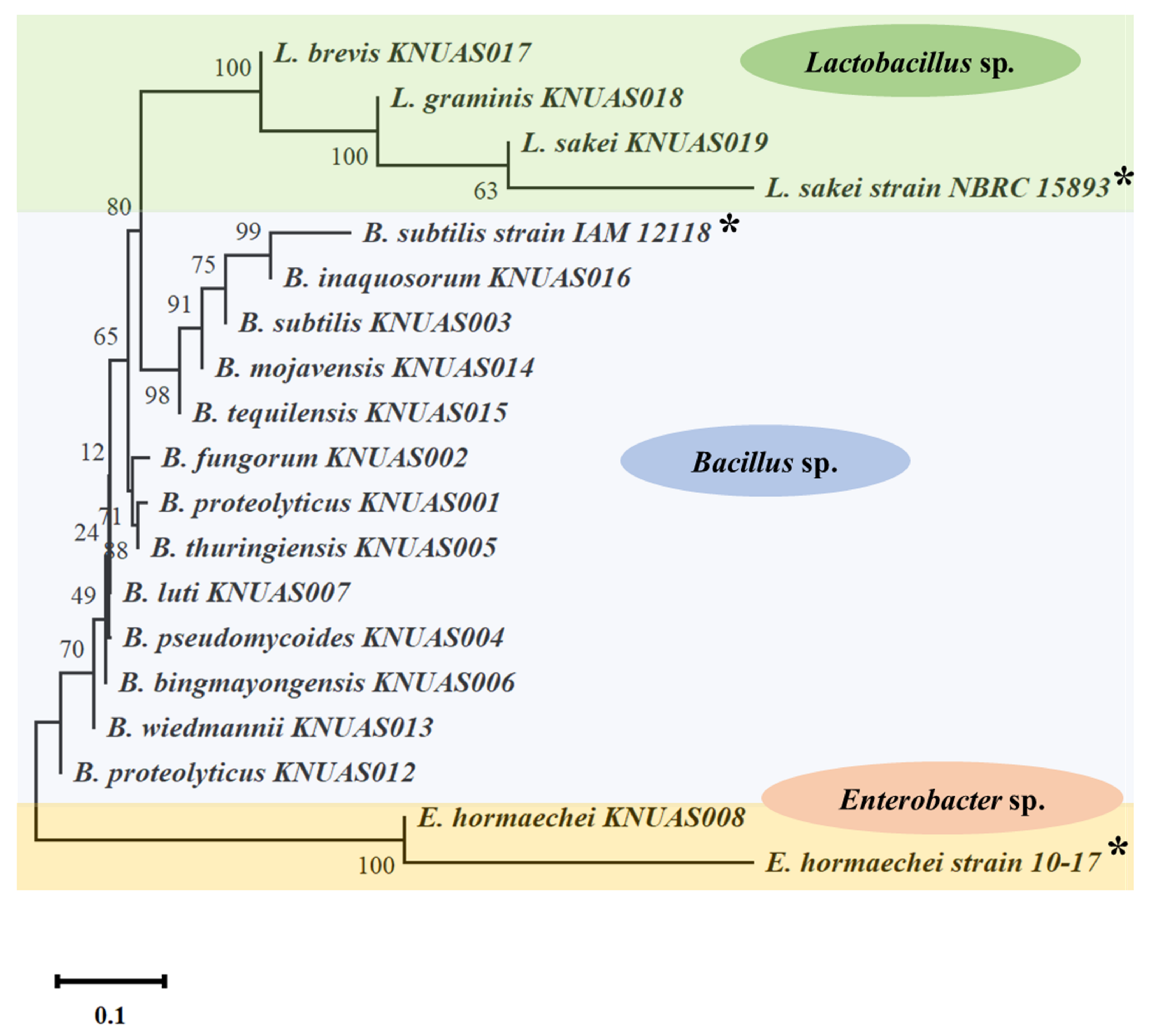
2.1. Identification of Probiotic Bacterial Strains

About 14 fermented Korean food samples were collected for the isolation of probiotics. Among the samples, except the fermented fruit juices (quince, crimson glory vine, David’s peach, and Japanese apricot green (old)), all other samples were observed the bacterial colonies. Sixteen bacterial isolates were isolated and identified using morphological properties and 16S rRNA gene sequencing primers (27F and 1492R) (Table 1). The results revealed that the 16 isolates were belonging to the genera of Lactobacillus (3 strains), Bacillus (12 strains), and Enterobacter (1 strain). The Bacillus sp. includes B. proteolyticus KNUAS001, B. fungorum KNUAS002, B. subtilis KNUAS003, B. pseudomycoides KNUAS004, B. thuringiensis KNUAS005, B. bingmayongensis KNUAS006, B. luti KNUAS007, B. proteolyticus KNUAS012, B. wiedmannii KNUAS013, B. mojavensis KNUAS014, B. tequilensis KNUAS015, and B. inaquosorum KNUAS016. The report supported that B. subtilis, B. coagulans, and B. clausii were commonly used as probiotic strains in many countries [40]. In addition, B. mojavensis, reported as an endophytic bacterium, was isolated from the plant Bacopa monnieri (Linn.) [41]. B. inaquosorum was reported as a sub-species of B. subtilis [42]. In this study, Lactobacillus sp. was isolated, including L. brevis KNUAS017, L. graminis KNUAS018, and L. sakei KNUAS019. Similarly, L. brevis B13-2 and L. sakei were reported from Chinese cabbage kimchi and young radish kimchi, respectively [32,43]. Enterobacter sp. (E. hormaechei KNUAS008) was identified in this study. Enterobacter sp. is generally recognized as a foodborne pathogen that is also reported in fermented food products [44].
Further, the phylogenetic tree displayed a total of three clades belonging to Bacillus sp., Lactobacillus sp., and Enterobacter sp., which indicated that there were close species relationships among the species and respective genera (Figure 1). The 16s rRNA sequencing results indicated that 15 isolates were Gram-positive, while one strain was Gram-negative, which was confirmed with Gram staining analysis. Further, the bromocresol purple assay evidenced that all isolates from Lactobacillus sp., produced lactic acid in the MRS agar medium.

2.2. Resistance to Biological Barriers

2.2.1. Tolerance in Gastric and Intestinal Fluids

The pH of the human stomach and intestinal fluids ranges from 1 to 4.5 and varies depending upon food consumption, contents, age, and disease condition [45,46]. In addition, the bile salt concentration is a major factor in determining microbial colonization. Survival in gastrointestinal conditions, including low pH and bile salts, is considered an important property of good probiotics. To evaluate the gastrointestinal pH tolerance, bacterial strains were incubated with simulated gastric juice and intestinal fluid at pH 3.0 (Table 2 and Figure S2). The results showed that all the strains survived at the simulated gastric pH for 0–3 h. The survivability ranged from 2.48–7.86 (log10 CFU/mL) and 0.8–8.54 (log10 CFU/mL) at 1 h and 3 h, respectively. Among the strains, the Bacillus sp. showed the highest survivability in gastric pH, while cell counts were reduced in Lactobacillus sp. (Table 2). In particular, the maximum survival rate of 8.25 ± 0.08 log10 CFU/mL was observed for B. inaquosorum KNUAS016 and the minimum survival rate of 0.8 ± 0.02 log10 CFU/mL for L. sakei KNUAS019 at 3 h of incubation. For the intestinal fluid tolerance test, the bacterial strains were incubated with bile salts (1%) and pancreatin (0.1%) for 0–4 h at pH 8.0 (Table 2 and Figure S3). The results showed that all the bacterial strains survived for 4 h in intestinal fluid. However, the Bacillus sp. exhibited higher survivability compared to Lactobacillus sp. (Table 2). Among them, B. inaquosorum KNUAS016 and L. brevis KNUAS017 had the highest survival rates of 6.86 ± 0.02 and 5.37 ± 0.01 log10 CFU/mL, respectively, in the intestinal fluid environment (Figure S3). According to GRAS status, B. inaquosorum was a subspecies of B. subtilis and a potential probiotic strain [47]. The earlier report also supported that L. brevis KU15153 isolated from kimchi exhibited higher viability under the gastric condition with probiotic potential [48]. Overall, the simulated tolerance studies found that harsh environmental viability highly depends on each strain.

2.2.2. Autoaggregation, Coaggregation, and Hydrophobicity Properties of Bacterial Strains

The autoaggregation and coaggregation ability assays were used to evaluate the potential of probiotics in intestinal colonization through cell-to-cell interaction, biofilm formation, and pathogen inhibition [49]. Hence, all the bacterial strains were tested for their autoaggregation ability at different time intervals (1, 3, and 6 h) (Table 3). All the strains exhibited increased autoaggregation (%) with increasing incubation time from 1 h to 6 h. The maximum autoaggregation (%) was observed at 6 h for L. sakei KNUAS019 (66.55 ± 0.33%), B. tequilensis KNUAS015 (64.56 ± 0.14%), and B. inaquosorum KNUAS016 (61.63 ± 0.19%) but was lower in L. brevis KNUAS017 (29.98 ± 0.09%). These results indicated that those bacterial strains could survive and adhere to the gastrointestinal tract. Moreover, the coaggregation ability of bacterial strains was tested at different time intervals (1, 3, and 6 h) (Table 3). At 6 h of incubation, the coaggregation was greater than 50% for B. subtilis KNUAS003 (63.84 ± 0.19%), B. tequilensis KNUAS015 (63.64 ± 0.35%), B. mojavensis KNUAS014 (61.67 ± 0.39%), and L. sakei KNUAS019 (55.69 ± 0.30%) but lower for B. proteolyticus KNUAS001 (30.02 ± 0.33%). The coaggregation results indicated that B. subtilis KNUAS003, B. tequilensis KNUAS015, B. mojavensis KNUAS014, and L. sakei KNUAS019 might have surface proteins that inhibited the pathogens, thereby maintaining the microbial balance in the gut [50].
Further cell surface properties of bacterial strains were determined through hydrophobicity. The hydrophobicity of bacterial strains indicated that they could interact with the intestinal mucosa and epithelial cells, thereby ensuring colonization [51]. Hence, the bacterial strains were incubated with an organic solvent (xylene), and their ability to adhere to the cell surface was tested (Table 3). Interestingly, all Lactobacillus spp., including L. brevis KNUAS017 (25.64 ± 0.28%), L. graminis KNUAS018 (18.81 ± 1.36%), and L. sakei KNUAS019 (14.91 ± 0.48%), and one Bacillus strain, such as B. inaquosorum KNUAS016 (16.76 ± 0.16%), exhibited promising hydrophobicity, while the lowest hydrophobicity was found for B. bingmayongensis KNUAS006 (0.09 ± 0.83%) and E. hormaechei KNUAS008 (0.24 ± 0.10%). Accordingly, the hydrophobicity may differ with each isolate even within species and with the types of hydrocarbons used in this assay [52].

2.3. Safety Assessment

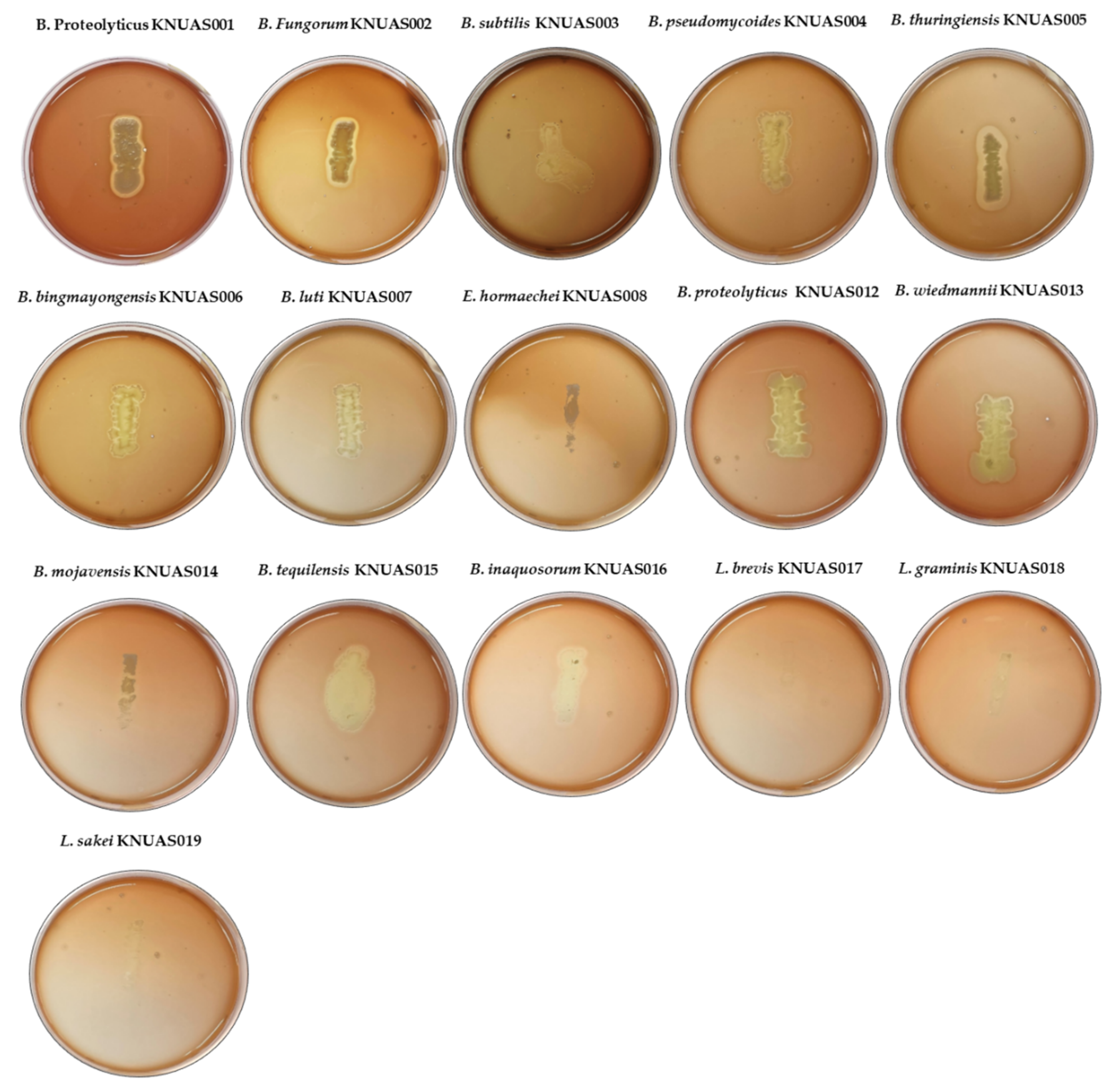
2.3.1. Hemolytic Property

For the safety assessment, all the bacterial strains isolated from Korean fermented foods were evaluated their hemolytic activity in 5% of sheep blood supplemented blood base agar (Figure 2). Results demonstrated that none of the bacterial strains exhibited the α-hemolysis, while some of the Bacillus strains (B. proteolyticus KNUAS001, B. fungorum KNUAS002, B. thuringiensis KNUAS005, and B. wiedmannii KNUAS013) showed β-hemolysis. However, the B. proteolyticus KNUAS012 did not show significant hemolytic activity compared to B. proteolyticus KNUAS001. Accordingly, the study reported that B. proteolyticus from Tibetan yaks did not show hemolytic activity but did find probiotic potential [53]. Interestingly, none of the Lactobacillus spp. and Enterobacter spp. showed any changes, which was considered to be non-hemolytic activity (γ-hemolysis).

2.3.2. Antibiotic Susceptibility

Another safety assessment of probiotics regards antibiotic resistance. According to the FAO and WHO, probiotics should not carry the antibiotic-resistant gene. The results showed that most of the Bacillus strains were susceptible to tested antibiotics, but B. mojavensis KNUAS014 and B. inaquosorum KNUAS016 showed complete resistance to all the tested antibiotics (Table 4 and Figure S4), followed by B. proteolyticus KNUAS012, which showed resistance toward vancomycin. The E. hormaechei KNUAS008 was susceptible to all the tested antibiotics, but Enterobacter sp. caused the pathogenicity and antimicrobial resistance [54]. In addition, the study reported that E. hormaechei subsp. Were susceptible to different antibiotics [55]. Interestingly, L. brevis KNUAS017 showed resistance to all the tested antibiotics, while L. graminis KNUAS018 and L. sakei KNUAS019 were susceptible to TCH, ERY, and AMP but resistant to VAN and GEN (Table 4). According to the earlier report, Lactobacillus spp. were generally resistant to vancomycin and gentamycin [56,57,58]. Furthermore, antibiotic-resistant genes were found in commercially available health-promoting probiotics [59]. The frequent use of antibiotic-resistant genes containing probiotics may negatively modulate the immune system and impact human health [25]. Most of the LAB strains showed intrinsic resistance to antibiotics that could not be transferred to other microbes. However, the selection of potential health-promoting probiotics is a challenging task that could be overcome by multiple analyses related to the toxicity assessment.

2.4. Characterization of Probiotic Potential

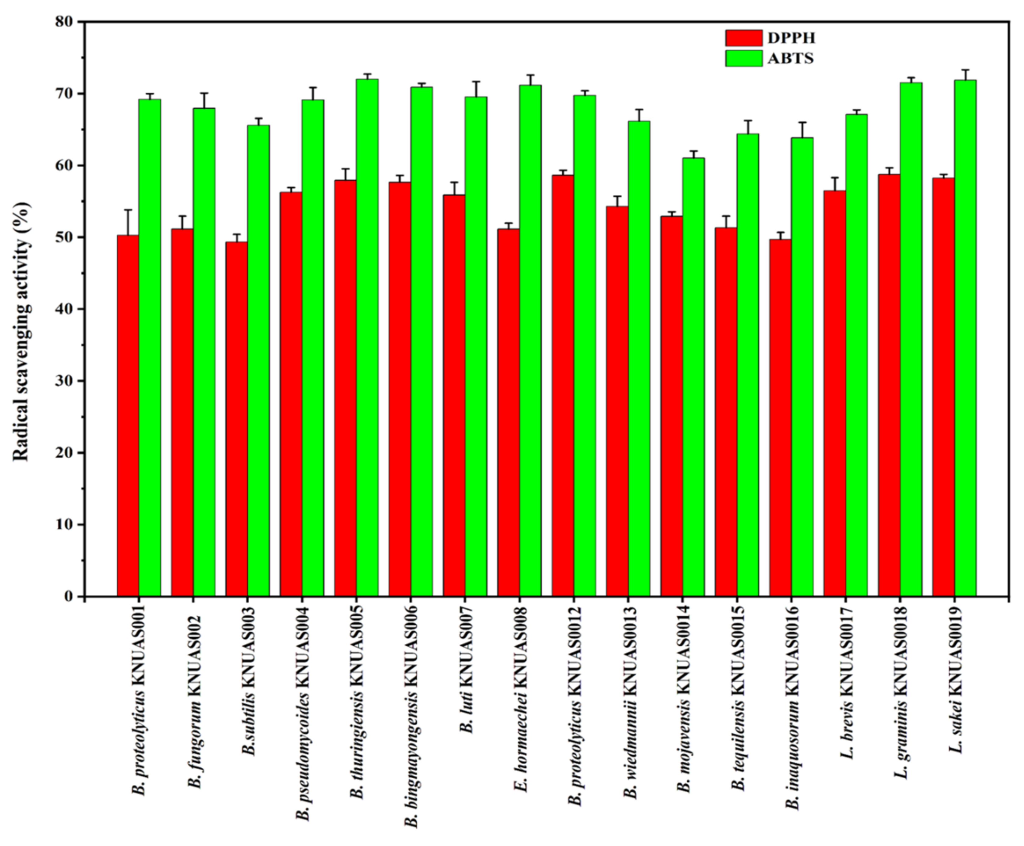
2.4.1. Antioxidant Activity

Antioxidant molecules play a major role in retaining the gut microbiome balance by modulating oxidative stress [60]. Evaluating the antioxidant properties of isolated bacterial strains, suspensions were determined in DPPH and ABTS free radical scavenging assay (Figure 3). The results indicated that the bacterial suspensions of all the strains showed a substantial DPPH and ABTS free radical scavenging ability (Figure 3). Among them, the highest DPPH and ABTS radical scavenging activity was observed in L. sakei KNUAS019 (58.25% and 71.88%), L. graminis KNUAS018 (58.74% and 71.51%), and B. proteolyticus KNUAS012 (58.62% and 71.17%). The B. mojavensis KNUAS014 showed the lowest ABTS radical scavenging activity at 61.02%. Besides, B. subtilis KNUAS003 showed the lowest DPPH radical scavenging activity at 49.34%. A study reports that Lactobacillus plantarum strains showed <10% DPPH radical scavenging activity [61]. In addition, a previous study reported that L. plantarum NJAU-01 significantly decreased lipid peroxidation by increasing enzyme activity in mice [62]. However, the antioxidant activity of isolated Bacillus sp. and Lactobacillus sp. was considerably good in this study compared to earlier reports [32,63].

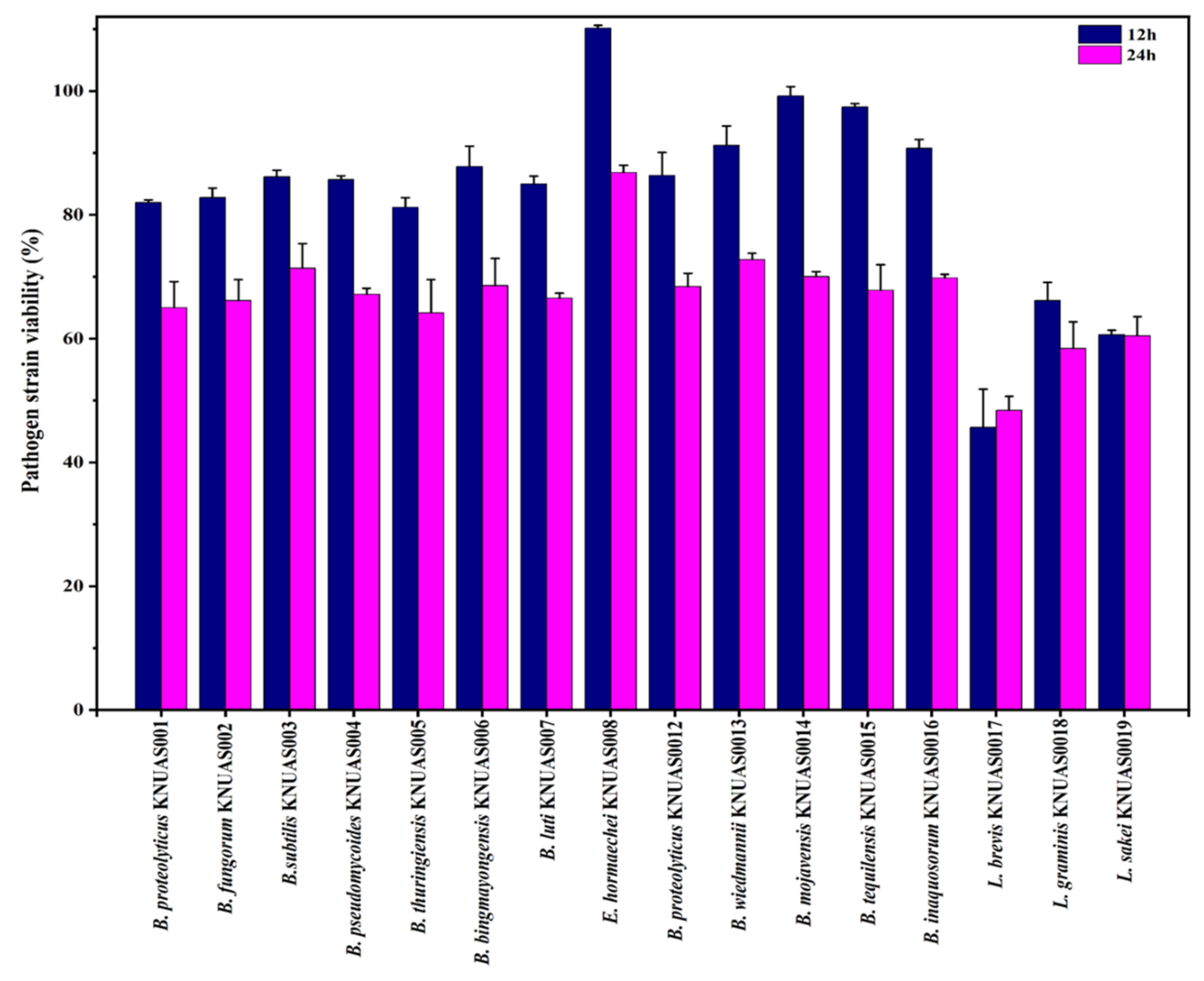
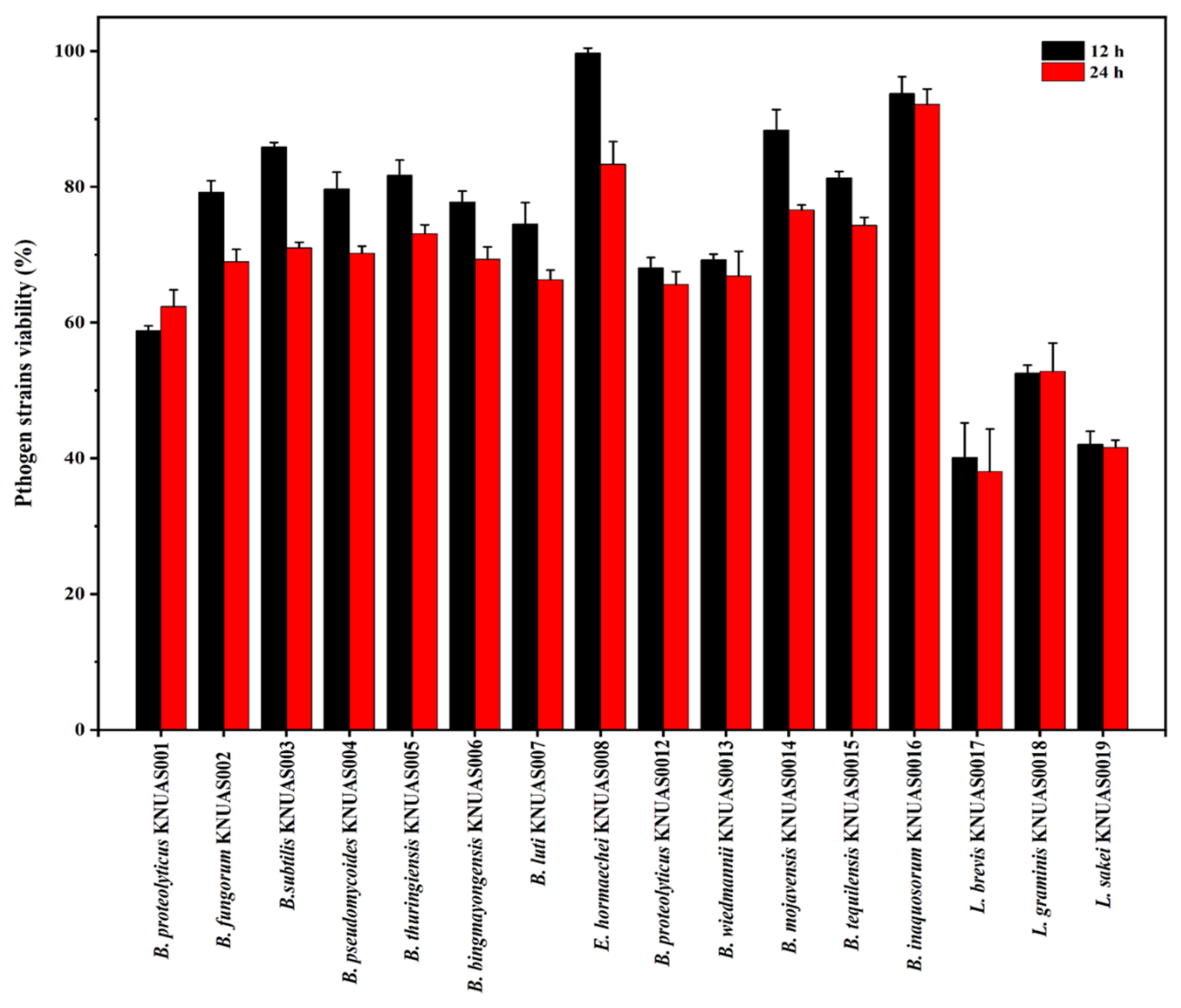
2.4.2. Antibacterial Properties

The antibacterial property of CFs of bacterial strains was evaluated with Gram-positive (S. aureus) and Gram-negative bacteria (E. coli) (Figure 4 and Figure 5). Results found that cell-free supernatant of Lactobacillus sp. considerably inhibited pathogenic growth, while cell-free supernatant of Bacillus sp. was moderately inhibited when incubated for 24 h. Accordingly, earlier studies reported that B. subtilis, B. mojavensis, and B. inaquosorum showed broad-spectrum antibacterial and antifungal activity [64,65,66]. In addition, earlier studies confirmed that B. subtilis and B. mojavensis had considerable probiotic potential with antibacterial activity against pathogenic bacteria such as S. aureus [41,67]. B. proteolyticus and B. thuringiensis exhibited pathogenic bacterial inhibition, and the production of antimicrobial peptide bacteriocin from B. thuringiensis was reported [68]. Further, the antilisterial peptide (Subtilosin A) showed the inhibition of invasion of the pathogen on human cells, which was isolated from B. tequilensis FR9 [69]. However, E. hormaechei KNUAS008 showed no significant inhibition of pathogenic growth. The 24 h incubation of CFs of L. brevis KNUAS017 considerably inhibited the growth of S. aureus and E. coli (51.59% and 61.96%), followed by L. sakei KNUAS019 (39.54% and 58.42%) and L. graminis KNUAS018 (41.56% and 47.19%), respectively. Similarly, the earlier study reported that L. crispatus strain exhibited significant inhibitory activity among the other LAB stains when co-cultured with a different bacterial pathogen, which might have a higher content of bacteriocin [70]. Similarly, several studies confirmed the antibacterial efficiency of LAB [58,71]. These results indicated that the antibacterial activity highly depends on the strains and their bioactive metabolites.

3. Materials and Methods

3.1. Isolation of Probiotic Bacterial Strains and Culture Condition

The bacterial cultures were isolated from the 14 types of fermented samples, including homemade soybean paste, commercial soybean paste, cabbage kimchi, green onion kimchi, leaf mustard kimchi, ginger (Zingiber officinale) juice, quince (Cydonia oblonga) fruit juice, plum (Prunus domestica) fruit juice, crimson glory vine (Vitis coignetiae) fruit juice, Korean bramble (Rubus coreanus) fruit juice, Japanese apricot (Prunus mume) red fruit juice, David’s peach (Prunus davidiana) fruit juice, Prunus mume green fruit juice (old), and Prunus mume green fruit juice (fresh) (Figure S1), were collected from different places in the Republic of Korea. For the isolation, fruit juices (1 mL), soybean paste (1 g), and kimchi (1 g) were inoculated in MRS (de Man, Rogosa, Sharpe) broth purchased from Oxoid LTD, England. The sample inoculated broth was incubated at 37 °C for 24 h under an anaerobic condition. Then, each sample was serially diluted (10−1 to 10−5) by the standard serial dilution method. At each concentration, 50 µL of the sample was inoculated on an MRS (de Man, Rogosa, Sharpe) agar purchased from Oxoid LTD, England. Then, the plates were incubated at 37 °C for 24–48 h. Further, morphologically distinct colonies were sub-cultured onto MRS agar plates, and pure cultures were stored for further analysis. To determine the lactic acid production, bacterial strains were inoculated in 0.12 g/L of bromocresol purple (Sigma-Aldrich, St. Louis, MO, USA), and MRS agar medium, pH 6.8, was added. The formation of the yellow zone around the colony was a visual indication of lactic acid production.

3.2. Identification of Bacterial Strains from Fermented Korean Foods

For identification, a total of 16 pure bacterial isolates were subjected to morphological and 16S rRNA sequencing analysis. Morphological characteristics were determined by Gram staining analysis. In molecular identification, the genomic DNA was extracted from the selected bacterial isolates using a bacterial DNA extraction kit (GenelixTM, Republic of Korea). Further, the isolated bacterial DNA was amplified using standard forward primer 27F (5′-AGA GTT TGA TCM TGG CTC AG-3′) and reverse primer 1492R (5′-TAC GGY TAC CTT GTT ACG ACT T-3′) by polymerase chain reaction (PCR). The PCR condition was used for pre-denaturation at 90 °C for 3 min, denaturation at 95 °C for the 30s, annealing at 56 °C for 30 s, and elongation at 72 °C for 60 s, 30 cycles, with a final extension at 72 °C for 10 min. The obtained PCR product was separated using agarose gel (1.2%) electrophoresis and purified. Then, 16S rDNA sequences were performed at COSMOGENTECH, Republic of Korea. The obtained 16S rDNA sequences were used for the identification of bacterial isolates by nucleotide BLAST search, and then, these sequences were deposited at the NCBI Gene bank (https://www.ncbi.nlm.nih.gov/ accessed on 21 January 2022). The phylogenetic relationship among the sequences was determined using a neighbor-joining method by MEGA X software (version 11).

3.3. Resistance to Biological Barriers

3.3.1. Tolerance in Gastric Juice and Intestinal Fluids

Bile salts and pancreatin from the porcine pancreas were purchased from Sigma-Aldrich (St. Louis, MO, USA). Pepsin from pig gastric mucosa was purchased from Roche (Basel, Switzerland). The simulated gastric juice and intestinal fluid tolerance of bacterial strains were determined according to the earlier report [72]. In brief, the bacterial strains were cultured in 30 mL of MRS broth and incubated in an incubator overnight at 37 °C. After incubation, the bacterial cells were collected by centrifugation at 6000× g for 10 min, washed with phosphate buffer saline (PBS, pH 7.4), and suspended in 10 mL of PBS. For simulated gastric juice tolerance, 1 mL of bacterial cell suspension was further suspended and incubated for 3 h with 10 mL of PBS (pH 3.0) containing pepsin (0.3%) and NaCl (0.5%). For intestinal fluid tolerance, the bacterial cell suspension was resuspended and incubated for 4 h with 10 mL PBS (pH 8.0) containing pancreatin (0.1%) and bile salts (1%). Bacterial tolerance at each condition was also determined by broth dilution assay by measuring the optical density (OD) at 600 nm under a UV–visible spectrophotometer and inoculating 50 µL of cell suspension onto the MRS agar plate. After incubation at 37 °C, the plates were enumerated, and the results were expressed as log CFU/mL.

3.3.2. Autoaggregation and Coaggregation Ability

The overnight cultured bacterial cells were collected, washed by centrifugation at 6000× g for 10 min, and resuspended in PBS (pH 7.4). Autoaggregation was determined by measuring the suspended bacterial cells absorbance (OD at 600 nm) at each predetermined time interval (1, 3, and 6 h), and the plates were kept in an incubator at 37 °C without any disturbance during the experiment. For coaggregation analysis, the bacterial cells were prepared according to the autoaggregation assay. Further, bacterial cells were resuspended in PBS and co-incubated with a cell suspension of Staphylococcus aureus (ATCC 19095) under the above-mentioned conditions. The autoaggregation and coaggregation percentages were determined according to the earlier work [61].

3.3.3. Hydrophobicity

To understand the microbial adhesion in the intestine, the bacterial cells were incubated with non-polar solvents and evaluated for their cell surface hydrophobicity. In brief, overnight bacterial cultures were collected by centrifugation at 6000× g for 10 min. Then, cells were washed and resuspended with 10 mL of PBS (pH 7.4). Next, 1 mL of bacterial cell suspensions (108 CFU/mL) were mixed with 3 mL of organic solvent (Xylene (98.5%)) and vortex for 60 s. Then, the bacterial and organic solvent mixture was left at room temperature for 60 min. After phase separation, the upper organic phase was removed, and the lower aqueous phase was measured at 600 nm and the hydrophobicity calculated according to an earlier report [73].

3.4. Safety Assessment

3.4.1. Hemolytic Property

Hemolytic activity was determined by streaking the bacterial strains on a blood base agar medium supplemented with 5% (v/v) of sheep blood. All the plates were incubated at 37 °C for 24 h. Hemolytic properties of bacterial strains were determined based on the following changes: α-hemolysis (green color in the medium); γ-hemolysis (no changes), considered non-hemolytic; and β-hemolysis (blood lysis, clear zone), considered hemolytic [74].

3.4.2. Antibiotic Susceptibility

Tetracycline hydrochloride (TCH), erythromycin (ERY), ampicillin sodium salt (AMP), gentamicin sulfate (GEN), and vancomycin hydrochloride (VAN) were purchased from Sigma-Aldrich (St. Louis, MO, USA). The antibiotic sensitivity of bacterial strains was evaluated by the disc diffusion method according to the regulations of the Clinical and Laboratory Standards Institute (CLSI) [75]. The overnight cultured bacterial cell counts were adjusted to 1 × 10⁷ CFU/mL and inoculated on MHA plates. Afterward, antibiotic discs (TCH (30 µg), VAN (30 µg), ERY (15 µg), GEN (10 µg), and AMP (10 µg)) were placed on the bacteria inoculated medium. Then, the plates were incubated at 37 °C for 24 h. The results with a zone of inhibition ≤15 mm were considered to be resistant, 15–21 mm to be intermediate, and ≥21 mm to be susceptible [75]. Experiments were repeated three times.

3.5. Characterization of Probiotic Potential

3.5.1. Antioxidant Activity

The antioxidant activity of bacterial strains was determined by DPPH and ABTS radical scavenging assay [32,76]. For 2,2-diphenyl-1-picrylhydrazyl (DPPH) (Sigma-Aldrich, St. Louis, MO, USA) assay, 100 µL of bacterial suspension (1 × 10⁹ CFU/mL) was mixed with DPPH (100 µM) solution, incubated for 15 min in dark condition, the pellet removed by centrifugation, and the supernatant measured at 517 nm by UV–vis spectrophotometer. For 2,2′-Azino-bis(3-ethylbenzothiazoline-6-sulfonic acid) diammonium salt (ABTS) (Sigma-Aldrich, St. Louis, MO, USA) radical scavenging assay, 100 µL of bacterial suspension mixed with 100 µL of ABTS+ solution was prepared according to the earlier report [77] and then incubated for 10 min at room temperature in a dark condition. ABTS+ scavenging ability was determined by measuring the absorbance of 734 nm. The percentage of DPPH and ABTS+ scavenging activity was determined according to an earlier report [76].

3.5.2. Preparation of Culture Free Supernatant

The bacterial strains were cultured in MRS broth at 37 °C for 24 h. The cell-free culture supernatant was collected from bacterial cell suspension cultures by centrifugation at 15,000× g for 10 min. To inactivate the organic acid in the culture supernatant, it was aseptically neutralized using NaOH (1 M) at pH 6.5 [78]. Further, the culture supernatant was filtered using a 0.22 µm syringe filter to obtain the cell-free supernatant.

3.5.3. Antibacterial Properties

The antibacterial activity of culture supernatant was determined against Gram-positive (S. aureus; ATCC 19095) and Gram-negative (E. coli; ATCC 43888) pathogens by agar well diffusion and broth dilution method. For agar well diffusion assay, pathogenic bacteria were initially cultured in nutrient broth (NB) medium overnight at 37 °C. The bacterial cells (1 × 10⁹ CFU/mL) were inoculated onto an MHA plate by sterile cotton swap uniformly, then wells were made using a cork borer. After 50 µL of each bacterial culture supernatant was added to each well, the plates were incubated overnight at 37 °C. Antibacterial ability is represented in terms of the zone of inhibition. For the broth dilution assay, pathogenic bacterial cells (1 × 10⁴ CFU/mL) were inoculated in BHI medium along with culture supernatant (30% v/v), added to 96-well plates, and kept at 37 °C for 24 h. The antibacterial ability was determined by measuring the absorbance at 620 nm. The percentage of growth inhibition is represented by the percentage of optical density (OD) according to the earlier report [78].

3.6. Statistical Analysis

The experimental results were expressed mean ± standard deviation. The statistical significance was determined through a one-way analysis of variance (ANOVA). p-value less than 0.05 is considered statistically significant.

4. Conclusions

In this study, we isolated sixteen bacterial isolates belonging to three genera, including Lactobacillus (3 strains), Bacillus (12 strains), and Enterobacter (1 strain), from Korean fermented food samples; identified using 16S rRNA gene sequencing; and studied their probiotic potential. Bacterial strains such as B. subtilis KNUAS003, B. proteolyticus KNUAS012, L. brevis KNUAS017, L. graminis KNUAS018, and L. sakei KNUAS019 exhibited several promising probiotic properties, including tolerance to the gastric environment (simulated gastric juice, bile salts, pancreatin) autoaggregation, coaggregation, hydrophobicity, and non-hemolytic activity. Further, isolated strains showed antibiotic susceptibility and resistance that may be used to develop a controlled therapeutic approach. Additionally, the antibacterial and antioxidant properties of probiotics permitted their utilization and commercialization.

Supplementary Materials

The supporting information can be downloaded at: https://www.mdpi.com/article/10.3390/ijms231710062/s1.

Author Contributions

Conceptualization, A.S.; data curation, A.S. and K.S.; formal analysis, A.S., K.S., K.H. and K.V.N.; investigation, A.S.; methodology, A.S.; visualization, A.S.; writing—original draft, A.S.; writing—review and editing, A.S. and K.S.; software, A.S.; funding acquisition, M.-H.W. and A.S.; project administration, M.-H.W. and A.S.; resources, M.-H.W.; supervision, M.-H.W. All authors have read and agreed to the published version of the manuscript.

Funding

This work was supported by the National Research Foundation of Korea (2021R1I1A1A01057742).

Institutional Review Board Statement

Not applicable.

Informed Consent Statement

Not applicable.

Data Availability Statement

The data presented in this study are available in supplementary material.

Conflicts of Interest

The authors declare no conflict of interest. The funders had no role in the design of the study; in the collection, analyses, or interpretation of data; in the writing of the manuscript, or in the decision to publish the results.

References

  1. Zheng, D.; Liwinski, T.; Elinav, E. Interaction between Microbiota and Immunity in Health and Disease. Cell Res. 2020, 30, 492–506. [Google Scholar] [CrossRef] [PubMed]
  2. Hacquard, S.; Garrido-Oter, R.; González, A.; Spaepen, S.; Ackermann, G.; Lebeis, S.; McHardy, A.C.; Dangl, J.L.; Knight, R.; Ley, R.; et al. Microbiota and Host Nutrition across Plant and Animal Kingdoms. Cell Host Microbe 2015, 17, 603–616. [Google Scholar] [CrossRef] [PubMed]
  3. Lynch, J.B.; Hsiao, E.Y. Microbiomes as Sources of Emergent Host Phenotypes. Science 2019, 365, 1405–1409. [Google Scholar] [CrossRef] [PubMed]
  4. Wu, H.J.; Wu, E. The Role of Gut Microbiota in Immune Homeostasis and Autoimmunity. Gut Microbes 2012, 3, 4. [Google Scholar] [CrossRef]
  5. Zhang, M.; Sun, K.; Wu, Y.; Yang, Y.; Tso, P.; Wu, Z. Interactions between Intestinal Microbiota and Host Immune Response in Inflammatory Bowel Disease. Front. Immunol. 2017, 8, 942. [Google Scholar] [CrossRef]
  6. Valitutti, F.; Cucchiara, S.; Fasano, A. Celiac Disease and the Microbiome. Nutrients 2019, 11, 2403. [Google Scholar] [CrossRef]
  7. Maeda, Y.; Takeda, K. Host-Microbiota Interactions in Rheumatoid Arthritis. Exp. Mol. Med. 2019, 51, 1–6. [Google Scholar] [CrossRef]
  8. Belizário, J.E.; Faintuch, J.; Garay-Malpartida, M. Gut Microbiome Dysbiosis and Immunometabolism: New Frontiers for Treatment of Metabolic Diseases. Mediat. Inflamm. 2018, 2018, 2037838. [Google Scholar] [CrossRef]
  9. Main, B.S.; Minter, M.R. Microbial Immuno-Communication in Neurodegenerative Diseases. Front. Neurosci. 2017, 11, 151. [Google Scholar] [CrossRef]
  10. Gopalakrishnan, V.; Helmink, B.A.; Spencer, C.N.; Reuben, A.; Wargo, J.A. The Influence of the Gut Microbiome on Cancer, Immunity, and Cancer Immunotherapy. Cancer Cell 2018, 33, 570–580. [Google Scholar] [CrossRef] [Green Version]
  11. Redondo-Useros, N.; Nova, E.; González-Zancada, N.; Díaz, L.E.; Gómez-Martínez, S.; Marcos, A. Microbiota and Lifestyle: A Special Focus on Diet. Nutrients 2020, 12, 1776. [Google Scholar] [CrossRef]
  12. Patangia, D.V.; Ryan, C.A.; Dempsey, E.; Ross, R.P.; Stanton, C. Impact of Antibiotics on the Human Microbiome and Consequences for Host Health. Microbiologyopen 2022, 11, e1260. [Google Scholar] [CrossRef]
  13. Belkaid, Y.; Hand, T.W. Role of the Microbiota in Immunity and Inflammation. Cell 2014, 157, 121. [Google Scholar] [CrossRef]
  14. Rostami, F.M.; Mousavi, H.; Mousavi, M.R.N.; Shahsafi, M. Efficacy of Probiotics in Prevention and Treatment of Infectious Diseases. Clin. Microbiol. Newsl. 2018, 40, 97–103. [Google Scholar] [CrossRef]
  15. Meurman, J.H.; Stamatova, I. Probiotics: Contributions to Oral Health. Oral Dis. 2007, 13, 443–451. [Google Scholar] [CrossRef]
  16. Morrow, L.E.; Gogineni, V.; Malesker, M.A. Probiotics in the Intensive Care Unit. Nutr. Clin. Pract. 2012, 27, 235–241. [Google Scholar] [CrossRef]
  17. Fontana, L.; Bermudez-Brito, M.; Plaza-Diaz, J.; Muñoz-Quezada, S.; Gil, A. Sources, Isolation, Characterisation and Evaluation of Probiotics. Br. J. Nutr. 2013, 109, S35–S50. [Google Scholar] [CrossRef]
  18. Dekaboruah, E.; Suryavanshi, M.V.; Chettri, D.; Verma, A.K. Human Microbiome: An Academic Update on Human Body Site Specific Surveillance and Its Possible Role. Arch. Microbiol. 2020, 202, 2147–2167. [Google Scholar] [CrossRef] [PubMed]
  19. Belizário, J.E.; Napolitano, M. Human Microbiomes and Their Roles in Dysbiosis, Common Diseases, and Novel Therapeutic Approaches. Front. Microbiol. 2015, 6, 1050. [Google Scholar] [CrossRef]
  20. Dubourg, G.; Lagier, J.C.; Armougom, F.; Robert, C.; Audoly, G.; Papazian, L.; Raoult, D. High-Level Colonisation of the Human Gut by Verrucomicrobia Following Broad-Spectrum Antibiotic Treatment. Int. J. Antimicrob. Agents 2013, 41, 149–155. [Google Scholar] [CrossRef]
  21. Sornplang, P.; Piyadeatsoontorn, S. Probiotic Isolates from Unconventional Sources: A Review. J. Anim. Sci. Technol. 2016, 58, 26. [Google Scholar] [CrossRef] [PubMed]
  22. Roobab, U.; Batool, Z.; Manzoor, M.F.; Shabbir, M.A.; Khan, M.R.; Aadil, R.M. Sources, Formulations, Advanced Delivery and Health Benefits of Probiotics. Curr. Opin. Food Sci. 2020, 32, 17–28. [Google Scholar] [CrossRef]
  23. Verna, E.C.; Lucak, S. Use of Probiotics in Gastrointestinal Disorders: What to Recommend? Ther. Adv. Gastroenterol. 2010, 3, 307. [Google Scholar] [CrossRef] [PubMed]
  24. Culligan, E.P.; Hill, C.; Sleator, R.D. Probiotics and Gastrointestinal Disease: Successes, Problems and Future Prospects. Gut Pathog. 2009, 1, 19. [Google Scholar] [CrossRef]
  25. Montassier, E.; Valdés-Mas, R.; Batard, E.; Zmora, N.; Dori-Bachash, M.; Suez, J.; Elinav, E. Probiotics Impact the Antibiotic Resistance Gene Reservoir along the Human GI Tract in a Person-Specific and Antibiotic-Dependent Manner. Nat. Microbiol. 2021, 6, 1043–1054. [Google Scholar] [CrossRef]
  26. Lee, J.H.; Jin, Y.H.; Park, Y.K.; Yun, S.J.; Mah, J.H. Formation of Biogenic Amines in Pa (Green Onion) Kimchi and Gat (Mustard Leaf) Kimchi. Foods 2019, 8, 109. [Google Scholar] [CrossRef]
  27. Lee, Y.C. Kimchi: The Famous Fermented Vegetable Product in Korea. Food Rev. Int. 2009, 7, 399–415. [Google Scholar] [CrossRef]
  28. Song, H.S.; Lee, S.H.; Ahn, S.W.; Kim, J.Y.; Rhee, J.K.; Roh, S.W. Effects of the Main Ingredients of the Fermented Food, Kimchi, on Bacterial Composition and Metabolite Profile. Food Res. Int. 2021, 149, 110668. [Google Scholar] [CrossRef]
  29. Petrova, P.; Arsov, A.; Tsvetanova, F.; Parvanova-mancheva, T.; Vasileva, E.; Tsigoriyna, L.; Petrov, K. The Complex Role of Lactic Acid Bacteria in Food Detoxification. Nutrients 2022, 14, 2038. [Google Scholar] [CrossRef]
  30. Jeong, D.W.; Lee, J.H. Antibiotic Resistance, Hemolysis and Biogenic Amine Production Assessments of Leuconostoc and Weissella Isolates for Kimchi Starter Development. LWT-Food Sci. Technol. 2015, 64, 1078–1084. [Google Scholar] [CrossRef]
  31. Park, S.; Saravanakumar, K.; Sathiyaseelan, A.; Park, S.J.; Hu, X.; Wang, M.H. Cellular Antioxidant Properties of Nontoxic Exopolysaccharide Extracted from Lactobacillales (Weissella Cibaria) Isolated from Korean Kimchi. LWT 2022, 154, 112727. [Google Scholar] [CrossRef]
  32. Song, M.W.; Chung, Y.; Kim, K.T.; Hong, W.S.; Chang, H.J.; Paik, H.D. Probiotic Characteristics of Lactobacillus Brevis B13-2 Isolated from Kimchi and Investigation of Antioxidant and Immune-Modulating Abilities of Its Heat-Killed Cells. LWT 2020, 128, 109452. [Google Scholar] [CrossRef]
  33. Cutting, S.M. Bacillus Probiotics. Food Microbiol. 2011, 28, 214–220. [Google Scholar] [CrossRef]
  34. Casula, G.; Cutting, S.M. Bacillus Probiotics: Spore Germination in the Gastrointestinal Tract. Appl. Environ. Microbiol. 2002, 68, 2344–2352. [Google Scholar] [CrossRef]
  35. Mazkour, S.; Shekarforoush, S.S.; Basiri, S.; Nazifi, S.; Yektaseresht, A.; Honarmand, M. Effects of Two Probiotic Spores of Bacillus Species on Hematological, Biochemical, and Inflammatory Parameters in Salmonella Typhimurium Infected Rats. Sci. Rep. 2020, 10, 8035. [Google Scholar] [CrossRef]
  36. Mingmongkolchai, S.; Panbangred, W. Bacillus Probiotics: An Alternative to Antibiotics for Livestock Production. J. Appl. Microbiol. 2018, 124, 1334–1346. [Google Scholar] [CrossRef]
  37. Ehling-Schulz, M.; Koehler, T.M.; Lereclus, D. The Bacillus cereus Group: Bacillus Species with Pathogenic Potential. Microbiol. Spectr. 2019, 7, GPP3-0032-2018. [Google Scholar] [CrossRef]
  38. Pilo, P.; Frey, J. Pathogenicity, Population Genetics and Dissemination of Bacillus Anthracis. Infect. Genet. Evol. 2018, 64, 115–125. [Google Scholar] [CrossRef]
  39. McFarland, L.V.; Evans, C.T.; Goldstein, E.J.C. Strain-Specificity and Disease-Specificity of Probiotic Efficacy: A Systematic Review and Meta-Analysis. Front. Med. (Lausanne) 2018, 5, 124. [Google Scholar] [CrossRef]
  40. Elshaghabee, F.M.F.; Rokana, N.; Gulhane, R.D.; Sharma, C.; Panwar, H. Bacillus as Potential Probiotics: Status, Concerns, and Future Perspectives. Front. Microbiol. 2017, 8, 1490. [Google Scholar] [CrossRef] [Green Version]
  41. Jasim, B.; Sreelakshmi, S.; Mathew, J.; Radhakrishnan, E.K. Identification of Endophytic Bacillus Mojavensis with Highly Specialized Broad Spectrum Antibacterial Activity. 3 Biotech 2016, 6, 187. [Google Scholar] [CrossRef]
  42. Dunlap, C.A.; Bowman, M.J.; Zeigler, D.R. Promotion of Bacillus Subtilis Subsp. Inaquosorum, Bacillus Subtilis Subsp. Spizizenii and Bacillus Subtilis Subsp. Stercoris to Species Status. Antonie Van Leeuwenhoek 2019, 113, 1–12. [Google Scholar] [CrossRef]
  43. Won, S.M.; Chen, S.; Park, K.W.; Yoon, J.H. Isolation of Lactic Acid Bacteria from Kimchi and Screening of Lactobacillus Sakei ADM14 with Anti-Adipogenic Effect and Potential Probiotic Properties. LWT 2020, 126, 109296. [Google Scholar] [CrossRef]
  44. Shaker, R.; Osaili, T.; Al-Omary, W.; Jaradat, Z.; Al-Zuby, M. Isolation of Enterobacter Sakazakii and Other Enterobacter Sp. from Food and Food Production Environments. Food Control 2007, 18, 1241–1245. [Google Scholar] [CrossRef]
  45. Riethorst, D.; Mols, R.; Duchateau, G.; Tack, J.; Brouwers, J.; Augustijns, P. Characterization of Human Duodenal Fluids in Fasted and Fed State Conditions. J. Pharm. Sci. 2016, 105, 673–681. [Google Scholar] [CrossRef]
  46. Evans, D.F.; Pye, G.; Bramley, R.; Clark, A.G.; Dyson, T.J.; Hardcastle, J.D. Measurement of Gastrointestinal PH Profiles in Normal Ambulant Human Subjects. Gut 1988, 29, 1035. [Google Scholar] [CrossRef]
  47. Hutt, C.A.; Keefe, D.M. GRAS Notice (GRN) No. 831. 2018. Available online: https://www.fda.gov/food/generally-recognized-safe-gras/gras-notice-inventory (accessed on 21 January 2022).
  48. Jang, H.J.; Lee, N.K.; Paik, H.D. Probiotic Characterization of Lactobacillus Brevis KU15153 Showing Antimicrobial and Antioxidant Effect Isolated from Kimchi. Food Sci. Biotechnol. 2019, 28, 1521. [Google Scholar] [CrossRef]
  49. Peres, C.M.; Alves, M.; Hernandez-Mendoza, A.; Moreira, L.; Silva, S.; Bronze, M.R.; Vilas-Boas, L.; Peres, C.; Malcata, F.X. Novel Isolates of Lactobacilli from Fermented Portuguese Olive as Potential Probiotics. LWT-Food Sci. Technol. 2014, 59, 234–246. [Google Scholar] [CrossRef]
  50. Pickard, J.M.; Zeng, M.Y.; Caruso, R.; Núñez, G. Gut Microbiota: Role in Pathogen Colonization, Immune Responses and Inflammatory Disease. Immunol. Rev. 2017, 279, 70. [Google Scholar] [CrossRef]
  51. Farid, W.; Masud, T.; Sohail, A.; Ahmad, N.; Naqvi, S.M.S.; Khan, S.; Ali, A.; Khalifa, S.A.; Hussain, A.; Ali, S.; et al. Gastrointestinal Transit Tolerance, Cell Surface Hydrophobicity, and Functional Attributes of Lactobacillus Acidophilus Strains Isolated from Indigenous Dahi. Food Sci. Nutr. 2021, 9, 5092. [Google Scholar] [CrossRef]
  52. Lakra, A.K.; Domdi, L.; Hanjon, G.; Tilwani, Y.M.; Arul, V. Some Probiotic Potential of Weissella Confusa MD1 and Weissella Cibaria MD2 Isolated from Fermented Batter. LWT 2020, 125, 109261. [Google Scholar] [CrossRef]
  53. Zeng, Z.; He, X.; Li, F.; Zhang, Y.; Huang, Z.; Wang, Y.; Li, K.; Bao, Y.; Iqbal, M.; Fakhar-e-Alam Kulyar, M.; et al. Probiotic Properties of Bacillus proteolyticus Isolated From Tibetan Yaks, China. Front. Microbiol. 2021, 12, 649207. [Google Scholar] [CrossRef] [PubMed]
  54. Davin-Regli, A.; Lavigne, J.P.; Pagès, J.M. Enterobacter Spp.: Update on Taxonomy, Clinical Aspects, and Emerging Antimicrobial Resistance. Clin. Microbiol. Rev. 2019, 32, e00002-19. [Google Scholar] [CrossRef] [PubMed]
  55. Hoffmann, H.; Stindl, S.; Ludwig, W.; Stumpf, A.; Mehlen, A.; Monget, D.; Pierard, D.; Ziesing, S.; Heesemann, J.; Roggenkamp, A.; et al. Enterobacter Hormaechei Subsp. Oharae Subsp. Nov., E. Hormaechei Subsp. Hormaechei Comb. Nov., and E. Hormaechei Subsp. Steigerwaltii Subsp. Nov., Three New Subspecies of Clinical Importance. J. Clin. Microbiol. 2005, 43, 3297. [Google Scholar] [CrossRef]
  56. Erginkaya, Z.; Turhan, E.U.; Tatli, D. Determination of Antibiotic Resistance of Lactic Acid Bacteria Isolated from Traditional Turkish Fermented Dairy Products. Iran. J. Vet. Res. 2018, 19, 53. [Google Scholar] [CrossRef]
  57. Szutowska, J.; Gwiazdowska, D. Probiotic Potential of Lactic Acid Bacteria Obtained from Fermented Curly Kale Juice. Arch. Microbiol. 2021, 203, 975–988. [Google Scholar] [CrossRef]
  58. Sornsenee, P.; Singkhamanan, K.; Sangkhathat, S.; Saengsuwan, P.; Romyasamit, C. Probiotic Properties of Lactobacillus Species Isolated from Fermented Palm Sap in Thailand. Probiotics Antimicrob. Proteins 2021, 13, 957–969. [Google Scholar] [CrossRef]
  59. Zheng, M.; Zhang, R.; Tian, X.; Zhou, X.; Pan, X.; Wong, A. Assessing the Risk of Probiotic Dietary Supplements in the Context of Antibiotic Resistance. Front. Microbiol. 2017, 8, 908. [Google Scholar] [CrossRef]
  60. Rajoka, M.S.R.; Thirumdas, R.; Mehwish, H.M.; Umair, M.; Khurshid, M.; Hayat, H.F.; Phimolsiripol, Y.; Pallarés, N.; Martí-Quijal, F.J.; Barba, F.J. Role of Food Antioxidants in Modulating Gut Microbial Communities: Novel Understandings in Intestinal Oxidative Stress Damage and Their Impact on Host Health. Antioxidants 2021, 10, 1563. [Google Scholar] [CrossRef]
  61. Kostelac, D.; Gerić, M.; Gajski, G.; Markov, K.; Domijan, A.M.; Čanak, I.; Jakopović, Ž.; Svetec, I.K.; Žunar, B.; Frece, J. Lactic Acid Bacteria Isolated from Equid Milk and Their Extracellular Metabolites Show Great Probiotic Properties and Anti-Inflammatory Potential. Int. Dairy J. 2021, 112, 104828. [Google Scholar] [CrossRef]
  62. Ge, Q.; Yang, B.; Liu, R.; Jiang, D.; Yu, H.; Wu, M.; Zhang, W. Antioxidant Activity of Lactobacillus Plantarum NJAU-01 in an Animal Model of Aging. BMC Microbiol. 2021, 21, 182. [Google Scholar] [CrossRef] [PubMed]
  63. Alkalbani, N.S.; Turner, M.S.; Ayyash, M.M. Isolation, Identification, and Potential Probiotic Characterization of Isolated Lactic Acid Bacteria and in Vitro Investigation of the Cytotoxicity, Antioxidant, and Antidiabetic Activities in Fermented Sausage. Microb. Cell Fact. 2019, 18, 188. [Google Scholar] [CrossRef]
  64. Avery, S.E.; Ruzbarsky, S.P.; Hise, A.M.; Schreier, H.J. Antibacterial Activity of Bacillus Inaquosorum Strain T1 against PirABVp-Bearing Vibrio Parahaemolyticus: Genetic and Physiological Characterization. Appl. Environ. Microbiol. 2020, 86, e01950-20. [Google Scholar] [CrossRef]
  65. Knight, C.A.; Bowman, M.J.; Frederick, L.; Day, A.; Lee, C.; Dunlap, C.A. The First Report of Antifungal Lipopeptide Production by a Bacillus Subtilis Subsp. Inaquosorum Strain. Microbiol. Res. 2018, 216, 40–46. [Google Scholar] [CrossRef]
  66. Caulier, S.; Nannan, C.; Gillis, A.; Licciardi, F.; Bragard, C.; Mahillon, J. Overview of the Antimicrobial Compounds Produced by Members of the Bacillus Subtilis Group. Front. Microbiol. 2019, 10, 302. [Google Scholar] [CrossRef]
  67. Park, J.H.; Saravanakumar, G.; Kim, K.; Kwon, I.C. Targeted Delivery of Low Molecular Drugs Using Chitosan and Its Derivatives. Adv. Drug Deliv. Rev. 2010, 62, 28–41. [Google Scholar] [CrossRef]
  68. Jung, W.J.; Mabood, F.; Souleimanov, A.; Zhou, X.; Jaoua, S.; Kamoun, F.; Smith, D.L. Stability and Antibacterial Activity of Bacteriocins Produced by Bacillus Thuringiensis and Bacillus Thuringiensis Ssp. Kurstaki. J. Microbiol. Biotechnol. 2008, 18, 1836–1840. [Google Scholar] [CrossRef]
  69. Rani, R.P.; Anandharaj, M.; Hema, S.; Deepika, R.; Ravindran, A.D. Purification of Antilisterial Peptide (Subtilosin A) from Novel Bacillus Tequilensis FR9 and Demonstrate Their Pathogen Invasion Protection Ability Using Human Carcinoma Cell Line. Front. Microbiol. 2016, 7, 1910. [Google Scholar] [CrossRef]
  70. Argentini, C.; Fontana, F.; Alessandri, G.; Lugli, G.A.; Mancabelli, L.; Ossiprandi, M.C.; van Sinderen, D.; Ventura, M.; Milani, C.; Turroni, F. Evaluation of Modulatory Activities of Lactobacillus Crispatus Strains in the Context of the Vaginal Microbiota. Microbiol. Spectr. 2022, 10, e02733-21. [Google Scholar] [CrossRef]
  71. Somashekaraiah, R.; Shruthi, B.; Deepthi, B.V.; Sreenivasa, M.Y. Probiotic Properties of Lactic Acid Bacteria Isolated from Neera: A Naturally Fermenting Coconut Palm Nectar. Front. Microbiol. 2019, 10, 1382. [Google Scholar] [CrossRef]
  72. Mohammed, S.; Çon, A.H. Isolation and Characterization of Potential Probiotic Lactic Acid Bacteria from Traditional Cheese. LWT 2021, 152, 112319. [Google Scholar] [CrossRef]
  73. Albayrak, Ç.B.; Duran, M. Isolation and Characterization of Aroma Producing Lactic Acid Bacteria from Artisanal White Cheese for Multifunctional Properties. LWT 2021, 150, 112053. [Google Scholar] [CrossRef]
  74. Vasiee, A.; Falah, F.; Behbahani, B.A.; Tabatabaee-yazdi, F. Probiotic Characterization of Pediococcus Strains Isolated from Iranian Cereal-Dairy Fermented Product: Interaction with Pathogenic Bacteria and the Enteric Cell Line Caco-2. J. Biosci. Bioeng. 2020, 130, 471–479. [Google Scholar] [CrossRef]
  75. Xia, A.N.; Meng, X.S.; Tang, X.J.; Zhang, Y.Z.; Lei, S.M.; Liu, Y.G. Probiotic and Related Properties of a Novel Lactic Acid Bacteria Strain Isolated from Fermented Rose Jam. LWT 2021, 136, 110327. [Google Scholar] [CrossRef]
  76. Sathiyaseelan, A.; Park, S.; Saravanakumar, K.; Mariadoss, A.V.A.; Wang, M.-H. Evaluation of Phytochemicals, Antioxidants, and Antidiabetic Efficacy of Various Solvent Fractions of Gynura procumbens (Lour.) Merr. Process Biochem. 2021, 111, 51–62. [Google Scholar] [CrossRef]
  77. Sathiyaseelan, A.; Saravanakumar, K.; Mariadoss, A.V.A.; Ramachandran, C.; Hu, X.; Oh, D.H.; Wang, M.H. Chitosan-Tea Tree Oil Nanoemulsion and Calcium Chloride Tailored Edible Coating Increase the Shelf Life of Fresh Cut Red Bell Pepper. Prog. Org. Coat. 2020, 151, 106010. [Google Scholar] [CrossRef]
  78. Estilarte, M.L.; Tymczyszyn, E.E.; de los Ángeles Serradell, M.; Carasi, P. Freeze-Drying of Enterococcus Durans: Effect on Their Probiotics and Biopreservative Properties. LWT 2021, 137, 110496. [Google Scholar] [CrossRef]
Figure 1. Phylogenetic relationship of 16S rRNA gene sequences of Lactobacillus, Bacillus, and Enterobacter strains were isolated from different Korean fermented foods. Phylogenetic tree constructed by neighbor-joining method. * Reference strains.
Ijms 23 10062 g001
Figure 2. Hemolytic properties of isolated bacterial strains. α-hemolysis, green color in the medium; β-hemolysis, blood lysis, clear zone and γ-hemolysis, no changes in the medium, non-hemolytic.
Ijms 23 10062 g002
Figure 3. Antioxidant activity of cell free supernatant (CFS) of isolated bacterial strains. DPPH and ABTS radical scavenging activity.
Ijms 23 10062 g003
Figure 4. Antibacterial activity of cell-free supernatant of isolated bacterial strains against Staphylococus aureus (S. aureus).
Ijms 23 10062 g004
Figure 5. Antibacterial activity of cell free supernatant of isolated bacterial strains against Escherichia coli (E. coli).
Ijms 23 10062 g005
Table 1. List of isolated bacterial strains from fermented Korean foods with 16S rRNA species identification and GenBank accession number. According to the new taxonomy of Lactobacillus, Lactobacillu brevis, Lacotbacillus graminis, and Lactobacillus sakei named as Levilactobacillus brevis, Latilactobacillus graminis, and Latilactobacillus sakei, respectively.
StrainsOrganismIsolation SourceGenBank Accession Number
KNUAS001Bacillus proteolyticusFruit juice of Prunus domesticaOM327557
KNUAS002Bacillus fungorumRhizome juice of Zingiber officinaleOM327558
KNUAS003Bacillus subtilisOM327559
KNUAS004Bacillus pseudomycoidesCommercial soybean pasteOM327560
KNUAS005Bacillus thuringiensisOM327561
KNUAS006Bacillus bingmayongensisOM327562
KNUAS007Bacillus lutiOM327563
KNUAS008Enterobacter hormaecheiFruit juice of red Prunus mumeOM327564
KNUAS012Bacillus proteolyticusHomemade soybean pasteOM327568
KNUAS013Bacillus wiedmanniiOM327569
KNUAS014Bacillus mojavensisGreen onion KimchiOM327570
KNUAS015Bacillus tequilensisOM327571
KNUAS016Bacillus inaquosorumMustard leaf KimchiOM327572
KNUAS017Lactobacillu brevis
(Levilactobacillus brevis)
OM327573
KNUAS018Lacotbacillus graminis (Latilactobacillus graminis)Cabbage KimchiOM327574
KNUAS019Lactobacillus sakei
(Latilactobacillus sakei)
OM327575
Table 2. Tolerance of isolated bacterial strains from fermented Korean foods to simulated gastric juice and intestinal fluid. The different superscript letters follow values indicating the significance among the samples (p < 0.05).
Bacterial IsolateCell Viability (log10 CFU/mL)
Simulated Gastric Juice at pH 3.0Intestinal Fluid (Bile Salts (1%) and Pancreatin (0.1%))
1 h3 h1 h2 h4 h
B. proteolyticus KNUAS0016.72 ± 0.04 b7.42 ± 0.05 b7.95 ± 0.02 a7.54 ± 0.02 a6.46 ± 0.06 a
B. fungorum KNUAS0026.93 ± 0.07 b7.89 ± 0.08 b6.96 ± 0.03 b6.47 ± 0.04 b6.19 ± 0.07 a
B. subtilis KNUAS0035.84 ± 0.02 c5.95 ± 0.02 d6.73 ± 0.02 b6.55 ± 0.03 b6.21 ± 0.05 a
B. pseudomycoides KNUAS0047.26 ± 0.05 a8.12 ± 0.08 a7.45 ± 0.04 a7.24 ± 0.05 a6.34 ± 0.02 a
B. thuringiensis KNUAS0056.21 ± 0.03 b6.76 ± 0.03 c6.21 ± 0.03 b6.12 ± 0.03 b6.02 ± 0.07 a
B. bingmayongensis KNUAS0067.21 ± 0.05 a8.14 ± 0.07 a7.45 ± 0.05 a7.08 ± 0.02 a6.28 ± 0.05 a
B. luti KNUAS0076.58 ± 0.02 b6.97 ± 0.03 c7.87 ± 0.02 a7.52 ± 0.04 a5.84 ± 0.04 b
E. hormaechei KNUAS0086.78 ± 0.04 b7.05 ± 0.05 b6.38 ± 0.04 b6.21 ± 0.08 b5.97 ± 0.03 b
B. proteolyticus KNUAS0127.46 ± 0.07 a6.59 ± 0.04 c7.46 ± 0.07 a7.07 ± 0.06 a6.54 ± 0.07 a
B. wiedmannii KNUAS0137.25 ± 0.08 a5.37 ± 0.01 d6.28 ± 0.04 b6.94 ± 0.02 b6.24 ± 0.08 a
B. mojavensis KNUAS0145.87 ± 0.04 c6.64 ± 0.05 c6.36 ± 0.02 b6.22 ± 0.07 b5.65 ± 0.05 b
B. tequilensis KNUAS0156.79 ± 0.02 b3.47 ± 0.02 e5.48 ± 0.05 c5.25 ± 0.05 c5.15 ± 0.04 b
B. inaquosorum KNUAS0167.86 ± 0.04 a8.25 ± 0.08 a7.98 ± 0.03 a7.74 ± 0.01 a6.86 ± 0.02 a
L. brevis KNUAS0176.31 ± 0.09 b6.17 ± 0.04 c6.24 ± 0.02 b6.09 ± 0.07 b5.37 ± 0.01 b
L. graminis KNUAS0182.48 ± 0.04 e1.56 ± 0.05 f2.81 ± 0.07 e2.61 ± 0.08 d2.21 ± 0.08 c
L. sakei KNUAS0193.48 ± 0.03 d0.8 ± 0.02 g3.65 ± 0.04 d2.82 ± 0.04 d2.14 ± 0.05 c
Table 3. Autoaggregation, coaggregation, and hydrophobicity of isolated bacterial strains from fermented Korean foods to simulated gastric juice and bile salts. The different superscript letters follow values indicating the significance among the samples (p < 0.05).
Bacterial IsolatesAutoaggregation (%)Coaggregation (%)Hydrophobicity (%)
1 h3 h6 h1 h3 h6 hXylene
B. proteolyticus KNUAS0013.87 ± 0.42 g13.83 ± 0.21 e34.44 ± 0.59 e0.62 ± 5.73 h9.61 ± 0.24 f30.02 ± 0.33 d7.44 ± 0.75 d
B. fungorum KNUAS00219.40 ± 0.55 b30.66 ± 0.25 b46.30 ± 0.14 d4.69 ± 0.40 f19.85 ± 0.08 e50.67 ± 0.26 b5.26 ± 1.28 e
B. subtilis KNUAS00318.71 ± 0.38 c33.67 ± 0.26 a52.27 ± 0.23 c36.13 ± 0.46 a39.75 ± 0.23 b63.84 ± 0.19 a1.64 ± 0.38 f
B. pseudomycoides KNUAS00418.60 ± 0.30 c24.89 ± 0.21 c49.08 ± 0.16 d5.82 ± 0.13 f22.37 ± 0.39 d46.09 ± 0.2615.41 ± 0.40 b
B. thuringiensis KNUAS00510.62 ± 0.39 d15.69 ± 0.41 e49.32 ± 0.04 d1.25 ± 0.24 g24.25 ± 0.22 c46.44 ± 0.45 c11.96 ± 0.73 c
B. bingmayongensis KNUAS0066.62 ± 0.08 f8.85 ± 0.12 g49.73 ± 0.10 d1.61 ± 0.44 g24.34 ± 0.72 c51.63 ± 0.35 b0.09 ± 0.83 g
B. luti KNUAS0072.54 ± 0.63 h20.41 ± 0.34 d48.86 ± 0.14 d2.68 ± 0.28 g20.82 ± 0.0647.67 ± 0.27 c13.17 ± 0.76 c
E. hormaechei KNUAS00819.45 ± 0.53 b26.19 ± 0.38 c56.85 ± 0.18 c26.02 ± 0.49 b48.46 ± 0.33 a62.58 ± 0.55 a0.24 ± 0.10 g
B. proteolyticus KNUAS0123.08 ± 0.30 g10.75 ± 0.31 f33.73 ± 0.12 e8.76 e16.03 e49.12 ± 0.34 c12. 46 ± 0.06 c
B. wiedmannii KNUAS0131.52 ± 0.22 i11.04 ± 0.33 f51.35 ± 0.27 c0.39 ± 6.049.68 ± 0.29 f50.39 ± 0.364.67 ± 0.17 e
B. mojavensis KNUAS0147.24 ± 0.44 e8.58 ± 0.27 g56.98 ± 0.10 c14.76 ± 0.12 d20.11 ± 0.40 e61.67 ± 0.39 a1.78 ± 1.00 f
B. tequilensis KNUAS01522.63 ± 0.43 a24.71 ± 0.18 c64.56 ± 0.14 a21.21 ± 0.29 c34.15 ± 0.55 b63.64 ± 0.35 a7.29 ± 0.63 d
B. inaquosorum KNUAS0168.51 ± 0.20 e10.82 ± 0.18 f61.63 ± 0.19 b5.83 ± 0.19 f7.36 ± 0.42 f57.63 ± 0.48 b16.76 ± 0.16 b
L. brevis KNUAS0178.58 ± 0.18 e21.58 ± 0.18 d29.98 ± 0.09 e0.75 ± 0.21 h27.45 ± 0.26 c33.86 ± 0.12 d25.64 ± 0.28 a
L. graminis KNUAS0180.95 ± 0.17 i3.06 ± 0.33 h31.11 ± 0.19 e5.56 f12.14 ± 0.29 f45.71 ± 0.23 c18.81 ± 1.36 b
L. sakei KNUAS0197.01 ± 0.23 e26.06 ± 0.11 c66.55 ± 0.330.66 ± 0.29 h24.71 ± 0.21 c55.69 ± 0.30 b14.91 ± 0.48 c
Table 4. Antibiotic susceptibility of isolated bacterial strains. Tetracycline hydrochloride (TCH, 30 µg); vancomycin hydrochloride (VAN, 30 µg); erythromycin (ERY, 15 µg); gentamicin sulfate (GEN, 10 µg); ampicillin sodium salt (AMP, 10 µg). ≤15 mm, resistance; 15–21 mm, intermediate; and ≥21 mm, susceptible. The different superscript letters follow values indicating the significance among the samples (p < 0.05).
Zone of Inhibition (mm)
Bacterial IsolatesTCHVANERYGENAMP
B. proteolyticus KNUAS00122.2 ± 0.4 b10.2 ± 0.3 c23.2 ± 0.3 c14.1 ± 0.1 b9.2 ± 0.3 d
B. fungorum KNUAS00223.7 ± 0.4 b14.9 ± 0.1 b22.9 ± 0.1 c14.3 ± 0.4 b8.2 ± 0.4 d
B. subtilis KNUAS00318.7 ± 1.1 c11.3 ± 0.4 b19.3 ± 0.5 d13.3 ± 0.5 b12.1 ± 0.1 d
B. pseudomycoides KNUAS00425.9 ± 0.1 b17.7 ± 0.4 a25.8 ± 0.3 a18.2 ± 0.3 a39.7 ± 0.4 a
B. thuringiensis KNUAS00522.2 ± 0.4 b17.8 ± 0.3 a27.1 ± 0.1 a12.2 ± 0.3 c21.8 ± 1.1 b
B. bingmayongensis KNUAS00612.2 ± 0.3 d13.1 ± 0.1 b28.1 ± 0.1 a21.5 ± 0.7 a10.1 ± 0.2 d
B. luti KNUAS00731.7 ± 0.4 a19.1 ± 0.2 a10.1 ± 0.1 e17.2 ± 0.3 a20.9 ± 0.1 b
E. hormaechei KNUAS00818.3 ± 0.4 c12.2 ± 0.4 c22.2 ± 0.3 c12.3 ± 0.5 c22.1 ± 0.1 b
B. proteolyticus KNUAS01228.8 ± 0.2 a022.3 ± 0.4 c17.4 ± 0.6 a39.4 ± 0.9 a
B. wiedmannii KNUAS01325.3 ± 0.4 b18.2 ± 0.4 a23.1 ± 0.1 c041.3 ± 0.9 a
B. mojavensis KNUAS01400000
B. tequilensis KNUAS01510.9 ± 0.1 d14.1 ± 0.2 b16.2 ± 0.3 d12.1 ± 0.1 c18.1 ± 0.1 c
B. inaquosorum KNUAS01600000
L. brevis KNUAS01700000
L. graminis KNUAS01824.8 ± 0.2 b024.9 ± 0.1 c023.2 ± 0.3 b
L. sakei KNUAS01921.4 ± 0.6 b017.1 ± 0.2 d018.0 ± 0.0 c
Publisher’s Note: MDPI stays neutral with regard to jurisdictional claims in published maps and institutional affiliations.

