Article

Limited Accuracy of Pan-Trk Immunohistochemistry Screening for NTRK Rearrangements in Follicular-Derived Thyroid Carcinoma

1
Department of Surgical, Medical, Molecular Pathology and Critical Area, University of Pisa, 56126 Pisa, Italy
2
Pathology Unit, USL Toscana Nord-Ovest, 54033 Carrara, Italy
3
Department of Clinical and Experimental Medicine, University of Pisa, 56126 Pisa, Italy
*
Author to whom correspondence should be addressed.
Int. J. Mol. Sci. 2022, 23(13), 7470; https://doi.org/10.3390/ijms23137470
Submission received: 7 June 2022 / Revised: 1 July 2022 / Accepted: 3 July 2022 / Published: 5 July 2022

Abstract

:
Patients with advanced thyroid cancer harboring NTRK rearrangements can be treated with highly effective selective inhibitors. Immunohistochemistry (IHC) analysis, to detect Trk protein expression, represents an appealing screening strategy for NTRK rearrangements, but its efficacy has been poorly explored in thyroid cancer. The aim of this study is to investigate the diagnostic utility of Trk IHC in the identification of NTRK rearrangements. A series of 26 follicular-derived thyroid tumors, positive for NTRK rearrangements, and 28 NTRK fusion-negative controls were retrospectively analyzed by IHC using the pan-Trk monoclonal antibody (clone EPR17341) on the Ventana system. Area under the curve (AUC), sensitivity and specificity were calculated by ROC analysis. Trk expression was detected in 25 samples, including 22 out of the 26 NTRK-rearranged (84.6%) and three out of 28 NTRK-negative samples (10.7%). Four out of twenty-six NTRK-rearranged thyroid tumors were negative for Trk expression (15.4%), all carrying the ETV6/NTRK3 fusion. The AUC, sensitivity and specificity were 0.87, 0.85 and 0.89, respectively. A screening based on IHC analysis showed limited sensitivity and specificity in the identification of NTRK-rearranged tumors. Since falsely negative results could preclude the administration of effective targeted drugs, alternative detection strategies should be considered for thyroid cancer.

1. Introduction

Structural rearrangements involving neurotrophic tyrosine receptor kinase (NTRK) cause a constitutive activation of Trk proteins, which represents a driving event in several cancer types. The Food and Drug Administration (FDA) tumor-agnostic approval of selective inhibitors targeting NTRK genes expanded the treatment options for patients with advanced tumors carrying these alterations [1]. Besides the relevant clinico-therapeutic implications, many authors focused on the identification of optimal NTRK fusion detection strategies to be implemented in the laboratory practice.
In thyroid cancer, NTRK rearrangements can be found in 2–4% of adult patients, with no evident differences among well-differentiated, poorly differentiated and undifferentiated histotypes [2,3,4,5,6,7,8]. The frequency in pediatric patients with papillary thyroid carcinoma (PTC) is higher, ranging from 8 to 15% [9,10,11,12].
The evaluation of NTRK rearrangements can be performed with a variety of techniques at different levels, by using chromosomal locus-specific probes (FISH, fluorescent in situ hybridization), DNA and RNA sequencing (NGS, next-generation sequencing-based testing), fusion transcript detection (RT-PCR, reverse-transcription polymerase chain reaction, nanoString system) and protein expression analysis (IHC, immunohistochemistry). Each methodology presents its own advantages and limitations, in terms of analytical sensitivity and specificity, technical equipment, time of execution, required expertise, amount of biological material and costs [13,14,15,16].
To ensure the optimal management of biological material and laboratory resources with a reasonable turn-around time, several NTRK testing algorithms have been proposed, specifically focused on thyroid cancer. Some authors would recommend performing IHC testing first, and then: in the case of protein expression, confirmation by RT-PCR or FISH; in the case of negative IHC staining, NGS testing [14,17]. Other authors suggest that no confirmation is needed in the case of IHC positivity, whilst IHC negative cases should undergo further confirmation, only in the presence of morphological tumor features indicative of NTRK fusion [18]. Some authors would encourage the use of NGS tests, mainly targeted RNA-based panels [13,15,19]; otherwise, IHC can be performed as a screening technique: IHC-negative cases can be considered as truly negative, while IHC-positive samples should be further analyzed for confirmation [13,19]. This variability in NTRK testing recommendations is in part due to the rarity of NTRK rearrangements. This has likely caused difficulties in designing robust and effective testing algorithms. In particular, Trk IHC interpretation appears to be inconsistent: positivity is at times presented as an affordable indicator of the presence of a rearrangement, and at others as weak evidence of NTRK fusion. Indeed, the IHC expression pattern of Trk proteins can be highly variable depending on the type of tumor tissue and also on the specific fusion event (NTRK gene and fusion partner involved) [14]. Moreover, IHC testing presents an intrinsic technical variability across laboratories and can be subjected to inter-observer interpretation variations [20].
Although NTRK testing algorithms based on IHC screening have already been proposed, only a few studies focused on Trk expression in thyroid cancer have been conducted. Herein, we performed Trk IHC analysis in a series of NTRK-positive and NTRK-negative thyroid tumors, with the aim to evaluate the diagnostic efficacy of IHC screening and identify peculiar Trk expression patterns.

2. Results

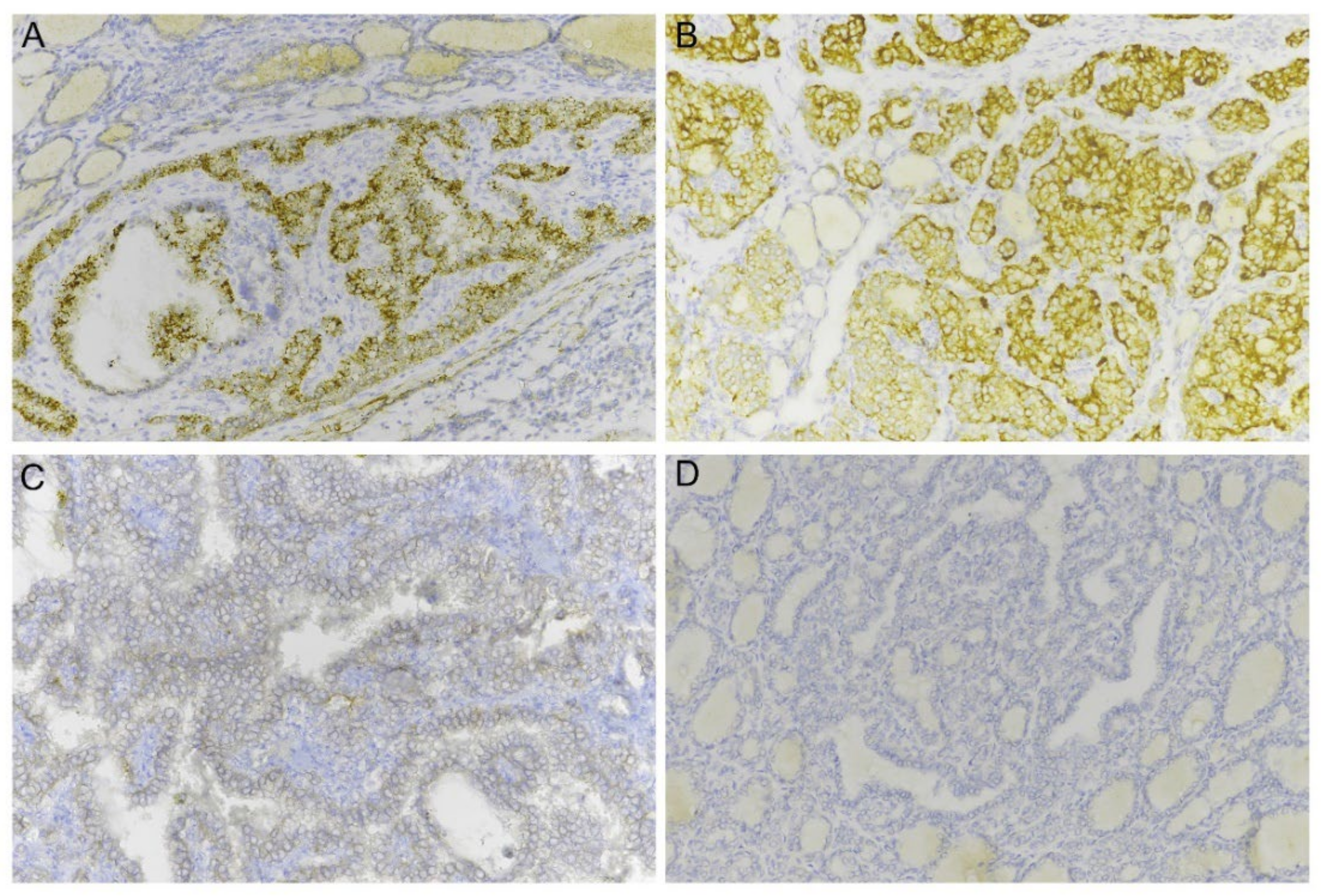
2.1. Pan-Trk Immunohistochemistry in Thyroid Cancer

A total of 25 samples out of 54 (46.3%) showed a positive Trk immunoreactivity, as shown in Table 1. In detail, IHC-positive cases included 22 out of the 26 NTRK fusion-positive (84.6%) and 3 out of the 28 NTRK fusion-negative samples (10.7%). The three control cases showing Trk proteins expression had RET fusion (classical PTC), HRAS point mutation (follicular variant PTC) and NRAS point mutation (local recurrence of PTC), respectively. In all IHC-positive samples, a signal was present in more than 10% of tumor cells. Signal intensity was mostly mild and strong (scores of 2+ and 3+) in 22 out of 25 cases (88%). Two out of the three IHC false-positive cases showed weak immunoreactivity (1+). The majority of samples showed cytoplasmic Trk expression (n = 20), with a granular pattern; in the remaining samples (n = 5), Trk staining was prevalently membranous (Figure 1).
The remaining 29 cases were negative for Trk expression (53.7%), including 4 out of 26 NTRK-rearranged cases which showed false-negative IHC staining (15.4%). These four discordant cases had all the same rearrangement, the ETV6/NTRK3, and all were PTCs (three classical and one follicular variant).

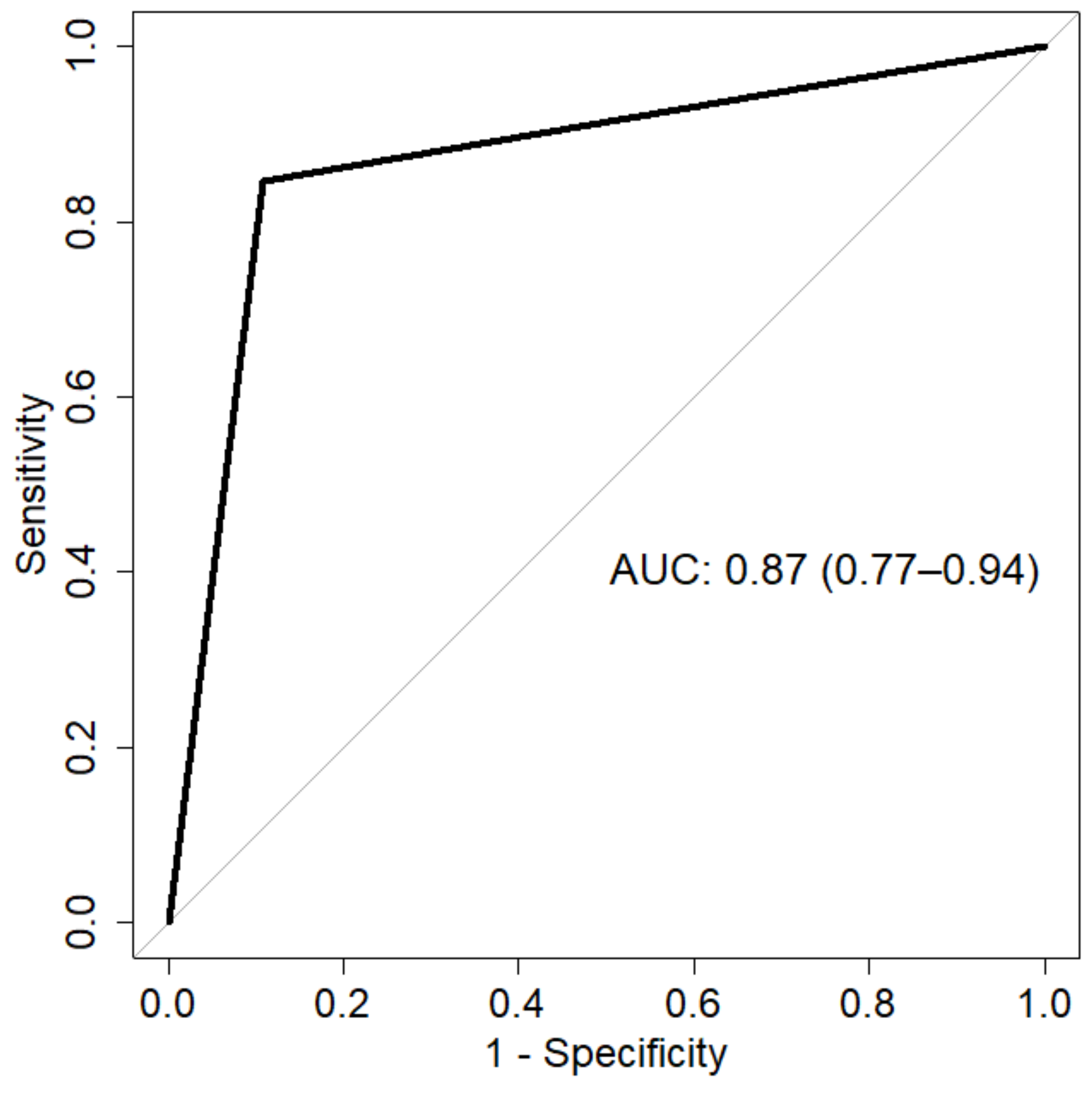
2.2. Pan-Trk Immunohistochemistry Test Performance

By ROC analysis, Trk expression testing showed 0.87 area under the curve (AUC; 95% CI, 0.77–0.94) (Figure 2), 0.85 sensitivity (95% CI, 0.69–0.96), 0.89 specificity (95% CI, 0.75–1), 0.87 accuracy (95% CI, 0.78–0.94), 0.88 positive predictive value (PPV; 95% CI, 0.77–1) and 0.87 negative predictive value (NPV; 95% CI, 0.76–0.96) in identifying NTRK-rearranged tumors. The calculated sensitivity and specificity values are below 90% due to the presence of both false-positive and false-negative cases. As a consequence, the overall accuracy of pan-Trk testing in the identification of NTRK rearrangements is relatively low.

3. Discussion

In adult thyroid cancer, NTRK rearrangement is not common, with a prevalence of 2–4% [21]. However, in advanced-stage thyroid tumors, the availability of life-saving drugs targeting rearranged NTRK has made its testing mandatory.
Several testing algorithms have been proposed, but no broad consensus has been reached on the optimal analysis strategy. In particular, an approach based on the IHC analysis of Trk protein expression is recommended by several authors, as a rapid and cost-effective screening tool for NTRK rearrangements. One of the most widely used antibodies (pan-Trk monoclonal antibody, clone EPR17341) was optimized to detect a C-terminal portion common to TrkA, TrkB and TrkC proteins. A certain rate of false positivity could be expected, since the antibody cannot distinguish between the native protein expression and its chimeric forms.
It is known that the Trk staining pattern is highly variable according to several factors: the specific tumor tissue, the NTRK gene involved and the partner gene. Some studies have reported a relationship between the signal localization and the specific fusion event. For instance, a prevalently nuclear staining was observed exclusively in ETV6/NTRK3 fusions cases [5,22]. In thyroid cancer, the described IHC signal pattern is generally cytoplasmic and/or membranous, with a cytoplasmic and nuclear staining in NTRK3-rearranged tumors [18,23,24]. In our study, the majority of Trk-positive cases presented a cytoplasmic staining.
Independently of the subcellular localization of signal, the pan-Trk antibody has demonstrated high levels of sensitivity and specificity for NTRK rearrangements in various cancer types [22,25,26,27]. In detail, Hechtman et al. reported a high level of concordance between pan-Trk testing and RNA-based NGS across 22 NTRK fusion-positive and 20 fusion-negative tumors of various histotypes, with one false-negative colorectal cancer sample carrying the ETV6/NTRK3 fusion (specificity—100%; sensitivity—95.2%) [22]. In 79 pediatric mesenchymal tumors analyzed by Rudzinski and collaborators, pan-Trk IHC showed 98% specificity and 97% sensitivity in the identification of NTRK fusions [26]. Moreover, pan-Trk antibody testing showed good diagnostic performances in secretory carcinoma of the breast [27] and secretory carcinoma of the salivary gland [28].
On the other hand, there are studies highlighting that Trk IHC testing has important limitations. In a recent study conducted on lung carcinoma, the authors found that 12 out of 387 (3.1%) cases showed positive IHC staining; however, for NGS testing, all these cases were negative for rearrangements [29]. Across 327 samples from multiple cancer types, Koopman and colleagues reported 84% specificity and 77% sensitivity for pan-Trk IHC compared to RNA- and DNA-based NGS [30]; with regard to false-negative cases, 6 out of 29 NTRK-rearranged tumors showed a negative pan-Trk stain (20.7%).
In thyroid cancer, Lee and colleagues found that pan-Trk IHC showed high specificity but moderate sensitivity for NTRK fusion-driven PTCs, due to the presence of false-negative staining in 5 out of 12 NTRK-rearranged tumors (41.7%) [18]. Similarly, Gatalica and collaborators demonstrated that IHC testing for Trk proteins can be challenging. Among 70 thyroid carcinomas, the authors found four NTRK3-rearranged cases (5.7%), of which only two (50%) showed positive pan-Trk staining [24]. Solomon and colleagues detected 13 NTRK fusions among 571 thyroid carcinomas (2.3%); pan-Trk IHC analysis showed 100% specificity but lower sensitivity (82%) for NTRK rearrangements [5].
In our study, pan-Trk IHC testing did not show satisfying specificity (89%) nor sensitivity (85%). The four cases showing false-negative IHC results in our series were positive for rearrangements involving the NTRK3 gene (ETV6/NTRK3). These findings are consistent with previous evidence indicating that pan-Trk reliability is poorer in NTRK3-rearranged tumors, compared with NTRK1 and NTRK2 genes [13,19]. With regard to false-positive cases, there are many possible causes: misinterpretation due to background signal; the overexpression of Trk proteins independent of structural rearrangements; non-specific antibody reaction; or cross-reaction. On the other hand, it is difficult to explain why, in some cases, the ETV6/NTRK3 is associated with negative IHC staining. In our study, all ETV6/NTRK3 rearrangements were detected at the RNA level, and thus the fusion transcript was sufficiently expressed to be measured. It is not known whether some biological factors could influence antibody reaction, or even the chimeric transcript translation; however, this would also affect the oncogenic potential of NTRK3 fusion. To our knowledge, no specific studies have been conducted to address this issue.
Beyond these considerations, it must be highlighted that the ETV6/NTRK3 is the most frequent structural rearrangement involving NTRK genes described in thyroid cancer [2,3,12,23]; therefore, a screening based on IHC testing could miss essential information in thyroid tumors. In fact, in our series, the IHC screening would have missed 4 out of 26 rearranged cases (15.4%). The NTRK testing algorithms that recommend using IHC analysis as a screening tool and NGS testing in the case of negative staining might overcome this poor performance in terms of sensitivity, allowing the recovery of eventual false-negative tumors. However, in practical terms, independently of the IHC results, samples should be further analyzed by a molecular test (i.e., RT-PCR, NGS) to exclude both false-positive and false-negative immunoreactions. Therefore, IHC-based testing does not represent an effective strategy in the screening of NTRK rearrangement in thyroid cancer.
This study presents some limitations. The sample size might appear too low to appropriately assess important diagnostic parameters, such as sensitivity and specificity. However, our sample series represents one of the largest ever reported including NTRK-rearranged thyroid tumors. In addition, differently from other studies focused on Trk IHC analysis, the negative cases included as controls were positive for other driver alterations, known to be mutually exclusive with NTRK rearrangements. Another limitation could be the lack of information of the fusion partner for 11 NTRK-rearranged tumors, due to the employed detection method (FISH, RT-PCR). Currently, the administration of drugs targeting rearranged NTRK is not related to the identification of the fusion partner; however, in the future, this aspect will likely be crucial to understanding whether the partner gene could influence not only IHC performance, but also treatment efficacy.
In conclusion, the recent approval of drugs targeting NTRK-rearranged tumors highlighted the necessity of developing new diagnostic algorithms to be applied in the molecular pathology setting. Our study demonstrated that using an IHC-based approach for the detection of Trk protein expression in thyroid cancer could present serious sensitivity issues. The diagnostic algorithm for testing NTRK rearrangements in this tumor model should include alternative analysis strategies, including in situ or nucleic acid-based detection methods.

4. Materials and Methods

4.1. Samples

A total of 54 thyroid tumors with available molecular profiles were selected from the archives of the Pathological Anatomy Unit of the University Hospital of Pisa. In detail, the case series was composed by 26 NTRK fusion-positive and 28 NTRK fusion-negative thyroid tumors, used as negative controls. To ensure NTRK negativity in control cases, besides a negative NTRK fusion test, only tumors carrying other driver genetic alterations were included. Cases were included only based on their molecular status, independent of eligibility for NTRK-targeted treatment. The most represented histological type was papillary thyroid carcinoma (PTC). Details on the sample series, including histo-pathological information, are shown in Table 2. All the experimental procedures were conducted on anonymous samples, according to the Declaration of Helsinki. Informed consent was waived due to the anonymous nature of the study. The study protocol received the institutional ethical committee approval (CEAVNO, protocol number 9989/2019).
NTRK status was assessed by different methodologies, as reported in Table 2. The most frequent fusion was the ETV6/NTRK3, detected in 13 out of 26 rearranged cases (50%). In all ETV6/NTRK3 cases analyzed by NGS, the specific rearrangement involved exon 4 of ETV6 and exon 14 of NTRK3 (COSF1534). The employed NGS panel (Myriapod NGS Cancer Panel RNA, Diatech Pharmacogenetics, Iesi, AN, Italy) allowed the detection of the most common NTRK fusion variants described in cancer, 243 in NTRK1, 330 in NTRK2, and 154 in NTRK3. In case of FISH analysis, the fusion partner was unknown (break apart probes, ZytoLight SPEC NTRK1/NTRK3 Dual Color Break Apart Probe, Zytovision GmbH, Bremerhaven, Germany). For samples analyzed by RT-PCR (easyPGX Ready NTRK Fusion Kit, Diatech Pharmacogenetics), the employed methodology was unable to identify the specific partner gene of NTRK1, due to the presence of multiple probes in the same well. No NTRK2-positive tumors were present in this study; in fact, no NTRK2 fusion has ever been detected in thyroid cancer [21]. Parts of positive cases were included in our previous study, where fusion testing was conducted by the nCounter system, and then confirmed by orthogonal techniques [12].

4.2. IHC Analysis

For each of the included cases, a 4 um-thick slice was obtained from FFPE tissue blocks for IHC analysis; the most representative tissue block, the same as previously employed for NTRK fusion detection, was used. The VENTANA pan-TRK (EPR17341) assay (Roche Diagnostics Spa, Monza, MB, Italy) was used to assess the expression of Trk proteins. In detail, this in vitro-validated assay enables the detection of C-terminal region of TrkA, TrkB and TrkC, which should be maintained in case of NTRK1, NTRK2 and NTRK3 rearrangements. All procedures were conducted according to the methodology protocol. A positive control (appendix tissue) was included in each experimental session, as recommended by the manufacturer.
IHC staining was interpreted by three qualified pathologists. Positivity was deemed in cases with signal above background in at least 1% of tumor cells, as indicated by the manufacturer. Signal intensity, percentage of positive cells and subcellular staining localization (membranous, cytoplasmic and nuclear) were recorded. In discordant cases, a consensus was reached by collegial discussion. Signal intensity was expressed as a score, from 1 to 3, corresponding to weak, mild and strong signals [31].

4.3. Data Analysis

The area under the curve (AUC), specificity, sensitivity, accuracy, positive predictive value (PPV) and negative predictive value (NPV) of IHC analysis for NTRK fusions were calculated by ROC analysis, along with related 95% confidence intervals (CI) by using 2000 bootstrap resampling. The analysis was performed following the procedures of the pROC R package v.1.18.0 in R environment (https://www.r-project.org, v.4.1.2; last accessed on 9 December 2021).

Author Contributions

E.M., A.P. and F.B. designed and conceived of the study; E.M., A.M.P. and A.P. developed the methodology; E.M., A.P., A.M.P., P.V., R.S., A.G. and A.B. provided acquisition, analysis, and interpretation of data; E.M. and A.M.P. performed statistical analysis; all authors performed writing, review, and revision of the paper. All authors have read and agreed to the published version of the manuscript.

Funding

Reagents and consumables were obtained with funds from the University of Pisa (no specific grant number available).

Institutional Review Board Statement

All the experimental procedures were performed in line with the principles of the Declaration of Helsinki. The study was approved by the Institutional Ethical Committee, CEAVNO (protocol number 9989/2019).

Informed Consent Statement

Patient consent was waived by the ethical committee due to the retrospective nature of the study, which was conducted on de-identified samples.

Data Availability Statement

All data generated or analyzed during this study are included in this published article.

Acknowledgments

We thank Cristina Niccoli and Serena Pelliccioni for their technical support.

Conflicts of Interest

The authors have no conflict of interest to declare.

References

  1. Belli, C.; Penault-Llorca, F.; Ladanyi, M.; Normanno, N.; Scoazec, J.-Y.; Lacroix, L.; Reis-Filho, J.S.; Subbiah, V.; Gainor, J.F.; Endris, V.; et al. ESMO Recommendations on the Standard Methods to Detect RET Fusions and Mutations in Daily Practice and Clinical Research. Ann. Oncol. 2021, 32, 337–350. [Google Scholar] [CrossRef] [PubMed]
  2. Cancer Genome Atlas Research Network. Integrated Genomic Characterization of Papillary Thyroid Carcinoma. Cell 2014, 159, 676–690. [Google Scholar] [CrossRef] [PubMed] [Green Version]
  3. Bastos, A.U.; de Jesus, A.C.; Cerutti, J.M. ETV6-NTRK3 and STRN-ALK Kinase Fusions Are Recurrent Events in Papillary Thyroid Cancer of Adult Population. Eur. J. Endocrinol. 2018, 178, 83–91. [Google Scholar] [CrossRef] [PubMed]
  4. Van der Tuin, K.; Ventayol Garcia, M.; Corver, W.E.; Khalifa, M.N.; Ruano Neto, D.; Corssmit, E.P.M.; Hes, F.J.; Links, T.P.; Smit, J.W.A.; Plantinga, T.S.; et al. Targetable Gene Fusions Identified in Radioactive Iodine Refractory Advanced Thyroid Carcinoma. Eur. J. Endocrinol. 2019, 180, 235–241. [Google Scholar] [CrossRef] [Green Version]
  5. Solomon, J.P.; Linkov, I.; Rosado, A.; Mullaney, K.; Rosen, E.Y.; Frosina, D.; Jungbluth, A.A.; Zehir, A.; Benayed, R.; Drilon, A.; et al. NTRK Fusion Detection across Multiple Assays and 33,997 Cases: Diagnostic Implications and Pitfalls. Mod. Pathol. 2020, 33, 38–46. [Google Scholar] [CrossRef]
  6. Pozdeyev, N.; Gay, L.M.; Sokol, E.S.; Hartmaier, R.; Deaver, K.E.; Davis, S.; French, J.D.; Borre, P.V.; LaBarbera, D.V.; Tan, A.-C.; et al. Genetic Analysis of 779 Advanced Differentiated and Anaplastic Thyroid Cancers. Clin. Cancer Res. 2018, 24, 3059–3068. [Google Scholar] [CrossRef] [Green Version]
  7. Duan, H.; Li, Y.; Hu, P.; Gao, J.; Ying, J.; Xu, W.; Zhao, D.; Wang, Z.; Ye, J.; Lizaso, A.; et al. Mutational Profiling of Poorly Differentiated and Anaplastic Thyroid Carcinoma by the Use of Targeted Next-generation Sequencing. Histopathology 2019, 75, 890–899. [Google Scholar] [CrossRef]
  8. Xu, B.; Fuchs, T.; Dogan, S.; Landa, I.; Katabi, N.; Fagin, J.A.; Tuttle, R.M.; Sherman, E.; Gill, A.J.; Ghossein, R. Dissecting Anaplastic Thyroid Carcinoma: A Comprehensive Clinical, Histologic, Immunophenotypic, and Molecular Study of 360 Cases. Thyroid 2020, 30, 1505–1517. [Google Scholar] [CrossRef]
  9. Cordioli, M.I.C.V.; Moraes, L.; Bastos, A.U.; Besson, P.; de Seixas Alves, M.T.; Delcelo, R.; Monte, O.; Longui, C.; Cury, A.N.; Cerutti, J.M. Fusion Oncogenes Are the Main Genetic Events Found in Sporadic Papillary Thyroid Carcinomas from Children. Thyroid 2017, 27, 182–188. [Google Scholar] [CrossRef]
  10. Pekova, B.; Sykorova, V.; Dvorakova, S.; Vaclavikova, E.; Moravcova, J.; Katra, R.; Astl, J.; Vlcek, P.; Kodetova, D.; Vcelak, J.; et al. RET, NTRK, ALK, BRAF, and MET Fusions in a Large Cohort of Pediatric Papillary Thyroid Carcinomas. Thyroid 2020, 30, 1771–1780. [Google Scholar] [CrossRef]
  11. Alzahrani, A.S.; Alswailem, M.; Alswailem, A.A.; Al-Hindi, H.; Goljan, E.; Alsudairy, N.; Abouelhoda, M. Genetic Alterations in Pediatric Thyroid Cancer Using a Comprehensive Childhood Cancer Gene Panel. J. Clin. Endocrinol. Metab. 2020, 105, 3324–3334. [Google Scholar] [CrossRef] [PubMed]
  12. Macerola, E.; Proietti, A.; Poma, A.M.; Ugolini, C.; Torregrossa, L.; Vignali, P.; Basolo, A.; Materazzi, G.; Elisei, R.; Santini, F.; et al. Molecular Alterations in Relation to Histopathological Characteristics in a Large Series of Pediatric Papillary Thyroid Carcinoma from a Single Institution. Cancers 2021, 13, 3123. [Google Scholar] [CrossRef] [PubMed]
  13. Marchiò, C.; Scaltriti, M.; Ladanyi, M.; Iafrate, A.J.; Bibeau, F.; Dietel, M.; Hechtman, J.F.; Troiani, T.; López-Rios, F.; Douillard, J.-Y.; et al. ESMO Recommendations on the Standard Methods to Detect NTRK Fusions in Daily Practice and Clinical Research. Ann. Oncol. 2019, 30, 1417–1427. [Google Scholar] [CrossRef] [PubMed] [Green Version]
  14. Zito Marino, F.; Pagliuca, F.; Ronchi, A.; Cozzolino, I.; Montella, M.; Berretta, M.; Errico, M.E.; Donofrio, V.; Bianco, R.; Franco, R. NTRK Fusions, from the Diagnostic Algorithm to Innovative Treatment in the Era of Precision Medicine. Int. J. Mol. Sci 2021, 21, 3718. [Google Scholar] [CrossRef] [PubMed]
  15. Perreault, S.; Chami, R.; Deyell, R.J.; El Demellawy, D.; Ellezam, B.; Jabado, N.; Morgenstern, D.A.; Narendran, A.; Sorensen, P.H.B.; Wasserman, J.D.; et al. Canadian Consensus for Biomarker Testing and Treatment of TRK Fusion Cancer in Pediatric Patients. Curr. Oncol. 2021, 28, 346–366. [Google Scholar] [CrossRef] [PubMed]
  16. Bruno, R.; Fontanini, G. Next Generation Sequencing for Gene Fusion Analysis in Lung Cancer: A Literature Review. Diagnostics 2020, 10, 521. [Google Scholar] [CrossRef]
  17. Hsiao, S.J.; Zehir, A.; Sireci, A.N.; Aisner, D.L. Detection of Tumor NTRK Gene Fusions to Identify Patients Who May Benefit from Tyrosine Kinase (TRK) Inhibitor Therapy. J. Mol. Diag. 2019, 21, 553–571. [Google Scholar] [CrossRef] [Green Version]
  18. Lee, Y.-C.; Chen, J.-Y.; Huang, C.-J.; Chen, H.-S.; Yang, A.-H.; Hang, J.-F. Detection of NTRK1/3 Rearrangements in Papillary Thyroid Carcinoma Using Immunohistochemistry, Fluorescent In Situ Hybridization, and Next-Generation Sequencing. Endocr. Pathol. 2020, 31, 348–358. [Google Scholar] [CrossRef]
  19. Penault-Llorca, F.; Rudzinski, E.R.; Sepulveda, A.R. Testing Algorithm for Identification of Patients with TRK Fusion Cancer. J. Clin. Pathol. 2019, 72, 460–467. [Google Scholar] [CrossRef]
  20. Kim, S.-W.; Roh, J.; Park, C.-S. Immunohistochemistry for Pathologists: Protocols, Pitfalls, and Tips. J. Pathol. Transl. Med. 2016, 50, 411–418. [Google Scholar] [CrossRef] [Green Version]
  21. Macerola, E.; Poma, A.M.; Vignali, P.; Basolo, A.; Ugolini, C.; Torregrossa, L.; Santini, F.; Basolo, F. Molecular Genetics of Follicular-Derived Thyroid Cancer. Cancers 2021, 13, 1139. [Google Scholar] [CrossRef] [PubMed]
  22. Hechtman, J.F.; Benayed, R.; Hyman, D.M.; Drilon, A.; Zehir, A.; Frosina, D.; Arcila, M.E.; Dogan, S.; Klimstra, D.S.; Ladanyi, M.; et al. Pan-Trk Immunohistochemistry Is an Efficient and Reliable Screen for the Detection of NTRK Fusions. Am. J. Surg. Pathol. 2017, 41, 1547–1551. [Google Scholar] [CrossRef] [PubMed]
  23. Gatalica, Z.; Xiu, J.; Swensen, J.; Vranic, S. Molecular Characterization of Cancers with NTRK Gene Fusions. Mod. Pathol. 2019, 32, 147–153. [Google Scholar] [CrossRef] [PubMed]
  24. Nozaki, Y.; Yamamoto, H.; Iwasaki, T.; Sato, M.; Jiromaru, R.; Hongo, T.; Yasumatsu, R.; Oda, Y. Clinicopathological Features and Immunohistochemical Utility of NTRK-, ALK-, and ROS1-Rearranged Papillary Thyroid Carcinomas and Anaplastic Thyroid Carcinomas. Hum. Pathol. 2020, 106, 82–92. [Google Scholar] [CrossRef]
  25. Murphy, D.A.; Ely, H.A.; Shoemaker, R.; Boomer, A.; Culver, B.P.; Hoskins, I.; Haimes, J.D.; Walters, R.D.; Fernandez, D.; Stahl, J.A.; et al. Detecting Gene Rearrangements in Patient Populations Through a 2-Step Diagnostic Test Comprised of Rapid IHC Enrichment Followed by Sensitive Next-Generation Sequencing. Appl. Immunohistochem. Mol. Morphol. 2017, 25, 513–523. [Google Scholar] [CrossRef] [Green Version]
  26. Rudzinski, E.R.; Lockwood, C.M.; Stohr, B.A.; Vargas, S.O.; Sheridan, R.; Black, J.O.; Rajaram, V.; Laetsch, T.W.; Davis, J.L. Pan-Trk Immunohistochemistry Identifies NTRK Rearrangements in Pediatric Mesenchymal Tumors. Am. J. Surg. Pathol. 2018, 42, 927–935. [Google Scholar] [CrossRef]
  27. Harrison, B.T.; Fowler, E.; Krings, G.; Chen, Y.-Y.; Bean, G.R.; Vincent-Salomon, A.; Fuhrmann, L.; Barnick, S.E.; Chen, B.; Hosfield, E.M.; et al. Pan-TRK Immunohistochemistry: A Useful Diagnostic Adjunct For Secretory Carcinoma of the Breast. Am. J. Surg. Pathol. 2019, 43, 1693–1700. [Google Scholar] [CrossRef]
  28. Bell, D.; Ferrarotto, R.; Liang, L.; Goepfert, R.P.; Li, J.; Ning, J.; Broaddus, R.; Weber, R.S.; El-Naggar, A.K. Pan-Trk immunohistochemistry reliably identifies ETV6-NTRK3 fusion in secretory carcinoma of the salivary gland. Virchows Arch. 2020, 476, 295–305. [Google Scholar] [CrossRef]
  29. Strohmeier, S.; Brcic, I.; Popper, H.; Liegl-Atzwanger, B.; Lindenmann, J.; Brcic, L. Applicability of Pan-TRK Immunohistochemistry for Identification of NTRK Fusions in Lung Carcinoma. Sci. Rep. 2021, 11, 9785. [Google Scholar] [CrossRef]
  30. Koopman, B.; Kuijpers, C.; Groen, H.; Timens, W.; Schuuring, E.; Willems, S.M.; van Kempen, L.C. Detection of NTRK Fusions and TRK Expression and Performance of pan-TRK Immunohistochemistry in Routine Diagnostics: Results from a Nationwide Community-Based Cohort. Diagnostics 2022, 12, 668. [Google Scholar] [CrossRef]
  31. Fedchenko, N.; Reifenrath, J. Different Approaches for Interpretation and Reporting of Immunohistochemistry Analysis Results in the Bone Tissue—A Review. Diagn. Pathol. 2014, 9, 221. [Google Scholar] [CrossRef] [PubMed] [Green Version]
Figure 1. Pan-Trk IHC in papillary thyroid carcinoma. A strong granular cytoplasmic immunoreactivity is evident in neoplastic cells of a classical PTC carrying a ETV6/NTRK3 rearrangement (A); strong immunoreactivity is clear and specific in cell membrane and cytoplasm of neoplastic cells in a case of NTRK3-rearranged classical PTC (B); weak and focal immunopositivity for pan-Trk in a case of classical PTC that was negative for NTRK rearrangements (C); no immunoreactivity is observed in a case of follicular variant PTC that was positive for the ETV6/NTRK3 rearrangement (D).
Ijms 23 07470 g001
Figure 2. ROC analysis. The AUC represents the performance of pan-Trk IHC analysis in the identification of NTRK fusion-positive tumors.
Ijms 23 07470 g002
Table 1. Characteristics of 25 thyroid tumors showing positive immunohistochemical Trk stain. The NTRK gene, the fusion partner gene (if known), the percentage of positive tumor cells, signal localization and intensity, and tumor histotype are reported. For samples positive for Trk expression but negative for NTRK rearrangements (N20, N24, N28), the driver alteration is indicated.
Sample NameDriver GeneFusion PartnerPositive Cells (%)Localization of Positive SignalSignal IntensityTumor Histotype
P1NTRK1unknown50Cytoplasmic (granular)2+PTC—diffuse sclerosing variant
P2NTRK1unknown50Cell membrane2+PTC—classical type
P3NTRK1unknown80Cytoplasmic (granular)3+PTC—classical type
P4NTRK1unknown60Cell membrane2+PTC—classical type
P5NTRK1unknown70Cytoplasmic (granular)2+PTC—classical type
P6NTRK1TPM370Cytoplasmic (granular)3+PDTC
P7NTRK3ETV640Cytoplasmic (granular)2+PTC—follicular variant
P8NTRK3unknown80Cytoplasmic (granular)2+PTC—classical type
P11NTRK3unknown60Cell membrane3+PTC—follicular variant
P12NTRK3unknown30Cytoplasmic (granular)3+PTC—classical type
P13NTRK3unknown70Cytoplasmic3+PTC—classical type
P14NTRK3ETV620Cytoplasmic (granular)1+PTC–solid variant
P15NTRK3ETV610Cell membrane2+PTC—classical type
P17NTRK3ETV630Cytoplasmic (granular)3+PTC—classical type
P18NTRK3unknown30Cytoplasmic (granular)3+PTC—classical type
P19NTRK3ETV640Cytoplasmic (granular)3+PTC—classical type
P20NTRK3ETV630Cytoplasmic (granular)2+PTC—classical type
P21NTRK3unknown70Cytoplasmic (granular)3+PTC—classical type
P22NTRK3SQSTM120Cytoplasmic (granular)3+PTC—classical type
P24NTRK3ETV620Cytoplasmic (granular)2+PTC—classical type
P25NTRK3ETV630Cytoplasmic (granular)3+PTC—classical type
P26NTRK3ETV680Cytoplasmic (granular)3+ATC
N20RET fusion/10Cell membrane1+PTC—classical type
N24HRAS p.Q61K/15Cytoplasmic (granular)1+PTC—follicular variant
N28NRAS p.Q61R/70Cytoplasmic (granular)2+PTC—metastasis
Abbreviations: PTC, papillary thyroid carcinoma; PDTC, poorly differentiated thyroid carcinoma; ATC, anaplastic thyroid carcinoma; IHC, immunohistochemistry.
Table 2. NTRK-rearranged thyroid tumors and NTRK fusion-negative controls. The methodology of NTRK fusion detection and the identity of the partner gene, if known, are reported. In cases negative for NTRK rearrangements, the detected driver genetic alteration is indicated. Histological diagnosis has been reported for each tumor. In cases of papillary thyroid carcinoma, the variant has been also indicated.
Sample NameNTRK GeneNTRK Status Assessed byFusion PartnerNon-NTRK Driver AlterationHistology
P1NTRK1FISHunknown/PTC—diffuse sclerosing variant
P2NTRK1RT-PCRunknown/PTC—classical type
P3NTRK1FISHunknown/PTC—classical type
P4NTRK1RT-PCRunknown/PTC—classical type
P5NTRK1FISHunknown/PTC—classical type
P6NTRK1NGSTPM3/PDTC
P7NTRK3RT-PCRETV6/PTC—follicular variant
P8NTRK3FISHunknown/PTC—classical type
P9NTRK3RT-PCRETV6/PTC—classical type
P10NTRK3RT-PCRETV6/PTC—follicular variant
P11NTRK3FISHunknown/PTC—follicular variant
P12NTRK3FISHunknown/PTC—classical type
P13NTRK3FISHunknown/PTC—classical type
P14NTRK3RT-PCRETV6/PTC—solid variant
P15NTRK3RT-PCRETV6/PTC—classical type
P16NTRK3RT-PCRETV6/PTC—classical type
P17NTRK3RT-PCRETV6/PTC—classical type
P18NTRK3FISHunknown/PTC—classical type
P19NTRK3RT-PCRETV6/PTC—classical type
P20NTRK3RT-PCRETV6/PTC—classical type
P21NTRK3FISHunknown/PTC—classical type
P22NTRK3NGSSQSTM1/PTC—classical type
P23NTRK3NGSETV6/PTC—classical type
P24NTRK3NGSETV6/PTC—classical type
P25NTRK3NGSETV6/PTC—classical type
P26NTRK3RT-PCRETV6/ATC
N1/FISH/BRAF p.V600EPTC—classical type
N2/FISH/BRAF p.V600EPTC—classical type
N3/FISH/BRAF p.V600EPTC—classical type
N4/FISH/BRAF p.V600EPTC—classical type
N5/FISH/BRAF p.V600EPTC—classical type
N6/FISH/BRAF p.V600EPTC—classical type
N7/FISH/BRAF p.V600EPTC—classical type
N8/FISH/NRAS p.Q61RPTC—follicular variant
N9/FISH/RET fusionPTC—solid variant
N10/FISH/RET fusionPTC—classical type
N11/FISH/PPARG fusionPTC—follicular variant
N12/FISH/NRAS p.Q61KPTC—follicular variant
N13/FISH/RET fusionPTC—classical type
N14/FISH/ALK fusionPTC—follicular variant
N15/FISH/BRAF p.V600EPTC—classical type
N16/FISH/RET fusionPTC—classical type
N17/FISH/BRAF p.V600EPTC—classical type
N18/FISH/BRAF p.V600EPTC—classical type
N19/FISH/ALK fusionPTC—follicular variant
N20/NGS/RET fusionPTC—classical type
N21/FISH/BRAF p.V600EPTC—classical type
N22/FISH/BRAF p.V600EPTC—classical type
N23/FISH/BRAF p.V600EPTC—classical type
N24/FISH/HRAS p.Q61KPTC—follicular variant
N25/FISH/BRAF p.V600EPDTC
N26/RT-PCR/BRAF p.V600EPTC—lymph node recurrence
N27/RT-PCR/BRAF p.V600EPTC—local recurrence
N28/RT-PCR/NRAS p. Q61RPTC—local recurrence
Abbreviations: PTC, papillary thyroid carcinoma; PDTC, poorly differentiated thyroid carcinoma; ATC, anaplastic thyroid carcinoma; FISH, fluorescent in situ hybridization; RT-PCR, reverse-transcription polymerase chain reaction; NGS, next-generation sequencing.
Publisher’s Note: MDPI stays neutral with regard to jurisdictional claims in published maps and institutional affiliations.

