Article

Verrucosidin Derivatives from the Deep Sea Cold-Seep-Derived Fungus Penicillium polonicum CS-252

1
CAS and Shandong Province Key Laboratory of Experimental Marine Biology, Institute of Oceanology, Chinese Academy of Sciences, Nanhai Road 7, Qingdao 266071, China
2
Laboratory of Marine Biology and Biotechnology, Qingdao National Laboratory for Marine Science and Technology, Wenhai Road 1, Qingdao 266237, China
3
College of Marine Science, University of Chinese Academy of Sciences, Yuquan Road 19A, Beijing 100049, China
4
Center for Ocean Mega-Science, Chinese Academy of Sciences, Nanhai Road 7, Qingdao 266071, China
*
Authors to whom correspondence should be addressed.
Int. J. Mol. Sci. 2022, 23(10), 5567; https://doi.org/10.3390/ijms23105567
Submission received: 9 April 2022 / Revised: 1 May 2022 / Accepted: 5 May 2022 / Published: 16 May 2022

Abstract

:
Six novel verrucosidin derivatives, namely, poloncosidins A–F (16), together with one known analogue (7), were isolated and identified from the deep-sea-derived fungus Penicillium polonicum CS-252, which was obtained from cold-seep sediments collected in the South China Sea at a depth of 1183 m. Their structures were mainly established on the basis of a detailed interpretation of NMR spectroscopic and mass spectrometric data. The relative and absolute configurations of compounds 16 were determined by ECD calculations and a DP4+ probability analysis. Compounds 15 represent the first examples of verrucosidins with a 2,5-dihydrofuran ring which is uncommon among the known analogues. These compounds exhibited inhibitory activities against several human and aquatic pathogens with MIC values ranging from 4 to 32 μg/mL.

1. Introduction

Verrucosidins belong to a family of highly reduced polyketides, generally sharing a methylated α-pyrone, a conjugated polyene linker, and an epoxidated tetrahydrofuran ring [1]. All verrucosidin derivatives previously isolated and characterized from the fungal genus Penicillium were found to possess bioactivities such as antibacterial and GRP78/BiP down-regulator activities [2,3]. Penicillium polonicum is a ubiquitous fungus found as saprophyte in diverse environments [4], from which bioactive molecules with diverse chemical structures have been reported including diketopiperazine, α-pyrone derivative, dimeric anthraquinone, and taxol [5,6,7,8,9,10]. The verrucosidin biosynthesis gene cluster was also reported in P. polonicum, which confirmed the ability of the fungus to produce related secondary metabolites [11,12].
In our ongoing research for the discovery of bioactive secondary metabolites from marine-derived fungi [13,14,15], the fungal strain P. polonicum CS-252, which was obtained from sediments collected at the deep-sea cold seep area in the South China Sea, attracted our attention due to its unique HPLC profiles which showed peaks with different UV absorptions from those in our HPLC database. A chemical investigation of the culture extract of P. polonicum CS-252 led to the characterizations of six new verrucosidin derivatives, designated as poloncosidins A–F (16, Figure 1), together with a known analogue, deoxyverrucosidin (7). Among them, compounds 15 represent the first examples of verrucosidins with a 2,5-dihydrofuran ring. Details of the isolation, purification, structure elucidation, and biological evaluation of compounds 17 are provided herein.

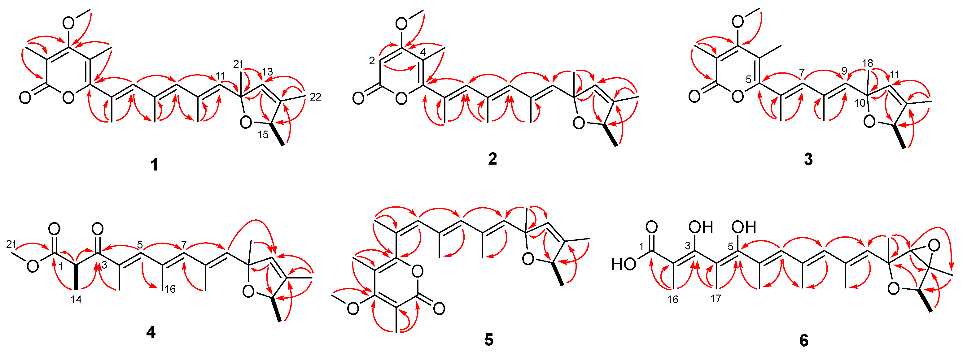
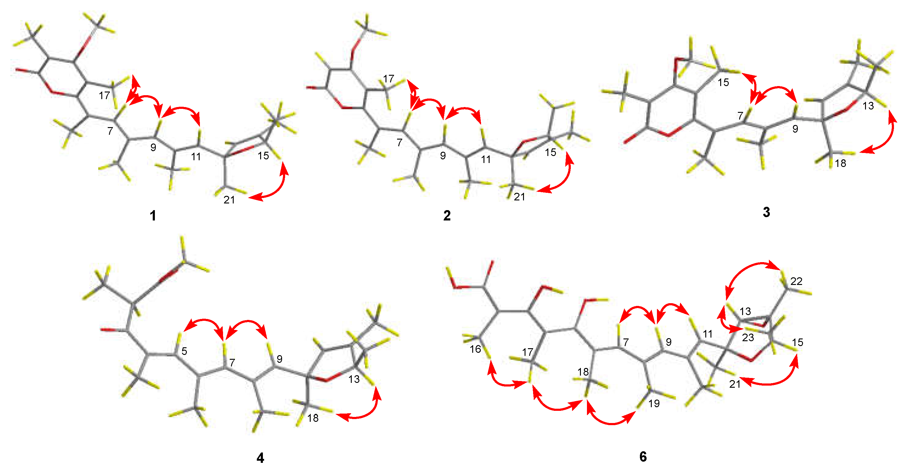
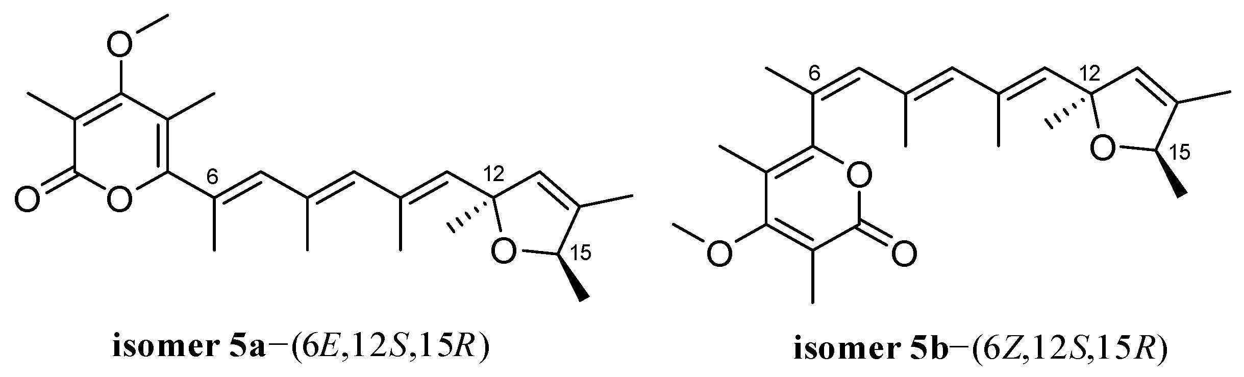
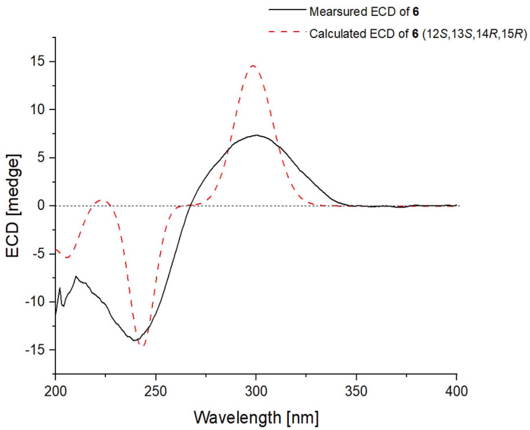
2. Results and Discussion

The fungus P. polonicum CS-252 was cultured on a rice solid medium for chemical investigation. The obtained culture was extracted thoroughly with EtOAc to produce an organic extract that was further fractionated and purified with a combination of column chromatography over Lobar LiChroprep RP-18, Si gel, and Sephadex LH-20 as well as by prep. TLC to yield compounds 17.
Poloncosidin A (1) was obtained as a colorless oil and its molecular formula was assigned as C24H32O4 on the basis of HRESIMS data, corresponding to nine indices of hydrogen deficiency. The 1H-NMR and HSQC spectra of 1 (Table 1 and Figure S5) revealed the signals of nine methyls with one doublet (δH 1.16, J = 6.4 Hz) eight singlets (δH 1.33, 1.66, 1.88, 1.93, 1.95, 1.96, 2.00, and 3.82), four singlet olefinic protons (δH 5.53, 5.61, 5.93, and 6.16), and a quartet proton at δH 4.62 (1H, q, J = 6.4 Hz). The 13C-NMR and DEPT data (Table 2 and Figure S3) indicated all 24 carbon resonances including nine methyls, five methines (with four olefinic at δC 127.2, 137.1, 137.2, and 139.1 as well as an oxygenated at δC 81.6), and ten nonprotonated carbons (with eight olefinic, one carbonyl, and one oxygenated). The aforementioned spectroscopic data suggested the characteristics of a verrucosidin derivative, which are similar to those of deoxyverrucosidin (7) previously described in Penicillium sp. in 2005, with the difference that the epoxide moiety in 7 was replaced by a double bond in 1. This difference was supported by the NMR evidence that signals for an oxygenated methine (δH 3.45; δC 67.5, CH-13) and an oxygenated nonprotonated carbon (δC 67.4, C-14) in the NMR spectra of deoxyverrucosidin (7) [3] were not present in those of 1, while resonances for an olefinic methine (δH 5.61, H-13; δC 127.2, C-13) and an olefinic nonprotonated carbon (δC 138.2, C-14) were observed in the NMR spectra of 1 (Table 1 and Table 2). These data indicated that the epoxidated tetrahydrofuran ring in deoxyverrucosidin was replaced by a 2,5-dihydrofuran ring in 1. This deduction was supported by the key HMBC from H-11 (δH 5.53) and H3-21 (δH 1.33) to C-13 (δC 127.2) and from H3-22 (δH 1.66) to C-13, C-14 (δC 138.2), and C-15 (δC 81.6) (Figure 2). The planar structure of 1 was thus identified as the first example of verrucosidins possessing a 2,5-dihydrofuran moiety.
The relative configuration of compound 1 was established by an analysis of its NOESY data (Figure 3). Key NOESY enhancement from H-15 to H3-21 indicated the cofacial orientation of these protons, which established the relative configuration of the 2,5-dihydrofuran moiety. The geometries of the double bonds at C-6, C-8, and C-10 were determined to be E configurations by the NOESY correlations from H-7 to H-9 and H3-17 and from H-9 to H-11 (Figure 3). To establish the absolute configuration of 1, the time-dependent density functional (TDDFT)-ECD calculations at the BH&HLYP/TZVP level were performed in Gaussian 09. The calculated ECD spectrum for the (12S,15R)-isomer of compound 1 matched well with the experimental curve, leading to the establishment of the absolute configuration of 1 as 12S, 15R (Figure 4).
Poloncosidin B (2) was obtained as a colorless oil and the molecular formula was assigned as C23H30O4 based on an analysis of the HRESIMS data with the same hydrogen deficiency index as that of 1. A comprehensive analysis of the 1H- and 13C-NMR data of 2 indicated a similar structure to that of 1, only differing in the absence of a substituent. Signals for a methyl (δH 1.93; δC 9.9, CH3-16) and an olefinic nonprotonated carbon (δC 108.6, C-2) in the NMR spectra of 1 were found to be absent in those of 2 (Table 1 and Table 2). In contrast, compound 2 exhibited an additional resonance at δH 5.61 (1H, s, H-2) in the 1H-NMR spectrum that correlates with an olefinic methine carbon at δC 87.9 (C-2), accounting for the shielding effect of the carbonyl group at C-1. The key HMBC correlations from H-2 to C-3 (δC 170.7) and C-4 (δC 106.0) as well as other key HMBC and COSY correlations (Figure 2) established the structure of compound 2 as shown.
The relative configuration of the 2,5-dihydrofuran ring and the geometries of the double bonds at C-6, C-8, and C-10 were determined to be the same as those of compound 1 with an analysis of the NOESY correlations (Figure 3). The absolute configuration of 2 was assigned as 12S and 15R, which was evidenced by a comparison of the overlaid experimental and calculated ECD curves at the BH&HLYP/TZVP level (Figure 5).
The molecular formula of poloncosidin C (3) was determined as C21H28O4 according to the HRESIMS ion peaks at m/z 345.2057 [M + H]+ (calcd for C21H29O4, 345.2060) and 367.1875 [M + Na]+ (calcd for C21H28O4Na, 367.1880), required eight indices of hydrogen deficiency. Detailed interpretation of the 1H- and 13C-NMR data revealed 3 to be structurally related to compounds 1 and 2, featuring a methylated α-pyrone moiety and a 2,5-dihydrofuran ring linked with a shorter polyene linker. This deduction was supported by the NMR evidence that resonances for one of the trisubstituted double bonds in the conjugated polyene linker, comprising a singlet methyl (δH 1.95; δC 17.9, CH3-19), a singlet olefinic methine (δH 5.93; δC 137.1, CH-9), and an olefinic nonprotonated carbon (δC 131.3, C-8) in the NMR spectra of 1 disappeared in those of 3. Combined with the key HMBC correlations from H-7 (δH 6.08) to C-5 (δC 158.4) and C-9 (δC 138.3) as well as from H3-18 (δH 1.33) to C-9 (δC 138.3), C-10 (δC 87.3) and C-11 (δC 127.1) (Figure 2), the planar structure of compound 3 was thus established.
The observed NOESY correlations between H-13 and H3-18 helped to establish the relative configuration of the 2,5-dihydrofuran moiety (Figure 3). The E-geometry of the double bonds at C-6 and C-8 was determined by the observed NOEs from H-7 to H-9 and H3-15 (Figure 3). The absolute configuration of 3 was assigned as 10S and 13R based on the results the of TDDFT-ECD calculations at the BH&HLYP/TZVP level as shown in Figure 6.
Poloncosidin D (4) was isolated as a colorless oil. The molecular formula was established as C21H30O4 from its HRESIMS data with seven indices of hydrogen deficiency. The 1H- and 13C-NMR spectra of 4 revealed a 2,5-dihydrofuran moiety with three sets of conjugated double bonds, conforming to the characteristics of verrucosidin derivatives. However, additional resonances for two carbonyl carbons (δC 198.5, C-3 and 171.4, C-1) were observed in the 13C-NMR spectrum of 4 that are quite different from those of compounds 13. The COSY correlation from H-2 (δH 4.52) to H3-14 (δH 1.21) as well as HMBC correlations from H3-14 to C-1 and C-3, from H-5 (δH 7.19) to C-3, C-7 (δC 140.8), and C-16 (δC 17.8), and from H3-21 (δH 3.60) to C-1 established the structure of 4, which possesses the same polyketide skeleton as that of compound 3, but without the formation of the α-pyrone moiety.
The relative configuration of 4 was established by the observed NOESY correlations (Figure 3), except for the stereogenic center of C-2. To establish the stereochemistry, TDDFT-ECD calculations for four possible isomers of 4 [4a-(2R,10S,13R), 4b-(2S,10S,13R), 4c-(2R,10R,13S), and 4d-(2S,10R,13S)] (Figure 7) were performed, and then two isomers [4a-(2R,10S,13R) and 4b-(2S,10S,13R)] were recommended for 4 (Figure 8), which were further clarified by the DP4+ probability analysis. Both the proton and carbon data of the two possible isomers were calculated based on the DP4+ protocol and the results were analyzed with the experimental values [16]. The statistical analysis indicated that the isomer [4a-(2R,10S,13R)] was the equivalent structure with a probability of 99.91% (Figure 8 and Figure S42). The absolute configuration of 4 was thus assigned.
The molecular formula of poloncosidin E (5) was assigned as C24H32O4 based on the accurate mass data measured by HRESIMS, which was identical to that of 1. The 1H- and 13C-NMR data of 5 were virtually identical to those obtained for 1 (Table 1 and Table 2), except for the shielded location of C-5 at δC 155.9 and de-shielded location of C-18 at δC 22.7 in the 13C-NMR spectrum of 5, conforming to the γ-gauche effect [17]. Moreover, resonances for C-6 (δC 125.6) and C-7 (δC 133.0) also moved to the shielded field in the 13C-NMR spectrum of 5. A detailed analysis of the COSY and HMBC correlations of 5 (Figure 2) revealed that it possessed the same planar structure as 1, but with Z-geometry for the double bond at C-6. The structure of compound 5 was thus established with the above NMR data.
Unfortunately, the NOESY spectrum of 5 was not obtained due to instability and low abundance, and compound 5 was actually isolated as a highly impure mixture. To further define the geometry of the double bond at C-6, a DP4+ probability analysis was performed. The steric configuration of the 2,5-dihydrofuran moiety was determined as the same as that of compounds 14 based on the NMR data comparisons and biosynthetic origins. Two possible isomers [5a-(6E,12S,15R) and 5b-(6Z,12S,15R)] of compound 5 were thus generated for DP4+ calculations (Figure 9). The 13C-NMR data calculated for the isomer [5b-(6Z,12S,15R)] matched well with the experimental data of 5 (probability = 100%), which led to the assignment of the steric configuration for 5.
Poloncosidin F (6) was obtained as a colorless oil with a molecular formula of C23H32O6 as determined by the HRESIMS data. Its 1H- and 13C-NMR data (Table 1 and Table 2) revealed a close structural relationship with deoxyverrucosidin (7) [3]. One of the visible differences were that the signals for a methoxy group 3-OCH3 (δH 3.83; δC 60.2) in the NMR spectra of deoxyverrucosidin (7) were absent in those of 6. Moreover, a de-shielded resonance for C-3 (δC 177.0) and a shielded field shift for C-2 (δC 89.9) were clearly observed in the 13C-NMR spectrum of 6 (Table 2), compared to those of 7 [3]. These spectroscopic features as well as in considering the molecular formula revealed that the ester linkage of the α-pyrone ring in deoxyverrucosidin (7) was hydrolyzed in 6. Supported by the HMBC correlations from H3-16 (δH 1.57) to C-1 (δC 165.3), C-2, and C-3 as well as from H3-17 (δC 1.72) to C-3, C-4 (δC 111.3), and C-5 (δC 155.4) (Figure 2), the planar structure of 6 was established.
The E-geometry of five conjugated double bonds at C-2, C-4, C-6, C-8, and C-10 were assigned by the NOE correlations from H3-17 to H3-16 and H3-18, from H3-18 to H3-19, and from H-9 to H-7 and H-11. NOE correlations from H-13 to H3-22 and H3-23 indicated the cofacial orientation of these groups, while the cross-peak from H-15 to H3-21 assigned these groups at the opposite face. Similarly, the absolute configuration of 6 was determined by the TDDFT-ECD calculations in Gaussian 09. The ECD spectrum calculated at the BH&HLYP/TZVP level for isomer (12S,13S,14R,15R)-6 matched well with the experimental curve (Figure 10), leading to the establishment of the absolute configuration of 6.
In addition to the new compounds 16, the known analogue deoxyverrucosidin (7) was also isolated and identified by a detailed spectroscopic analysis and comparisons with the reported data [3].
The obtained compounds, except for compound 5, were assayed for antimicrobial activities against human-, aqua-, and plant-pathogenic microbes. These compounds showed inhibitory activity against some of the tested bacterial strains (MIC ≤ 32 μg/mL, Table 3). Among them, compounds 4 and 6 displayed inhibitory activity against Escherichia coli EMBLC-1, Klebsiella pneumoniae EMBLC-3, Pseudomonas aeruginosa QDIO-4, Vibrio alginolyticus QDIO-5, and V. parahemolyticus QDIO-8 with MIC values ranging from 4 to 32 μg/mL. Compound 4 also inhibited methicillin-resistant Staphylococcus aureus (MRSA) EMBLC-4 with an MIC value of 16 μg/mL. Moreover, compound 1 showed activity against P. aeruginosa QDIO-4, V. alginolyticus QDIO-5, and V. parahemolyticus QDIO-8 with MICs 8.0, 8.0, and 4.0 μg/mL, respectively, whereas compound 7 was bactericidal against P. aeruginosa QDIO-4 and V. parahemolyticus QDIO-8 (each with an MIC value of 8.0 μg/mL). These data indicated that verrucosidin derivatives without the formation of the α-pyrone moiety exhibited a broad spectrum of antimicrobial activity against the tested strains (4 and 6 vs. 13, and 7). In addition, the methylation at C-2 and the 2,5-dihydrofuran ring in these compounds enhanced their activities (1 vs. 2 and 7), while the absence of a trisubstituted double bond in the polyene linker (compound 3) likely had an adverse effect on the antibacterial activity.

3. Experimental Section

3.1. General Experimental Procedures

The instruments, organic solvents, and reagents used in this work were the same as those used in our previous reports [13,14,15].

3.2. Fungal Material

The fungus P. polonicum CS-252 was isolated from the cold seep sediments, which were collected from South China Sea (depth 1183 m) in September 2020. The fungal strain was identified as Penicillium polonicum based on the ITS region sequence, which is the same (100%) as that of P. polonicum FNG3 (with accession no. MZ301257.1). The sequence data were deposited in GenBank with the accession no. OK175489.1. The strain is being preserved at the Key Laboratory of Experimental Marine Biology, Institute of Oceanology, Chinese Academy of Sciences (IOCAS).

3.3. Fermentation, Extraction and Isolation

For chemical investigations, 1 L Erlenmeyer flasks with a solid medium containing rice were autoclaved at 120 °C for 20 min before inoculation. Each flask contained 70 g rice, 0.3 g peptone, 0.5 g yeast extract, 0.2 g corn steep liquor, 0.1 g monosodium glutamate, and 100 mL of naturally sourced and filtered seawater obtained from the Huiquan Gulf of the Yellow Sea near the campus of IOCAS. The fresh mycelia of P. polonicum CS-252 were grown on a PDA medium at 28 °C for seven days, inoculated into the autoclaved medium and then statically cultured for 30 days at room temperature. The fermented rice substrate was fragmented mechanically and extracted thoroughly with EtOAc. The above EtOAc solutions were combined and concentrated under reduced pressure to produce 70.0 g organic extract.
The extract was subjected to vacuum liquid chromatography (VLC) over Si gel and fractionated using solvent mixtures of increasing polarity consisting of petroleum ether (PE) and EtOAc (20:1 to 1:1) and finally with CH2Cl2-MeOH (50:1 to 1:1) to yield 10 fractions (Frs. 1−10). Purification of Fr. 3 (4.1 g), performed with column chromatography (CC) over Lobar LiChroprep RP-18 with a MeOH-H2O gradient (from 1:9 to 10:0), yielded 10 subfractions (Frs. 3.1−3.10). Fr. 3.7 (113 mg) was purified with CC on Sephadex LH-20 (MeOH) to obtain compounds 4 (7.8 mg) and 6 (3.8 mg). Fr. 3.8 (215.0 mg) was subjected to CC on Si gel (CH2Cl2-MeOH, 200:1 to 150:1), and then purified by prep. TLC and CC on Sephadex LH-20 (MeOH) provided compounds 1 (52.4 mg), 2 (6.2 mg), and 3 (4.0 mg). Fr. 3.9 (75 mg) was purified with CC on Si gel (CH2Cl2-MeOH, 300:1), and then purified with CC on Sephadex LH-20 (MeOH) to obtain compound 7 (23.1 mg). Fr. 4 (10.4 g) was further fractionated with CC over Lobar LiChroprep RP-18 elution with a MeOH-H2O gradient (from 1:9 to 10:0) to yield 10 subfractions (Frs. 4.1−4.10). Fr. 4.3 (87 mg) was separated with CC on Si gel and Sephadex LH-20 (MeOH) to obtain compound 5 (7.4 mg).
Poloncosidin A (1) colorless oil; [ α ] D 25 = +65, c 0.10, MeOH; UV (MeOH) λmax (log ε) 229 (2.64), 303 (2.23) nm; ECD (1.04 mM, MeOH) λmaxε) 200 (–1.39), 223 (+1.39), 265 (+1.01), 321 (+3.56) nm; 1H- and 13C-NMR data, Table 1 and Table 2; HRESIMS m/z 385.2373 [M + H]+ (calcd for C24H33O4, 385.2373).
Poloncosidin B (2) colorless oil; [ α ] D 25 = +51, c 0.10, MeOH; UV (MeOH) λmax (log ε) 230 (2.66), 304 (2.25) nm; ECD (1.10 mM, MeOH) λmaxε) 221 (+1.03) nm, 261 (+0.32) nm, 308 (+5.77) nm; 1H- and 13C-NMR data, Table 1 and Table 2; HRESIMS m/z 371.2216 [M + H]+ (calcd for C24H31O4, 371.2217).
Poloncosidin C (3) colorless oil; [ α ] D 25 = +33, c 0.15, MeOH; UV (MeOH) λmax (log ε) 246 (2.68), 315 (3.03) nm; ECD (1.16 mM, MeOH) λmax (Δε) 201 (–1.86), 217 (+2.24), 250 (+1.09), 311 (+0.49) nm; 1H- and 13C-NMR data, Table 1 and Table 2; HRESIMS m/z 345.2057 [M + H]+ (calcd for C21H29O4, 345.2060), 367.1875 [M + Na]+ (calcd for C21H28O4Na, 367.1880).
Poloncosidin D (4) colorless oil; [ α ] D 25 = +38, c 0.14, MeOH; UV (MeOH) λmax (log ε) 232 (2.63) nm, 299 (2.32) nm; ECD (1.16 mM, MeOH) λmax (Δε) 200 (–2.93), 223 (+0.05), 251 (–0.72), 296 (+1.64) nm; 1H- and 13C-NMR data, Table 1 and Table 2; HRESIMS m/z 369.2031 [M + Na]+ (calcd for C21H30O4Na, 369.2036).
Poloncosidin E (5) colorless oil; 1H- and 13C-NMR data, Table 1 and Table 2; HRESIMS m/z 385.2372 [M + H]+ (calcd for C24H33O4, 385.2373), 407.2190 [M + Na]+ (calcd for C24H32O4Na, 407.2193).
Poloncosidin F (6) colorless oil; [ α ] D 25 = +30, c 0.10, MeOH; UV (MeOH) λmax (log ε) 232 (2.95) nm, 301 (2.68) nm; ECD (1.98 mM, MeOH) λmaxε) 210 (–3.36), 239 (–6.43), 300 (+3.40) nm; 1H- and 13C-NMR data, Table 1 and Table 2; HRESIMS m/z 405.2277 [M + H]+ (calcd for C23H33O6, 405.2272).

3.4. Fermentation, Extraction and Isolation

Conformational searches were carried out using molecular mechanics with the MM+ method and HyperChem 8.0 software. Afterward, the geometries were optimized at the gas-phase B3LYP/6-31G(d) level in Gaussian09 software to identify the energy-minimized conformers. Then, the optimized conformers were subjected to the calculations of ECD spectra using the TD-DFT at the BH&HLYP/TZVP level. Simultaneously, the solvent effects of the MeOH solution were evaluated at the same DFT level using the SCRF/PCM method [18].

3.5. ECD Calculations

Conformational searches were carried out via molecular mechanics with the MM+ method in HyperChem 8.0 software. Afterward, the geometries were optimized at the gas-phase B3LYP/6-31G(d) level in Gaussian09 software to identify the energy-minimized conformers. Then, the optimized conformers were subjected to calculations of the ECD spectra using the TDDFT at the BH&HLYP/TZVP level. Simultaneously, solvent effects of the MeOH solution were evaluated at the same DFT level using the SCRF/PCM method. [18]

3.6. Computational NMR Chemical Shift Calculations and DP4+ Analyses

All the theoretical calculations were performed using the Gaussian 09 program package. Conformational searches for possible isomers were conducted with molecular mechanics using the MMFF method with Macromodel software (Schrödinger, LLC) and the corresponding stable conformer, from which distributions higher than 2% were collected. Then, the B3LYP/6-31G(d) PCM level in DMSO was used to optimize the conformers. After that, the NMR shielding tensors of all optimized conformers were calculated using the DFT method at the mPW1PW91\6-31+G(d) PCM level on DMSO and then averaged based on Boltzmann’s distribution theory [16]. GIAO (gauge-independent atomic orbital) NMR chemical calculations were performed using an equation described previously [19]. Finally, the NMR chemical shifts and shielding tensors (1H and 13C) were analyzed and compared with the experimental chemical shifts using DP4+ probability [16].

3.7. Antibacterial Assay

The antimicrobial activities of the isolated compounds against the human and aquatic pathogenic bacteria, Aeromonas hydrophilia QDIO-1, Edwardsiella ictarda QDIO-9, E. tarda QDIO-2, Escherichia coli EMBLC-1, Klebsiella pneumoniae EMBLC-3, S. aureus (MASA) EMBLC-4, Micrococcus luteus QDIO-3, Pseudomonas aeruginosa QDIO-4, Vibrio alginolyticus QDIO-5, V. anguillarum QDIO-6, V. harveyi QDIO-7, V. parahemolyticus QDIO-8, and V. vulnificus QDIO-10, and the plant pathogenic fungi Alternaria solani QDAU-1, Colletotrichum gloeosporioides QDAU-2, Fusarium graminearum QDAU-4, F. oxysporum QDAU-8, Gaeumannomyces graminis QDAU-21, Rhizoctonia cerealis QDAU-20, and Valsa mali QDAU-16 were determined with a serial dilution technique using 96-well microtiter plates as in our previous report [14]. The human and aquatic pathogenic bacteria and plant pathogenic fungi were supplied by the Institute of Oceanology, Chinese Academy of Sciences.

4. Conclusions

In summary, six new verrucosidin derivatives (16) were identified from the deep sea cold-seep-derived Penicillium polonicum CS-252. Compounds 15 represent the first examples of verrucosidins with a 2,5-dihydrofuran ring. The structures of these compounds were determined using a combination of methods (NMR, MS, ECD calculations and DP4+ probability analysis).
All these compounds were tested for antimicrobial activities. Compounds 4 and 6 displayed broad-spectrum inhibitory activity against Escherichia coli EMBLC-1, Klebsiella pneumoniae EMBLC-3, Pseudomonas aeruginosa QDIO-4, Vibrio alginolyticus QDIO-5, and V. parahemolyticus QDIO-8 with MIC values ranging from 4 to 32 μg/mL. Compound 1 showed activity against P. aeruginosa QDIO-4, V. alginolyticus QDIO-5, and V. parahemolyticus QDIO-8 with MIC values of 8.0, 8.0, and 4.0 μg/mL, respectively. Moreover, compound 7 was bactericidal against P. aeruginosa QDIO-4 and V. parahemolyticus QDIO-8 (each with an MIC value of 8.0 μg/mL).

Supplementary Materials

The following supporting information can be downloaded at: https://www.mdpi.com/article/10.3390/ijms23105567/s1. Selected 1D and 2D NMR and HRESIMS spectra of compounds 16; DP4+ probability analyses of compounds 4 and 5.

Author Contributions

Conceptualization, Y.L., H.L. and B.W.; Measurement, X.L. (Xiaoming Li); Data analysis, Y.L., X.L. (Xin Li) and S.Y.; Funding acquisition and project administration, B.W.; Writing—original draft preparation, Y.L.; Writing—Review and editing, H.L. and B.W. All authors have read and agreed to the published version of the manuscript.

Funding

This work was financially supported by the Strategic Priority Research Program of the Chinese Academy of Sciences (XDA22050401), by the National Natural Science Foundation of China (U2006203 and 42076090), by the Senior User Project of RV KEXUE (KEXUE2020GZ02), and by the Shandong Provincial Natural Science Foundation (ZR2021ZD28).

Institutional Review Board Statement

Not applicable.

Informed Consent Statement

Not applicable.

Data Availability Statement

Not applicable.

Acknowledgments

B.-G.W. acknowledges the support of Oceanographic Data Center at IOCAS (for CPU time) and the RV KEXUE of the National Major Science and Technology Infrastructure from the Chinese Academy of Sciences (for sampling).

Conflicts of Interest

The authors declare no conflict of interest.

References

  1. Li, W.; Ma, Z.H.; Chen, L.; Yin, W.B. Synthesis and production of the antitumor polyketide aurovertins and structurally related compounds. Appl. Microbiol. Biotechnol. 2018, 102, 6373–6381. [Google Scholar] [CrossRef] [PubMed]
  2. Pan, C.Q.; Shi, Y.T.; Auckloo, B.N.; Chen, X.G.; Chen, C.T.A.; Tao, X.Y.; Wu, B. An unusual conformational isomer of verrucosidin backbone from a hydrothermal vent fungus, Penicillium sp. Y-50-10. Mar. Drugs 2016, 14, 156. [Google Scholar] [CrossRef] [PubMed] [Green Version]
  3. Choo, S.J.; Park, H.R.; Ryoo, I.J.; Kim, J.P.; Yun, B.S.; Kim, C.J.; Shin-ya, K.; Yoo, I.D. Deoxyverrucosidin, a novel GRP78/BiP down-regulator, produced by Penicillium sp. J. Antibiot. 2005, 58, 210–213. [Google Scholar] [CrossRef] [PubMed] [Green Version]
  4. Sonjak, S.; Frisvad, J.C.; Gunde-Cimerman, N. Penicillium mycobiota in arctic subglacial ice. Microb. Ecol. 2006, 52, 207–216. [Google Scholar] [CrossRef] [PubMed]
  5. Guo, X.C.; Zhang, Y.H.; Gao, W.B.; Pan, L.; Zhu, H.J.; Cao, F. Absolute configurations and chitinase inhibitions of quinazoline-containing diketopiperazines from the marine-derived fungus Penicillium polonicum. Mar. Drugs 2020, 14, 156. [Google Scholar] [CrossRef] [PubMed]
  6. Liu, S.Z.; Tang, X.X.; He, F.M.; Jia, J.X.; Hu, H.; Xie, B.Y.; Li, M.Y.; Qiu, Y.K. Two new compounds from a mangrove sediment-derived fungus Penicillium polonicum H175. Nat. Prod. Res. 2020, 36, 2370–2378. [Google Scholar] [CrossRef] [PubMed]
  7. Ma, Y.R.; Wen, Y.Z.; Cheng, H.T.; Deng, J.T.; Peng, Y.; Bahetejiang, Y.; Huang, H.Q.; Wu, C.Q.; Yang, X.Z.; Pang, K.J. Penpolonin A–E, cytotoxic α-pyrone derivatives from Penicillium polonicum. Bioorg. Med. Chem. Lett. 2021, 40, 127921. [Google Scholar] [CrossRef] [PubMed]
  8. Ding, G.Z.; Liu, J.; Wang, J.M.; Fang, L.; Yu, S.S. Secondary metabolites from the endophytic fungi Penicillium polonicum and Aspergillus fumigatus. J. Asian. Nat. Prod. Res. 2013, 15, 446–452. [Google Scholar] [CrossRef] [PubMed]
  9. Elsunni, M.A.; Yang, Z.D. Secondary metabolites of the endophytic fungi Penicillium polonicum and their monoamine oxidase inhibitory activity. Chem. Nat. Compd. 2018, 54, 1018–1019. [Google Scholar] [CrossRef]
  10. Abdel-Fatah, S.S.; El-Batal, A.I.; El-Sherbiny, G.M.; Khalafa, M.A.; El-Sayed, A.S. Production, bioprocess optimization and γ-irradiation of Penicillium polonicum, as a new Taxol producing endophyte from Ginko biloba. Biotechnol. Rep. 2021, 30, e00623. [Google Scholar] [CrossRef] [PubMed]
  11. Valente, S.; Piombo, E.; Schroeckh, V.; Meloni, G.R.; Heinekamp, T.; Brakhage, A.A.; Spadaro, D. CRISPR-Cas9-based discovery of the verrucosidin biosynthesis gene cluster in Penicillium polonicum. Front. Microbio. 2021, 12, 660871. [Google Scholar] [CrossRef] [PubMed]
  12. Aranda, E.; Rodríguez, M.; Benito, M.J.; Asensio, M.A.; Córdoba, J.J. Molecular cloning of verrucosidin-producing Penicillium polonicum genes by differential screening to obtain a DNA probe. Int. J. Food Microbiol. 2002, 76, 55–61. [Google Scholar] [CrossRef]
  13. Meng, L.H.; Li, X.M.; Zhang, F.Z.; Wang, Y.N.; Wang, B.G. Talascortenes A−G, highly oxygenated diterpenoid acids from the sea-anemone-derived endozoic fungus Talaromyces scorteus AS-242. J. Nat. Prod. 2020, 83, 2528–2536. [Google Scholar] [CrossRef] [PubMed]
  14. Cao, J.; Li, X.M.; Li, X.; Li, H.L.; Konuklugil, B.; Wang, B.G. Uncommon N-methoxyindolediketopiperazines from Acrostalag-mus luteoalbus, a marine algal isolate of endophytic fungus. Chin. J. Chem. 2021, 39, 2808–2814. [Google Scholar] [CrossRef]
  15. Li, Y.H.; Li, X.M.; Li, X.; Yang, S.Q.; Shi, X.S.; Li, H.L.; Wang, B.G. Antibacterial alkaloids and polyketides from the deep sea-derived fungus Penicillium cyclopium SD-413. Mar. Drugs 2020, 18, 553. [Google Scholar] [CrossRef] [PubMed]
  16. Grimblat, N.; Zanardi, M.M.; Sarotti, A.M. Beyond DP4: An improved probability for the stereochemical assignment of isomeric compounds using quantum chemical calculations of NMR shifts. J. Org. Chem. 2015, 80, 12526–12534. [Google Scholar] [CrossRef] [PubMed]
  17. Zhang, H.P.; Timmermann, B.N. Withanolide structural revisions by 13C NMR spectroscopic analysis inclusive of the γ-gauche effect. J. Nat. Prod. 2016, 79, 732–742. [Google Scholar] [CrossRef] [PubMed]
  18. Frisch, M.J.; Trucks, G.W.; Schlegel, H.B.; Scuseria, G.E.; Robb, M.A.; Cheeseman, J.R.; Scalmani, G.; Barone, V.; Mennucci, B.; Petersson, G.A.; et al. Gaussian09, RevisionC.01, Gaussian, Inc.: Wallingford, CT, USA, 2010.
  19. Lee, S.R.; Lee, D.; Park, M.; Lee, J.C.; Park, H.; Kang, K.S.; Kim, C.; Beemelmanns, C.; Kim, K.H. Absolute configuration and corrected NMR assignment of 17-hydroxycyclooctatin, a fused 5–8–5 tricyclic diterpene. J. Nat. Prod. 2020, 83, 354–361. [Google Scholar] [CrossRef] [PubMed]
Figure 1. Chemical structures of compounds 17.
Ijms 23 05567 g001
Figure 2. COSY (black bold lines) and key HMBC (red arrows) cross-peaks of compounds 16.
Ijms 23 05567 g002
Figure 3. Key NOESY correlations of compounds 14 and 6.
Ijms 23 05567 g003
Figure 4. Experimental ECD spectrum of 1 (black line) compared with that calculated for (12S,15R)–isomer of 1 at BH&HLYP/TZVP level (red dashed line).
Ijms 23 05567 g004
Figure 5. Experimental ECD spectrum of 2 (black line) comparing with that calculated for (12S,15R)–isomer of 2 at BH&HLYP/TZVP level (red dashed line).
Ijms 23 05567 g005
Figure 6. Experimental ECD spectrum of 3 (black line) comparing with that calculated for (10S,13R)–isomer of 3 at BH&HLYP/TZVP level (red dashed line).
Ijms 23 05567 g006
Figure 7. Experimental ECD spectrum of 4 (black line) compared with those calculated for 4a–(2R,10S,13R), 4b–(2S,10S,13R), 4c–(2R,10R,13S), and 4d–(2S,10R,13S) of 4 at BH&HLYP/TZVP level (red, blue, purple and green dashed lines, respectively).
Ijms 23 05567 g007
Figure 8. Two possible isomers of compound 4 for DP4+ probability analysis.
Ijms 23 05567 g008
Figure 9. Two possible isomers of compound 5 for DP4+ probability analysis.
Ijms 23 05567 g009
Figure 10. Experimental ECD spectrum of 6 (black line) compared with that calculated for (12S,13S,14R,15R)–isomer of 6 at BH&HLYP/TZVP level (red dashed line).
Ijms 23 05567 g010
Table 1. 1H-NMR Data of Compounds 16 (500 MHz, DMSO-d6, δ in ppm, J in Hz).
No123456
2 5.61, s 4.52, q (7.0)
5 7.19, s
76.16, s6.13, s6.08, s6.14, s6.26, s5.92, s
95.93, s5.93, s5.63, s5.60, s5.84, s5.83, s
115.53, s5.53, s5.62, s5.61, s5.39, s5.54, s
135.61, s5.61, s4.63, q (6.4)4.63, q (6.4)5.56, s3.63, s
14 1.92, s1.21, d (7.0)
154.62, q (6.4)4.61, q (6.4)1.94, s1.89, s4.59, q (6.5)4.00, q (6.7)
161.93, s 1.95, s2.00, s1.94, s1.57, s
171.96, s1.89, s1.92, s1.90, s1.76, s1.72, s
182.00, s2.00, s1.33, s1.33, s1.94, s1.93, s
191.95, s1.95, s1.67, s1.66, s1.58, s1.94, s
201.88, s1.87, s1.17, d (6.4)1.16, d (6.4)1.79, s1.92, s
211.33, s1.32, s3.81, s3.60, s1.28, s1.28, s
221.66, s1.66, s 1.63, s1.39, s
231.16, d (6.4)1.16, d (6.4) 1.12, d (6.5)1.11, d (6.7)
243.82, s3.84, s 3.80, s
Table 2. 13C-NMR Data of Compounds 16 (125 MHz, DMSO-d6, δ in ppm).
No123456
1164.0, C162.5, C164.0, C171.4, C164.4, C165.3, C
2108.6, C87.9, CH108.8, C45.6, CH108.8, C89.9, C
3168.0, C170.7, C168.0, C198.5, C168.5, C177.0, C
4108.9, C106.0, C108.8, C132.9, C109.5, C111.3, C
5158.6, C160.6, C158.4, C145.5, CH155.9, C155.4, C
6126.3, C126.3, C126.3, C131.8, C125.6, C132.3, C
7139.1, CH139.1, CH138.9, CH140.8, CH133.0, CH135.2, CH
8131.3, C131.2, C130.1, C130.6, C131.3, C129.7, C
9137.1, CH137.1, CH138.3, CH138.3, CH137.3, CH134.6, CH
10130.7, C130.7, C87.3, C87.3, C130.6, C134.4, C
11137.2, CH137.2, CH127.1, CH127.1, CH137.7, CH132.8, CH
1287.3, C87.3, C138.4, C138.4, C87.3, C79.4, C
13127.2, CH127.3, CH81.6, CH81.6, CH127.2, CH66.4, CH
14138.2, C138.3, C9.9, CH314.3, CH3138.3, C66.9, C
1581.6, CH81.6, CH11.6, CH313.0, CH381.6, CH75.9, CH
169.9, CH3 15.9, CH317.8, CH310.0, CH39.9, CH3
1711.7, CH310.8, CH317.4, CH317.6, CH310.5, CH312.3, CH3
1816.2, CH316.2, CH327.6, CH327.6, CH322.7, CH316.7, CH3
1917.9, CH317.9, CH311.8, CH311.8, CH315.7, CH318.4, CH3
2017.8, CH317.9, CH320.5, CH320.5, CH317.7, CH318.3, CH3
2127.6, CH327.7, CH360.2, CH351.9, CH327.6, CH321.9, CH3
2211.6, CH311.8, CH3 11.8, CH313.4, CH3
2320.5, CH320.5, CH3 20.5, CH318.6, CH3
2460.2, CH356.7, CH3 60.3, CH3
Table 3. Antimicrobial Activities of Compounds 14, 6, and 7 (MIC, μg/mL).
StrainsCompounds
123467Chloramphenicol b
E. coli16a4.032321.0
K. pneumoniae3232164
MRSA164
P. aeruginosa8.0168.016168.01.0
V. alginolyticus8.0168.08.0320.5
V. parahemolyticus4.08.016168.01.0
a (–) = MIC > 32 μg/mL. b Chloramphenicol as positive control.
Publisher’s Note: MDPI stays neutral with regard to jurisdictional claims in published maps and institutional affiliations.

