
Skin Pigmentation Abnormalities and Their Possible Relationship with Skin Aging

Abstract
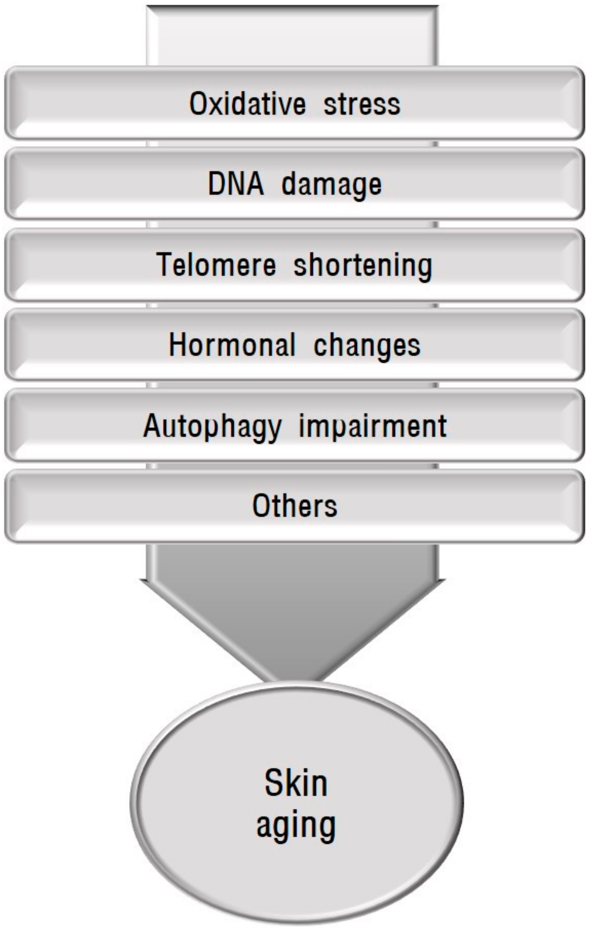
:1. Introduction
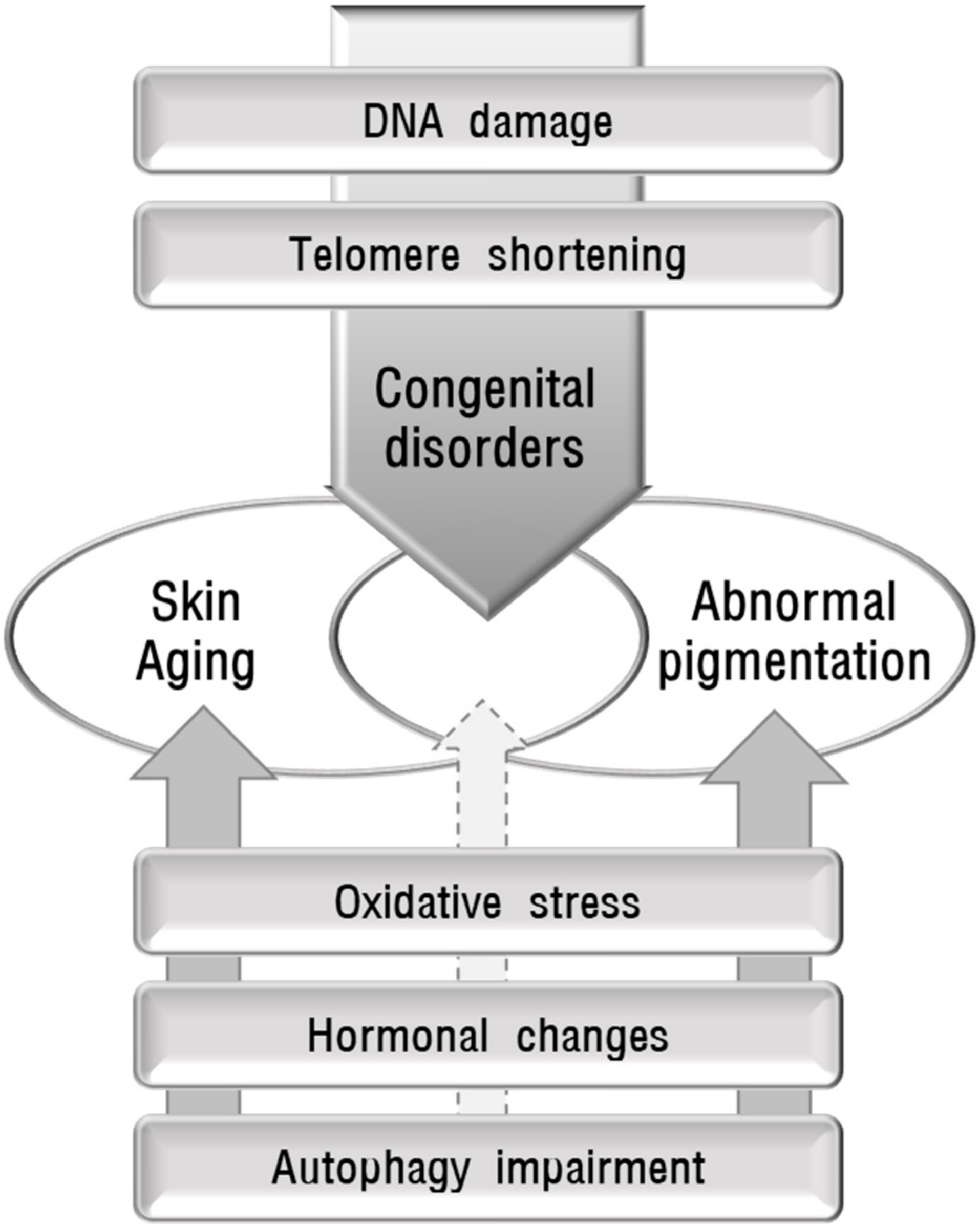
2. Melanin Pigmentation and Its Abnormalities Accompanied by Skin Aging
3. Association of Mechanisms Implicated in Skin Aging with Skin Pigmentation Abnormalities
3.1. Role of Oxidative Stress in Skin Pigmentation Abnormalities
3.1.1. Oxidative Stress and Skin Hypermelanosis
3.1.2. Oxidative Stress and Skin Hypomelanosis

| Pigment Change | Mutated Gene | Dysfunction | Skin Symptom | Reference |
|---|---|---|---|---|
| Hyper | COX7B | Oxidative phosphorylation defect, Mitochondrial respiration deficiency | Linear skin defects with hyperpigmented streaks | [28,42,43] |
| ERCC6 | Accumulation of damaged mitochondria, Mitophagy defect | Photosensitivity, Hyperpigmentation, freckling, and dryness in sun-exposed areas | [44,45] | |
| FANCA | Reduction of electron transfer between respiratory complex I-III and ROS detoxification enzymes, Mitophagy impairment | Morphological abnormalities including hyperpigmentation | [28,46,47,48] | |
| HRAS | Mitochondrial dysfunction, Oxidative phosphorylation defect | Nevi Phacomatosis pigmentokeratotica | [49,50,51,52,53] | |
| HCCS | Reduction of oxidative phosphorylation efficiency | Linear skin defects syndrome exhibiting skin pigmentation | [54,55] | |
| PPOX | Mitochondrial abnormalities | Variegate porphyria characterized by photosensitivity, skin fragility, hypertrichosis, and hyperpigmentation | [56,57,58,59] | |
| POLG1 | Mitochondrial DNA depletion | Hyperpigmentation in the antecubital and popliteal fossae and dorsa of the feet | [62,63] | |
| Hypo | ATP7A | Mitochondrial redox imbalance by copper accumulation | Melanogenesis impairment | [72,73] |
| SURF1 | Mitochondrial cytopathy | Skin and hair abnormalities including hypopigmentation | [74] | |
| Mitochondrial DNA deletion | Mitochondrial multisystem disorder | Hypopigmented patches like hypomelanosis of Ito | [75] | |
| KIT | H2O2-mediated stress | Piebaldism | [76] |
| Pigment Change | Disease/Condition | Evidence for Oxidative Stress-Related | Reference |
|---|---|---|---|
| Hyper | UV-induced melanogenesis | Improvement of hyperpigmentation by antioxidant therapy | [64,65,66] |
| Hyperpigmentation on skin equivalents by squalene monohydroperoxides, which is ameliorated by 12-hydroxystearic acid | [65] | ||
| Melasma | Increased malondialdehyde with decreased catalase in serum | [67,68] | |
| Clinical trial using antioxidant drugs | [69,70] | ||
| Seborrheic keratosis | Role of guanine deaminase in UV-induced keratinocyte senescence via ROS generation | [71] | |
| Hypo | Vitiligo | Elevated superoxide dismutase and malondialdehyde | [80] |
| Reduced catalase or catalase gene polymorphism | [81,82,83] | ||
| Nrf2 gene polymorphism or reduced Nrf2 activation | [85,86,87] | ||
| Increased mitochondrial DNA copy numbers or mitochondrial dysfunction | [88,89] | ||
| Upregulated TRPM2 | [90] | ||
| Autophagy impairment | [91,92] |
3.2. Role of DNA Damage in Skin Pigmentation Abnormalities
3.2.1. DNA Damage and Skin Hypermelanosis
| Pigment Change | Mutated Gene | Dysfunction | Skin Symptom | Reference |
|---|---|---|---|---|
| Hyper | STK11 /LKB1 | Abnormal regulation of UV-induced DNA damage response | Hyperpigmentation of mucous membranes and the skin | [102] |
| Hyper and hypo | RECQL4 | Defects in many aspects of DNA metabolism | Accelerated aging such as atrophic skin and pigment changes | [97,98] |
| XPA XPB, XPC, XPD, XPE, XPF, XPG, or XPV | Defects in damaged DNA repair | Photosensitivity, Lentigines, Hyperpigmentation and hypopigmentation, Accelerated photoaging | [98,99] | |
| Fanconi anemia | Defects in interstrand crosslink repair and telomere maintenance | Poikilodermatic change with hypopigmentation, hyperpigmentation, and telangiectasia | [100,101] | |
| Hypo | Deletion on 15q11.2-q13 | Leukocyte telomere length shortening | Hypopigmentation | [106,107] |
| Oculocutaneous albinism and Hermansky-Pudlak syndrome | Increased tyrosinase degradation through ubiquitin-proteasome system | Oculocutaneous Albinism, Photoaging | [108,109,110,111] |
| Pigment Change | Disease/Condition | Evidence for DNA Damage-Related | Reference |
|---|---|---|---|
| Hyper | UV-induced melanogenesis | Role of α-MSH secreted from UV-damaged keratinocytes in melanogenesis | [103,104] |
| Hypo | Leukotrichia | Associated with APE1 polymorphism | [112] |
3.2.2. DNA Damage and Skin Hypomelanosis
3.3. Role of Telomere Shortening in Skin Pigmentation Abnormalities
3.3.1. Telomere Shortening and Skin Hypermelanosis
3.3.2. Telomere Shortening and Skin Hypomelanosis
3.4. Role of Hormones in Skin Pigmentation Abnormalities
3.4.1. Hormones and Skin Hypermelanosis
3.4.2. Hormones and Skin Hypomelanosis
3.5. Role of Autophagy in Skin Pigmentation Abnormalities
3.5.1. Autophagy and Skin Hypermelanosis
3.5.2. Autophagy and Skin Hypomelanosis
4. Conclusions
Funding
Conflicts of Interest
Abbreviations
| α-MSH | α–melanocyte-stimulating hormone |
| APE1 | apurinic/apyrimidinic endonuclease 1 |
| Atg | autophagy-related |
| ATP7A | ATPase copper transporting alpha |
| CAT | catalase |
| COX7B | cytochrome c oxidase subunit 7B |
| DKC1 | dyskeratosis congenita 1 |
| EPG5 | ectopic P-granules autophagy protein 5 homolog |
| ERCC6 | excision repair cross-complementing group 6 |
| FANCA | Fanconi anemia complementation group A |
| GPNMB | glycoprotein nonmetastatic melanoma protein B |
| H2O2 | hydrogen peroxide |
| HCCS | holocytochrome c synthase |
| HRAS | Harvey rat sarcoma viral oncogene homolog |
| Hsp70-1A | heat shock 70kDa protein 1A |
| LMNA | lamin A |
| MC1R | melanocortin 1 receptor |
| MITF | microphthalmia-associated transcription factor |
| Nrf2 | nuclear factor erythroid 2-like 2 |
| POLG1 | polymerase gamma |
| PPOX | protoporphyrinogen oxidase |
| RECQL4 | RecQ protein-like 4 |
| ROS | reactive oxygen species |
| SIRT3 | sirtuin3 |
| STK11/LKB1 | serine-threonine kinase 11/liver kinase B1 |
| SURF1 | SURF1 cytochrome C oxidase assembly factor |
| TERT | telomerase reverse transcriptase |
| TRP | tyrosine-related protein |
| TRPM2 | transient receptor potential cation channel subfamily M member 2 |
| TSC | tuberous sclerosis complex |
| UV | ultraviolet |
| XPC | Xeroderma pigmentosum complementation group C |
References
- McCullough, J.L.; Kelly, K.M. Prevention and treatment of skin aging. In Aging Interventions and Therapies; World Scientific Publishing: Singapore, 2006; Volume 1067, pp. 323–331. [Google Scholar]
- Okazaki, M.; Yoshimura, K.; Uchida, G.; Harii, K. Correlation between age and the secretions of melanocyte-stimulating cytokines in cultured keratinocytes and fibroblasts. Br. J. Dermatol. 2005, 153 (Suppl. 2), 23–29. [Google Scholar] [CrossRef] [PubMed]
- Nakama, M.; Murakami, Y.; Tanaka, H.; Nakata, S. Decrease in nicotinamide adenine dinucleotide dehydrogenase is related to skin pigmentation. J. Cosmet. Dermatol. 2012, 11, 3–8. [Google Scholar] [CrossRef] [PubMed]
- Duval, C.; Cohen, C.; Chagnoleau, C.; Flouret, V.; Bourreau, E.; Bernerd, F. Key regulatory role of dermal fibroblasts in pigmentation as demonstrated using a reconstructed skin model: Impact of photo-aging. PLoS ONE 2014, 9, e114182. [Google Scholar] [CrossRef] [PubMed]
- Lee, E.J.; Kim, J.Y.; Oh, S.H. Advanced glycation end products (AGEs) promote melanogenesis through receptor for AGEs. Sci. Rep. 2016, 6, 27848. [Google Scholar] [CrossRef] [Green Version]
- Krutmann, J.; Bouloc, A.; Sore, G.; Bernard, B.A.; Passeron, T.J. The skin aging exposome. J. Dermatol. Sci. 2017, 85, 152–161. [Google Scholar] [CrossRef] [Green Version]
- Schikowski, T.; Hüls, A. Air Pollution and Skin Aging. Curr. Environ. Health Rep. 2020, 7, 58–64. [Google Scholar] [CrossRef]
- Fisher, G.J.; Kang, S.; Varani, J.; Bata-Csorgo, Z.; Wan, Y.; Datta, S.; Voorhees, J.J. Mechanisms of photoaging and chronological skin aging. Arch. Dermatol. 2002, 138, 1462–1470. [Google Scholar] [CrossRef]
- Tobin, D.J. Introduction to skin aging. J. Tissue Viability 2017, 26, 37–46. [Google Scholar] [CrossRef] [Green Version]
- Schuch, A.P.; Moreno, N.C.; Schuch, N.J.; Menck, C.F.M.; Garcia, C.C.M. Sunlight damage to cellular DNA: Focus on oxidatively generated lesions. Free Radic. Biol. Med. 2017, 107, 110–124. [Google Scholar] [CrossRef]
- Deruy, E.; Nassour, J.; Martin, N.; Vercamer, C.; Malaquin, N.; Bertout, J.; Chelli, F.; Pourtier, A.; Pluquet, O.; Abbadie, C. Level of macroautophagy drives senescent keratinocytes into cell death or neoplastic evasion. Cell Death Dis. 2014, 5, e1577. [Google Scholar] [CrossRef]
- Zhang, C.F.; Gruber, F.; Ni, C.; Mildner, M.; Koenig, U.; Karner, S.; Barresi, C.; Rossiter, H.; Narzt, M.S.; Nagelreiter, I.M.; et al. Suppression of autophagy dysregulates the antioxidant response and causes premature senescence of melanocytes. J. Investig. Dermatol. 2015, 135, 1348–1357. [Google Scholar] [CrossRef] [Green Version]
- Ito, S.; Wakamatsu, K.; Sarna, T. Photodegradation of Eumelanin and Pheomelanin and Its Pathophysiological Implications. Photochem. Photobiol. 2018, 94, 409–420. [Google Scholar] [CrossRef] [Green Version]
- Nasti, T.H.; Timares, L. MC1R, eumelanin and pheomelanin: Their role in determining the susceptibility to skin cancer. Photochem. Photobiol. 2015, 91, 188–200. [Google Scholar] [CrossRef] [Green Version]
- Slominski, A.; Tobin, D.J.; Shibahara, S.; Wortsman, J. Melanin pigmentation in mammalian skin and its hormonal regulation. Physiol. Rev. 2004, 84, 1155–1228. [Google Scholar] [CrossRef]
- Ko, H.; Kim, M.M. H2O2 promotes the aging process of melanogenesis through modulation of MITF and Nrf2. Mol. Biol. Rep. 2019, 46, 2461–2471. [Google Scholar] [CrossRef]
- Murase, D.; Kusaka-Kikushima, A.; Hachiya, A.; Fullenkamp, R.; Stepp, A.; Imai, A.; Ueno, M.; Kawabata, K.; Takahashi, Y.; Hase, T.; et al. Autophagy Declines with Premature Skin Aging resulting in Dynamic Alterations in Skin Pigmentation and Epidermal Differentiation. Int. J. Mol. Sci. 2020, 21, 5708. [Google Scholar] [CrossRef]
- Narurkar, V.A.; Alster, T.S.; Bernstein, E.F.; Lin, T.J.; Loncaric, A. Safety and Efficacy of a 1550 nm/1927 nm Dual Wavelength Laser for the Treatment of Photodamaged Skin. J. Drugs Dermatol. 2018, 17, 41–46. [Google Scholar]
- Boo, Y.C. Human Skin Lightening Efficacy of Resveratrol and Its Analogs: From in Vitro Studies to Cosmetic Applications. Antioxidants 2019, 8, 332. [Google Scholar] [CrossRef] [Green Version]
- Lee, A.Y. Recent progress in melasma pathogenesis. Pigment Cell Melanoma Res. 2015, 28, 648–660. [Google Scholar] [CrossRef]
- Kim, N.H.; Choi, S.H.; Lee, T.R.; Lee, C.H.; Lee, A.Y. Cadherin 11 involved in basement membrane damage and dermal changes in melasma. Acta Derm. Venereol. 2016, 96, 635–640. [Google Scholar] [CrossRef] [Green Version]
- Bellei, B.; Picardo, M. Premature cell senescence in human skin: Dual face in chronic acquired pigmentary disorders. Ageing Res. Rev. 2020, 57, 100981. [Google Scholar] [CrossRef] [PubMed]
- Rani, S.; Kumar, R.; Kumarasinghe, P.; Bhardwaj, S.; Srivastava, N.; Madaan, A.; Parsad, D. Melanocyte abnormalities and senescence in the pathogenesis of idiopathic guttate hypomelanosis. Int. J. Dermatol. 2018, 57, 559–565. [Google Scholar] [CrossRef] [PubMed]
- Bogdan, A.I.; Baumann, L. Antioxidants used in skin care formulations. Skin Therapy Lett. 2008, 13, 5–9. [Google Scholar]
- Sasaki, M.; Kajiya, H.; Ozeki, S.; Okabe, K.; Ikebe, T. Reactive oxygen species promotes cellular senescence in normal human epidermal keratinocytes through epigenetic regulation of p16(INK4a.). Biochem. Biophys. Res. Commun. 2014, 452, 622–628. [Google Scholar] [CrossRef]
- Kammeyer, A.; Luiten, R.M. Oxidation events and skin aging. Ageing Res. Rev. 2015, 21, 16–29. [Google Scholar] [CrossRef]
- De Jager, T.L.; Cockrell, A.E.; Du Plessis, S.S. Ultraviolet Light Induced Generation of Reactive Oxygen Species. Adv. Exp. Med. Biol. 2017, 996, 15–23. [Google Scholar]
- Sreedhar, A.; Aguilera-Aguirre, L.; Singh, K.K. Mitochondria in skin health, aging, and disease. Cell Death Dis. 2020, 11, 444. [Google Scholar] [CrossRef]
- Zhang, X.; Rosenstein, B.S.; Wang, Y.; Lebwohl, M.; Wei, H. Identification of possible reactive oxygen species involved in ultraviolet radiation-induced oxidative DNA damage. Free Radic. Biol. Med. 1997, 23, 980–985. [Google Scholar] [CrossRef]
- Trouba, K.J.; Hamadeh, H.K.; Amin, R.P.; Germolec, D.R. Oxidative stress and its role in skin disease. Antioxid. Redox Signal. 2002, 4, 665–673. [Google Scholar] [CrossRef]
- Hudson, L.; Bowman, A.; Rashdan, E.; Birch-Machin, M.A. Mitochondrial damage and ageing using skin as a model organ. Maturitas 2016, 93, 34–40. [Google Scholar] [CrossRef]
- Shen, J.; Wan, J.; Huff, C.; Fang, S.; Lee, J.E.; Zhao, H. Mitochondrial DNA 4977-base pair common deletion in blood leukocytes and melanoma risk. Pigment Cell Melanoma Res. 2016, 29, 372–378. [Google Scholar] [CrossRef] [Green Version]
- Birch-Machin, M.A.; A Bowman, A. Oxidative stress and ageing. Br. J. Dermatol. 2016, 175 (Suppl. 2), 26–29. [Google Scholar] [CrossRef] [Green Version]
- Gu, Y.; Han, J.; Jiang, C.; Zhang, Y. Biomarkers, oxidative stress and autophagy in skin aging. Ageing Res. Rev. 2020, 59, 101036. [Google Scholar] [CrossRef]
- Masaki, H. Role of antioxidants in the skin: Anti-aging effects. J. Dermatol. Sci. 2010, 58, 85–90. [Google Scholar] [CrossRef]
- Lephart, E.D. Skin aging and oxidative stress: Equol’s anti-aging effects via biochemical and molecular mechanisms. Ageing Res. Rev. 2016, 31, 36–54. [Google Scholar] [CrossRef]
- Zouboulis, C.C.; Ganceviciene, R.; Liakou, A.I.; Theodoridis, A.; Elewa, R.; Makrantonaki, E. Aesthetic aspects of skin aging, prevention, and local treatment. Clin. Dermatol. 2019, 37, 365–372. [Google Scholar] [CrossRef]
- Schallreuter, K.U.; Kothari, S.; Chavan, B.; Spencer, J.D. Regulation of melanogenesis--controversies and new concepts. Exp. Dermatol. 2008, 17, 395–404. [Google Scholar] [CrossRef]
- Park, D.J.; Sekhon, S.S.; Yoon, J.; Kim, Y.H.; Min, J. Color reduction of melanin by lysosomal and peroxisomal enzymes isolated from mammalian cells. Mol. Cell Biochem. 2016, 413, 119–125. [Google Scholar] [CrossRef]
- Jiang, R.; Xu, X.H.; Wang, K.; Yang, X.Z.; Bi, Y.F.; Yan, Y.; Liu, J.Z.; Chen, X.N.; Wang, Z.Z.; Guo, X.L.; et al. Ethyl acetate extract from Panax ginseng C.A. Meyer and its main constituents inhibit α-melanocyte-stimulating hormone-induced melanogenesis by suppressing oxidative stress in B16 mouse melanoma cells. J. Ethnopharmacol. 2017, 208, 149–156. [Google Scholar] [CrossRef]
- Rodboon, T.; Okada, S.; Suwannalert, P. Germinated Riceberry Rice Enhanced Protocatechuic Acid and Vanillic Acid to Suppress Melanogenesis through Cellular Oxidant-Related Tyrosinase Activity in B16 Cells. Antioxidants 2020, 9, 247. [Google Scholar] [CrossRef] [Green Version]
- Gronow, S.; Noah, C.; Blumenthal, A.; Lindner, B.; Brade, H. Construction of a deep-rough mutant of Burkholderia cepacia ATCC 25416 and characterization of its chemical and biological properties. J. Biol. Chem. 2003, 278, 1647–1655. [Google Scholar] [CrossRef] [PubMed] [Green Version]
- Temple, I.K.; Hurst, J.A.; Hing, S.; Butler, L.; Baraitser, M. De novo deletion of Xp22.2-pter in a female with linear skin lesions of the face and neck, microphthalmia, and anterior chamber eye anomalies. J. Med. Genet. 1990, 27, 56–58. [Google Scholar] [CrossRef] [PubMed]
- Scheibye-Knudsen, M.; Ramamoorthy, M.; Sykora, P.; Maynard, S.; Lin, P.-C.; Minor, R.K.; Wilson, D.M., 3rd; Cooper, M.; Spencer, R.; de Cabo, R.; et al. Cockayne syndrome group B protein prevents the accumulation of damaged mitochondria by promoting mitochondrial autophagy. J. Exp. Med. 2012, 209, 855–869. [Google Scholar] [CrossRef] [PubMed] [Green Version]
- Sin, Y.; Makimura, F.; Saijo, M.; Obika, S. Generation of splice switching oligonucleotides targeting the Cockayne syndrome group B gene product in order to change the diseased cell state. Biochem. Biophys. Res. Commun. 2018, 500, 163–169. [Google Scholar] [CrossRef]
- Cappelli, E.; Ravera, S.; Vaccaro, D.; Cuccarolo, P.; Bartolucci, M.; Panfoli, I.; Dufour, C.; Degan, P. Mitochondrial respiratory complex I defects in Fanconi anemia. Trends Mol. Med. 2013, 19, 513–514. [Google Scholar] [CrossRef]
- Ravera, S.; Vaccaro, D.; Cuccarolo, P.; Columbaro, M.; Capanni, C.; Bartolucci, M.; Panfoli, I.; Morelli, A.; Dufour, C.; Cappelli, E.; et al. Mitochondrial respiratory chain Complex I defects in Fanconi anemia complementation group A. Biochimie 2013, 95, 1828–1837. [Google Scholar] [CrossRef]
- Sumpter, R., Jr.; Sirasanagandla, S.; Fernández, Á.F.; Wei, Y.; Dong, X.; Franco, L.; Zou, Z.; Marchal, C.; Lee, M.Y.; Clapp, D.W.; et al. Fanconi Anemia Proteins Function in Mitophagy and Immunity. Cell 2016, 165, 867–881. [Google Scholar] [CrossRef] [Green Version]
- Aeby, A.; Sznajer, Y.; Cavé, H.; Rebuffat, E.; Coster, R.V.; Rigal, O.; Bogaert, P.V. Cardiofaciocutaneous (CFC) syndrome associated with muscular coenzyme Q10 deficiency. J. Inherit. Metab. Dis. 2007, 30, 827. [Google Scholar] [CrossRef]
- Champion, K.J.; Bunag, C.; Estep, A.L.; Jones, J.R.; Bolt, C.H.; Rogers, R.C.; Rauen, K.A.; Everman, D.B. Germline mutation in BRAF codon 600 is compatible with human development: De novo p.V600G mutation identified in a patient with CFC syndrome. Clin. Genet. 2011, 79, 468–474. [Google Scholar] [CrossRef]
- Groesser, L.; Herschberger, E.; Sagrera, A.; Shwayder, T.; Flux, K.; Ehmann, L.; Wollenberg, A.; Torrelo, A.; Bagazgoitia, L.; Blanca Diaz-Ley, B.; et al. Phacomatosis pigmentokeratotica is caused by a postzygotic HRAS mutation in a multipotent progenitor cell. J. Investig. Dermatol. 2013, 133, 1998–2003. [Google Scholar] [CrossRef] [Green Version]
- Sarin, K.Y.; McNiff, J.M.; Kwok, S.; Kim, J.; Khavari, P.A. Activating HRAS mutation in nevus spilus. J. Investig. Dermatol. 2014, 134, 1766–1768. [Google Scholar] [CrossRef] [Green Version]
- Martin, R.J.; Arefi, M.; Splitt, M.; Redford, L.; Moss, C.; Rajan, N. Phacomatosis pigmentokeratotica and precocious puberty associated with HRAS mutation. Br. J. Dermatol. 2018, 178, 289–291. [Google Scholar] [CrossRef] [Green Version]
- Amary, M.F.; Damato, S.; Halai, D.; Eskandarpour, M.; Berisha, F.; Bonar, F.; McCarthy, S.; Fantin, V.R.; Straley, K.S.; Lobo, S.; et al. Ollier disease and Maffucci syndrome are caused by somatic mosaic mutations of IDH1 and IDH2. Nat. Genet. 2011, 43, 1262–1265. [Google Scholar] [CrossRef]
- Slavotinek, A.M. Eye development genes and known syndromes. Mol. Genet. Metab. 2011, 104, 448–456. [Google Scholar] [CrossRef] [Green Version]
- Lange, H.; Mühlenhoff, U.; Denzel, M.; Kispal, G.; Lill, R. The heme synthesis defect of mutants impaired in mitochondrial iron-sulfur protein biogenesis is caused by reversible inhibition of ferrochelatase. J. Biol. Chem. 2004, 279, 29101–29108. [Google Scholar] [CrossRef] [Green Version]
- Nilsson, R.; Schultz, I.J.; Pierce, E.L.; Soltis, K.A.; Naranuntarat, A.; Ward, D.M.; Baughman, J.M.; Paradkar, P.N.; Kingsley, P.D.; Culotta, V.C.; et al. Discovery of genes essential for heme biosynthesis through large-scale gene expression analysis. Cell Metab. 2009, 10, 119–130. [Google Scholar] [CrossRef] [Green Version]
- Bareth, B.; Dennerlein, S.; Mick, D.U.; Nikolov, M.; Urlaub, H.; Rehling, P. The heme a synthase Cox15 associates with cytochrome c oxidase assembly intermediates during Cox1 maturation. Mol. Cell Biol. 2013, 33, 4128–4137. [Google Scholar] [CrossRef] [Green Version]
- Dawe, R. An overview of the cutaneous porphyrias. F1000Reserach 2017, 6, 1906. [Google Scholar] [CrossRef]
- Deichmann, M.; Kahle, B.; Benner, A.; Thome, M.; Helmke, B.; Helmut Näher, H. Somatic mitochondrial mutations in melanoma resection specimens. Int. J. Oncol. 2004, 24, 137–141. [Google Scholar] [CrossRef]
- Dutton-Regester, K.; Irwin, D.; Hunt, P.; Aoude, L.G.; Tembe, V.; Pupo, G.M.; Lanagan, C.; Carter, C.D.; O’Connor, L.; O’Rourke, M.; et al. A high-throughput panel for identifying clinically relevant mutation profiles in melanoma. Mol. Cancer Ther. 2012, 11, 888–897. [Google Scholar] [CrossRef] [Green Version]
- Saneto, R.P. Alpers-Huttenlocher syndrome: The role of a multidisciplinary health care team. J. Multidiscip. Healthc. 2016, 9, 323–333. [Google Scholar] [CrossRef] [Green Version]
- Campuzano-García, A.E.; Rodríguez-Arámbula, A.; Torres-Alvarez, B.; Castanedo-Cázares, J.P. Hyperpigmentation and atrophy in folds as cutaneous manifestation in a case of mitochondrial myopathy. Dermatol. Online J. 2015, 21, 5. [Google Scholar]
- Onkoksoong, T.; Jeayeng, S.; Poungvarin, N.; Limsaengurai, S.; Thamsermsang, O.; Tripatara, P.; Akarasereenont, P.; Panich, U. Thai herbal antipyretic 22 formula (APF22) inhibits UVA-mediated melanogenesis through activation of Nrf2-regulated antioxidant defense. Phytother. Res. 2018, 32, 1546–1554. [Google Scholar] [CrossRef]
- Mi, T.; Dong, Y.; Santhanam, U.; Huang, N. Niacinamide and 12-hydroxystearic acid prevented benzo(a)pyrene and squalene peroxides induced hyperpigmentation in skin equivalent. Exp. Dermatol. 2019, 28, 742–746. [Google Scholar] [CrossRef]
- Nahhas, A.F.; Abdel-Malek, Z.A.; Kohli, I.; Braunberger, T.L.; Lim, H.W.; Hamzavi, I.H. The potential role of antioxidants in mitigating skin hyperpigmentation resulting from ultraviolet and visible light-induced oxidative stress. Photodermatol. Photoimmunol. Photomed. 2019, 35, 420–428. [Google Scholar] [CrossRef] [Green Version]
- Choubey, V.; Sarkar, R.; Garg, V.; Kaushik, S.; Ghunawat, S.; Sonthalia, S. Role of oxidative stress in melasma: A prospective study on serum and blood markers of oxidative stress in melasma patients. Int. J. Dermatol. 2017, 56, 939–943. [Google Scholar] [CrossRef]
- Sarkar, R.; Devadasan, S.; Choubey, V.; Goswami, B. Melatonin and oxidative stress in melisma—An unexplored territory; a prospective study. Int. J. Dermatol. 2020, 59, 572–575. [Google Scholar] [CrossRef]
- Nofal, A.; Ibrahim, A.M.; Nofal, E.; Gamal, N.; Osman, S. Topical silymarin versus hydroquinone in the treatment of melasma: A comparative study. J. Cosmet. Dermatol. 2019, 18, 263–270. [Google Scholar] [CrossRef] [PubMed] [Green Version]
- Babbush, K.M.; Babbush, R.A.; Khachemoune, A. The Therapeutic Use of Antioxidants for Melasma. J. Drugs Dermatol. 2020, 19, 788–792. [Google Scholar] [CrossRef] [PubMed]
- Cheong, K.A.; Lee, A.Y. Guanine Deaminase Stimulates Ultraviolet-induced Keratinocyte Senescence in Seborrhoeic Keratosis via Guanine Metabolites. Acta. Derm. Venereol. 2020, 100, adv00109. [Google Scholar] [CrossRef] [PubMed]
- Bhattacharjee, A.; Yang, H.; Duffy, M.; Robinson, E.; Conrad-Antoville, A.; Lu, Y.-W.; Capps, T.; Braiterman, L.; Wolfgang, M.; Murphy, M.P.; et al. The Activity of Menkes Disease Protein ATP7A Is Essential for Redox Balance in Mitochondria. J. Biol. Chem. 2016, 291, 16644–16658. [Google Scholar] [CrossRef] [Green Version]
- Petris, M.J.; Strausak, D.; Mercer, J.F. The Menkes copper transporter is required for the activation of tyrosinase. Hum. Mol. Genet. 2000, 9, 2845–2851. [Google Scholar] [CrossRef] [Green Version]
- Sonam, K.; Khan, N.A.; Bindu, P.S.; Taly, A.B.; Gayathri, N.; Bharath, M.M.S.; Govindaraju, C.; Arvinda, H.R.; Nagappa, M.; Sinha, S.; et al. Clinical and magnetic resonance imaging findings in patients with Leigh syndrome and SURF1 mutations. Brain Dev. 2014, 36, 807–812. [Google Scholar] [CrossRef]
- Kakourou, T.; Garoufi, A.; Nicolaidou, P.; Dafni, E.; Tsamouri, M.; Papadimitriou, A.; Karpathios, T. Kearns Sayre syndrome initially presenting as hypomelanosis of Ito. Arch. Dis. Child. 1999, 81, 280–281. [Google Scholar]
- Vafaee, T.; Rokos, H.; Salem, M.M.; Schallreuter, K.U. In vivo and in vitro evidence for epidermal H2O2-mediated oxidative stress in piebaldism. Exp. Dermatol. 2010, 19, 883–887. [Google Scholar] [CrossRef]
- Xie, H.; Zhou, F.; Liu, L.; Zhu, G.; Li, Q.; Li, C.; Gao, T. Vitiligo: How do oxidative stress-induced autoantigens trigger autoimmunity? J. Dermatol. Sci. 2016, 81, 3–9. [Google Scholar] [CrossRef]
- Wang, Y.; Li, S.; Li, C. Perspectives of New Advances in the Pathogenesis of Vitiligo: From Oxidative Stress to Autoimmunity. Med. Sci. Monit. 2019, 25, 1017–1023. [Google Scholar] [CrossRef]
- Speeckaert, R.; Dugardin, J.; Lambert, J.; Lapeere, H.; Verhaeghe, E.; Speeckaert, M.M.; van Geel, N. Critical appraisal of the oxidative stress pathway in vitiligo: A systematic review and meta-analysis. J. Eur. Acad. Dermatol. Venereol. 2018, 32, 1089–1098. [Google Scholar] [CrossRef]
- Shi., M.-H.; Wu, Y.; Li, L.; Cai, Y.-F.; Liu, M.; Gao, X.-H.; Chen, H.-D. Meta-analysis of the association between vitiligo and the level of superoxide dismutase or malondialdehyde. Clin. Exp. Dermatol. 2017, 42, 21–29. [Google Scholar] [CrossRef] [Green Version]
- Mansuri, M.S.; Jadeja, S.D.; Singh, M.; Laddha, N.C.; Dwivedi, M.; Begum, R. The catalase gene promoter and 5′-untranslated region variants lead to altered gene expression and enzyme activity in vitiligo. Br. J. Dermatol. 2017, 177, 1590–1600. [Google Scholar] [CrossRef]
- Caputo, V.; Niceta, M.; Fiorella, S.; Vecchia, M.L.; Bastonini, E.; Bongiorno, M.R.; Pistone, G. Vitiligo susceptibility and catalase gene polymorphisms in Sicilian population. G. Ital. Dermatol. Venereol. 2018, 153, 619–623. [Google Scholar] [CrossRef] [PubMed]
- Ochoa-Ramírez, L.A.; Díaz-Camacho, S.P.; Becerra-Loaiza, D.S.; Verdugo-Nieto, L.; Muñoz-Estrada, V.F.; Servín-Vázquez, L.A.; Osuna-Ramírez, I.; Rodríguez-Millán, J.; Velarde-Félix, J.S. Catalase but not vitamin D receptor gene polymorphisms are associated with nonsegmental vitiligo in Northwestern Mexicans. Int. J. Dermatol. 2019, 58, 1264–1269. [Google Scholar] [CrossRef] [PubMed]
- Vaccaro, M.; Bagnato, G.; Cristani, M.; Borgia, F.; Spatari, G.; Tigano, V.; Saja, A.; Guarneri, F.; Cannavò, S.P.; Gangemi, S. Oxidation products are increased in patients affected by non-segmental generalized vitiligo. Arch. Dermatol. Res. 2017, 309, 485–490. [Google Scholar] [CrossRef] [PubMed]
- Song, P.; Li, K.; Liu, L.; Wang, X.; Jian, Z.; Zhang, W.; Wang, G.; Li, C.; Gao, T. Genetic polymorphism of the Nrf2 promoter region is associated with vitiligo risk in Han Chinese populations. J. Cell Mol. Med. 2016, 20, 1840–1850. [Google Scholar] [CrossRef] [Green Version]
- Kim, H.; Park, C.S.; Lee, A.Y. Reduced Nrf2 activation in PI3K phosphorylation-impaired vitiliginous keratinocytes increases susceptibility to ROS-generating chemical-induced apoptosis. Environ. Toxicol. 2017, 32, 2481–2491. [Google Scholar] [CrossRef]
- Arowojolu, O.A.; Orlow, S.J.; Elbuluk, N.; Manga, P. The nuclear factor (erythroid-derived 2)-like 2 (NRF2) antioxidant response promotes melanocyte viability and reduces toxicity of the vitiligo-inducing phenol monobenzone. Exp. Dermatol. 2017, 26, 637–644. [Google Scholar] [CrossRef]
- Vaseghi, H.; Houshmand, M.; Jadali, Z. Increased levels of mitochondrial DNA copy number in patients with vitiligo. Clin. Exp. Dermatol. 2017, 42, 749–754. [Google Scholar] [CrossRef]
- Yi, X.; Guo, W.; Shi, Q.; Yang, Y.; Zhang, W.; Chen, X.; Kang, P.; Chen, J.; Cui, T.; Ma, J.; et al. SIRT3-Dependent Mitochondrial Dynamics Remodeling Contributes to Oxidative Stress-Induced Melanocyte Degeneration in Vitiligo. Theranostics 2019, 9, 1614–1633. [Google Scholar] [CrossRef]
- Kang, P.; Zhang, W.; Chen, X.; Yi, X.; Song, P.; Chang, Y.; Zhang, S.; Gao, T.; Li, C.; Li, S. TRPM2 mediates mitochondria-dependent apoptosis of melanocytes under oxidative stress. Free Radic. Biol. Med. 2018, 126, 259–268. [Google Scholar] [CrossRef]
- Qiao, Z.; Wang, X.; Xiang, L.; Zhang, C. Dysfunction of Autophagy: A Possible Mechanism Involved in the Pathogenesis of Vitiligo by Breaking the Redox Balance of Melanocytes. Oxid. Med. Cell Longev. 2016, 2016, 3401570. [Google Scholar] [CrossRef]
- He, Y.; Li, S.; Zhang, W.; Dai, W.; Cui, T.; Wang, G.; Gao, T.; Li, C. Dysregulated autophagy increased melanocyte sensitivity to H2O2-induced oxidative stress in vitiligo. Sci. Rep. 2017, 7, 42394. [Google Scholar] [CrossRef]
- Moriwaki, S.; Takahashi, Y. Photoaging and DNA repair. J. Dermatol. Sci. 2008, 50, 169–176. [Google Scholar] [CrossRef]
- Ashapkin, V.V.; Kutueva, L.I.; Kurchashova, S.Y.; Kireev, I.I. Are There Common Mechanisms Between the Hutchinson-Gilford Progeria Syndrome and Natural Aging? Front. Genet. 2019, 10, 455. [Google Scholar] [CrossRef] [Green Version]
- Foo, M.X.R.; Ong, P.F.; Dreesen, O. Premature aging syndromes: From patients to mechanism. J. Dermatol. Sci. 2019, 96, 58–65. [Google Scholar] [CrossRef] [Green Version]
- Cestari, T.; Buster, K. Photoprotection in specific populations: Children and people of color. J. Am. Acad. Dermatol. 2017, 76, S110–S121. [Google Scholar] [CrossRef] [Green Version]
- Lu, L.; Jin, W.; Wang, L.L. Aging in Rothmund-Thomson syndrome and related RECQL4 genetic disorders. Ageing Res. Rev. 2017, 33, 30–35. [Google Scholar] [CrossRef]
- Walsh, M.F.; Chang, V.Y.; Kohlmann, W.K.; Scott, H.S.; Cunniff, C.; Bourdeaut, F.; Molenaar, J.J.; Porter, C.C.; Sandlund, J.T.; Plon, S.E.; et al. Recommendations for Childhood Cancer Screening and Surveillance in DNA Repair Disorders. Clin. Cancer Res. 2017, 23, e23–e31. [Google Scholar] [CrossRef] [Green Version]
- Lehmann, A.R.; McGibbon, D.; Stefanini, M. Xeroderma pigmentosum. Orphanet. J. Rare Dis. 2011, 6, 70. [Google Scholar] [CrossRef] [Green Version]
- Che, R.; Zhang, J.; Nepal, M.; Han, B.; Fei, P. Multifaceted Fanconi Anemia Signaling. Trends Genet. 2018, 34, 171–183. [Google Scholar] [CrossRef]
- Ruggiero, J.L.; Dodds, M.; Freese, R.; Polcari, I.C.; Maguiness, S.; Hook, K.P.; Boull, C. Cutaneous Findings in Fanconi Anemia. J. Am. Acad. Dermatol. 2020. [Google Scholar] [CrossRef]
- Esteve-Puig, R.; Gil, R.; González-Sánchez, E.; Bech-Serra, J.J.; Grueso, J.; Hernández-Losa, J.; Moliné, T.; Canals, F.; Ferrer, B.; Cortés, J.; et al. A mouse model uncovers LKB1 as an UVB-induced DNA damage sensor mediating CDKN1A (p21WAF1/CIP1) degradation. PLoS Genet. 2014, 10, e1004721. [Google Scholar] [CrossRef]
- Nguyen, N.T.; Fisher, D.E. MITF and UV responses in skin: From pigmentation to addiction. Pigment Cell Melanoma Res. 2019, 32, 224–236. [Google Scholar] [CrossRef] [Green Version]
- Jarrett, S.G.; Horrell, E.M.W.; Boulanger, M.C.; D’Orazio, J.A. Defining the Contribution of MC1R Physiological Ligands to ATR Phosphorylation at Ser435, a Predictor of DNA Repair in Melanocytes. J. Investig. Dermatol. 2015, 135, 3086–3095. [Google Scholar] [CrossRef] [Green Version]
- Takata, M.; Shirasaki, F.; Nakatani, T.; Takehara, K. Hereditary non-polyposis colorectal cancer associated with disseminated superficial porokeratosis. Microsatellite instability in skin tumours. Br. J. Dermatol. 2000, 143, 851–855. [Google Scholar] [CrossRef]
- Donze, S.H.; Codd, V.; Damen, L.; Goedegebuure, W.J.; Denniff, M.; Samani, N.J.; van der Velden, J.A.E.M.; Hokken-Koelega, A.C.S. Evidence for Accelerated Biological Aging in Young Adults with Prader-Willi Syndrome. J. Clin. Endocrinol. Metab. 2020, 105, 2053–2059. [Google Scholar] [CrossRef]
- Ehrhart, F.; Janssen, K.J.M.; Coort, S.L.; Evelo, C.T.; Curfs, L.M.G. Prader-Willi syndrome and Angelman syndrome: Visualisation of the molecular pathways for two chromosomal disorders. World J. Biol. Psychiatry 2019, 20, 670–682. [Google Scholar] [CrossRef]
- Seward, S.L., Jr.; Gahl, W.A. Hermansky-Pudlak syndrome: Health care throughout life. Pediatrics 2013, 132, 153–160. [Google Scholar] [CrossRef] [PubMed] [Green Version]
- Kamaraj, B.; Purohit, R. Mutational analysis of oculocutaneous albinism: A compact review. Biomed. Res. Int. 2014, 2014, 905472. [Google Scholar] [CrossRef] [Green Version]
- Ando, H.; Ichihashi, M.; Hearing, V.J. Role of the ubiquitin proteasome system in regulating skin pigmentation. Int. J. Mol. Sci. 2009, 10, 4428–4434. [Google Scholar] [CrossRef] [Green Version]
- Hossy, B.H.; da Costa Leitão, A.A.; Luz, F.B.; dos Santos, E.P.; Allodi, S.; de Pádula, M.; de Oliveira Miguel, N.C. Effects of a sunscreen formulation on albino hairless mice: A morphological approach. Arch. Dermatol. Res. 2013, 305, 535–544. [Google Scholar] [CrossRef]
- Aydin, A.F.; Aydıngöz, I.E.; Doğru-Abbasoğlu, S.; Vural, P.; Uysal, M. Association of Leukotrichia in Vitiligo and Asp148Glu Polymorphism of Apurinic/Apyrimidinic Endonuclease 1. Int. J. Trichol. 2017, 9, 171–176. [Google Scholar]
- Mangaonkar, A.A.; Patnaik, M.M. Short Telomere syndromes in clinical practice: Bridging bench and bedside. Mayo Clin. Proc. 2018, 93, 904–916. [Google Scholar] [CrossRef] [PubMed] [Green Version]
- Wang, A.S.; Dreesen, O. Biomarkers of Cellular Senescence and Skin Aging. Front. Genet. 2018, 9, 247. [Google Scholar] [CrossRef]
- Opresko, P.L.; Shay, J.W. Telomere-associated aging disorders. Ageing Res. Rev. 2017, 33, 52–66. [Google Scholar] [CrossRef] [PubMed]
- AlSabbagh, M.M. Dyskeratosis congenita: A literature review. J. Dtsch. Dermatol. Ges. 2020, 18, 943–967. [Google Scholar] [CrossRef] [PubMed]
- Stout, G.J.; Blasco, M.A. Telomere length and telomerase activity impact the UV sensitivity syndrome xeroderma pigmentosum C. Cancer Res. 2013, 73, 1844–1854. [Google Scholar] [CrossRef] [Green Version]
- Abdollahi, M.; Gao, M.Y.M.; Munoz, D.G. Distinct pattern of neostriatal calcifications in dyskeratosis congenita: A case report and literature review. Clin. Neuropathol. 2018, 37, 277–282. [Google Scholar] [CrossRef]
- Chojnowski, A.; Ong, P.F.; Wong, E.S.M.; Lim, J.S.Y.; Mutalif, R.A.; Navasankari, R.; Dutta, B.; Yang, H.; Liow, Y.Y.; Sze, S.K.; et al. Progerin reduces LAP2α-telomere association in Hutchinson-Gilford progeria. eLife 2015, 4, e07759. [Google Scholar] [CrossRef]
- Li, Y.; Zhou, G.; Bruno, I.G.; Cooke, J.P. Telomerase mRNA Reverses Senescence in Progeria Cells. J. Am. Coll. Cardiol. 2017, 70, 804–805. [Google Scholar] [CrossRef]
- Rork, J.F.; Huang, J.T.; Gordon, L.B.; Kleinman, M.; Kieran, M.W.; Liang, M.G. Initial cutaneous manifestations of Hutchinson-Gilford progeria syndrome. Pediatr. Dermatol. 2014, 31, 196–202. [Google Scholar] [CrossRef]
- Moftah, N.H.; El-Barbary, E.; Rashed, L.; El-Sammad, N. Assessment of telomerase activity in nonsegmental vitiligo tissue: A pilot study. Clin. Exp. Dermatol. 2019, 44, 747–752. [Google Scholar] [CrossRef]
- Bocheva, G.; Slominski, R.M.; Slominski, A.T. Neuroendocrine Aspects of Skin Aging. Int. J. Mol. Sci. 2019, 20, 2798. [Google Scholar] [CrossRef] [Green Version]
- Rusanova, I.; Martínez-Ruiz, L.; Florido, J.; Rodríguez-Santana, C.; Guerra-Librero, A.; Acuña-Castroviejo, D.; Escames, G. Protective Effects of Melatonin on the Skin: Future Perspectives. Int. J. Mol. Sci. 2019, 20, 4948. [Google Scholar] [CrossRef] [Green Version]
- Scheuer, C. Melatonin for prevention of erythema and oxidative stress in response to ultraviolet radiation. Dan. Med. J. 2017, 64, B5358. [Google Scholar]
- Skobowiat, C.; Brożyna, A.A.; Janjetovic, Z.; Jeayeng, S.; Oak, A.S.W.; Kim, T.K.; Panich, U.; Reiter, R.J.; Slominski, A.T. Melatonin and its derivatives counteract the ultraviolet B radiation-induced damage in human and porcine skin ex vivo. J. Pineal Res. 2018, 65, e12501. [Google Scholar] [CrossRef]
- Seleit, I.; Bakry, O.A.; Repey, H.S.E.; Ali, R. Intrinsic versus Extrinsic Aging: A Histopathological, Morphometric and Immunohistochemical Study of Estrogen Receptor β and Androgen Receptor. Skin Pharmacol. Physiol. 2016, 29, 178–189. [Google Scholar] [CrossRef]
- Draelos, Z.D. A Double-Blind Randomized Pilot Study Evaluating the Safety and Efficacy of Topical MEP in the Facial Appearance Improvement of Estrogen Deficient Females. J. Drugs Dermatol. 2018, 17, 1186–1189. [Google Scholar]
- Röck, K.; Joosse, S.A.; Müller, J.; Heinisch, N.; Fuchs, N.; Meusch, M.; Zipper, P.; Reifenberger, J.; Pantel, K.; Fischer, J.W. Chronic UVB-irradiation actuates perpetuated dermal matrix remodeling in female mice: Protective role of estrogen. Sci. Rep. 2016, 6, 30482. [Google Scholar] [CrossRef]
- Cohen, J.L. Evaluation of Efficacy of a Skin Care Regimen Containing Methyl Estradiolpropanoate (MEP) for Treating Estrogen Deficient Skin. J. Drugs Dermatol. 2019, 18, 1226–1230. [Google Scholar]
- Cario, M. How hormones may modulate human skin pigmentation in melasma: An in vitro perspective. Exp. Dermatol. 2019, 28, 709–718. [Google Scholar] [CrossRef] [Green Version]
- Mobasher, P.; Foulad, D.P.; Raffi, J.; Zachary, C.; Fackler, N.; Zohuri, N.; Juhasz, M.; Mesinkovska, N.A. Catamenial Hyperpigmentation: A Review. J. Clin. Aesthet. Dermatol. 2020, 13, 18–21. [Google Scholar] [PubMed]
- Patel, S.; Rauf, A.; Khan, H.; Meher, B.R.; Hassan, S.S.U. A holistic review on the autoimmune disease vitiligo with emphasis on the causal factors. Biomed. Pharmacother. 2017, 92, 501–508. [Google Scholar] [CrossRef] [PubMed]
- Paolino, G.; Bearzi, P.; Mercuri, S.R. Onset of vitiligo in a patient with acquired secondary hypogonadism under treatment with testosterone gel 2%: Inside the pathogenesis. An. Bras. Dermatol. 2020, 95, 661–662. [Google Scholar] [CrossRef] [PubMed]
- Gürpınar, A.; Günaydın, S.D.; Kılıç, C.; Karaduman, A. Association of serum cortisol and dehydroepiandrosterone sulfate (DHEAS) levels with psychological stress in patients with vitiligo. Turk. J. Med. Sci. 2019, 49, 832–837. [Google Scholar] [CrossRef] [PubMed]
- Kalfalah, F.; Janke, L.; Schiavi, A.; Tigges, J.; Ix, A.; Ventura, N.; Boege, F.; Reinke, H. Crosstalk of clock gene expression and autophagy in aging. Aging 2016, 8, 1876–1895. [Google Scholar] [CrossRef] [PubMed] [Green Version]
- Jeong, D.; Qomaladewi, N.P.; Lee, J.; Park, S.H.; Cho, J.Y. The Role of Autophagy in Skin Fibroblasts, Keratinocytes, Melanocytes, and Epidermal Stem Cells. J. Investig. Dermatol. 2020, 140, 1691–1697. [Google Scholar] [CrossRef] [PubMed]
- Majora, M.; Sondenheimer, K.; Knechten, M.; Uthe, I.; Esser, C.; Schiavi, A.; Ventura, N.; Krutmann, J. HDAC inhibition improves autophagic and lysosomal function to prevent loss of subcutaneous fat in a mouse model of Cockayne syndrome. Sci. Transl. Med. 2018, 10, eaam7510. [Google Scholar] [CrossRef] [Green Version]
- Huang, Y.; Li, Y.; Qu, Y.; Zheng, Y.; Ouyang, M.; Zhang, Y.; Lai, W.; Xu, Q. UVA-induced photoaging inhibits autophagic degradation by impairing lysosomal function in dermal fibroblasts. Biochem. Biophys. Res. Commun. 2019, 518, 611–618. [Google Scholar] [CrossRef]
- Endo, K.; Katsuyama, Y.; Taira, N.; Yoshioka, M.; Okano, Y.; Masaki, H. Impairment of the autophagy system in repetitively UVA-irradiated fibroblasts. Photodermatol. Photoimmunol. Photomed. 2020, 36, 111–117. [Google Scholar] [CrossRef]
- Song, X.; Narzt, M.S.; Nagelreiter, I.M.; Hohensinner, P.; Terlecki-Zaniewicz, L.; Tschachler, E.; Grillari, J.; Gruber, F. Autophagy deficient keratinocytes display increased DNA damage, senescence and aberrant lipid composition after oxidative stress in vitro and in vivo. Redox Biol. 2017, 11, 219–230. [Google Scholar] [CrossRef]
- Li, Y.F.; Ouyang, S.H.; Tu, L.F.; Wang, X.; Yuan, W.L.; Wang, G.E.; Wu, Y.P.; Duan, W.J.; Yu, H.M.; Fang, Z.Z.; et al. Caffeine Protects Skin from Oxidative Stress-Induced Senescence through the Activation of Autophagy. Theranostics. 2018, 8, 5713–5730. [Google Scholar] [CrossRef]
- Zhu, W.; Zhao, Z.; Cheng, B. The role of autophagy in skin pigmentation. Eur. J. Dermatol. 2020, 30, 655–662. [Google Scholar]
- Murase, D.; Hachiya, A.; Fullenkamp, R.; Beck, A.; Moriwaki, S.; Hase, T.; Takema, Y.; Manga, P. Variation in Hsp70-1A Expression Contributes to Skin Color Diversity. J. Investig. Dermatol. 2016, 136, 1681–1691. [Google Scholar] [CrossRef] [Green Version]
- Kim, N.H.; Choi, S.H.; Yi, N.; Lee, T.R.; Lee, A.Y. Arginase-2, a miR-1299 target, enhances pigmentation in melasma by reducing melanosome degradation via senescence-induced autophagy inhibition. Pigment. Cell Melanoma Res. 2017, 30, 521–530. [Google Scholar] [CrossRef]
- Cho, Y.H.; Park, J.E.; Lim, D.S.; Lee, J.S. Tranexamic acid inhibits melanogenesis by activating the autophagy system in cultured melanoma cells. J. Dermatol. Sci. 2017, 88, 96–102. [Google Scholar] [CrossRef] [Green Version]
- Lee, K.W.; Ryu, H.W.; Oh, S.S.; Park, S.; Madhi, H.; Yoo, J.; Park, K.H.; Kim, K.D. Depigmentation of α-melanocyte-stimulating hormone-treated melanoma cells by β-mangostin is mediated by selective autophagy. Exp. Dermatol. 2017, 26, 585–591. [Google Scholar] [CrossRef]
- Katsuyama, Y.; Taira, N.; Yoshioka, M.; Okano, Y.; Masaki, H. 3-O-Glyceryl-2-O-hexyl Ascorbate Suppresses Melanogenesis through Activation of the Autophagy System. Biol. Pharm. Bull. 2018, 41, 824–827. [Google Scholar] [CrossRef] [Green Version]
- Park, H.J.; Jo, D.S.; Choi, H.; Ji-Eun Bae, J.-E.; Park, N.Y.; Kim, J.B.; Choi, J.Y.; Kim, Y.H.; Oh, G.S.; Chang, J.H.; et al. Melasolv induces melanosome autophagy to inhibit pigmentation in B16F1 cells. PLoS ONE 2020, 15, e0239019. [Google Scholar] [CrossRef]
- Chen, L.; Xu, Z.; Jiang, M.; Zhang, C.; Wang, X.; Xiang, L. Light-emitting diode 585 nm photomodulation inhibiting melanin synthesis and inducing autophagy in human melanocytes. J. Dermatol. Sci. 2018, 89, 11–18. [Google Scholar] [CrossRef] [Green Version]
- Byrne, S.; Dionisi-Vici, C.; Smith, L.; Gautel, M.; Jungbluth, H. Vici syndrome: A review. Orphanet. J. Rare Dis. 2016, 11, 21. [Google Scholar] [CrossRef] [Green Version]
- Hori, I.; Otomo, T.; Nakashima, M.; Miya, F.; Negishi, Y.; Shiraishi, H.; Nonoda, Y.; Magara, S.; Tohyama, J.; Okamoto, N.; et al. Defects in autophagosome-lysosome fusion underlie Vici syndrome, a neurodevelopmental disorder with multisystem involvement. Sci. Rep. 2017, 7, 3552. [Google Scholar] [CrossRef] [Green Version]
- Cao, J.; Tyburczy, M.E.; Moss, J.; Darling, T.N.; Widlund, H.R.; Kwiatkowski, D.J. Tuberous sclerosis complex inactivation disrupts melanogenesis via mTORC1 activation. J. Clin. Investig. 2017, 127, 349–364. [Google Scholar] [CrossRef] [Green Version]
- Yang, F.; Yang, L.; Wataya-Kaneda, M.; Hasegawa, J.; Yoshimori, T.; Tanemura, A.; Tsuruta, D.; Katayama, I. Dysregulation of autophagy in melanocytes contributes to hypopigmented macules in tuberous sclerosis complex. J. Dermatol. Sci. 2018, 89, 155–164. [Google Scholar] [CrossRef]
- Yang, C.-F.; Lin, S.-P.; Chiang, C.-P.; Wu, Y.-H.; H’ng, W.S.; Chang, C.-P.; Chen, Y.-T.; Wu, J.-Y. Loss of GPNMB Causes Autosomal-Recessive Amyloidosis Cutis Dyschromica in Humans. Am. J. Hum. Genet. 2018, 102, 219–232. [Google Scholar] [CrossRef] [PubMed] [Green Version]
- Hoashi, T.; Sato, S.; Yamaguchi, Y.; Passeron, T.; Tamaki, K.; Hearing, V.J. Glycoprotein nonmetastatic melanoma protein b, a melanocytic cell marker, is a melanosome-specific and proteolytically released protein. FASEB J. 2010, 24, 1616–1629. [Google Scholar] [CrossRef] [PubMed] [Green Version]
- Zhang, P.; Liu, W.; Zhu, C.; Yuan, X.; Li, D.; Gu, W.; Ma, H.; Xie, X.; Gao, T. Silencing of GPNMB by siRNA inhibits the formation of melanosomes in melanocytes in a MITF-independent fashion. PLoS ONE 2012, 7, e42955. [Google Scholar] [CrossRef] [PubMed] [Green Version]
- Biswas, K.B.; Takahashi, A.; Mizutani, Y.; Takayama, S.; Ishitsuka, S.; Yang, L.; Yang, F.; Iddamalgoda, A.; Katayama, I.; Inoue, S. GPNMB is expressed in human epidermal keratinocytes but disappears in the vitiligo lesional skin. Sci. Rep. 2020, 10, 4930. [Google Scholar] [CrossRef]
- Grönniger, E.; Weber, B.; Heil, O.; Peters, N.; Stäb, F.; Wenck, H.; Korn, B.; Winnefeld, M.; Lyko, F. Aging and chronic sun exposure cause distinct epigenetic changes in human skin. PLoS Genet. 2010, 6, e1000971. [Google Scholar] [CrossRef] [Green Version]
- Sturm, G.; Cardenas, A.; Bind, M.-A.; Horvath, S.; Wang, S.; Wang, Y.; Hägg, S.; Hirano, M.; Picard, M. Human aging DNA methylation signatures are conserved but accelerated in cultured fibroblasts. Epigenetics 2019, 14, 961–976. [Google Scholar] [CrossRef]
- Gerasymchuk, M.; Cherkasova, V.; Kovalchuk, O.; Kovalchuk, I. The Role of microRNAs in Organismal and Skin Aging. Int. J. Mol. Sci. 2020, 21, 5281. [Google Scholar] [CrossRef]


| Pigment Change | Mutated Gene | Dysfunction | Skin Symptom | Reference |
|---|---|---|---|---|
| Hyper | DKC1 | Telomere maintenance defect | Reticular hyperpigmentation, Oral leukoplakia, Nail dystrophy | [116,117,118] |
| Hyper and hypo | XPC | Defect in telomere stability and recognition of DNA photoproducts | Photosensitivity, Lentigines, Hyperpigmentation and hypopigmentation, Accelerated photoaging | [99,117] |
| Fanconi anemia | Defect in DNA repair for telomere maintenance | Poikilodermatic change with hypopigmentation, hyperpigmentation, and telangiectasia | [100,101] | |
| LMNA | Rapid telomere erosion | Hyperpigmentation and hypopigmentation over areas of sclerodermoid change | [95,119,120,121] |
| Pigment Change | Causative Factor | Dysfunction | Skin Symptom | Reference |
|---|---|---|---|---|
| Hyper | Reduced Hsp70-1A | Decrease in autophagic melanosome degradation | Skin color | [144] |
| Accelerated keratinocyte senescence caused by arginase-2 upregulation | Decrease in autophagic melanosome degradation | Hyperpigmented lesion of melasma | [145] | |
| Premature skin aging | Significant decreases in autophagy | Hyperpigmented lesion of senile lentigo | [17] | |
| Chemical agents | Autophagy activation | Lightening of skin hyperpigmentation | [146,147,148,149] | |
| Light-emitting diodes | Autophagy activation | Lightening of skin hyperpigmentation | [150] | |
| Hypo | EPG5 mutations | Autophagosome-lysosome fusion defect, leading to antioxidant defense system disruption | Oculocutaneous hypopigmentation | [151,152] |
| TSC mutations | Autophagic dysregulation by mTOR signaling pathway activation | Hypopigmented macules | [153,154] | |
| Downregulated GPNMB | Defect in melanosome formation and autophagy | Hypopigmented macules | [155,156,157] | |
| Reduced melanocyte survival | Vitiligo | [158] |
Publisher’s Note: MDPI stays neutral with regard to jurisdictional claims in published maps and institutional affiliations. |
© 2021 by the author. Licensee MDPI, Basel, Switzerland. This article is an open access article distributed under the terms and conditions of the Creative Commons Attribution (CC BY) license (https://creativecommons.org/licenses/by/4.0/).
Share and Cite
Lee, A.-Y. Skin Pigmentation Abnormalities and Their Possible Relationship with Skin Aging. Int. J. Mol. Sci. 2021, 22, 3727. https://doi.org/10.3390/ijms22073727
Lee A-Y. Skin Pigmentation Abnormalities and Their Possible Relationship with Skin Aging. International Journal of Molecular Sciences. 2021; 22(7):3727. https://doi.org/10.3390/ijms22073727
Chicago/Turabian StyleLee, Ai-Young. 2021. "Skin Pigmentation Abnormalities and Their Possible Relationship with Skin Aging" International Journal of Molecular Sciences 22, no. 7: 3727. https://doi.org/10.3390/ijms22073727