Article

Root-to-Shoot Long-Distance Mobile miRNAs Identified from Nicotiana Rootstocks

1
Hubei Collaborative Innovation Center for Grain Industry, School of Agriculture, Yangtze University, Jingzhou 434023, China
2
Engineering Research Center of Ecology and Agricultural Use of Wetland, Ministry of Education, Yangtze University, Jingzhou 434023, China
3
Hubei Collaborative Innovation Center for Green Transformation of BioResources, State Key Lab of Biocatalysis and Enzyme Engineering, Hubei Key Laboratory of Industrial Biotechnology, College of Life Sciences, Hubei University, Wuhan 430062, China
4
National Center of Plant Gene Research (Wuhan), National Key Laboratory of Crop Genetic Improvement, Huazhong Agricultural University, Wuhan 430070, China
5
Western Barley Genetics Alliance, Murdoch University, Murdoch, WA 6150, Australia
*
Author to whom correspondence should be addressed.
Int. J. Mol. Sci. 2021, 22(23), 12821; https://doi.org/10.3390/ijms222312821
Submission received: 7 November 2021 / Revised: 24 November 2021 / Accepted: 24 November 2021 / Published: 26 November 2021
(This article belongs to the Special Issue Plant Genomics and Genome Editing)

Abstract

:
Root-derived mobile signals play critical roles in coordinating a shoot’s response to underground conditions. However, the identification of root-to-shoot long-distance mobile signals has been scant. In this study, we aimed to characterize root-to-shoot endogenous mobile miRNAs by using an Arabidopsis/Nicotiana interfamilial heterograft in which these two taxonomically distant species with clear genetic backgrounds had sufficient diversity in differentiating miRNA sources. Small RNA deep sequencing analysis revealed that 82 miRNAs from the Arabidopsis scion could travel through the graft union to reach the rootstock, whereas only a very small subset of miRNA (6 miRNAs) preferred the root-to-shoot movement. We demonstrated in an ex vivo RNA imaging experiment that the root-to-shoot mobile Nb-miR164, Nb-miR395 and Nb-miR397 were targeted to plasmodesmata using the bacteriophage coat protein MS2 system. Furthermore, the Nb-miR164 was shown to move from the roots to the shoots to induce phenotypic changes when its overexpressing line was used as rootstock, strongly supporting that root-derived Nb-miR164 was able to modify the scion trait via its long-distance movement.

1. Introduction

One of the most fascinating aspects of plant grafting is the movement of macromolecules between a scion and rootstock (e.g., small RNA-mediated long-distance silencing movement [1,2], protein shuttling [3] and genetic material transfer [4,5,6,7,8]). The macromolecule movement may occur locally through cell-to-cell transfer via plasmodesmata (PD) or systematically through vasculature-mediated inter-tissue translocation. There are two specialized vascular tissues, namely the phloem and xylem, that usually serve as the superhighway for the long-distance transport of water, photoassimilates, nutrients, minerals and other signaling molecules. Emerging studies show that macromolecules are also moving through these superhighways. For instance, the florigen signal FLOWERING LOCUS T (FT) is generated in leaf tissue but is transmitted to the floral meristem via phloem [9,10,11,12]. This movement is critical for plants to exert their reproductive functions in response to environmental input, which in this case is light. However, new studies have found a certain type of transfer from a scion to rootstock may be physically inconclusive. For example, some proteins that are targeted toward suborganelles are also loaded into phloem and transmitted down to the rootstock, and the chloroplast-localized ferredoxin-NADPH oxidoreductase fused with GFP controlled by the 35S promoter in the scion was found down to the root meristem of a non-transgenic rootstock [13]. Further experiments have found those proteins during their transit to their target suborganelles, rather than via a secreted pathway, can be engulfed into the phloem stream and brought into the rootstock [13], implying that some proteins could be passively transported via phloem during the process of transit.
The passive long-distance transport could also occur to RNA. The majority of the RNAs with abundant expression and longer half-lives were detected as mobile RNAs, and only a small proportion were identified as the low-abundance mobile RNAs, suggesting the passive movement mechanism plays an important role in the intraspecific scion–rootstock exchange [14]. Abundance-driven RNA mobility was also observed in the parasite–host system (e.g., the ArabidopsisCuscuta and tomato–Cuscuta interaction) [15]. RNAs in high abundance from either the host or parasite tend to be detected in the tissues from the respective opponent, implying that the movement of highly abundant RNAs might not be selective.
Aside from the abundance model, the structural model provides another mechanism for intercellular RNA movement. A tRNA-like structure is capable of moving between cells, such that the non-mobile GUS mRNA can be, when adorned at the 5′ UTR, transmitted to the shoot tip in the grafting assay [16]. These insightful results could spur further investigation on how this particular structure can impart mRNA movement and its underlying mechanisms.
Apparently, the small RNAs, typically 20–24 nt and including small interfering RNA (siRNA) and miRNA [17], do not possess the tRNA-like structure to perform non-autonomous movement. For example, siRNA-mediated post-transcript gene silencing can move from root to shoot or shoot to root without the help of a special RNA structure [1,2,18]. However, the genetic components for small RNA processing, particularly RNA amplification, are required in the recipient tissues (e.g., DICER-like 3 (DCL3) or RNA-dependent RNA polymerase (RDR2)) [1] and also in the sending tissues (e.g., RDR6) [2]. These results further emphasized that some small RNAs could make their move in a quantity-dependent manner.
On the contrary, the endogenous miRNAs are RDR-independent small RNAs (sRNAs) and tend to exert their functions locally [19,20]. However, many studies have shown a good number of miRNAs can perform long-distance movement from a scion to a rootstock [21,22]. We need to point out that so far, the systemically mobile miRNAs have been validated through overexpression of the target miRNA in the scion or examination in a rootstock hen1 mutant background that is unable to accumulate mature miRNAs. These verifying methods again alluded to the importance of abundance through artificial enrichment of the target miRNAs, thus compromising the biological significance of scion-to-rootstock mobile miRNAs. Questions arise as to whether any miRNA can perform long-distance movement without amplification or phloem streaming or if any miRNA can move from the rootstock to the scion, which is against the phloem stream.
Many studies have used homografts to demonstrate the movement of miRNAs and other macromolecules. However, the conserved sequence in the miRNAs and other RNAs would complicate the distinction of the mobile sequence from the non-mobile. To address the above-mentioned questions and further explore the long-distance macromolecule translocation between the scion and rootstock across the graft union, we used two taxonomically distinct plants, Arabidopsis thaliana and Nicotiana benthamiana, to construct an interfamilial heterograft [23]. With this system, we demonstrated using small RNA deep sequencing that scion–rootstock communication could be achieved via long-distance mobile miRNAs. Further analysis showed that the mobile miRNAs preferred directional movement and might perform their biological functions in the destination to modify the biological traits.

2. Results

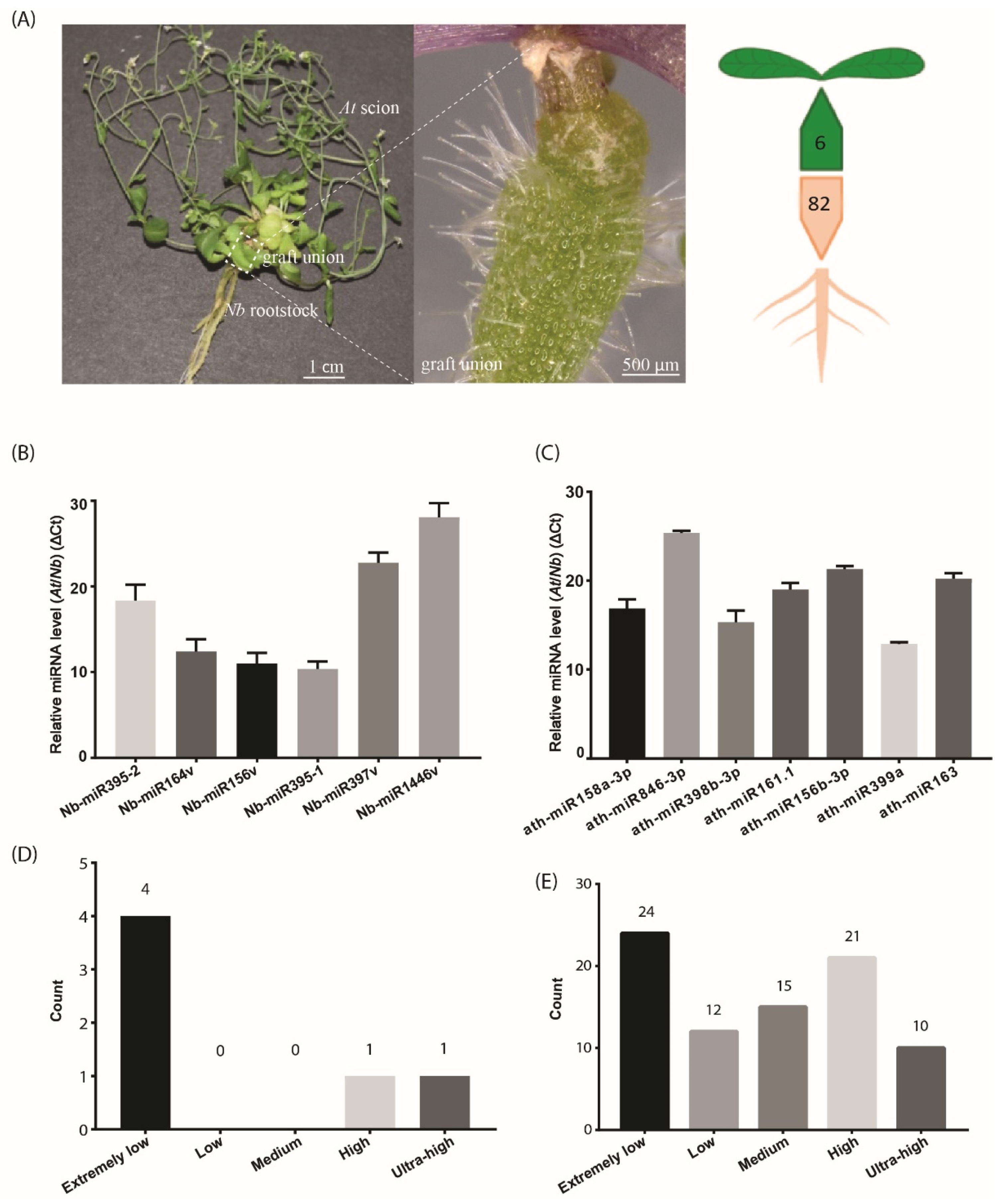
2.1. Hypocotyl Grafting and Small RNA Deep Sequencing in At Scions and Nb Rootstocks

To stringently characterize the small RNAs that move from root to shoot, we adopted hypocotyl grafting, in which the graft union occurred at the hypocotyl region of At and Nb (Figure 1A). This type of graft, unlike those exploring the source-to-sink movement, separated the shoot and root spatially, thus avoiding the leaf source-derived signals being included. We found that the life span was extensively extended in the At/Nb grafts [23], and we hypothesized that any small RNAs from the rootstock could partially represent contributing factors to the observed phenotypic changes. With this premise, we collected the samples at the mature stage of the At/Nb graft or 90 days after grafting (DAG) for small RNA library construction. Because sample collection in a similar timescale is not applicable for At/At or Nb/Nb self-grafts, we chose the similarly developmental stage of self-grafts (e.g., 40–45 DAG) for a negative control.
Four groups of materials that were labeled as AGS (Arabidopsis grafting scion in At/Nb heterograft), ACS (Arabidopsis control scion in At/At homograft), NGR (Nicotiana grafting rootstock in At/Nb heterograft) and NCR (Nicotiana control rootstock in Nb/Nb homograft) were harvested for RNA deep sequencing. More than 13.85 million clean reads in the size of 18–30 nt for each sample were obtained by removing the low-quality reads (e.g., reads of a length <18 nt or >30 nt, reads with more than three unknown bases or reads without adaptors) (Table S1). Bowtie analysis was performed to determine the read distribution in the clean reads. The results showed that the unannotated reads accounted for more than 50% of all reads in all samples, and the miRNA reads accounted for about 6.3%, 4.4%, 1.9% and 2% in the AGS, ACS, NGR and NCR, respectively (Figure S1). More than 60% of the total sRNA reads including miRNA and unannotated RNAs in the AGS and ACS were further mapped to the Arabidopsis genome, and less than 1% of the remaining unmapped reads were re-mapped onto the Nb genome (Table 1). Similarly, in the Nb rootstock, about 0.29% and 0.05% of the total sRNA reads from NGR and NCR, respectively, were mapped to the Arabidopsis genome (Table 2). These results suggested that a small fraction of the total small RNAs could potentially move across the graft union.

2.2. Small RNA Movement across the Graft Union

The overall identified potential mobile miRNAs are listed in Tables S2 and S3. A very simple method to recover the high-confidence mobile sRNAs dictates that RNA reads from either the scion or rootstock that are mapped to the opposite part of the grafts should not appear in the corresponding part of self-grafts. Under this stringent purview, we recovered six candidates from Nb in the At scions (Table 3). These miRNAs were highly conserved, belonging to five miRNA families, including miR156, miR164, miR395, miR397 and miR1446. Since these miRNAs showed sequence variation to the existing miRNA members, they were referred to as Nb-miR156v, Nb-miR164v, Nb-miR395-1, Nb-miR395-2, Nb-miR397v and Nb-miR1446v, respectively, in this study (Table 3). The precursor sequences were also identified from the Nb genome (Table S4). They may be ascribed as high-confidence root-to-shoot mobile candidates, as they repeatedly appeared in all heterografting samples.
Likewise, the same selection criteria applied to the Nb rootstock led to the identification of 82 At miRNAs (Table S3), a much higher number than those in the rootstock-to-scion migrating direction. This in turn agreed with the bulk flow in the source-to-sink direction. These potentially mobile miRNAs accounted for nearly one quarter of the total Arabidopsis miRNAs (miRbase), suggesting the movement of substantial miRNAs via the phloem. We further compared the miRNAs in the AGS samples with those in the Nb samples and found 31 At miRNAs that shared the exact sequence with those in the Nb (Table S5). Thus, we could not determine their mobility in this study. This might further imply that the number of mobile miRNAs through phloem could potentially increase with an improved technique.
We also noticed that the two subsets of mobile miRNAs shared no essential overlaps. Although the miR156 family member appeared in both subsets (Table 3 and Table S3), the Nb miR156 sequence corresponded to the sense strand, while the At miR156 sequence recovered in the rootstock was located in the antisense strand, agreeing with previous studies showing that the miR156* strand could be detected in the phloem [24]. Taken together, the non-overlaps between the two mobile miRNA subsets implied the divergence of the regulatory mechanisms of the top-down and down-top miRNA movement.

2.3. Mobile miRNA Detection

To detect the mobile miRNAs, we adopted the stem-loop RT-qPCR procedure which proved to be specific and sensitive to miRNA detection [25]. In the scion tissues, five out of the six selected miRNAs were detected (Figure 1B), including Nb-miR395-1, -2, Nb-miR164v, Nb-miR156v, Nb-miR397v and Nb-miR1446v (primers in Table S6). In the rootstock, the seven selected miRNAs from At were detected (Figure 1C). All these detected miRNAs showed a very low accumulation relative to the U6 reference. We further checked the expression of these mobile miRNAs in the non-grafting tissues and found nearly one third of the root-to-shoot miRNAs (29%) belonged to the extremely low expression group (Figure 1D). In the shoot-to-root miRNA subset, nearly one third of them belonged to the extremely low expression group, and around 38% of the miRNAs were from the high or ultra-high expression group (Figure 1E), suggesting the abundance model [14] could partially explain the disparity in the miRNA numbers between the two subsets, given that the high expression miRNAs could easily be brought down to the rootstock through the phloem bulk flow.

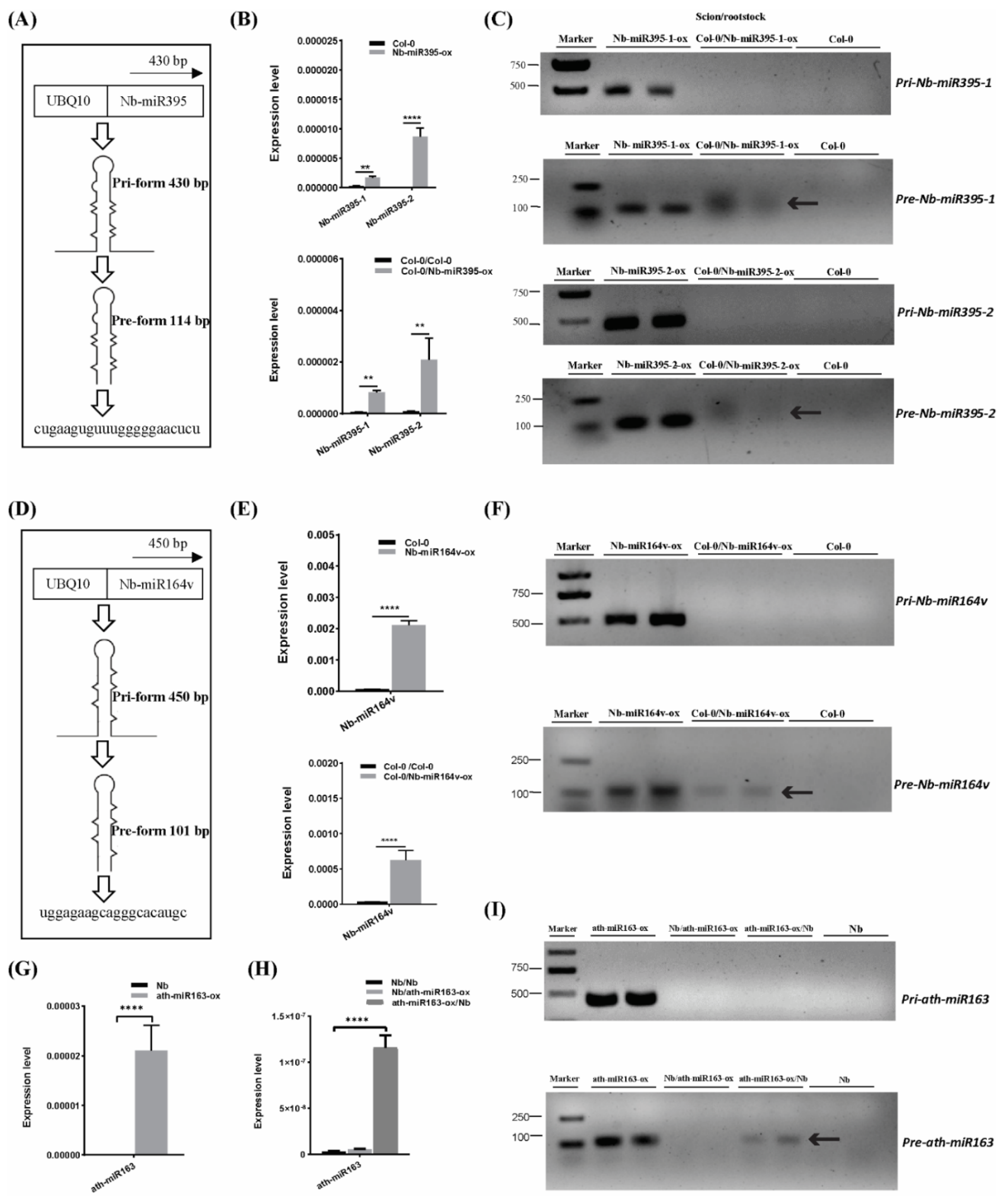
2.4. Pre- and Mature miRNA, but Not the Pri-miRNA, Can Be Detected in the Scion or Rootstock

To further confirm the mobility of these miRNAs, we overexpressed the pri-miRNA coding sequence of Nb-miR395-1, Nb-miR395-2 and Nb-miR164v in Arabidopsis to check whether they sustained their mobility in Arabidopsis (Figure 2A,D). The stem-loop QPCR results revealed that the mature miRNAs were expressed in Arabidopsis and could be detected in the scion tissues in which the miRNA-overexpressing Arabidopsis was used as the rootstock (Figure 2B,E), suggesting the mature Nb-miR395-1, Nb-miR395-2 and Nb-miR164v could move to the scion as shown in the heterografts (Figure 1B). We found that the transcribed pri-miRNAs in all these selected miRNA loci (Nb-miR395-1, Nb-miR395-2 and Nb-miR164v) were beyond detection in the scions (Figure 2C,F), but the pre-miRNAs could be detected in the Arabidopsis scions (two independent materials), suggesting the pre-miRNAs together with the mature miRNAs could be transferred from the roots to the shoots. To further evaluate the specificity of the miRNA movement, we overexpressed an Arabidopsis-specific miRNA gene, miR163, which showed a potential for rootward mobility (Table S3) in N. benthamiana and used this transgenic Nb line (ath-miR163-ox) as the scion or rootstock (Figure 2G–I). QPCR showed that miR163 could not be detected in the wild-type (WT) Nb background but was highly expressed in the Nb transgenic line (Figure 2G). The grafting experiment revealed that miR163 from the Nb overexpressing line could be detected in the WT Nb rootstock rather than the Nb scion (Figure 2H), suggesting the specificity of its directional movement. Likewise, the pre-miRNA (but not the pri-miRNA) of miR163 could be detected in the WT Nb rootstock (Figure 2I), supporting the mobility of the miR163 precursor.

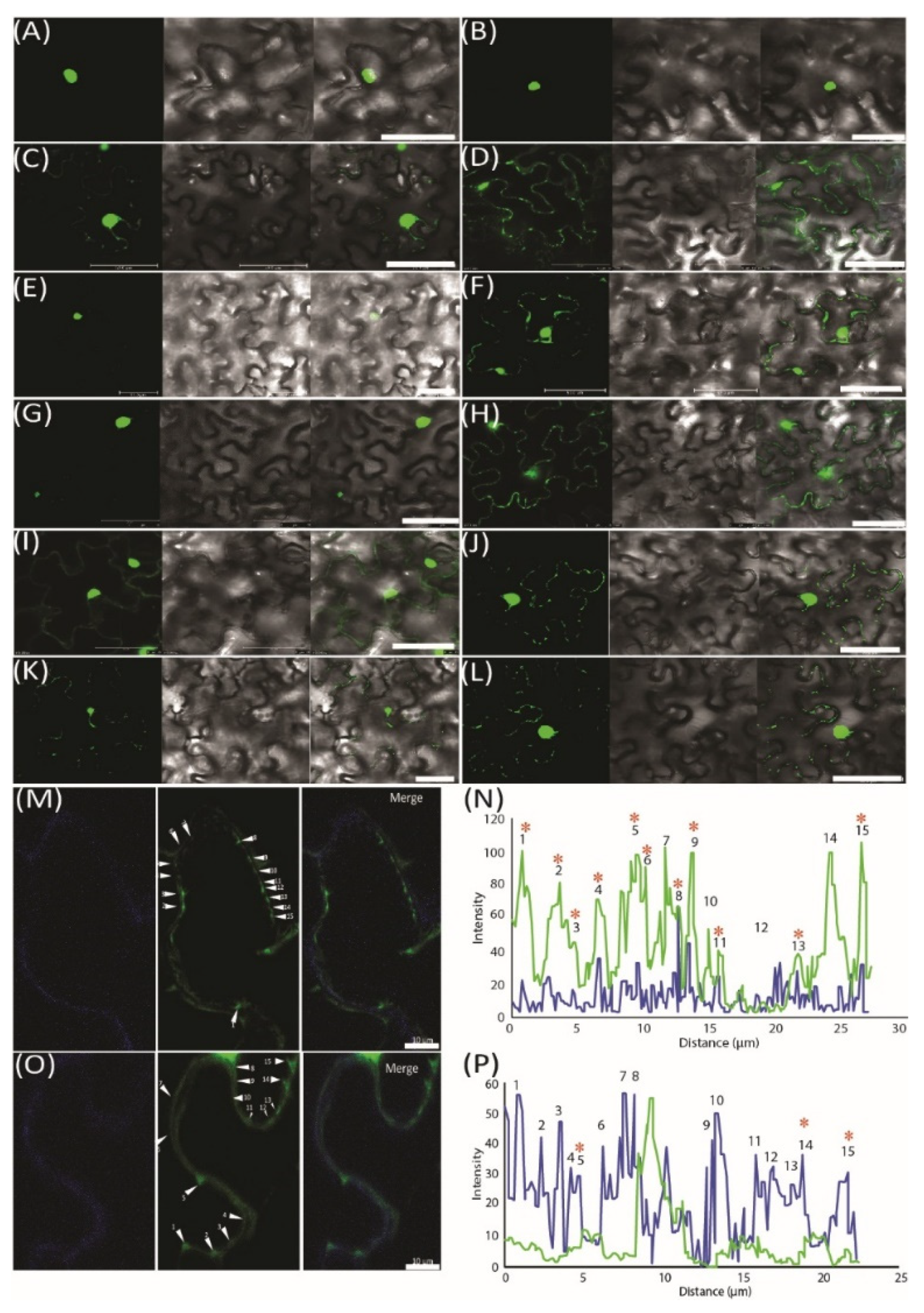
2.5. Validation of miRNA Movement Using the MS2 System

To further demonstrate the movement of miRNAs, we used an MS2 RNA visualizing system, which was adopted to visualize the RNA movement in the plants [26]. MS2FD-GFP was only detected in the nucleus when transiently co-expressed with the nonmobile RNA Actin2-SL24 [27] or SL24 blank vector (Figure 3A–C). To test how the root-to-shoot miRNAs could be mobile between cells, we fused two different forms of miRNAs (i.e., the pri and pre forms) with SL24. As seen in Figure 3E–G, the full-length pri forms of Nb-miR395-1 and Nb-miR397v were mainly accumulated in the nucleus, giving out a similar pattern to that of MS2FD-GFP or that of nonmobile RNAs (Figure 3A–C,E,G). However, the pre forms of Nb-miR395-1 and Nb-miR397v were localized to the punctate foci around the cell periphery (Figure 3F,H) in a pattern similar to that of FLOWERING LOCUS T (FT) mRNA FT-SL24 (Figure 3D). In pri-miR164-SL24, the GFP signal was simply uniformly detected at the cell periphery (Figure 3I). However, in pre-Nb-miR164v-SL24, the strong signal of the punctate green fluorescent foci appeared around the cell periphery (Figure 3J). Similarly, the mature Nb-miR164v and three tandem repeats of Nb-miR164v (3xNb-miR164v-SL24) could also be located in the punctate foci (Figure 3K,L), agreeing with the miRNA detection results (Figure 2E,F). To further confirm the punctate foci were overlapping with PD, we examined the localization of 3xNb-miR164V-SL24 GFP fluorescence and the signal of aniline blue that was used to indicate callose deposition at the PD neck, and we found the two signals were co-localized in the majority of the examined foci (Figure 3M,N). In contrast, the Actin2-SL24 GFP signal, shown in Figure 3O,P, was very scarce and only partly overlapping with the aniline blue signal (3 of 15 stains). These findings suggest that the mature and pre forms of the selected miRNAs were mobile and most probably targeted to PD to be transferred to the adjacent cell.

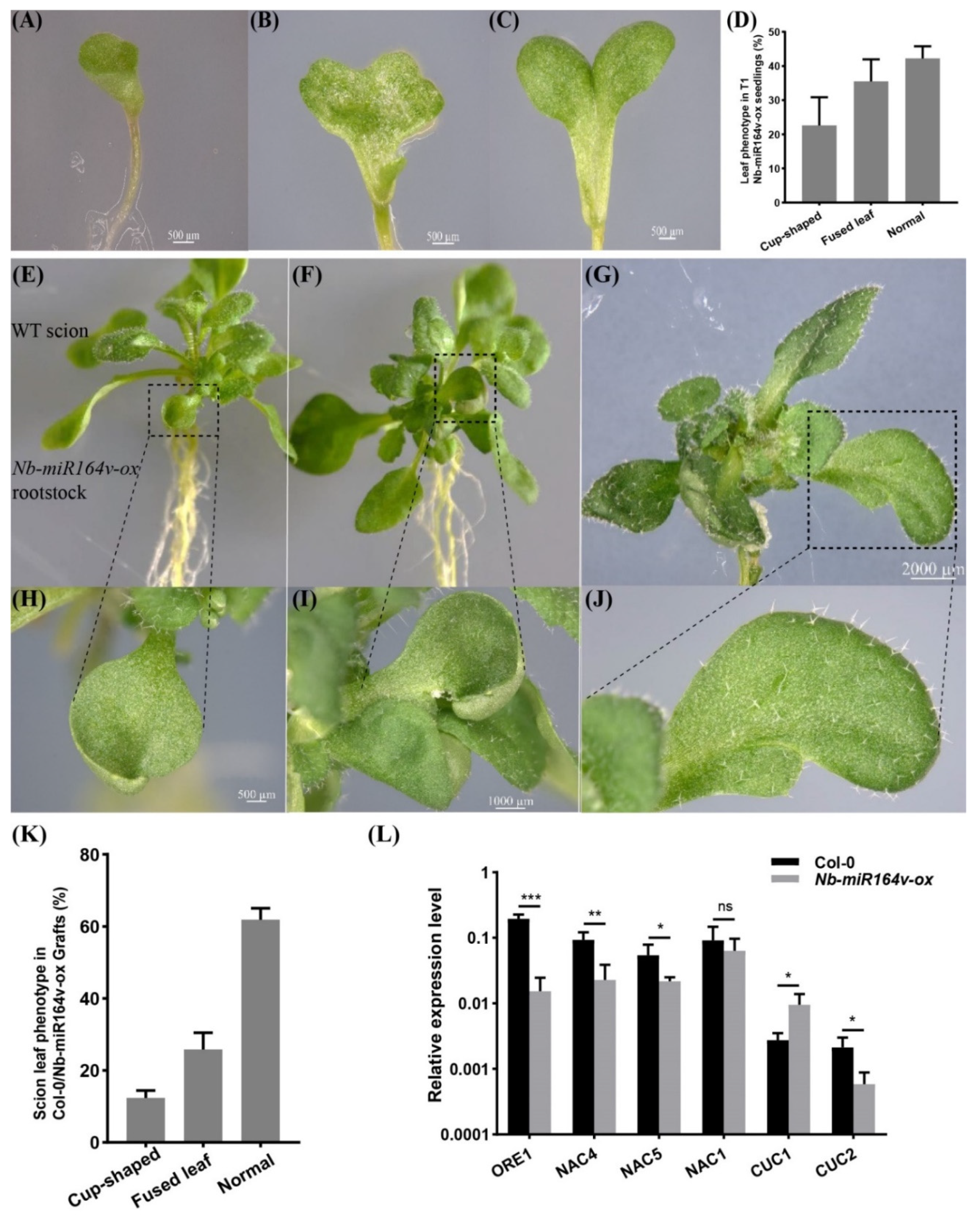
2.6. Phenotypic Modification in the Scion by Root-to-Shoot Mobile Nb-miR164v

We next asked whether these root-to-shoot mobile miRNAs could cause any biological consequences on the scion morphology after moving into the scion. For this, we overexpressed Nb-miR395-1, Nb-miR395-2, Nb-miR397v, Nb-miR1446v and Nb-miR164v in Arabidopsis to check if they could cause any phenotypic changes. In the T2 generation of the Nb-miR395-1, Nb-miR395-2, Nb-miR397v and Nb-miR1446v overexpressing lines, no visual phenotype or developmental defects were observed (Figure S2). The grafting experiment involving these lines as rootstocks did not show any observable phenotypic difference with the Col-0 self-grafts (Figure S2). As a representative example, Nb-miR164v was chosen for further investigation, since the overexpression of Nb-miR164v in Arabidopsis gave rise to copious morphological defects, such as the fused and misshapen leaves (Figure 4A–D). These phenotypes were exactly similar to the Arabidopsis‘s own miR164 overexpressing lines [28,29,30], suggesting the similar role of Nb-miR164v in leaf development.
We then grafted the Nb-miR164v overexpressing line as a rootstock with the WT Col-0 scion. The first emerging leaves in these grafts showed the typical defects (Figure 4E–J), and the fusion of two different leaves could also occur (Figure 4G,J). In total, around 38% of the grafts showed the typical leaf phenotypes (Figure 4K). Since these defects are characteristic for plants with reduced CUC1 or CUC2 activity [28,29,30], we quantified the levels of the miR164 targets in the WT and the grafts. The results showed that the majority of the miR164 targets were significantly downregulated in the WT scion (Figure 4L), which was consistent with the expectations. These results strongly indicated that the Nb-miR164v in the rootstock moved into the scion to modify the leaf shape.

3. Discussion

3.1. Phloem-Mediated Bulk Flow of Mobile RNAs

Macromolecules are able to carry out their function in a non-autonomous way. This phenomenon has become increasingly appreciated for their roles in intercellular and inter-tissue communication by using a grafting technique [31,32,33]. By such means, Notaguchi et al. [34] identified 138 mobile mRNAs by constructing a graft between At and Nb at the bolting stage. A comparative number of mobile transcripts (183 Nb transcripts) was identified to move into a tomato rootstock using an Nb/tomato heterograft system [35]. Recently, with the long-read RNA-seq technology, Li et al. [36] were able to detect a very small subset of mRNAs moving between the heterografting partners involving soybean and common bean (163 mRNAs from soybean and 129 mRNAs from common bean), but with only around 56% of each subset being full-length coding sequences (91 from soybean and 77 from common bean). Aside from that, this study identified 100 miRNAs that were predominantly produced in shoots and transported to the roots. Our results also showed a comparable number of miRNAs (82 miRNAs) moving down to the rootstock in the At/Nb heterograft at 90 DAG, implying the common regulatory mechanisms for miRNA movement. In addition, many miRNAs moving from scion to rootstock have been reported before. For example, the well-documented shoot-to-root mobile sRNA miR399 [37,38,39,40] was recovered in the samples. Meanwhile, several miRNAs, such as miR156 [41,42,43], miR167 [41,42,44,45], miR168 [39], miR169 [39], miR172 [42,45], miR829 [39], miR2111 [40,43,46] and miR403 [39,44], that were phloem-mobile sRNAs were also found in the Nb rootstock, particularly miR2111, which was recently confirmed to move from shoot to root to regulate nodulation in soybean [46].
Interestingly, the number of mobile mRNA transcripts seemed to be vastly increased in the homograft or closely related species. For example, there were about 2006 RNA transcripts transported between 2 At genotypes [27], more than 3000 mobile mRNAs across the graft union in 2 grapevine species [47] and more than 3500 mRNAs exchanged between cucumber and watermelon [48]. A discrepancy of this magnitude could arise from the process of vascular connection between the scion and rootstock; that is, the phloem connection in the grafts involving closely related species performed much better than in those distantly related species, thus allowing more RNA transcripts to move into the sink tissues through the phloem. For example, phloem translocation of [14C]-sucrose could occur 5–10 days after grafting in autografts and in closely related grafting partners involving Lycopersicon and Solanum (compatible combination) but not in the Vicia/Helianthus heterograft (less compatible combination), and a strong correlation between phloem connection and assimilate transport across the graft interface could be found [49]. Similarly, [14C]-sorbitol translocation in the incompatible pear/quince heterograft was greatly reduced compared with the compatible graft or the autograft [50]. Given that the majority of mobile RNAs move through the phloem by bulk flow, the number of identified mobile RNAs could strongly rely on the phloem connection between the scion and rootstock.
Compared with the shoot-to-root mobile RNAs, the number of root-to-shoot mobile RNAs was extremely low. In our case, there were only six miRNAs being detected (Figure 1A). The immediate question that arises is how these miRNAs can be transmitted to the shoot, given that they are moving against phloem streaming. One possible route is the cell-to-cell transmission from the bottom, as shown in long-distance mobile silencing [2]. We indeed showed that the NbmiR395, 397 and 164 could be transported through PD (Figure 3), which may constitute the first step of shootward non-cell-autonomy. Because the cell-to-cell transmission of miRNA usually operates locally to form a gradient for its function [51], the graft-transmissible long-distance movement of miRNA could hardly be achieved simply through local transmission. That aside, miRNA movement is an amplification-independent process, and thus root-derived miRNA movement into shoots warrants further explanation.
Another seemingly remote possibility could be attributed to xylem transport. Given the root-to-shoot transport against phloem streaming, the possibility for bottom-to-top transport via xylem might not be irrefutably excluded, as several studies have observed before. For instance, the StBEL5 RNA can be detected in the micro-dissected xylem in the potato [52], and RNA profiling in xylem tissue also identifies many non-coding RNAs and mRNAs [53,54,55]. Furthermore, small peptides can signal their physiological status to the shoots by traveling through xylem [56]. Nevertheless, Buhtz et al. [42] found no RNAs in the xylem sap of Brassica, apparently contradicting the results mentioned above. This contradiction could come from a technological difference and could be resolved through technique improvement, particularly the improvement of xylem isolation while maintaining RNA integrity.

3.2. Robustness of Heterografts in the Identification of Root-to-Shoot Long-Distance Mobile miRNA

Bioinformatic analysis of the mobile RNA dataset indicated that smaller transcripts tend to be more mobile [14]. Thus, it is reasonable to infer that small RNA (including miRNAs) should have a high tendency to move. Indeed, more and more studies have shown that many small RNAs are mobile [36,57,58]. However, the identification of bottom-to-top mobile miRNAs has been lacking. In this study, we have identified six candidates that can move from rootstock to shoot (Table 3). Among them, miR395 and miR397 are very likely possessing the property of root-derived signals: highly or exclusively expressed or inducibly expressed in the root but partially accumulated in the shoot. Taking the Arabidopsis data as an example, the promoter activity of miR395a and miR395b, miR395c and miR395d indicates that they are highly induced in the Arabidopsis roots [59]. Since miR395 regulates sulfate accumulation and allocation in Arabidopsis [60], the expression pattern of the miR395 family implied that root-to-shoot movement might represent a physiologically relevant event in which the sulfate availability sensed by the roots would coordinate the nutritional redistribution in the shoot by emanating root-derived functional macromolecules (e.g., miR395 in this case) to regulate the sulfate-associated metabolic networks.
Similarly, miR397 also showed the above property. The expression of miR397 was strongly induced in the Cd-treated roots but only partially elevated in the shoots [61], a very promising sign for the root-derived signal for regulating shoot response. miR397 is also induced by other stress stimuli, such as H2O2 [62] and heat stress [63,64]. Since our previous study also showed that H2O2 plays critical roles in regulating root-to-shoot mobile silencing [65], the correlation of miR395 expression with cellular H2O2 or other stressor-induced H2O2 fluctuations could reflect its root-to-shoot mobility.

3.3. Mobile Small RNA and Trait Modification in Grafts

Grafting-induced phenotypic changes have been widely documented [66,67,68]. Various traits, such as the flowering time, plant architecture, fruit flavor and quality, as well as the response to biotic and abiotic stress, can be modified by grafting. The mechanisms of underlying scion or rootstock phenotypic changes could be very diverse and have been extensively explored, with the focus on what signals communicate between the scion and rootstock to have their impact on phenotypes [67,69]. Grafting experiments combined with next-generation sequencing technology and genetic studies have shown that many sRNAs, particularly the 23–24-nt siRNAs associated with epigenetic regulation in the scion, moved into the rootstock to induce DNA methylation in the target loci [20,70,71]. In terms of scion trait modification, one recent study showed that an enhanced growth vigor conferred by the msh1 rootstock in both Arabidopsis and tomato graft progenies was due to RdDM-dependent epigenetic changes in 1380 differentially methylated genes, including many auxin-related gene pathways [72].
The data from this study showed that several miRNAs could move from rootstock to scion, and we further found that miR164 could deliver a phenotypic impact on scion leaf development (Figure 4). Actually, miR164 is mainly expressed in the roots (and almost absent in the leaves of Arabidopsis) to regulate the root morphology through the miR164-NAC1 module [73], suggesting the property of root-derived signals for miR164. Interestingly, further study shows that it also regulates the leaf senescence through the miR164-ORE1 module in the leaf [74]. Given the mobility of miR164 from rootstock to scion identified in this study, it is very tempting to speculate that root development could be coordinated with leaf development via the long-distance movement of miR164. Further experiments need to be designed to reveal this potential relationship.

4. Materials and Methods

4.1. Plant Material and Growth Conditions

Seeds of the wild-type (WT) Arabidopsis thaliana (Col-0) and the WT Nicotiana benthamiana, as well as transgenic lines including Nb-miR395-1, -2, Nb-miR397v and Nb-miR164v overexpressing Arabidopsis lines and ath-miR163 overexpressing Nb lines, were surface-sterilized in chlorine gas for 1 h and then plated on sterile Murashige and Skoog (MS) medium supplemented with 3% (w/v) sucrose. The plants were grown vertically under long day conditions (16 h light, 8 h dark, 22–23 °C).

4.2. Grafting

The micro-grafting procedure was essentially described in detail by Andersen et al. [75]. Briefly, the Nb young seedlings that were no more than 8 mm long were used as rootstocks, and the At seedlings that were 2 cm long were used as scions. The cut was made halfway from the base of each hypocotyl, and the cut surfaces were pushed against each other with a certain tension. The grafts were grown on moisturized Whatman paper for 2 days, and then the grafts were gently lifted with forceps and placed vertically on the MS medium with 1% agar and 3% sucrose (w/v) in the growth room (16 h light, 8 h dark) at 22–23 °C.

4.3. Tissue Collection and RNA Extraction

Ninety DAG grafts were used for each tissue collection. Four different types of tissues were collected from an At/Nb heterograft and a self-graft control (At/At and Nb/Nb), which were the scion samples of AGS (At grafting shoots) and ACS (At control shoots) and rootstock samples of NGR (Nb grafting roots) and NCR (Nb control roots). For the aerial parts, the old and yellowing leaves, including rosette leaves, were removed. The stems, cauline leaves and flowers were harvested. For the root tissue, we collected the whole Nb roots, excluding the hypocotyl part of the graft union. Each sample was approximately 200 mg and was immediately frozen in liquid nitrogen and stored at −80 °C in a freezer. Three independent biological samples for each type were harvested. The total RNA was obtained using the TRIzol reagent according to Invitrogen’s instructions. The RNA concentration was measured using NanoDrop 2000 (ThermoFisher Scientific, Waltham, MA, USA).

4.4. Small RNA Sequencing and Analysis

The RNA quantity and integrity were measured with Qubit 2.0 (Life Technologies, ThermoFisher Scientific, Waltham, MA, USA) and a 2100 Bioanalyzer (Agilent Technologies, Santa Clara, CA, USA), respectively. A small RNA library was prepared using a Next Ultra small RNA Sample Library Prep Kit for Illumina (New England Biolabs, Ipswich, MA, USA). The total RNA was ligated with a 5′ and 3′ adapter sequentially and then reversely transcribed into cDNA. After cDNA PCR amplification and gel purification, the insert size and quantity of the constructed library were assessed with a 2100 Bioanalyzer (Agilent Technologies, Santa Clara, CA, USA) and Qubit 2.0 (Life Technologies, ThermoFisher Scientific, Waltham, MA, USA), respectively. The sequencing procedure was performed with an Illumina HiSeq Xten system at BioMarker Technologies in Beijing, China. The total sRNA reads, including miRNAs and unannotated RNAs, were then obtained by removing sequences from other non-coding RNAs, such as rRNA, tRNA, snRNA and snoRNA in a repetitive sequence using the bowtie tool to align against the Silva, GtRNAdb, Rfam and Repbase databases.

4.5. miRNA Detection

Stem-loop RT-qPCR was used to quantify the mobile miRNAs. The stem-loop RT primers and forward qPCR primers were designed using miRNA Design V1.01 software (Vazyme Biotech, Nanjing, China). The first cDNA was synthesized from 1 µg of the total RNA using an miRNA 1st Strand cDNA Synthesis Kit (Vazyme, MR101, Nanjing, China) following the manufacturer’s instruction. The target miRNA was detected by QPCR using the kit’s miRNA Universal SYBR qPCR Master Mix (2×) containing mQ primer R (Vazyme, MQ101, Nanjing, China). Each QPCR reaction was performed in a volume of 20 µL containing 1 µL cDNA, 0.4 µL of each forward qPCR primer (10 µM), 0.4 µL of mQ primer R (10 µM) and 10 µL of 2× miRNA Universal SYBR qPCR Master Mix (Vazyme, MQ101, Nanjing, China). The nuclear small RNA U6 was used as the internal reference gene [76]. QPCR was performed on a Bio-Rad CFX cycler, and the cycling conditions were as follows: 5 min at 95 °C, followed by 40 cycles at 95 °C for 10 s and 60 °C for 30 s. All reactions were run in biological triplicate. A list of the stem-loop and qPCR primers used in this study is given in Supplementary Table S6.

4.6. RT-PCR on Grafted Arabidopsis and Nicotiana Transgenic Lines

Three biological replicates of the grafting samples were used to test the mobile transcripts. The total RNA was extracted from the aerial parts (excluding rosette leaves) at 30 DAG using TRIzol reagent. The RNA was quantified with a NanoDrop 2000 spectrophotometer and then pretreated with gDNase to remove any DNA. The 1st strand cDNA from 1 µg RNA was synthesized in a 20-µL volume using a FastQuant RT Kit (TIANGEN, Beijing, China). Then, 1 µL cDNA was used for PCR with 40 cycles. The primers for Figure 2C,F,I are provided in Supplementary Table S6.

4.7. Plasmid Construction

The pri- and pre-sequence of Nb-miR395-1, -2, Nb-miR397v and Nb-miR164v were amplified from Nb genomic DNA using the corresponding primers shown in Supplementary Table S6. These amplified genomic DNA fragments were then purified and cloned downstream of ubiquitin 10 promoter in a binary vector p2S3CherryUniH and then confirmed by Sanger sequencing. All plasmids were then transformed into Agrobacterium tumefaciens strain GV3101. The transgenic plants were selected using 50 mg/L hygromycin.
The precursor and primary sequences of miRNAs Nb-miR395-1, Nb-miR397v and Nb-miR164v were amplified from Nb genomic DNA using primers Nb-miR395-1-SL24-F1, Nb-miR395-1-SL24-R1, Nb-miR395-1-SL24-F2, Nb-miR395-1-SL24-R2, Nb-miR397-SL24-F1, Nb-miR397-SL24-R1, Nb-miR397-SL24-F2, Nb-miR397-SL24-R2, Nb-miR164-SL24-F1,Nb-miR164-SL24-R1,Nb-miR164-SL24-F2 and Nb-miR164-SL24-R2 (Table S6). A 332-bp fragment of an Actin2 gene was amplified using primer AT-Actin2-SL24-F and AT-Actin2-SL24-R. A partial FT cDNA fragment corresponding to the mobile part [77] was amplified from Arabidopsis cDNA using primer FT-SL24-F and FT-SL24-R (Table S6). The amplified sequences were purified and cloned into the MfeI-linearized pUbik-SL24 vector individually and confirmed by sequencing. All constructs were transformed into Agrobacterium tumefaciens strain GV3101.
For miR164 tandem repeat construction, 100 bp of a partial GUS fragment fused with an Nb-miR164v mature sequence was amplified using primers Nb-miR164 P1 and Nb-miR164 P8 (1 repeat). The other fragments were amplified with the following primers: Nb-miR164 P1, Nb-miR164 P2, Nb-miR164 P3, Nb-miR164 P4, Nb-miR164 P5 and Nb-miR164 P8 (3 repeats), as well as Nb-miR164 P1, Nb-miR164 P2, Nb-miR164 P3, Nb-miR164 P4, Nb-miR164 P5, Nb-miR164 P6, Nb-miR164 P7 and Nb-miR164 P8 (4 repeats) (Table S6). After BsaI digestion, these fragments were ligated by T4 ligase (NEB). We then obtained the Nb-miR164 repeat fragment by PCR amplification with primers Nb-miR164 P1 and Nb-miR164 P8. The amplified fragment was purified and cloned into the MfeI-linearized pUbik-SL24 vector and then confirmed by Sanger sequencing. All plasmids were then transformed into Agrobacterium tumefaciens strain GV3101.

4.8. Infiltration and Confocal Microscopy

The above constructs were individually transformed into Agrobacterium tumefaciens strain GV3101. When the optical density of the A. tumefaciens cells was 0.6 (OD600nm), the bacterial cells were pelleted and resuspended in infiltration solution (10 mM MgCl2, 10 mM MES, 20 µM 5-azacytidine, 0.5 mM ascorbic acid, 0.03% tween-20 and 150 mM acetosyringone, pH 5.6). The suspension was adjusted to an OD600nm of 0.3. For the mixed infiltration, each suspension was adjusted to an OD600nm of 0.6. The two suspensions were equally mixed before injection. The 6-week-old N. benthamiana plants to be used were infiltrated by using a needleless syringe. The infiltrated areas in the leaves were imaged at 3 d after infiltration with a confocal laser scanning microscope (Leica TCS SP8 MP) using a 40× water immersion lens.

5. Conclusions

Heterografts involving distantly related species provided us with a valuable offer to uncover macromolecule signaling in plants, particularly for those highly conserved RNAs. We used an At/Nb grafting system to identify six promising miRNAs that were moving from root to shoot, providing a foundation for understanding the shootward signaling mechanisms. Among them, miR164 could work as a shootward signaling macromolecule to coordinate root and shoot development.

Supplementary Materials

The following are available online at https://www.mdpi.com/article/10.3390/ijms222312821/s1.

Author Contributions

Conceptualization, D.L.; methodology, Z.D., H.W., D.L. (Dongyi Li), L.L. and Z.W.; formal analysis, Z.D., W.Y., Y.X., C.L. and D.L. (Dacheng Liang); data curation, Z.D., W.Y., Y.X., C.L. and D.L. (Dacheng Liang); writing—review and editing, D.L. (Dacheng Liang); project administration, D.L. (Dacheng Liang). All authors have read and agreed to the published version of the manuscript.

Funding

This work was partially supported by the National Natural Science Foundation of China (31671257) and Hubei Collaborative Innovation Center for Grain Industry (LXT-16-18).

Institutional Review Board Statement

Not applicable.

Informed Consent Statement

Not applicable.

Data Availability Statement

The small RNA deep sequencing data are available at the NCBI database under the accession ID PRJNA744705.

Acknowledgments

We would like to thank Tien-Shin Yu for the generous gifts of p1390-35S-MS2FD-GFP and p1390-35S-AtFTSL24 containing SL24. We would like to thank Ruochao Hao and Xingping Zhang at the Life Science Instrument Center of Yangtze University for their help with the confocal microscopy.

Conflicts of Interest

The authors declare no conflict of interest.

References

  1. Brosnan, C.A.; Mitter, N.; Christie, M.; Smith, N.A.; Waterhouse, P.M.; Carroll, B.J. Nuclear gene silencing directs reception of long-distance mRNA silencing in Arabidopsis. Proc. Natl. Acad. Sci. USA 2007, 104, 14741–14746. [Google Scholar] [CrossRef] [Green Version]
  2. Liang, D.; White, R.; Waterhouse, P. Gene silencing in Arabidopsis spreads from the root to the shoot, through a gating barrier, by template-dependent, nonvascular, cell-to-cell movement. Plant Physiol. 2012, 159, 984–1000. [Google Scholar] [CrossRef] [PubMed] [Green Version]
  3. Gallagher, K.L.; Sozzani, R.; Lee, C.M. Intercellular protein movement: Deciphering the language of development. Annu. Rev. Cell Dev. Biol. 2014, 30, 207–233. [Google Scholar] [CrossRef]
  4. Lough, T.; Lucas, W. Integrative plant biology role of phloem long- distance macromolecular trafficking. Annu. Rev. Plant Biol. 2006, 57, 203–232. [Google Scholar] [CrossRef] [Green Version]
  5. Stegemann, S.; Bock, R. Exchange of genetic material between cells in plant tissue grafts. Science 2009, 324, 649–651. [Google Scholar] [CrossRef] [PubMed]
  6. Atkins, C.A.; Smith, P.M.; Rodriguez-Medina, C. Macromolecules in phloem exudates—A review. Protoplasma 2011, 248, 165–172. [Google Scholar] [CrossRef]
  7. Fuentes, I.; Stegemann, S.; Golczyk, H.; Karcher, D.; Bock, R. Horizontal genome transfer as an asexual path to the formation of new species. Nature 2014, 511, 232. [Google Scholar] [CrossRef] [PubMed]
  8. Yu, Y.; Jia, T.; Chen, X. The ‘how’ and ‘where’ of plant microRNAs. New Phytol. 2017, 216, 1002–1017. [Google Scholar] [CrossRef] [PubMed] [Green Version]
  9. Corbesier, L.; Vincent, C.; Jang, S.; Fornara, F.; Fan, Q.; Searle, I.; Giakountis, A.; Farrona, S.; Gissot, L.; Turnbull, C.; et al. FT Protein Movement Contributes to Long-Distance Signaling in Floral Induction of Arabidopsis. Science 2007, 316, 1030–1033. [Google Scholar] [CrossRef] [Green Version]
  10. Lin, M.K.; Belanger, H.; Lee, Y.J.; Varkonyi-Gasic, E.; Taoka, K.; Miura, E.; Xoconostle-Cazares, B.; Gendler, K.; Jorgensen, R.A.; Phinney, B.; et al. FLOWERING LOCUS T protein may act as the long-distance florigenic signal in the cu-curbits. Plant Cell 2007, 19, 1488–1506. [Google Scholar] [CrossRef] [Green Version]
  11. Yoo, S.; Hong, S.; Jung, H.; Ahn, J. The cotyledons produce sufficient FT protein to induce flowering: Evidence from cotyledon micrografting in Arabidopsis. Plant Cell Physiol. 2013, 54, 119–128. [Google Scholar] [CrossRef] [Green Version]
  12. Zhu, Y.; Liu, L.; Shen, L.; Yu, H. NaKR1 regulates long-distance movement of FLOWERING LOCUS T in Arabidopsis. Nat. Plants 2016, 2, 16075. [Google Scholar] [CrossRef]
  13. Paultre, D.; Gustin, M.; Molnar, A.; Oparka, K. Lost in transit: Long-distance trafficking and phloem unloading of protein signals in Arabidopsis homografts. Plant Cell 2016, 28, 2016–2025. [Google Scholar] [CrossRef] [Green Version]
  14. Calderwood, A.; Kopriva, S.; Morris, R.J. Transcript Abundance Explains mRNA Mobility Data in Arabidopsis thaliana. Plant Cell 2016, 28, 610–615. [Google Scholar] [CrossRef] [Green Version]
  15. Kim, G.; LeBlanc, M.L.; Wafula, E.K.; dePamphilis, C.W.; Westwood, J.H. Genomic-scale exchange of mRNA between a parasitic plant and its hosts. Science 2014, 345, 808–811. [Google Scholar] [CrossRef]
  16. Zhang, W.; Thieme, C.J.; Kollwig, G.; Apelt, F.; Yang, L.; Winter, N.; Andresen, N.; Walther, D.; Kragler, F. tRNA-Related Sequences Trigger Systemic mRNA Transport in Plants. Plant Cell 2016, 28, 1237–1249. [Google Scholar] [CrossRef] [PubMed] [Green Version]
  17. Axtell, M. Classification and comparison of small RNAs from plants. Annu. Rev. Plant Biol. 2013, 64, 137–159. [Google Scholar] [CrossRef] [Green Version]
  18. Molnar, A.; Melnyk, C.W.; Bassett, A.; Hardcastle, T.J.; Dunn, R.; Baulcombe, D.C. Small Silencing RNAs in Plants Are Mobile and Direct Epigenetic Modification in Recipient Cells. Science 2010, 328, 872–875. [Google Scholar] [CrossRef] [PubMed] [Green Version]
  19. Alvarez, J.P.; Pekker, I.; Goldshmidt, A.; Blum, E.; Amsellem, Z.; Eshed, Y. Endogenous and Synthetic MicroRNAs Stimulate Simultaneous, Efficient, and Localized Regulation of Multiple Targets in Diverse Species. Plant Cell 2006, 18, 1134–1151. [Google Scholar] [CrossRef] [PubMed] [Green Version]
  20. Polydore, S.; Axtell, M.J. Analysis of RDR1/RDR2/RDR6-independent small RNAs in Arabidopsis thaliana improves MIRNA annotations and reveals unexplained types of short interfering RNA loci. Plant J. 2018, 4, 1051–1063. [Google Scholar] [CrossRef] [Green Version]
  21. Marin-Gonzalez, E.; Suarez-Lopez, P. “And yet it moves”: Cell-to-cell and long-distance signaling by plant microRNAs. Plant Sci. 2012, 196, 18–30. [Google Scholar] [CrossRef] [PubMed]
  22. Liu, L.; Chen, X. Intercellular and systemic trafficking of RNAs in plants. Nat. Plants 2018, 4, 869–878. [Google Scholar] [CrossRef] [PubMed]
  23. Deng, Z.; Wu, H.; Jin, T.; Cai, T.; Jiang, M.; Wang, M.; Liang, D. A Sequential Three-Phase Pathway Constitutes Tracheary Element Connection in the Arabidopsis/Nicotiana Interfamilial Grafts. Front. Plant Sci. 2021, 12, 664342. [Google Scholar] [CrossRef] [PubMed]
  24. Bhogale, S.; Mahajan, A.; Natarajan, B.; Rajabhoj, M.; Thulasiram, H.; Banerjee, A. MicroRNA156: A potential graft-transmissible microRNA that modulates plant architecture and tuberization in Solanum tuberosum ssp. andigena. Plant Physiol. 2014, 164, 1011–1027. [Google Scholar] [CrossRef] [Green Version]
  25. Varkonyi-Gasic, E.; Wu, R.; Wood, M.; Walton, E.F.; Hellens, R.P. Protocol: A highly sensitive RT-PCR method for detection and quantification of microRNAs. Plant Methods 2007, 3, 12. [Google Scholar] [CrossRef] [Green Version]
  26. Luo, K.-R.; Huang, N.-C.; Yu, T.-S. Selective Targeting of Mobile mRNAs to Plasmodesmata for Cell-to-Cell Movement. Plant Physiol. 2018, 177, 604–614. [Google Scholar] [CrossRef] [PubMed] [Green Version]
  27. Thieme, C.; Rojas-Triana, M.; Stecyk, E.; Schudoma, C.; Zhang, W.; Yang, L.; Miñambres, M.; Walther, D.; Schulze, W.; Paz-Ares, J.; et al. Endogenous Arabidopsis messenger RNAs transported to distant tissues. Nat. Plants 2015, 1, 15025. [Google Scholar] [CrossRef]
  28. Laufs, P.; Peaucelle, A.; Morin, H.; Traas, J. MicroRNA regulation of the CUC genes is required for boundary size control in Arabidopsis meristems. Development 2004, 131, 4311–4322. [Google Scholar] [CrossRef] [PubMed] [Green Version]
  29. Raman, S.; Greb, T.; Peaucelle, A.; Blein, T.; Laufs, P.; Theres, K. Interplay of miR164, CUP-SHAPED COTYLEDON genes and LATERAL SUPPRESSOR controls axillary meristem formation in Arabidopsis thaliana. Plant J. 2008, 55, 65–76. [Google Scholar] [CrossRef]
  30. Berger, Y.; Harpaz-Saad, S.A.; Melnik, H.N.; Alvarez, J.P.; Zinder, M.; Samach, A.; Eshed, Y.; Ori, N. The NAC-domain transcription factor GOBLET specifies leaflet boundaries in compound tomato leaves. Development 2009, 136, 823–832. [Google Scholar] [CrossRef] [Green Version]
  31. Harada, T. Grafting and RNA transport via phloem tissue in horticultural plants. Sci. Hortic. 2010, 125, 545–550. [Google Scholar] [CrossRef]
  32. Gaion, L.A.; Carvalho, R.F. Long-Distance Signaling: What Grafting has Revealed? J. Plant Growth Regul. 2017, 37, 694–704. [Google Scholar] [CrossRef] [Green Version]
  33. Wang, J.; Jiang, L.; Wu, R. Plant grafting: How genetic exchange promotes vascular reconnection. New Phytol. 2017, 214, 56–65. [Google Scholar] [CrossRef] [Green Version]
  34. Notaguchi, M.; Higashiyama, T.; Suzuki, T. Identification of mRNAs that move over long distances using an RNA-Seq analysis of Arabidopsis/Nicotiana benthamiana heterografts. Plant Cell Physiol. 2015, 56, 311–321. [Google Scholar] [CrossRef] [Green Version]
  35. Xia, C.; Zheng, Y.; Huang, J.; Zhou, X.; Li, R.; Zha, M.; Wang, S.; Huang, Z.; Lan, H.; Turgeon, R.; et al. Elucidation of the Mechanisms of Long-Distance mRNA Movement in a Nicotiana benthamiana/Tomato Heterograft System. Plant Physiol. 2018, 177, 745–758. [Google Scholar] [CrossRef] [PubMed] [Green Version]
  36. Li, S.; Wang, X.; Xu, W.; Liu, T.; Cai, C.; Chen, L.; Clark, C.B.; Ma, J. Unidirectional movement of small RNAs from shoots to roots in interspecific heterografts. Nat. Plants 2021, 7, 50–59. [Google Scholar] [CrossRef]
  37. Lin, S.; Chiang, S.; Lin, W.; Chen, J.; Tseng, C.; Wu, P.; Chiou, T. Regulatory Network of MicroRNA399 and PHO2 by Systemic Signaling. Plant Physiol. 2008, 147, 732–746. [Google Scholar] [CrossRef] [Green Version]
  38. Pant, B.D.; Buhtz, A.; Kehr, J.; Scheible, W.R. MicroRNA399 is a long-distance signal for the regulation of plant phosphate homeostasis. Plant J. 2008, 53, 731–738. [Google Scholar] [CrossRef] [PubMed] [Green Version]
  39. Buhtz, A.; Pieritz, J.; Springer, F.; Kehr, J. Phloem small RNAs, nutrient stress responses, and systemic mobility. BMC Plant Biol. 2010, 10, 64. [Google Scholar] [CrossRef] [Green Version]
  40. Huen, A.; Rodriguez-Medina, C.; Ho, A.; Atkins, C.; Smith, P.M.C. Long-distance movement of phosphate starvation-responsive microRNAs in Arabidopsis. Plant Biol. 2017, 19, 643–649. [Google Scholar] [CrossRef] [PubMed]
  41. Yoo, B.-C.; Kragler, F.; Varkonyi-Gasic, E.; Haywood, V.; Archer-Evans, S.; Lee, Y.M.; Lough, T.J.; Lucas, W.J. A Systemic Small RNA Signaling System in Plants. Plant Cell 2004, 16, 1979–2000. [Google Scholar] [CrossRef] [Green Version]
  42. Buhtz, A.; Springer, F.; Chappell, L.; Baulcombe, D.; Kehr, J. Identification and characterization of small RNAs from the phloem of Brassica napus. Plant J. 2008, 53, 739–749. [Google Scholar] [CrossRef] [PubMed]
  43. Pant, B.D.; Musialak-Lange, M.; Nuc, P.; May, P.; Buhtz, A.; Kehr, J.; Walther, D.; Scheible, W.-R. Identification of Nutri-ent-Responsive Arabidopsis and Rapeseed MicroRNAs by Comprehensive Real-Time Polymerase Chain Reaction Profiling and Small RNA Sequencing. Plant Physiol. 2009, 150, 1541–1555. [Google Scholar] [CrossRef] [Green Version]
  44. Rodriguez-Medina, C.; Atkins, C.A.; Mann, A.J.; Jordan, M.E.; Smith, P.M. Macromolecular composition of phloem exudate from white lupin (Lupinus albus L.). BMC Plant Biol. 2011, 11, 36. [Google Scholar] [CrossRef] [PubMed] [Green Version]
  45. Gai, Y.-P.; Zhao, H.-N.; Zhao, Y.-N.; Zhu, B.-S.; Yuan, S.-S.; Li, S.; Guo, F.-Y.; Ji, X.-L. MiRNA-seq-based profiles of miRNAs in mulberry phloem sap provide insight into the pathogenic mechanisms of mulberry yellow dwarf disease. Sci. Rep. 2018, 8, 812. [Google Scholar] [CrossRef] [Green Version]
  46. Tsikou, D.; Yan, Z.; Holt, D.B.; Abel, N.B.; Reid, D.E.; Madsen, L.H.; Bhasin, H.; Sexauer, M.; Stougaard, J.; Markmann, K. Systemic control of legume susceptibility to rhizobial infection by a mobile microRNA. Science 2018, 362, eaat6907. [Google Scholar] [CrossRef] [PubMed]
  47. Yang, Y.; Mao, L.; Jittayasothorn, Y.; Kang, Y.; Jiao, C.; Fei, Z.; Zhong, G. Message RNA exchange between scions and rootstocks in grafted grapevines. BMC Plant Biol. 2015, 15, 251. [Google Scholar] [CrossRef] [Green Version]
  48. Zhang, Z.; Zheng, Y.; Ham, B.K.; Chen, J.; Yoshida, A.; Kochian, L.V.; Fei, Z.; Lucas, W.J. Vascular-mediated signalling involved in early phosphate stress response in plants. Nat. Plants 2016, 2, 16033. [Google Scholar] [CrossRef] [PubMed]
  49. Schöning, U.; Kollmann, R. Phloem translocation in regenerating in vitro- heterografts of different compatibility. J. Exp. Bot. 1997, 48, 289–295. [Google Scholar] [CrossRef] [Green Version]
  50. Espen, L.; Cocucci, M.; Sacchi, G.A. Differentiation and functional connection of vascular elements in compatible and incompatible pear/quince internode micrografts. Tree Physiol. 2005, 25, 1419–1425. [Google Scholar] [CrossRef] [Green Version]
  51. Benkovics, A.H.; Timmermans, M.C. Developmental patterning by gradients of mobile small RNAs. Curr. Opin. Genet. Dev. 2014, 27, 83–91. [Google Scholar] [CrossRef]
  52. Banerjee, A.K.; Chatterjee, M.; Yu, Y.; Suh, S.-G.; Miller, W.A.; Hannapel, D.J. Dynamics of a Mobile RNA of Potato Involved in a Long-Distance Signaling Pathway. Plant Cell 2006, 18, 3443–3457. [Google Scholar] [CrossRef] [PubMed] [Green Version]
  53. Bao, H.; Li, E.; Mansfield, S.D.; Cronk, Q.C.B.; El-Kassaby, Y.A.; Douglas, C.J. The developing xylem transcriptome and ge-nome-wide analysis of alternative splicing in Populus trichocarpa (black cottonwood) populations. BMC Genom. 2013, 14, 359. [Google Scholar] [CrossRef] [PubMed] [Green Version]
  54. Chen, J.; Quan, M.; Zhang, D. Genome-wide identification of novel long non-coding RNAs in Populus tomentosa tension wood, opposite wood and normal wood xylem by RNA-seq. Planta 2015, 241, 125–143. [Google Scholar] [CrossRef]
  55. Cai, M.; Huang, H.; Ni, F.; Tong, Z.; Lin, E.; Zhu, M. RNA-Seq analysis of differential gene expression in Betula luminifera xylem during the early stages of tension wood formation. PeerJ 2018, 6, e5427. [Google Scholar] [CrossRef] [PubMed] [Green Version]
  56. Notaguchi, M.; Okamoto, S. Dynamics of long-distance signaling via plant vascular tissues. Front. Plant Sci. 2015, 6, 161. [Google Scholar] [CrossRef] [PubMed] [Green Version]
  57. Knip, M.; Constantin, M.E.; Thordal-Christensen, H. Trans-kingdom Cross-Talk: Small RNAs on the Move. PLoS Genet. 2014, 10, e1004602. [Google Scholar] [CrossRef]
  58. Ham, B.-K.; Lucas, W.J. Phloem-Mobile RNAs as Systemic Signaling Agents. Annu. Rev. Plant Biol. 2017, 68, 173–195. [Google Scholar] [CrossRef] [PubMed]
  59. Kawashima, C.G.; Yoshimoto, N.; Maruyama-Nakashita, A.; Tsuchiya, Y.N.; Saito, K.; Takahashi, H.; Dalmay, T. Sulphur starvation induces the expression of microRNA-395 and one of its target genes but in different cell types. Plant J. 2009, 57, 313–321. [Google Scholar] [CrossRef]
  60. Liang, G.; Yang, F.; Yu, D. MicroRNA395 mediates regulation of sulfate accumulation and allocation in Arabidopsis thaliana. Plant J. 2010, 62, 1046–1057. [Google Scholar] [CrossRef]
  61. Zhou, Z.S.; Song, J.B.; Yang, Z.M. Genome-wide identification of Brassica napus microRNAs and their targets in response to cadmium. J. Exp. Bot. 2012, 63, 4597–4613. [Google Scholar] [CrossRef] [Green Version]
  62. Li, T.; Li, H.; Zhang, Y.X.; Liu, J.Y. Identification and analysis of seven H(2)O(2)-responsive miRNAs and 32 new miRNAs in the seedlings of rice (Oryza sativa L. ssp. indica). Nucleic Acids Res. 2011, 39, 2821–2833. [Google Scholar] [CrossRef] [PubMed] [Green Version]
  63. Jeong, D.-H.; Park, S.; Zhai, J.; Gurazada, S.G.R.; De Paoli, E.; Meyers, B.C.; Green, P.J. Massive Analysis of Rice Small RNAs: Mechanistic Implications of Regulated MicroRNAs and Variants for Differential Target RNA Cleavage. Plant Cell 2011, 23, 4185–4207. [Google Scholar] [CrossRef] [PubMed] [Green Version]
  64. Yu, X.; Wang, H.; Lu, Y.; de Ruiter, M.; Cariaso, M.; Prins, M.; van Tunen, A.; He, Y. Identification of conserved and novel mi-croRNAs that are responsive to heat stress in Brassica rapa. J. Exp. Bot. 2012, 63, 1025–1038. [Google Scholar] [CrossRef] [PubMed]
  65. Liang, D.; White, R.G.; Waterhouse, P.M. Mobile gene silencing in Arabidopsis is regulated by hydrogen peroxide. PeerJ 2014, 2, e701. [Google Scholar] [CrossRef] [PubMed] [Green Version]
  66. Goldschmidt, E.E. Plant grafting: New mechanisms, evolutionary implications. Front. Plant Sci. 2014, 5, 727. [Google Scholar] [CrossRef] [Green Version]
  67. Albacete, A.; Martínez-Andújar, C.; Martínez-Pérez, A.; Thompson, A.J.; Dodd, I.C.; Pérez-Alfocea, F. Unravelling root-stock×scion interactions to improve food security. J. Exp. Bot. 2015, 66, 2211–2226. [Google Scholar] [CrossRef] [Green Version]
  68. Warschefsky, E.J.; Klein, L.L.; Frank, M.H.; Chitwood, D.H.; Londo, J.P.; von Wettberg, E.J.B.; Miller, A.J. Rootstocks: Diversity, Domestication, and Impacts on Shoot Phenotypes. Trends Plant Sci. 2016, 21, 418–437. [Google Scholar] [CrossRef]
  69. Gautier, A.T.; Chambaud, C.; Brocard, L.; Ollat, N.; Gambetta, G.A.; Delrot, S.; Cookson, S.J. Merging genotypes: Graft union formation and scion-rootstock interactions. J. Exp. Bot. 2019, 70, 747–755. [Google Scholar] [CrossRef]
  70. Melnyk, C.W.; Molnar, A.A.; Baulcombe, D.C. Mobile 24 nt small RNAs direct transcriptional gene silencing in the root meristems of Arabidopsis thaliana. Curr. Biol. 2011, 21, 1678–1683. [Google Scholar] [CrossRef] [Green Version]
  71. Lewsey, M.G.; Hardcastle, T.J.; Melnyk, C.W.; Molnar, A.; Valli, A.; Urich, M.A.; Nery, J.R.; Baulcombe, D.C.; Ecker, J.R. Mobile small RNAs regulate genome-wide DNA methylation. Proc. Natl. Acad. Sci. USA 2016, 113, E801–E810. [Google Scholar] [CrossRef] [Green Version]
  72. Kundariya, H.; Yang, X.; Morton, K.; Sanchez, R.; Axtell, M.J.; Hutton, S.F.; Fromm, M.; Mackenzie, S.A. MSH1-induced heritable enhanced growth vigor through grafting is associated with the RdDM pathway in plants. Nat. Commun. 2020, 11, 5343. [Google Scholar] [CrossRef]
  73. Guo, H.-S.; Xie, Q.; Fei, J.-F.; Chua, N.-H. MicroRNA Directs mRNA Cleavage of the Transcription Factor NAC1 to Downregulate Auxin Signals for Arabidopsis Lateral Root Development. Plant Cell 2005, 17, 1376–1386. [Google Scholar] [CrossRef] [PubMed] [Green Version]
  74. Li, Z.; Peng, J.; Wen, X.; Guo, H. Ethylene-insensitive3 is a senescence-associated gene that accelerates age-dependent leaf se-nescence by directly repressing miR164 transcription in Arabidopsis. Plant Cell 2013, 25, 3311–3328. [Google Scholar] [CrossRef] [PubMed] [Green Version]
  75. Andersen, T.G.; Liang, D.; Halkier, B.A.; White, R. Grafting Arabidopsis. BioProtocol 2014, 4, e1164. [Google Scholar] [CrossRef]
  76. Turner, M.; Adhikari, S.; Subramanian, S. Optimizing stem-loop qPCR assays through multiplexed cDNA synthesis of U6 and miRNAs. Plant Signal. Behav. 2013, 8, e24918. [Google Scholar] [CrossRef] [Green Version]
  77. Li, C.; Gu, M.; Shi, N.; Zhang, H.; Yang, X.; Osman, T.; Liu, Y.; Wang, H.; Vatish, M.; Jackson, S.; et al. Mobile FT mRNA con-tributes to the systemic florigen signalling in floral induction. Sci. Rep. 2011, 1, 73. [Google Scholar] [CrossRef] [Green Version]
Figure 1. The number of identified mobile miRNAs in the Arabidopsis/Nicotiana interfamilial graft. (A) A representative At/Nb interfamilial graft showing the scion, rootstock and graft union. In total, 6 and 82 miRNAs were identified to move from root to shoot and shoot to root, respectively, in Nicotiana benthamiana and Arabidopsis thaliana. (B) Expression levels of the selected Nb miRNAs were determined in the At scion by quantitative real-time PCR. (C) Expression levels of the selected At miRNAs were determined in the Nb rootstock by quantitative real-time PCR. (D) Classification of expression level for the identified mobile miRNA in the scion. (E) Classification of expression level for the identified mobile miRNA in the rootstock. Data in (B,C) (mean ± standard deviation) were generated from three biological replicates and two technical replicates for each. U6 RNA was used as the internal reference. ΔCt values were the difference between the Ct values of the selected miRNA and U6 and thus were inversely proportional to the amount of the target miRNA in the samples.
Ijms 22 12821 g001
Figure 2. Pre- and mature miRNA detection in the scion. (A) Pri and pre forms of Nb-miR395-1. (B) Nb-miR395-1, -2 detection in the Arabidopsis overexpressing line (top) and in the WT Col-0 scion (bottom). (C) RT-PCR assays on pri and pre forms of Nb-miR395-1, -2. (D) Pri and pre forms of Nb-miR164v. (E) Nb-miR164v detection in the Arabidopsis overexpressing line (top) and in the WT Col-0 scion (bottom). (F) RT-PCR assays on pri and pre forms of Nb-miR164v. (G) QPCR quantification of ath-miR163 in the Nb overexpressing line. (H) QPCR quantification of ath-miR163 in the WT Nb scion and rootstock. (I) RT-PCR assays on pri and pre forms of ath-miR163 in the WT Nb scion and rootstock. The arrow indicates the precursor transcript in the WT scion or rootstock. The bars represent the means and standard deviations of six replicates (three biological replicates, each with two technical replicates). The two asterisks indicate p < 0.01, and four asterisks indicate p < 0.0001 (t-test).
Ijms 22 12821 g002
Figure 3. Pre form (rather than pri form) of root-to-shoot mobile miRNAs that were targeted toward plasmodesmata (PD). (A) MS2FD-GFP localization in the nucleus of Nb epidermal cells 2 days after Agrobacterium infiltration. (B) Co-infiltration of MS2FD-GFP and an SL24 empty vector. (C) Co-infiltration of MS2FD-GFP and an Actin2-SL24 negative control. (D) Co-infiltration of MS2FD-GFP and SL24-FT (FLOWERING LOCUS T) used as a positive control. (E) Pri-Nb-miR395-1 co-expressed with MS2FD-GFP. (F) Pre-Nb-miR395-1 co-expressed with MS2FD-GFP. (G) Pri-Nb-miR397v co-expressed with MS2FD-GFP. (H) Pre-Nb-miR397v co-expressed with MS2FD-GFP. (I) Pri-Nb-miR164v co-expressed with MS2FD-GFP. (J) Pre-Nb-miR164v co-expressed with MS2FD-GFP. (K) Mature Nb-miR164v fused with SL24 and its co-expression with MS2FD-GFP. (L) Three tandem repeats of Nb-miR164v (3xNb-miR164v-SL24) fused with SL24 and its co-expression with MS2FD-GFP. (M) Co-expression of 3xNb-miR164v-SL24 and MS2FD-GFP in aniline blue-stained N. benthamiana leaves. Left: image taken in the aniline blue channel. Middle: image taken in the GFP channel. Right: merged image from left and middle image. (N) Co-localization analysis of the GFP foci in 3xNb-miR164v-SL24 transiently expressing leaves. (O) Co-expression of non-mobile Actin2-SL24 RNA and MS2FD-GFP in aniline blue-stained N. benthamiana leaves. Left: image taken in the aniline blue channel. Middle: image taken in the GFP channel. Right: merged image from left and middle image. (P) Co-localization analysis of the GFP foci in Actin2-SL24 transiently expressing leaves. The numbers in (MP) represent individual GFP spots. Co-localization of the GFP signal with aniline blue is indicated by a star sign. Scale bar in (AL) represents 50 µm and scale bar in (M,O) represents 10 µm.
Ijms 22 12821 g003
Figure 4. Phenotypic changes in a scion by root-to-shoot transmissible Nb-miR164v. (A–D) Overexpression of Nb-miR164v driven by a ubiquitin10 promoter in Arabidopsis, resulting in leaf defects such as the cup-shaped cotyledon (A), fully fused cotyledon (B) and partially fused cotyledon (C). (D) The percentage of Nb-miR-164v-ox seedlings showing the leaf phenotype as in (AC). The bars represent the means and standard deviations of the two experiments (N = 50 in each experiment). (EG) The WT Arabidopsis was used as a scion and grafted to the above plants with leaf defects. (HJ) Cup-shaped and partially fused leaves from the scion. Note that not all the leaves from the scion showed defects. (K) The percentage of grafts showing the altered leaf phenotype. The bars represent the means and standard deviations of three experiments (N = 15 in each experiment). (L) Expression of Nb-miR164v targets in the scion leaves. The bars represent the means and standard deviations of six replicates (three biological replicates, each with two technical replicates). *, **, and *** indicate p < 0.05, <0.001, and <0.0001, respectively.
Ijms 22 12821 g004
Table 1. Deep sequencing of small RNA libraries from the At scion and mapping to the At and Nb genomes.
SamplesSpeciesTissue SamplesTotal ReadsClean Reads (%)Total sRNA Reads from At
ScionRootstocksRNA ReadsMapped sRNA Reads (%)Unmapped sRNA ReadsRe-Mapping to Nb (%)
AGSA.thalianaN.benthamianacauline leaf,
stem, flower
77,896,72062,481,229 (80.21%)41,991,40025,206,466 (60.03%)16,784,934298,739 (0.71%)
ACSA.thalianaA.thalianacauline leaf,
stem, flower
76,187,70057,001,571 (74.82%)34,161,42120,766,148 (60.79%)13,395,273237,220 (0.69%)
Table 2. Deep sequencing of small RNA libraries from Nb rootstock and mapping to At and Nb genome.
SampleSpeciesTissue SamplesTotal ReadsClean Reads (%)Total sRNA Reads from Nb
ScionRootstocksRNA ReadsMapped sRNA Reads (%)Unmapped sRNA ReadsRe-Mapping to At (%)
NGRA.thalianaN.benthamianaroot68,325,04962,307,593 (91.19%)52,616,67836,897,829 (70.13%)15,718,849152,412 (0.29%)
NCRN.benthamianaN.benthamianaroot256,050,792223,052,992 (87.11%)166,724,440112,256,797 (67.33%)54,467,64389,082 (0.05%)
Table 3. Mobile Nb miRNAs identified from the At scion in an At/Nb heterograft.
Sequencing IDmiRNA FamilyMature miRNA SequenceLengthAGS Read CountsACS Read Counts
AGS1AGS2AGS3ACS1ACS2ACS3
conservative_Niben101Scf00647_2272 (Nb-miR156 variant or Nb-miR156v)miR156UGACAGAAGAGAGUGGGC18496000
conservative_Niben101Scf00747_2488 (Nb-miR164 variant or Nb-miR164v)miR164UGGAGAAGCAGGGCACAUGC20111000
conservative_Niben101Scf02279_7619 (Nb-miR395-1)miR395CUGAAGUGUUUGGGGGAACUCU223131000
conservative_Niben101Scf02027_6631 (Nb-miR1446 variant or Nb-miR1446v)miR1446UUCUGAACUCUCUCCCUCAAU21030000
conservative_Niben101Scf02778_9073 (Nb-miR397 variant or Nb-miR397v)miR397UCAUUGAGUGCAGCGUUGAUGA22135000
conservative_Niben101Scf01112_4153 (Nb-miR395-2)miR395CUGAAGUGUUUGGGGGAACUCCG23254745000
Publisher’s Note: MDPI stays neutral with regard to jurisdictional claims in published maps and institutional affiliations.

