Review

Targeting NF-κB-Inducing Kinase (NIK) in Immunity, Inflammation, and Cancer

by
Kathryn M. Pflug
1,2 and
Raquel Sitcheran
1,2,*
1
Interdisciplinary Program in Genetics, Texas A&M University, College Station, TX 77843, USA
2
Department of Molecular & Cellular Medicine, Texas A&M University Health Science Center, Bryan, TX 77002, USA
*
Author to whom correspondence should be addressed.
Int. J. Mol. Sci. 2020, 21(22), 8470; https://doi.org/10.3390/ijms21228470
Submission received: 27 September 2020 / Revised: 3 November 2020 / Accepted: 7 November 2020 / Published: 11 November 2020
(This article belongs to the Special Issue NF-κB and Disease)

Abstract

:
NF-κB-inducing kinase (NIK), the essential upstream kinase, which regulates activation of the noncanonical NF-κB pathway, has important roles in regulating immunity and inflammation. In addition, NIK is vital for maintaining cellular health through its control of fundamental cellular processes, including differentiation, growth, and cell survival. As such aberrant expression or regulation of NIK is associated with several disease states. For example, loss of NIK leads to severe immune defects, while the overexpression of NIK is observed in inflammatory diseases, metabolic disorders, and the development and progression of cancer. This review discusses recent studies investigating the therapeutic potential of NIK inhibitors in various diseases.

1. Introduction

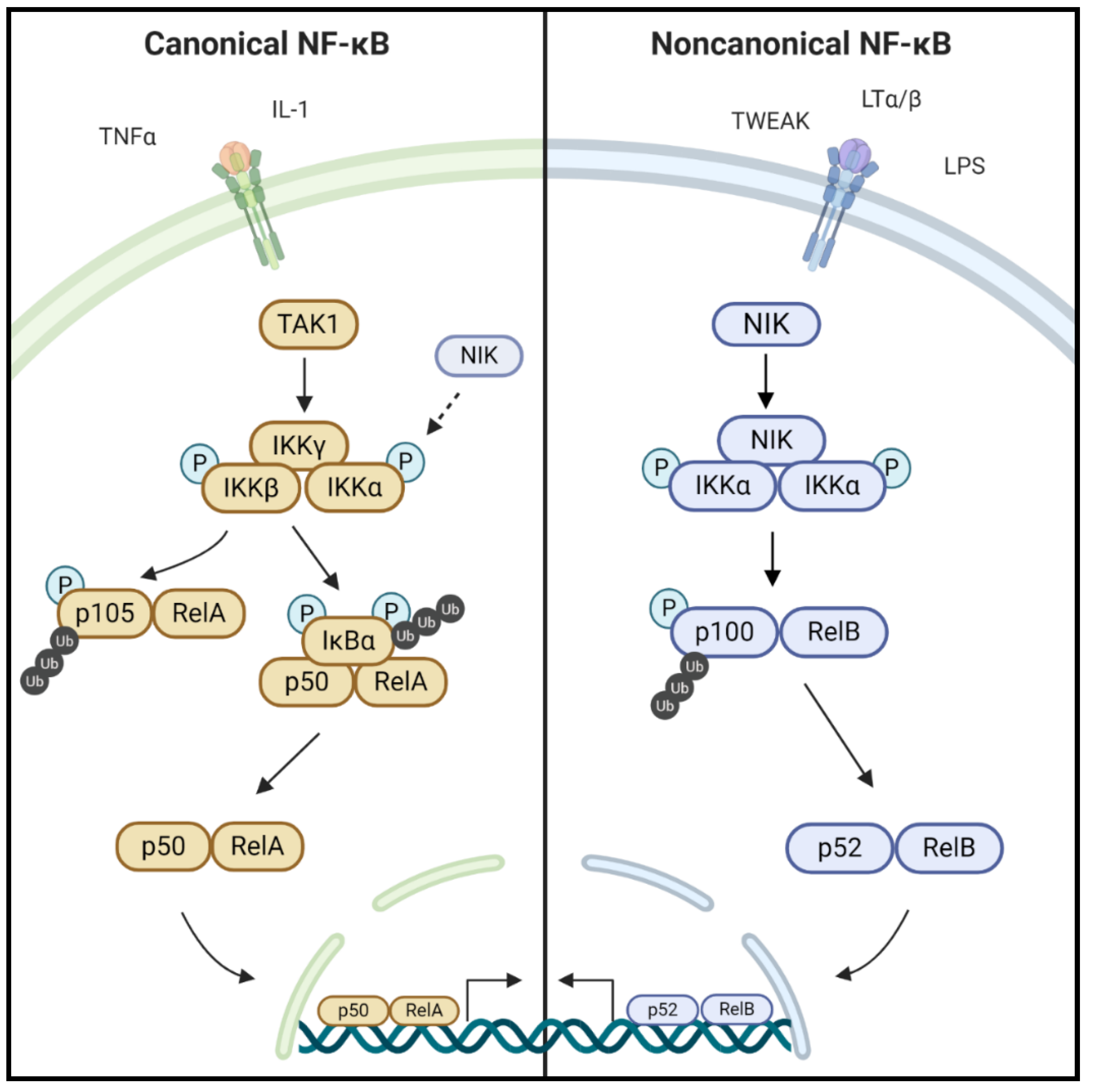
1.1. NF-κB Signaling

Nuclear factor-kappa light chain enhancer of activated B cells (NF-κB) refers to a family of evolutionarily conserved transcription factors that regulate a wide range of biological processes, including cell growth and survival to immunity and inflammation [1]. In mammals, there are five NF-κB family members: RelA (p65), RelB, c-Rel, NF-κB1 (p105/p50) and NF-κB2 (p100/p52). Activation of NF-κB occurs through two distinct signaling pathways, referred to as the canonical and noncanonical NF-κB pathways, and is often triggered by inflammatory, pathogenic, or stress signals (Figure 1) [2,3]. The canonical NF-κB pathway is activated by several inflammatory cytokines, the most well-known being tumor necrosis factor alpha (TNFα). Stimulation of the TNF receptors, TLRs (Toll-like receptors), T-cell receptors, and interleukin receptors typically leads to the activation of this pathway and its downstream transcription factors, RelA/p65 and p50 [1]. The canonical NF-κB pathway is well characterized for having an inhibitor of kappa B kinase (IKK) complex consisting of IKKα, IKKβ, and IKKγ (also known as NEMO) [4]. IKKα and IKKβ are the catalytic units of the complex, and IKKγ is a regulatory subunit that is required for activation of IKKβ. After receptor activation, various kinases such as RIP1 (receptor interacting kinase 1), TAK1 (transforming growth factor beta-activated kinase 1), or TBK1 (TANK-binding kinase) phosphorylate and activate the IKK complex, preferentially on IKKβ [5]. Although constitutive proteasomal processing of p105 generates a subset pool of p50, p105 processing can also be induced through phosphorylation by IKKβ [6,7,8]. Activated IKKβ phosphorylates and induces the proteasomal degradation of IκBα (inhibitory kappa B alpha), which sequesters RelA-p50 complexes in the cytoplasm under basal, unstimulated conditions. Degradation of IκBα releases RelA-p50 complexes to translocate to the nucleus and regulate NF-κB-dependent gene transcription [9].

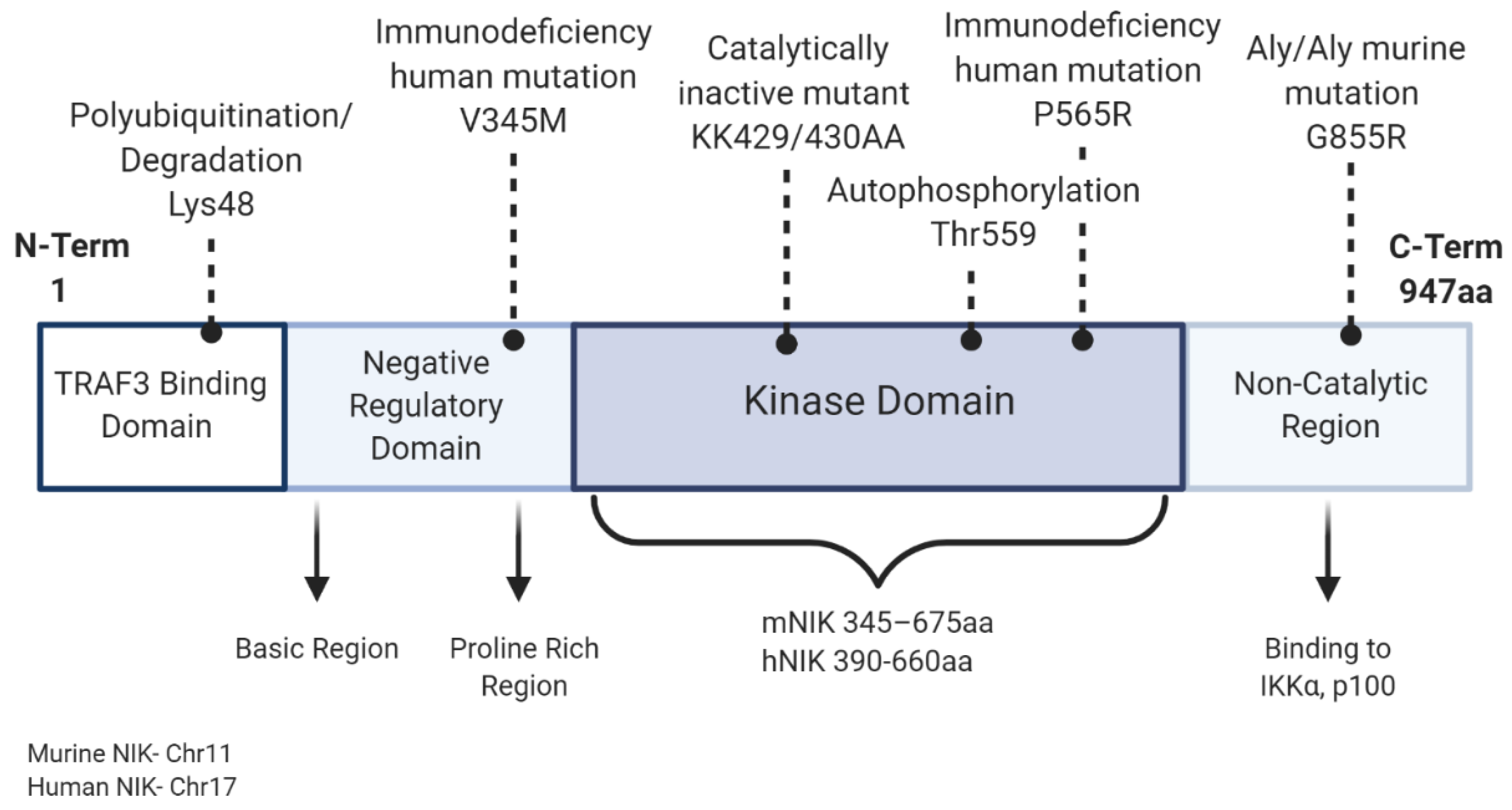
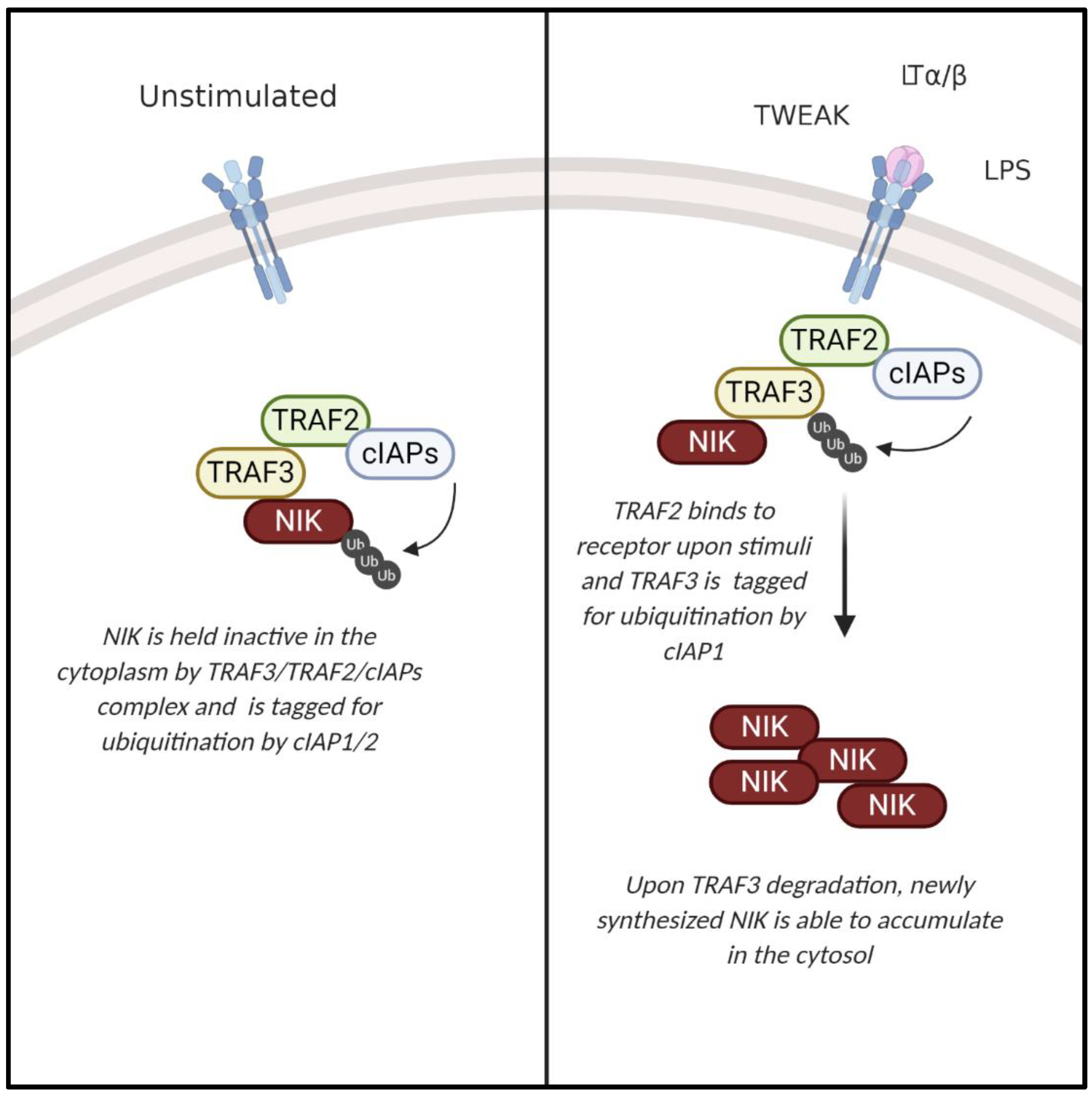
1.2. NIK and the Noncanonical NF-κB Pathway

The noncanonical NF-κB pathway ultimately leads to the activation of RelB-p52 dimers and is activated by a subset of tumor necrosis factor superfamily receptors (TNFSFRs), including TNFSFR12A (Fn14, Tweak receptor), lymphotoxin β receptor (LTβR), B-cell activating factor receptor (BAFF-R), receptor activator of NF-κB (RANK), CD40, and CD27 [10]. NF-κB-inducing kinase (NIK; gene name Map3k14) is a key upstream regulator of the noncanonical NF-κB signaling pathway [11,12]. Though initially identified by its ability to activate the canonical NF-κB pathway, loss of NIK did not impair TNF-induced activation of IKKβ/p65/p50, and NIK was subsequently shown to be essential for activation of noncanonical NF-κB signaling [13,14]. Regulation of NIK activity occurs primarily at the post-translational level. This serine/threonine kinase has four domains including a N-terminal region for TRAF3 binding, a negative regulatory domain (NRD), a core kinase domain, and a C-terminal domain responsible for binding with proteins such as IKKα and p100 (Figure 2) [15,16]. Under steady state conditions, NIK is bound to tumor necrosis factor (TNF) receptor-associated factor 2 and 3 (TRAF2/3) and cellular inhibitor of apoptosis protein 1 and 2 (cIAP1/2), resulting in its ubiquitination and continuous degradation. Stimulation with cytokines, such as CD40L, TWEAK (TNF-like weak inducer of apoptosis), LTα/β (lymphotoxin alpha/beta), or the endotoxin LPS (lipopolysaccharide), and specific binding to their membrane receptors sequesters TRAF2/3, which is then tagged for ubiquitination by cIAP1. Subsequent TRAF3 degradation allows accumulation of newly synthesized NIK within the cell (Figure 3) [1,2,3]. NIK stabilization and accumulation is critical for subsequent activation of the noncanonical NF-κB pathway [17]. Following receptor activation, NIK phosphorylates IKKα at Ser-176 and Ser-180 resulting in its activation and subsequent phosphorylation of p100 [18]. Phosphorylation of p100 induces binding of ubiquitin ligase, β-TrCP (beta transducin repeat containing proteins), and induces partial proteasomal processing to p52 [19,20]. Proteasomal processing of p100 removes the C-term ankyrin repeat domain, which functions similar to the inhibitory nature of mature IκB proteins, holding the noncanonical transcription factor RelB inactive in the cytoplasm. After processing of p100 into p52, p52-RelB translocates to the nucleus to regulate transcription [10] (Figure 1). Although IKKα is the key regulator of p100 phosphorylation and proteolytic processing, NIK functions to activate IKKα and also serves as an adaptor protein to recruit IKKα to p100. Ultimately, the NIK-IKKα-p100 complex is required for generation of mature, transcriptionally active p52 protein [20].
NIK activity is tightly regulated by several levels of negative regulatory feedback mechanisms. For example, activation of the noncanonical NF-κB pathway requires NIK and IKKα, but not NEMO. However, NEMO was found to suppress levels of NIK protein, suggesting a role for NEMO in limiting noncanonical NF-κB signaling [21]. TANK-binding kinase 1 (TBK1) directly interacts with NIK in a signal-dependent manner, inducing NIK phosphorylation on Ser-862, triggering degradation independently of TRAF3, and resulting in class switching to the immunoglobulin A (IgA) isotype [22,23]. Additionally, IKKα also functions to phosphorylate NIK at three residues in the C-terminus (Ser-809, Ser-812, and Ser-815), which are required for degradation of NIK downstream of BAFF-R and LTβR ligation [24]. Notably, IKKα-mediated negative regulation of NIK stability is independent of TRAF-cIAP complex, which limits basal levels of NIK in unstimulated cells. Similarly, the E3 ligase CRL4DCAF2 promoted the polyubiquitination and, consequently, the degradation of NIK in dendritic cells, independent of TRAF3 degradation [25].

2. NIK Regulation of Lymphoid Organogenesis, Immune Cell Development, and Hematopoiesis

NIK is best known for its regulation of the immune system development and function. It plays critical roles in both the development of lymphoid organs, as well as the function of different immune cells, including B-cells, T-cells, macrophages, dendritic cells, and hematopoietic stem cells (HSCs).

2.1. Lymphoid Organogenesis and B-Cell Development

The importance of NIK in lymphoid development was highlighted in the study of alymphoplasia (Map3k14aly/aly) mice, which lacked lymph nodes and Peyer’s patches [26]. This phenotype was linked to a spontaneous, autosomal recessive point mutation in the Map3k14 gene that altered the C-terminal end of NIK, inhibiting its binding to proteins such as IKKα. In addition to lacking lymph nodes and Peyer’s patches, these mice displayed abnormal spleen and thymic structure with a reduction in regions of white blood cell housing specifically in the marginal zone of the spleen and medulla/cortex border of the thymus [14]. Map3k14aly/aly or complete NIK-knockout mice also exhibited stunted ability to mount an immune response with a reduction in B-cells and immunoglobulin development [13,14]. Maturation and viability of B-cells, particularly lymphocytes, are hindered, contributing to abnormal thymic and spleen structure. Many studies have shown that basal levels of immunoglobulins, specifically IgM and IgA, are found to be reduced in mice lacking NIK, and these mice are further unable to upregulate immunoglobulins in response to outside stressors [13,14]. Deletion of NIK in early B-cell development also produced a phenotype of inactive B-cells, particularly in peripheral tissues, further demonstrating the importance of NIK in all stages of B-cell development. The B-cell phenotype in NIK-knockout mice mimics that of BAFF (B-cell activating factor) or BAFF-R-knockout mice showing NIK is a critical downstream component of this signaling pathway to regulate B-cell development [27]. Moreover, consistent with murine models, dysregulation of immune function caused by mutations in NIK has also been identified in human patients with primary immunodeficiencies (see Section 3, below).

2.2. T-Cell Functions

In addition to regulating B-cells, loss of NIK also impacts T-cell maturation and activation. In T-cell-specific NIK-knockout mice, thymocytes underwent normal development, but NIK was observed to be critical for maintaining the homeostasis and function of peripheral T-cells [28]. Specifically, development of memory and regulatory T-cells were significantly reduced in the lymph nodes and spleen, reflected by an increase in naïve T-cells. NIK was shown to regulate the differentiation of T helper type Th1 and Th17 cells, as well as the recall responses of the effector T-cells to protein antigens in vivo [28]. Moreover, T-cell-specific NIK-knockout mice were resistant to the development of a T-cell-dependent autoimmune disease, experimental autoimmune encephalomyelitis [29]. T-cells lacking NIK exhibited reduced antigen responses, cytokine activation, and altered cytoskeletal dynamics compared to wild-type cells [29]. Indeed, reduced antiviral CD8+ T-cell immunity was observed in Map3k14aly/aly mice infected with lymphocytic choriomeningitis virus (LCMV) [30]. In another study, a T cell-intrinsic requirement for NIK was found in graft versus host disease (GVHD), whereby NIK-deficient T-cells transferred to major histocompatibility complex (MHC) class II mismatched mice failed to induce GVHD [31]. NIK has also been shown to have anti-inflammatory functions that are essential for Th2 cell function and is essential for protection from inflammatory disease arising from increased eosinophil accumulation in blood and tissues [32]. Interestingly, NIK-knockout mice spontaneously develop this hypereosinophilic-like disease independently of its phosphorylation and activation of IKKα [32]. Together these findings established NIK as an important cell-intrinsic mediator of T-cell functions in both immune and autoimmune responses.

2.3. Dendritic Cells

NIK was found to play a cell-intrinsic role in regulating intestinal homeostasis, promoting mucosal immunity against pathogens, as well as promoting chronic inflammation associated with inflammatory bowel disease (IBD) [33]. Although NIK was shown to be dispensable for dendritic cell development, a dendritic-cell-specific deletion of NIK altered the profile of commensal bacteria in the gut, specifically the segmented filamentous bacteria and Enterococcus spp. NIK promoted TLR-stimulated IL-23 production, which maintained an increased population of Th17 cells and innate lymphoid cells. These Th17 and innate lymphoid cells are major sources of IL-17, which, in turn, positively regulates IgA secretion and plays important roles in promoting the chronic inflammation associated with inflammatory bowel disease [33]. Interestingly, NIK signaling in dendritic cells was shown to be required for effector T-cell lineages [34], and impaired dendritic cell functions were observed in Map3k14aly/aly mice [35].

2.4. Macrophages

In addition to the aberrant development and paucity of B-cells, including marginal zone B-cells in Map3k14aly/aly mice, a lack of CD169+ macrophages was also observed, whereas other macrophage populations were not affected [30]. As macrophages are potent immunoregulatory cells of the innate immune system that fight pathogens, the loss of CD169+ macrophages was directly correlated with reduced immune activation in response to vesicular stomatitis virus (VSV) or lymphocytic choriomeningitis virus (LCMV) infection [30]. In another study, the sine oculis homeobox (SIX) homologue family transcription factors SIX1 and SIX2, which are developmentally silenced, were shown to function as negative regulators of noncanonical NF-κB signaling. Specifically, in differentiated macrophages, cytokine stimulation or pathogen infection resulted in reactivation of developmentally silenced SIX proteins by NIK-mediated suppression of the ubiquitin proteosome pathway. The net result of SIX1/2 reactivation was attenuation of inflammatory gene expression in response to persistent activation of the noncanonical NF-κB signaling cascade [36]. Together, these studies demonstrate that NIK plays important roles in regulating innate, as well as adaptive, immunity through macrophage functions in the pathogenesis of human disease.

2.5. Hematopoietic Stem Cells (HSCs)

Though normal under steady-state conditions, NIK-induced noncanonical NF-κB signaling has been shown to be essential for maintaining normal hematopoiesis, particularly in response to stressors, including excessive cytokines, chemotherapeutic agents, and hematopoietic transplantation [37]. The absence of NIK results in the dysregulation of hematopoietic stem/progenitor cell self-renewal and expansion capacity resulting from decreased proliferation and increased apoptosis. Therefore, disruption in normal inflammatory responses and the bone marrow microenvironment in NIK-deficient mice are major factors contributing to the development of bone marrow failure after stress [37].

3. NIK in Immune, Inflammatory, and Metabolic Diseases

Pathogen-sensing, immune system signaling, and metabolic signaling pathways are evolutionarily conserved and highly interconnected physiological processes. Indeed, as a central mediator of immune responses, aberrant NIK activation or expression has been linked to several immune and inflammatory, as well as metabolic diseases.

3.1. Immunodeficiency and Autoimmune Disorders

Defects in NIK and noncanonical NF-κB signaling are associated with severe immune deficiencies. Similar to Map3k14aly/aly mice, and other mouse models of NIK deficiency, mutations in NIK have been identified in patients with combined immunodeficiency [38,39]. Patients with homozygous recessive mutations in NIK (NIKPro565Arg) displayed B-cell lymphopenia, decreased frequencies of class-switched memory B-cells, and impaired B-cell survival. Additionally, patients exhibited defects in both follicular helper and memory T-cells populations, as well as natural killer cells [38]. The NIKPro565Arg mutation resulted in impaired IKKα phosphorylation, and similarly, patients with a homozygous recessive NIKVal345Met variant exhibited combined immunodeficiency, resulting from impaired NIK kinase activity [38,39].

3.2. Systemic Lupus Erythematosus

With regard to autoimmune disease, NIK was recently shown to be a potential therapeutic target for treatment of systemic lupus erythematosus (SLE) [40]. Inhibition of NIK using a NIK-specific small molecule inhibitor (NIK SMI1) increased survival in an experimental murine models of lupus by restricting overactive immune cells through a reduction in OX40, BAFF, and CD40 signaling [40].

3.3. Rheumatoid Arthritis

Rheumatoid arthritis (RA), another autoimmune disease characterized by chronic inflammation of the synovial joints, has also been linked to an increased NIK expression or activity. RA synovial fibroblasts secrete pro-inflammatory cytokines and growth factors, creating an abnormal microenvironment that supports activation of NIK and noncanonical NF-κB signaling that drive inflammation [41]. NIK was recently shown to promote inflammatory activation of human endothelial cells by RA synovial fluid and has previously been shown to promote inflammation-induced angiogenesis associated with RA endothelial cells [42]. In a GWAS study, NIK was found to be a genetic marker of RA susceptibility in different ethnic populations [43]. In another study of RA, significantly elevated NIK expression was observed in tertiary lymphoid organs, which are abnormal lymph node-like structures that form in peripheral tissues at sites of high chronic inflammation [44]. Similar to systemic lupus erythematosus, elevated BAFF signaling promotes the inflammatory RA microenvironment and antagonists have shown promising efficacy in the treatment of RA [41,45]. Therefore, as a downstream regulator of BAFF signaling, along with several other cytokine activated receptors, NIK is an apt target for RA therapeutics (see Table 1). Collectively, these data illustrate the nonredundant role for NIK in human immune responses, demonstrating that overactivation or loss-of-function mutations in NIK can cause multiple immune or inflammatory diseases.

3.4. Bone Disorders

Osteoporosis and generalized bone loss are characteristic features associated with both established and early rheumatoid arthritis [56]. RANK and RANK-ligand (RANKL) are key regulators of bone remodeling, and NIK has been shown to regulate osteoclast differentiation through RANK activation [57,58,59]. In one study, transgenic mice expressing constitutively active NIK showed a significant increase in osteoclast formation and increased bone erosion around the joints of the mice, suggesting that targeting NIK may also improve bone loss associated with RA [59]. Indeed, a small-molecule NIK inhibitor, Cpd33, was shown to inhibit osteoclastogenesis and bone resorption, preventing bone loss in ovariectomized mice [50].

3.5. Aging and Cardiovascular Disease

Aging, as well as associated increased inflammation and stress responses, is an important risk factor for several diseases. Recently, the FK506-binding protein 51 (FKBP51/FKBP5), a protein implicated in stress physiology, was found to significantly increase NIK-IKKα signaling to promote inflammation. Moreover, this correlation was strongly associated with increased risk of cardiovascular disease, specifically acute myocardial infarction, which is caused by chronic inflammation.

3.6. Metabolic Disorders

It has become increasingly evident that inflammation is a hallmark feature of metabolic disorders, including obesity and type 2 diabetes. Increased NIK expression has been linked to metabolic disorders associated with increased inflammation such as obesity, insulin resistance, and diabetes. Studies have shown that NIK is hyperactivated in livers of obese mice [60]. Overactivation of NIK in the liver of these obese mice led to glucose intolerance by inducing hyperglycemia through increased CREB-regulated glucagon activity [60]. Obesity-induced glucose intolerance by NIK was also observed in pancreatic β-cells leading to β-cell dysfunction and a decrease in insulin production [61]. Notably, tissue-specific deletion of NIK in the liver protected obese mice from liver inflammation and alcoholic steatosis. Consistent with that finding, pharmacological inhibition of NIK with B022 attenuated ethanol-induced hepatic steatosis and suppression of PPARα and fatty acid oxidation [46,62]. With regard to obesity, increased adiposity induces recruitment of pro-inflammatory M1 macrophages that are classically activated by LPS and INF-γ, and secrete inflammatory cytokines, most commonly TNFα [63]. Thus, stimulation of NF-κB is critical for macrophage activation and leads to subsequent elevation of NF-κB activation in surrounding cells, potentiating insulin resistance and type II diabetes [64]. Inhibition of NF-κB pathways, including NIK itself, is a promising therapeutic intervention to protect against diet-induced metabolic disorders [65].

4. Aberrant NIK Regulation in Cancer

Similar to other inflammatory diseases, cancer is marked by chronic inflammation and aberrant activation of immune signaling pathways. For example, cancerous cells recruit a variety of immune cells including T-cells and macrophages, resulting in elevated levels of proinflammatory cytokines that stimulate activation of NF-κB, promoting several hallmarks of cancer, including survival, proliferation, and metastasis [66]. Aberrant, constitutive activation of canonical NF-κB signaling is a feature of several cancers [67,68]. In these cases, although NF-κB typically functions as a tumor promoter, it can also function as a tumor suppressor [69]. In contrast to the canonical NF-κB pathway, which has been extensively studied in cancer, roles for NIK and the noncanonical NF-κB pathway in cancer are still emerging. Nevertheless, it is evident that similar to canonical NF-κB signaling, dysregulation of NIK and noncanonical NF-κB can lead to the formation and progression of several types of cancer, while in some cases, NIK suppresses tumorigenesis [70]. The initiation and progression of various cancers by NF-κB has been studied by dysregulation of individual subunits or of the pathways as a whole. Cancer models for NIK and the noncanonical NF-κB pathway have been analyzed at various stages from TRAF2/3 or cIAP degradation, to accumulation of NIK itself, to IKKα and its phosphorylation by NIK, or finally by increased expression or translocation of the downstream transcription factor RelB [71]. As with inflammatory and immune diseases, investigation of therapeutic effects of targeting NIK in cancer has been the focus of several studies (Table 1).

4.1. Cancer Stem Cells

Upregulation of NIK mRNA was shown to play an important role in maintaining the population of breast cancer stem cells (CSCs), which possess intrinsic properties of self-renewal and are important for tumor development, recurrence, and metastasis, particularly in the context of therapy resistance [72,73]. Knockdown of NIK reduced expression of breast CSC markers, reduced stem cell clonogenicity, and impaired tumorigenic potential in xenograft breast cancer mouse models [73]. Furthermore, a separate study demonstrated that specific deletion of RANK or IKKα in mouse mammary epithelial cells decreased tumorigenesis and invasive carcinoma [74]. In GBM initiating cells (GICs), it was found that NIK accumulation and noncanonical NF-κB activation was induced through epithelial V-like antigen (Eva1) degradation of TRAF2 and cIAP. This study further showed that the knockdown of RelB inhibited the self-renewal properties of human GICs and abolished tumorigenesis of mouse stem cells in vivo [75]. Increased expression of RelA, RelB, and IKKα was also observed in ovarian CSCs, and knockdown of IKKα decreased stem cell marker expression in those cells [76].

4.2. Leukemias and Lymphomas

As NIK has a crucial role in hematopoiesis, its dysregulation has been well studied in leukemias and lymphomas. Indeed, NIK inhibition has been shown to be a therapeutically efficacious treatment option for immune cell-derived cancers. For example, NIK and the noncanonical NF-κB pathway was shown to be stably activated in Hodgkin lymphoma (HL) cell lines and tumors, and treatment with a NIK small molecule inhibitor, 4H-isoquinoline-1,3-dione, significantly reduced HL cell viability [53]. In Mantel cell lymphoma (MCL), an aggressive B-cell malignancy that is refractory to treatment, NIK was identified as a new therapeutic target, particularly for lymphomas that are resistant to B-cell receptor pathway inhibitors [77]. On the other hand, studies in acute myeloid leukemia (AML) highlight the dual effects of regulating NIK stabilization as a therapeutic approach. AML cells, expressing a common gene fusion mutation in mixed-lineage leukemia 1 (MLL-ALL), exhibit constitutive activation of NIK and the noncanonical NF-κB pathway. Moreover, the stable knockdown of NIK increased the sensitivity of AML cells to chemotherapeutics and promoted apoptosis [78]. In contrast, a separate study demonstrated that activating NIK was beneficial in treating AML. Specifically, this group showed that the MLL-ALL fusion resulted in the stabilization of NIK and mice receiving hematopoietic stem cells transduced with MLL-ALL exhibited a significant delay in the initiation of AML due to increased NIK protein levels [79]. Notably, this myeloid-leukemia suppressive role of NIK differs from its tumor-promoting role in other lymphomas and functions to repress canonical NF-κB signaling, indicating that canonical and noncanonical NF-κB signaling may have opposing roles in AML.
Second mitochondria-derived activator of caspases (SMAC) mimetics target inhibitor of apoptosis proteins (IAPs) and are actively being studied for their chemotherapeutic potential through induction of apoptosis [80,81]. As cIAP1/2 are key ubiquitin ligases that bind NIK, use of SMAC mimetics increases NIK stabilization and activation [82]. As NIK stabilization can lead to canonical NF-κB activation and induction of apoptosis, SMAC mimetics have been used to treat several cancers including AML [83]. However, these effects are context dependent, and many cancer cells are resistant to SMAC-mediated apoptosis [84]. Similar to MML-ALL leukemias described above, in other cancers or cell types, NIK stabilization and activation by SMACs has been shown to protect cells from SMAC-induced death [85] (and see Section 4.5 below).
The use of SMAC mimetics is also being optimized in combinational therapies for treatment of tumors that take advantage of the immune checkpoint blockade. By expressing PD-L1, tumor cells are often able to evade cell death from surrounding T-cells. Upregulation of noncanonical NF-κB was seen in tumor dendritic cells, which increased IL-12 production, improving the synergistic relationship between dendritic cells and CD8+ T-cells. The priming of these T-cells upon noncanonical NF-κB activation and induction of IL-12 in neighboring dendritic cells improved T-cell sensitivity to aPD-1 treatment. This study further showed that the use of a cIAP antagonist on murine tumors increased invading dendritic cells and that chimera mice with NIK-deficient bone marrow had poor response to aPD-1 treatment [86]. Though this study highlighted the importance of the noncanonical NF-κB on the efficacy of tumor invading T-cells, further investigation is needed to understand the effects on the inactivation or overactivation of NIK on other tumor-invading immune cells such as macrophages. Furthermore, though separate studies identified NF-κB proteins, such as RelA, the downstream transcription factor of the canonical NF-κB pathway, as increasing cancer cell resistance to T-cell-induced death or increasing PD-L1 expression in ovarian cancer cells, little is known about the role of the noncanonical NF-κB pathway in regulating this immune checkpoint blockade in cancer cells [87,88].

4.3. Melanoma

Aberrant NIK activation has been observed in several solid cancer types, including melanoma, which is characterized by dysregulation of multiple signaling and tumor suppressor/oncogene pathways, such as BRAF [89]. For example, NIK expression and activity are increased by AKT and ERK signaling in melanoma cells compared with normal epidermal melanocytes, and knockdown of NIK reduced melanoma cell survival through reduction in noncanonical NF-κB pathway activation and expression of pro-survival genes [90,91]. Interestingly, mangiferin (1,3,6,7-tetrahydroxyxanthone-C2-β-D-glucoside), a natural bioactive agent with potent antioxidant, anti-inflammatory, and immunomodulatory effects, was recently identified as a NIK inhibitor [55]. Mangiferin and another NIK inhibitor, N-Acetyl-3-aminopyrazole, were shown to induce apoptosis and inhibit tumor growth and metastasis in a melanoma mouse model.

4.4. Breast Cancer

In addition to regulating breast cancer stem cells, elevated NIK expression was observed in breast carcinoma tissue vs. tumor-adjacent normal tissue surgically resected from human patients, and high NIK expression was associated with clinical stage and patient prognosis [92]. Recent gene co-expression network analysis identified NIK/NF-κB signaling pathway enrichment in chemotherapy-resistant breast carcinoma-associated fibroblasts [93]. However, another study found that expression of canonical and alternative NF-κB proteins are associated with improved relapse-free survival of breast cancer patients [94]. Taken together, these results highlight a critical need to understand how these pathways intersect in specific cellular and disease contexts.

4.5. Brain Cancer

Work from our group has elucidated tumor-promoting functions for NIK in glioma, including aggressive high-grade gliomas, such as glioblastoma, which are notoriously invasive and therapy resistant. We previously demonstrated that NIK promotes glioma invasion through TWEAK and noncanonical NF-κB activation, which induced expression of MMP9 (matrix metalloprotease 9). NIK also induced matrix metalloproteinase 14 (MT1-MMP) upregulation in invadopodia and promoted glioma cell invasion in a manner that was dependent on noncanonical NF-κB, but independent of canonical NF-κB signaling [95]. Additionally, overexpression of NIK significantly induced tumor cell invasion and increased tumor size [96]. Subsequently, we showed that NIK can regulate cancer cell invasion through regulation of mitochondrial dynamics and mitochondrial trafficking at the leading edge of migrating glioma cancer cells [97]. With regard to therapeutics, although the SMAC mimetic and IAP agonist BV6 has demonstrated potential for inducing cell death in a large number of cancers, it was shown to induce accumulation of NIK, resulting in increased cell elongation, migration, and invasion in primary glioblastoma cells [85]. Together with our findings that TWEAK treatment induced NIK expression and promoted NIK-dependent glioblastoma cell invasion, these results suggest that therapy-induced inhibition of NIK should be considered as a potential determinant of treatment efficacy in the context of glioblastoma.

4.6. Other Solid Tumors

NIK overexpression has been observed in human pancreatic cancer samples, where TRAF2 degradation increased NIK stabilization [98]. Furthermore, NIK overactivation was observed to have a role in cell survival and proliferation in pancreatic cancer cell lines, implicating NIK as therapeutic target for growth inhibition [99]. In a human study of patients biopsied with gastric cancer, NIK was not only highly expressed in tumor tissue but it was found to be significantly associated with tumor differentiation and had a higher positive rate in later stages of the cancer [100].

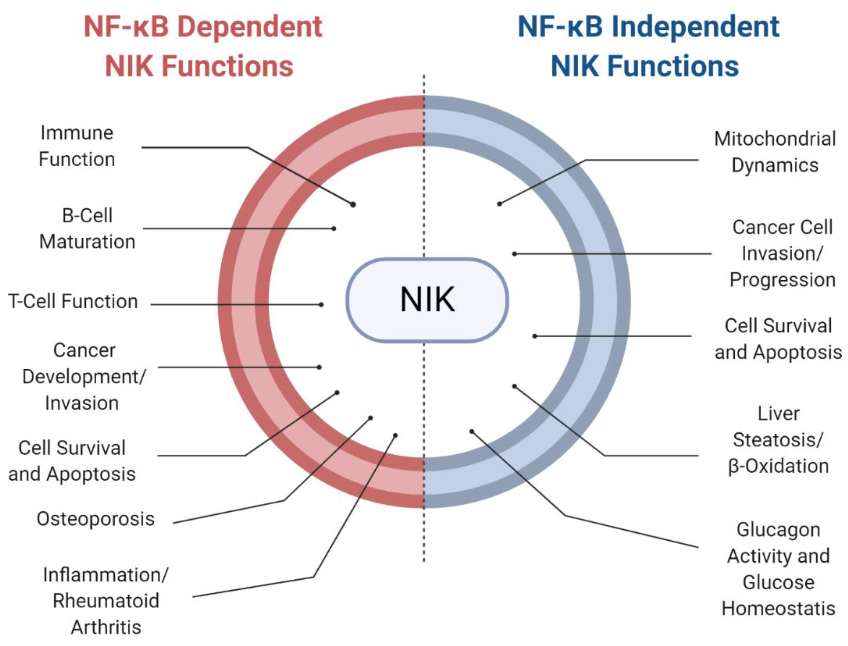
5. NF-κB-Independent Roles for NIK in Disease

Finally, several studies highlighted here have observed NF-κB-independent NIK functions in regulating cellular processes and disease (Figure 4). As previously discussed, NIK induction of liver steatosis was observed through regulation of PPARα phosphorylation and recruitment of ERK1/2 and MEK1/2. NF-κB-independent roles for NIK were observed in the regulation of beta oxidation and inflammation in the liver [46]. NIK was also observed to induce apoptosis independent of the noncanonical NF-κB pathway through phosphorylation of RIP1 [101]. Additionally, NIK independently regulates glucose homeostasis by direct phosphorylation of CREB, increasing its stabilization to regulate glucagon activity [60]. In cancer models, increased NIK activity has been linked to cell survival and tumorigenesis through association with β-catenin and upregulation of pro-survival genes [91]. Furthermore, NIK can regulate mitochondrial function through dynamin-related protein 1 (DRP1) activity, in a manner that is independent of downstream IKK/NF-κB signaling, resulting in amplification of tumorigenic effects and deregulated cancer metabolism [97]. These studies present a strong rationale for specific therapeutic targeting of NIK in various inflammatory diseases and cancer.

6. Conclusions

While much remains to be learned about the full spectrum of NIK’s functions in health and disease, particularly with regard to NF-κB-independent signaling, it is evident that NIK and noncanonical NF-κB signaling are important for many diverse functions of immune cells, inflammatory disease pathogenesis, and malignancies. Although the dual roles that NIK plays in promoting or attenuating cell survival depending on cell context must be an important consideration in therapeutic approaches targeting NIK, small-molecule pharmacological inhibitors of NIK have tremendous potential to be potent stand-alone or combinatorial therapeutics in a wider range of diseases than previously appreciated.

Author Contributions

Conceptualization, R.S.; writing—original draft preparation, K.M.P., writing—review and editing, K.M.P. and R.S.; visualization: K.M.P.; funding acquisition: R.S.; supervision, R.S. All authors have read and agreed to the published version of the manuscript.

Funding

NIH-1R01NS082554 and CPRIT-RP160842 to R.S.

Acknowledgments

BioRender (BioRender.com) software was used for generating figures.

Conflicts of Interest

The authors declare no conflict of interest.

Abbreviations

ALLacute lymphoblastic leukemia
AMLacute myeloid leukemia
Alyalymphoplasia
BAFF/BAFFRB cell activating factor/B cell activating factor receptor
β TrCPbeta transducin repeats containing proteins
CDK4cyclin-dependent kinase 4
cIAP1/2cellular inhibitor of apoptosis protein 1 and 2
CMLchronic myeloid leukemia
Cpd33compound 33
CREBcAMP response element binding
CSCcancer stem cells
DRP1dynamine-related protein 1
ERKextracellular signal regulated kinase
Eva1epithelial V-like antigen 1
FKBP5FK506-binding protein 51 (FKBP51)
GBMglioblastoma multiforme
GICGBM initiating cells
GVHDgraft versus host disease
GWASgenome-wide association study
HLHodgkin lymphoma
hNIKhuman NIK
IC50inhibitory concentration (half of maximal)
IGF-1Rinsulin like growth factor 1 receptor
IBDinflammatory bowel disease
IκBαinhibitory kappa B alpha
IKKαinhibitory kappa B kinase alpha (aka CHUK)
IKKβinhibitory kappa B kinase beta
IKKγinhibitory kappa B kinase gamma (aka NEMO)
ILinterleukin
INF-γinterferon gamma
LCMVlymphocytic choriomeningitis virus
LPSlipopolysaccharide
LTα/βlymphotoxin alpha/beta
MAP3K14mitogen activated protein kinase, kinase, kinase 14 (aka NIK)
MCLMantle cell lymphoma
MHCmajor histocompatibility complex
MLL1mixed-lineage leukemia 1
MMP9matrix metalloproteinase 9
mNIKmouse NIK
MT1-MMPmembrane-type matrix metalloproteinase
NFATc1nuclear factor of activated T-cells 1
NF-κBnuclear factor kappa-light-chain-enhancer of activated B cells
NIKNF-κB-inducing kinase
NRDnegative regulatory domain
OVXovariectomy
PD1programmed cell death protein 1
PPARαperoxisome proliferator-activated receptor alpha
RArheumatoid arthritis
RANKreceptor activator of NF-κB
RANKLreceptor activator of NF-κB ligand
RIP1receptor interacting kinase 1
SIXsine oculis homeobox homologue family transcription factors
SMACsecond mitochondria-derived activator of caspases
SMI1small molecule Inhibitor 1
SPPseveral species
TAK1transforming growth factor beta-activated kinase 1
TBK1TANK-binding kinase 1
TLRToll-like receptor
TNFtumor necrosis factor
TNFSFRtumor necrosis factor superfamily receptor
TRAF2/3TNF-receptor associated factor 2 and 3
TWEAKtumor necrosis factor-like weak inducer of apoptosis
VSVvesicular stomatitis virus

References

  1. Zhang, Q.; Lenardo, M.J.; Baltimore, D. 30 Years of NF-κB: A Blossoming of Relevance to Human Pathobiology. Cell 2017, 168, 37–57. [Google Scholar] [CrossRef] [PubMed] [Green Version]
  2. Bonizzi, G.; Karin, M. The two NF-κB activation pathways and their role in innate and adaptive immunity. Trends Immunol. 2004, 25, 280–288. [Google Scholar] [CrossRef] [PubMed]
  3. Gilmore, T.D. Introduction to NF-kappaB: Players, pathways, perspectives. Oncogene 2006, 25, 6680–6684. [Google Scholar] [CrossRef] [PubMed] [Green Version]
  4. Häcker, H.; Karin, M. Regulation and function of IKK and IKK-related kinases. Sci. STKE Signal Transduct. Knowl. Environ. 2006, 2006, re13. [Google Scholar] [CrossRef]
  5. Israël, A. The IKK complex, a central regulator of NF-kappaB activation. Cold Spring Harb. Perspect. Biol. 2010, 2, a000158. [Google Scholar] [CrossRef] [Green Version]
  6. Salmerón, A.; Janzen, J.; Soneji, Y.; Bump, N.; Kamens, J.; Allen, H.; Ley, S.C. Direct Phosphorylation of NF-κB1 p105 by the IκB Kinase Complex on Serine 927 Is Essential for Signal-induced p105 Proteolysis. J. Biol. Chem. 2001, 276, 22215–22222. [Google Scholar] [CrossRef] [Green Version]
  7. Orian, A.; Gonen, H.; Bercovich, B.; Fajerman, I.; Eytan, E.; Iwai, K.; Schwartz, A.L.; Ciechanover, A. SCFb-TrCP ubiquitin ligase-mediated processing of NF-kB p105 requires phosphorylation of its C-terminus by IkB kinase. EMBO J. 2020, 19, 2580–2591. [Google Scholar] [CrossRef] [Green Version]
  8. Lin, L.; DeMartino, G.N.; Greene, W.C. Cotranslational biogenesis of NF-kappaB p50 by the 26S proteasome. Cell 1998, 92, 819–828. [Google Scholar] [CrossRef] [Green Version]
  9. Baldwin, A.S. The NF-kappa B and I kappa B proteins: New discoveries and insights. Annu. Rev. Immunol. 1996, 14, 649–683. [Google Scholar] [CrossRef] [Green Version]
  10. Sun, S.C. The noncanonical NF-κB pathway. Immunol. Rev. 2012, 246, 125–140. [Google Scholar] [CrossRef] [Green Version]
  11. Sun, S.C. The non-canonical NF-κB pathway in immunity and inflammation. Nat. Rev. Immunol. 2017, 17, 545–558. [Google Scholar] [CrossRef] [PubMed]
  12. Cildir, G.; Low, K.C.; Tergaonkar, V. Noncanonical NF-κB Signaling in Health and Disease. Trends Mol. Med. 2016, 22, 414–429. [Google Scholar] [CrossRef] [PubMed]
  13. Yin, L.; Wu, L.; Wesche, H.; Arthur, C.D.; White, J.M.; Goeddel, D.V.; Schreiber, R.D. Defective lymphotoxin-beta receptor-induced NF-kappaB transcriptional activity in NIK-deficient mice. Sci. N. Y. 2001, 291, 2162–2165. [Google Scholar] [CrossRef] [PubMed]
  14. Shinkura, R.; Kitada, K.; Matsuda, F.; Tashiro, K.; Ikuta, K.; Suzuki, M.; Kogishi, K.; Serikawa, T.; Honjo, T. Alymphoplasia is caused by a point mutation in the mouse gene encoding Nf-kappa b-inducing kinase. Nat. Genet. 1999, 22, 74–77. [Google Scholar] [CrossRef]
  15. de Leon-Boenig, G.; Bowman, K.K.; Feng, J.A.; Crawford, T.; Everett, C.; Franke, Y.; Oh, A.; Stanley, M.; Staben, S.T.; Starovasnik, M.A.; et al. The crystal structure of the catalytic domain of the NF-κB inducing kinase reveals a narrow but flexible active site. Structure 2012, 20, 1704–1714. [Google Scholar] [CrossRef] [Green Version]
  16. Liu, J.; Sudom, A.; Min, X.; Cao, Z.; Gao, X.; Ayres, M.; Lee, F.; Cao, P.; Johnstone, S.; Plotnikova, O.; et al. Structure of the nuclear factor κB-inducing kinase (NIK) kinase domain reveals a constitutively active conformation. J. Biol. Chem. 2012, 287, 27326–27334. [Google Scholar] [CrossRef] [Green Version]
  17. Qing, G.; Qu, Z.; Xiao, G. Stabilization of basally translated NF-kappaB-inducing kinase (NIK) protein functions as a molecular switch of processing of NF-kappaB2 p100. J. Biol. Chem. 2005, 280, 40578–40582. [Google Scholar] [CrossRef] [Green Version]
  18. Ling, L.; Cao, Z.; Goeddel, D.V. NF-kappaB-inducing kinase activates IKK-alpha by phosphorylation of Ser-176. Proc. Natl. Acad. Sci. USA 1998, 95, 3792–3797. [Google Scholar] [CrossRef] [Green Version]
  19. Heusch, M.; Lin, L.; Geleziunas, R.; Greene, W.C. The generation of nfkb2 p52: Mechanism and efficiency. Oncogene 1999, 18, 6201–6208. [Google Scholar] [CrossRef] [Green Version]
  20. Xiao, G.; Fong, A.; Sun, S.C. Induction of p100 Processing by NF-κB-inducing Kinase Involves Docking IκB Kinase α (IKKα) to p100 and IKKα-mediated Phosphorylation. J. Biol. Chem. 2004, 279, 30099–30105. [Google Scholar] [CrossRef] [Green Version]
  21. Gray, C.M.; Remouchamps, C.; McCorkell, K.A.; Solt, L.A.; Dejardin, E.; Orange, J.S.; May, M.J. Noncanonical NF-κB signaling is limited by classical NF-κB activity. Sci. Signal. 2014, 7, ra13. [Google Scholar] [CrossRef] [PubMed] [Green Version]
  22. Bram, R.J. TBK1 suppression of IgA in the NIK of time. Nat. Immunol. 2012, 13, 1027–1029. [Google Scholar] [CrossRef] [PubMed]
  23. Jin, J.; Xiao, Y.; Chang, J.H.; Yu, J.; Hu, H.; Starr, R.; Brittain, G.C.; Chang, M.; Cheng, X.; Sun, S.C. The kinase TBK1 controls IgA class switching by negatively regulating noncanonical NF-κB signaling. Nat. Immunol. 2012, 13, 1101–1109. [Google Scholar] [CrossRef] [Green Version]
  24. Razani, B.; Zarnegar, B.; Ytterberg, A.J.; Shiba, T.; Dempsey, P.W.; Ware, C.F.; Loo, J.A.; Cheng, G. Negative Feedback in Noncanonical NF- B Signaling Modulates NIK Stability Through IKK -Mediated Phosphorylation. Sci. Signal. 2010, 3, ra41. [Google Scholar] [CrossRef] [PubMed] [Green Version]
  25. Huang, T.; Gao, Z.; Zhang, Y.; Fan, K.; Wang, F.; Li, Y.; Zhong, J.; Fan, H.Y.; Cao, Q.; Zhou, J.; et al. CRL4DCAF2 negatively regulates IL-23 production in dendritic cells and limits the development of psoriasis. J. Exp. Med. 2018, 215, 1999–2017. [Google Scholar] [CrossRef] [PubMed] [Green Version]
  26. Miyawaki, S.; Nakamura, Y.; Suzuka, H.; Koba, M.; Yasumizu, R.; Ikehara, S.; Shibata, Y. A new mutation, aly, that induces a generalized lack of lymph nodes accompanied by immunodeficiency in mice. Eur. J. Immunol. 1994, 24, 429–434. [Google Scholar] [CrossRef] [PubMed]
  27. Myles, A.; Cancro, M.P. The NIK of time for B cells. Eur. J. Immunol. 2016, 46, 547–551. [Google Scholar] [CrossRef] [Green Version]
  28. Li, Y.; Wang, H.; Zhou, X.; Xie, X.; Chen, X.; Jie, Z.; Zou, Q.; Hu, H.; Zhu, L.; Cheng, X.; et al. Cell intrinsic role of NF-κB-inducing kinase in regulating T cell-mediated immune and autoimmune responses. Sci. Rep. 2016, 6, 22115. [Google Scholar] [CrossRef] [Green Version]
  29. Lacher, S.M.; Thurm, C.; Distler, U.; Mohebiany, A.N.; Israel, N.; Kitic, M.; Ebering, A.; Tang, Y.; Klein, M.; Wabnitz, G.H.; et al. NF-κB inducing kinase (NIK) is an essential post-transcriptional regulator of T-cell activation affecting F-actin dynamics and TCR signaling. J. Autoimmun. 2018. [Google Scholar] [CrossRef]
  30. Hamdan, T.A.; Bhat, H.; Cham, L.B.; Adomati, T.; Lang, J.; Li, F.; Murtaza, A.; Hardt, C.; Lang, P.A.; Duhan, V.; et al. Map3k14 as a Regulator of Innate and Adaptive Immune Response during Acute Viral Infection. Pathogens 2020, 9, 96. [Google Scholar] [CrossRef] [Green Version]
  31. Murray, S.E.; Polesso, F.; Rowe, A.M.; Basak, S.; Koguchi, Y.; Toren, K.G.; Hoffmann, A.; Parker, D.C. NF-κB–inducing kinase plays an essential T cell–intrinsic role in graft-versus-host disease and lethal autoimmunity in mice. J. Clin. Investig. 2011, 121, 4775. [Google Scholar] [CrossRef] [PubMed]
  32. Häcker, H.; Chi, L.; Rehg, J.E.; Redecke, V. NIK Prevents the Development of Hypereosinophilic Syndrome-like Disease in Mice Independent of IKKα Activation. J. Immunol. 2012, 188, 4602–4610. [Google Scholar] [CrossRef] [PubMed] [Green Version]
  33. Jie, Z.; Yang, J.Y.; Gu, M.; Wang, H.; Xie, X.; Li, Y.; Liu, T.; Zhu, L.; Shi, J.; Zhang, L.; et al. NIK signaling axis regulates dendritic cell function in intestinal immunity and homeostasis. Nat. Immunol. 2018, 19, 1224–1235. [Google Scholar] [CrossRef] [PubMed]
  34. Hofmann, J.; Mair, F.; Greter, M.; Schmidt-Supprian, M.; Becher, B. NIK signaling in dendritic cells but not in T cells is required for the development of effector T cells and cell-mediated immune responses. J. Exp. Med. 2011, 208, 1917–1929. [Google Scholar] [CrossRef] [PubMed]
  35. Tamura, C.; Nakazawa, M.; Kasahara, M.; Hotta, C.; Yoshinari, M.; Sato, F.; Minami, M. Impaired function of dendritic cells in alymphoplasia (aly/aly) mice for expansion of CD25+CD4+ regulatory T cells. Autoimmunity 2006, 39, 445–453. [Google Scholar] [CrossRef] [PubMed]
  36. Liu, Z.; Mar, K.B.; Hanners, N.W.; Perelman, S.S.; Kanchwala, M.; Xing, C.; Schoggins, J.W.; Alto, N.M. A NIK–SIX signalling axis controls inflammation by targeted silencing of non-canonical NF-κB. Nature 2019, 22, 414–426. [Google Scholar] [CrossRef]
  37. González-Murillo, Á.; Fernández, L.; Baena, S.; Melen, G.J.; Sánchez, R.; Sánchez-Valdepeñas, C.; Segovia, J.C.; Liou, H.C.; Schmid, R.; Madero, L.; et al. The NFKB Inducing Kinase Modulates Hematopoiesis During Stress. Stem Cells 2015, 33, 2825–2837. [Google Scholar] [CrossRef] [PubMed] [Green Version]
  38. Willmann, K.L.; Klaver, S.; Doğu, F.; Santos-Valente, E.; Garncarz, W.; Bilic, I.; Mace, E.; Salzer, E.; Conde, C.D.; Sic, H.; et al. Biallelic loss-of-function mutation in NIK causes a primary immunodeficiency with multifaceted aberrant lymphoid immunity. Nat. Commun. 2014, 5, 5360. [Google Scholar] [CrossRef]
  39. Schlechter, N.; Glanzmann, B.; Hoal, E.G.; Schoeman, M.; Petersen, B.S.; Franke, A.; Lau, Y.L.; Urban, M.; van Helden, P.D.; Esser, M.M.; et al. Exome Sequencing Identifies a Novel MAP3K14 Mutation in Recessive Atypical Combined Immunodeficiency. Front. Immunol. 2017, 8, 1624. [Google Scholar] [CrossRef] [Green Version]
  40. Brightbill, H.D.; Suto, E.; Blaquiere, N.; Ramamoorthi, N.; Sujatha-Bhaskar, S.; Gogol, E.B.; Castanedo, G.M.; Jackson, B.T.; Kwon, Y.C.; Haller, S.; et al. NF-κB inducing kinase is a therapeutic target for systemic lupus erythematosus. Nat. Commun. 2018, 9, 179. [Google Scholar] [CrossRef]
  41. Noort, A.R.; Tak, P.P.; Tas, S.W. Non-canonical NF-κB signaling in rheumatoid arthritis: Dr Jekyll and Mr Hyde? Arthritis Res. Ther. 2015, 17, 15. [Google Scholar] [CrossRef] [PubMed] [Green Version]
  42. Kucharzewska, P.; Maracle, C.X.; Jeucken, K.C.M.; van Hamburg, J.P.; Israelsson, E.; Furber, M.; Tas, S.W.; Olsson, H.K. NIK–IKK complex interaction controls NF-κB-dependent inflammatory activation of endothelium in response to LTβR ligation. J. Cell Sci. 2019, 132, jcs225615. [Google Scholar] [CrossRef] [PubMed] [Green Version]
  43. Arya, R.; del Rincon, I.; Farook, V.S.; Restrepo, J.F.; Winnier, D.A.; Fourcaudot, M.J.; Battafarano, D.F.; de Almeida, M.; Kumar, S.; Curran, J.E.; et al. Genetic Variants Influencing Joint Damage in Mexican Americans and European Americans With Rheumatoid Arthritis: Genetic Variants Influencing Joint Damage. Genet. Epidemiol. 2015, 39, 678–688. [Google Scholar] [CrossRef] [PubMed]
  44. Noort, A.R.; van Zoest, K.P.M.; van Baarsen, L.G.; Maracle, C.X.; Helder, B.; Papazian, N.; Romera-Hernandez, M.; Tak, P.P.; Cupedo, T.; Tas, S.W. Tertiary Lymphoid Structures in Rheumatoid Arthritis. Am. J. Pathol. 2015, 185, 1935–1943. [Google Scholar] [CrossRef] [PubMed]
  45. Zhou, B.; Zhang, H.; Su, X.; Luo, Y.; Li, X.; Yu, C.; Xie, Q.; Xia, X.; He, G.; Yang, L. Therapeutic effects of a novel BAFF blocker on arthritis. Signal Transduct. Target. Ther. 2019, 4, 19. [Google Scholar] [CrossRef] [PubMed] [Green Version]
  46. Li, Y.; Chen, M.; Zhou, Y.; Tang, C.; Zhang, W.; Zhong, Y.; Chen, Y.; Zhou, H.; Sheng, L. NIK links inflammation to hepatic steatosis by suppressing PPARα in alcoholic liver disease. Theranostics 2020, 10, 3579–3593. [Google Scholar] [CrossRef]
  47. Ren, X.; Li, X.; Jia, L.; Chen, D.; Hou, H.; Rui, L.; Zhao, Y.; Chen, Z. A small-molecule inhibitor of NF-κB-inducing kinase (NIK) protects liver from toxin-induced inflammation, oxidative stress, and injury. FASEB J. 2017, 31, 711–718. [Google Scholar] [CrossRef] [Green Version]
  48. Li, Z.; Li, X.; Su, M.B.; Gao, L.X.; Zhou, Y.B.; Yuan, B.; Lyu, X.; Yan, Z.; Hu, C.; Zhang, H.; et al. Discovery of a Potent and Selective NF-κB-Inducing Kinase (NIK) Inhibitor That Has Anti-inflammatory Effects in Vitro and in Vivo. J. Med. Chem. 2020, 63, 4388–4407. [Google Scholar] [CrossRef]
  49. Blaquiere, N.; Castanedo, G.M.; Burch, J.D.; Berezhkovskiy, L.M.; Brightbill, H.; Brown, S.; Chan, C.; Chiang, P.C.; Crawford, J.J.; Dong, T.; et al. Scaffold-Hopping Approach To Discover Potent, Selective, and Efficacious Inhibitors of NF-κB Inducing Kinase. J. Med. Chem. 2018, 61, 6801–6813. [Google Scholar] [CrossRef]
  50. Takakura, N.; Matsuda, M.; Khan, M.; Hiura, F.; Aoki, K.; Hirohashi, Y.; Mori, K.; Yasuda, H.; Hirata, M.; Kitamura, C.; et al. A novel inhibitor of NF-κB-inducing kinase prevents bone loss by inhibiting osteoclastic bone resorption in ovariectomized mice. Bone 2020, 135, 115316. [Google Scholar] [CrossRef]
  51. Cheng, G.; Mei, X.; Yan, Y.; Chen, J.; Zhang, B.; Li, J.; Dong, X.; Lin, N.; Zhou, Y. Identification of new NIK inhibitors by discriminatory analysis-based molecular docking and biological evaluation. Arch. Pharm. 2019, 352, 1800374. [Google Scholar] [CrossRef] [PubMed]
  52. Mortier, J.; Masereel, B.; Remouchamps, C.; Ganeff, C.; Piette, J.; Frederick, R. NF-kappaB inducing kinase (NIK) inhibitors: Identification of new scaffolds using virtual screening. Bioorg. Med. Chem. Lett. 2010, 20, 4515–4520. [Google Scholar] [CrossRef] [PubMed]
  53. Ranuncolo, S.M.; Pittaluga, S.; Evbuomwan, M.O.; Jaffe, E.S.; Lewis, B.A. Hodgkin lymphoma requires stabilized NIK and constitutive RelB expression for survival. Blood 2012, 120, 3756–3763. [Google Scholar] [CrossRef] [Green Version]
  54. Pippione, A.C.; Sainas, S.; Federico, A.; Lupino, E.; Piccinini, M.; Kubbutat, M.; Contreras, J.M.; Morice, C.; Barge, A.; Ducime, A.; et al. N-Acetyl-3-aminopyrazoles block the non-canonical NF-kB cascade by selectively inhibiting NIK †Electronic supplementary information (ESI) available: Additional biochemical data, chemistry, NMR characterization of final compounds, and biochemical protocols. MedChemComm 2018, 9, 963–968. [Google Scholar] [CrossRef] [PubMed]
  55. Takeda, T.; Tsubaki, M.; Sakamoto, K.; Ichimura, E.; Enomoto, A.; Suzuki, Y.; Itoh, T.; Imano, M.; Tanabe, G.; Muraoka, O.; et al. Mangiferin, a novel nuclear factor kappa B-inducing kinase inhibitor, suppresses metastasis and tumor growth in a mouse metastatic melanoma model. Toxicol. Appl. Pharmacol. 2016, 306, 105–112. [Google Scholar] [CrossRef]
  56. Gough, A.K.S.; Emery, P.; Holder, R.L.; Lilley, J.; Eyre, S. Generalised bone loss in patients with early rheumatoid arthritis. Lancet 1994, 344, 23–27. [Google Scholar] [CrossRef]
  57. Zeng, R.; Faccio, R.; Novack, D.V. Alternative NF-κB Regulates RANKL-Induced Osteoclast Differentiation and Mitochondrial Biogenesis via Independent Mechanisms. J. Bone Miner. Res. Off. J. Am. Soc. Bone Miner. Res. 2015, 30, 2287–2299. [Google Scholar] [CrossRef] [Green Version]
  58. Aya, K.; Alhawagri, M.; Hagen-Stapleton, A.; Kitaura, H.; Kanagawa, O.; Veis Novack, D. NF-κB–inducing kinase controls lymphocyte and osteoclast activities in inflammatory arthritis. J. Clin. Investig. 2005, 115, 1848–1854. [Google Scholar] [CrossRef]
  59. Yang, C.; McCoy, K.; Davis, J.L.; Schmidt-Supprian, M.; Sasaki, Y.; Faccio, R.; Novack, D.V. NIK Stabilization in Osteoclasts Results in Osteoporosis and Enhanced Inflammatory Osteolysis. PLoS ONE 2010, 5, e15383. [Google Scholar] [CrossRef]
  60. Sheng, L.; Zhou, Y.; Chen, Z.; Ren, D.; Cho, K.W.; Jiang, L.; Shen, H.; Sasaki, Y.; Rui, L. NF-κB–inducing kinase (NIK) promotes hyperglycemia and glucose intolerance in obesity by augmenting glucagon action. Nat. Med. 2012, 18, 943–949. [Google Scholar] [CrossRef] [Green Version]
  61. Malle, E.K.; Zammit, N.W.; Walters, S.N.; Koay, Y.C.; Wu, J.; Tan, B.M.; Villanueva, J.E.; Brink, R.; Loudovaris, T.; Cantley, J.; et al. Nuclear factor κB-inducing kinase activation as a mechanism of pancreatic β cell failure in obesity. J. Exp. Med. 2015, 212, 1239–1254. [Google Scholar] [CrossRef] [PubMed]
  62. Liu, Y.; Sheng, L.; Xiong, Y.; Shen, H.; Liu, Y.; Rui, L. Liver NF-κB-Inducing Kinase Promotes Liver Steatosis and Glucose Counterregulation in Male Mice With Obesity. Endocrinology 2017, 158, 1207–1216. [Google Scholar] [CrossRef] [PubMed]
  63. Russo, L.; Lumeng, C.N. Properties and functions of adipose tissue macrophages in obesity. Immunology 2018, 155, 407–417. [Google Scholar] [CrossRef] [PubMed]
  64. Dorrington, M.G.; Fraser, I.D.C. NF-κB Signaling in Macrophages: Dynamics, Crosstalk, and Signal Integration. Front. Immunol. 2019, 10, 705. [Google Scholar] [CrossRef]
  65. Zeng, T.; Zhou, J.; He, L.; Zheng, J.; Chen, L.; Wu, C.; Xia, W. Blocking Nuclear Factor-Kappa B Protects against Diet-Induced Hepatic Steatosis and Insulin Resistance in Mice. PLoS ONE 2016, 11, e0149677. [Google Scholar] [CrossRef]
  66. Hoesel, B.; Schmid, J.A. The complexity of NF-κB signaling in inflammation and cancer. Mol. Cancer 2013, 12, 86. [Google Scholar] [CrossRef] [Green Version]
  67. Bassères, D.S.; Baldwin, A.S. Nuclear factor-κB and inhibitor of κB kinase pathways in oncogenic initiation and progression. Oncogene 2006, 25, 6817–6830. [Google Scholar] [CrossRef] [Green Version]
  68. Taniguchi, K.; Karin, M. NF-κB, inflammation, immunity and cancer: Coming of age. Nat. Rev. Immunol. 2018, 18, 309–324. [Google Scholar] [CrossRef]
  69. Xia, Y.; Shen, S.; Verma, I.M. NF-κB, an active player in human cancers. Cancer Immunol. Res. 2014, 2, 823–830. [Google Scholar] [CrossRef] [Green Version]
  70. Maubach, G.; Feige, M.H.; Lim, M.C.C.; Naumann, M. NF-kappaB-inducing kinase in cancer. Biochim. Biophys. Acta BBA Rev. Cancer 2019, 1871, 40–49. [Google Scholar] [CrossRef]
  71. Kaltschmidt, B.; Greiner, J.F.W.; Kadhim, H.M.; Kaltschmidt, C. Subunit-Specific Role of NF-κB in Cancer. Biomedicines 2018, 6, 44. [Google Scholar] [CrossRef] [PubMed] [Green Version]
  72. Pattabiraman, D.R.; Weinberg, R.A. Tackling the cancer stem cells—What challenges do they pose? Nat. Rev. Drug Discov. 2014, 13, 497–512. [Google Scholar] [CrossRef] [PubMed] [Green Version]
  73. Vazquez-Santillan, K.; Melendez-Zajgla, J.; Jimenez-Hernandez, L.E.; Gaytan-Cervantes, J.; Muñoz-Galindo, L.; Piña-Sanchez, P.; Martinez-Ruiz, G.; Torres, J.; Garcia-Lopez, P.; Gonzalez-Torres, C.; et al. NF-kappaΒ-inducing kinase regulates stem cell phenotype in breast cancer. Sci. Rep. 2016, 6, 37340. [Google Scholar] [CrossRef] [PubMed] [Green Version]
  74. Schramek, D.; Leibbrandt, A.; Sigl, V.; Kenner, L.; Pospisilik, J.A.; Lee, H.J.; Hanada, R.; Joshi, P.A.; Aliprantis, A.; Glimcher, L.; et al. Osteoclast differentiation factor RANKL controls development of progestin-driven mammary cancer. Nature 2010, 468, 98–102. [Google Scholar] [CrossRef] [Green Version]
  75. Ohtsu, N.; Nakatani, Y.; Yamashita, D.; Ohue, S.; Ohnishi, T.; Kondo, T. Eva1 Maintains the Stem-like Character of Glioblastoma-Initiating Cells by Activating the Noncanonical NF- B Signaling Pathway. Cancer Res. 2016, 76, 171–181. [Google Scholar] [CrossRef] [Green Version]
  76. Gonzalez-Torres, C.; Gaytan-Cervantes, J.; Vazquez-Santillan, K.; Mandujano-Tinoco, E.A.; Ceballos-Cancino, G.; Garcia-Venzor, A.; Zampedri, C.; Sanchez-Maldonado, P.; Mojica-Espinosa, R.; Jimenez-Hernandez, L.E.; et al. NF-κB Participates in the Stem Cell Phenotype of Ovarian Cancer Cells. Arch. Med. Res. 2017, 48, 343–351. [Google Scholar] [CrossRef]
  77. Rahal, R.; Frick, M.; Romero, R.; Korn, J.M.; Kridel, R.; Chun Chan, F.; Meissner, B.; Bhang, H.; Ruddy, D.; Kauffmann, A.; et al. Pharmacological and genomic profiling identifies NF-κB-targeted treatment strategies for mantle cell lymphoma. Nat. Med. 2014, 20, 87–92. [Google Scholar] [CrossRef]
  78. Studencka-Turski, M.; Maubach, G.; Feige, M.H.; Naumann, M. Constitutive activation of nuclear factor kappa B-inducing kinase counteracts apoptosis in cells with rearranged mixed lineage leukemia gene. Leukemia 2018, 32, 2498–2501. [Google Scholar] [CrossRef]
  79. Xiu, Y.; Dong, Q.; Li, Q.; Li, F.; Borcherding, N.; Zhang, W.; Boyce, B.; Xue, H.; Zhao, C. Stabilization of NF-κB-Inducing Kinase Suppresses MLL-AF9-Induced Acute Myeloid Leukemia. Cell Rep. 2018, 22, 350–358. [Google Scholar] [CrossRef]
  80. Wu, H.; Tschopp, J.; Lin, S.C. Smac mimetics and TNFalpha: A dangerous liaison? Cell 2007, 131, 655–658. [Google Scholar] [CrossRef] [Green Version]
  81. Morrish, E.; Brumatti, G.; Silke, J. Future Therapeutic Directions for Smac-Mimetics. Cells 2020, 9, 406. [Google Scholar] [CrossRef] [PubMed] [Green Version]
  82. Bai, L.; Smith, D.C.; Wang, S. Small-Molecule SMAC Mimetics as New Cancer Therapeutics. Pharmacol. Ther. 2014, 144, 82–95. [Google Scholar] [CrossRef] [PubMed] [Green Version]
  83. Lueck, S.C.; Russ, A.C.; Botzenhardt, U.; Schlenk, R.F.; Zobel, K.; Deshayes, K.; Vucic, D.; Döhner, H.; Döhner, K.; Fulda, S.; et al. Smac mimetic induces cell death in a large proportion of primary acute myeloid leukemia samples, which correlates with defined molecular markers. Oncotarget 2016, 7, 49539–49551. [Google Scholar] [CrossRef] [PubMed] [Green Version]
  84. Cheung, H.H.; St Jean, M.; Beug, S.T.; Lejmi-Mrad, R.; LaCasse, E.; Baird, S.D.; Stojdl, D.F.; Screaton, R.A.; Korneluk, R.G. SMG1 and NIK regulate apoptosis induced by Smac mimetic compounds. Cell Death Dis. 2011, 2, e146. [Google Scholar] [CrossRef] [PubMed]
  85. Tchoghandjian, A.; Jennewein, C.; Eckhardt, I.; Rajalingam, K.; Fulda, S. Identification of non-canonical NF-κB signaling as a critical mediator of Smac mimetic-stimulated migration and invasion of glioblastoma cells. Cell Death Dis. 2013, 4, e564. [Google Scholar] [CrossRef] [PubMed] [Green Version]
  86. Garris, C.S.; Arlauckas, S.P.; Kohler, R.H.; Trefny, M.P.; Garren, S.; Piot, C.; Engblom, C.; Pfirschke, C.; Siwicki, M.; Gungabeesoon, J.; et al. Successful anti-PD-1 cancer immunotherapy requires T cell-dendritic cell crosstalk involving the cytokines IFN-γ and IL-12. Immunity 2018, 49, 1148–1161. [Google Scholar] [CrossRef] [Green Version]
  87. Pan, D.; Kobayashi, A.; Jiang, P.; de Andrade, L.F.; Tay, R.E.; Luoma, A.M.; Tsoucas, D.; Qiu, X.; Lim, K.; Rao, P.; et al. A major chromatin regulator determines resistance of tumor cells to T cell–mediated killing. Science 2018, 359, 770–775. [Google Scholar] [CrossRef] [Green Version]
  88. Peng, J.; Hamanishi, J.; Matsumura, N.; Abiko, K.; Murat, K.; Baba, T.; Yamaguchi, K.; Horikawa, N.; Hosoe, Y.; Murphy, S.K.; et al. Chemotherapy Induces Programmed Cell Death-Ligand 1 Overexpression via the Nuclear Factor- B to Foster an Immunosuppressive Tumor Microenvironment in Ovarian Cancer. Cancer Res. 2015, 75, 5034–5045. [Google Scholar] [CrossRef] [Green Version]
  89. Davies, H.; Bignell, G.R.; Cox, C.; Stephens, P.; Edkins, S.; Clegg, S.; Teague, J.; Woffendin, H.; Garnett, M.J.; Bottomley, W.; et al. Mutations of the BRAF gene in human cancer. Nature 2002, 417, 949–954. [Google Scholar] [CrossRef]
  90. Dhawan, P.; Richmond, A. A novel NF-kappa B-inducing kinase-MAPK signaling pathway up-regulates NF-kappa B activity in melanoma cells. J. Biol. Chem. 2002, 277, 7920–7928. [Google Scholar] [CrossRef] [Green Version]
  91. Thu, Y.M.; Su, Y.; Yang, J.; Splittgerber, R.; Na, S.; Boyd, A.; Mosse, C.; Simons, C.; Richmond, A. NF-κB inducing kinase (NIK) modulates melanoma tumorigenesis by regulating expression of pro-survival factors through the β-catenin pathway. Oncogene 2012, 31, 2580–2592. [Google Scholar] [CrossRef] [PubMed] [Green Version]
  92. Zhang, X.; Wang, Y.; Mao, Z.; Huang, D.; Zhou, J.; Wang, X. Expression of NF-κB-inducing kinase in breast carcinoma tissue and its clinical significance. Int. J. Clin. Exp. Pathol. 2015, 8, 14824–14829. [Google Scholar] [PubMed]
  93. Xu, Y.; Zhang, Z.; Zhang, L.; Zhang, C. Novel module and hub genes of distinctive breast cancer associated fibroblasts identified by weighted gene co-expression network analysis. Breast Cancer 2020, 27, 1017–1028. [Google Scholar] [CrossRef] [PubMed]
  94. Differential Impact of Classical and Non-Canonical NF-κB Pathway-Related Gene Expression on the Survival of Breast Cancer Patients. Available online: https://www.ncbi.nlm.nih.gov/pmc/articles/PMC6775609/ (accessed on 21 September 2020).
  95. Duran, C.L.; Lee, D.W.; Jung, J.U.; Ravi, S.; Pogue, C.B.; Toussaint, L.G.; Bayless, K.J.; Sitcheran, R. NIK regulates MT1-MMP activity and promotes glioma cell invasion independently of the canonical NF-κB pathway. Oncogenesis 2016, 5, e231. [Google Scholar] [CrossRef] [Green Version]
  96. Cherry, E.M.; Lee, D.W.; Jung, J.U.; Sitcheran, R. Tumor necrosis factor-like weak inducer of apoptosis (TWEAK) promotes glioma cell invasion through induction of NF-κB-inducing kinase (NIK) and noncanonical NF-κB signaling. Mol. Cancer 2015, 14, 9. [Google Scholar] [CrossRef] [Green Version]
  97. Jung, J.U.; Ravi, S.; Lee, D.W.; McFadden, K.; Kamradt, M.L.; Toussaint, L.G.; Sitcheran, R. NIK/MAP3K14 Regulates Mitochondrial Dynamics and Trafficking to Promote Cell Invasion. Curr. Biol. 2016, 26, 3288–3302. [Google Scholar] [CrossRef] [Green Version]
  98. Döppler, H.; Liou, G.Y.; Storz, P. Downregulation of TRAF2 Mediates NIK-Induced Pancreatic Cancer Cell Proliferation and Tumorigenicity. PLoS ONE 2013, 8, e53676. [Google Scholar] [CrossRef]
  99. Nishina, T.; Yamaguchi, N.; Gohda, J.; Semba, K.; Inoue, J. NIK is involved in constitutive activation of the alternative NF-κB pathway and proliferation of pancreatic cancer cells. Biochem. Biophys. Res. Commun. 2009, 388, 96–101. [Google Scholar] [CrossRef]
  100. Teng, H.; Xue, L.; Wang, Y.; Ding, X.; Li, J. Nuclear factor κB -inducing kinase is a diagnostic marker of gastric cancer. Medicine 2020, 99, e18864. [Google Scholar] [CrossRef]
  101. Boutaffala, L.; Bertrand, M.J.M.; Remouchamps, C.; Seleznik, G.; Reisinger, F.; Janas, M.; Bénézech, C.; Fernandes, M.T.; Marchetti, S.; Mair, F.; et al. NIK promotes tissue destruction independently of the alternative NF-κB pathway through TNFR1/RIP1-induced apoptosis. Cell Death Differ. 2015, 22, 2020–2033. [Google Scholar] [CrossRef] [Green Version]
Figure 1. Nuclear factor-kappa light chain enhancer of activated B cells (NF-κB) pathways. NF-κB pathways are stimulated by various cytokines, bacteria, or viruses on different receptors. Activation of the canonical NF-κB pathway leads to various kinases, including transforming growth factor beta-activated kinase 1 (TAK1), phosphorylating the inhibitor of kappa B kinase (IKK) complex preferentially activating IKKβ. Inhibitory kappa B kinase beta (IKKβ) then phosphorylates p105 bound to RelA targeting p105 for partial proteasomal degradation into p50. Additionally, IKKβ phosphorylates IκBα targeting it for proteasomal degradation allowing p50-RelA to translocate to the nucleus. Activation of the noncanonical NF-κB pathway leads to NF-κB-inducing kinase (NIK) activation and subsequent phosphorylation of IKKα. Inhibitory kappa B kinase alpha (IKKα) then phosphorylates p100 marking it for partial proteasomal degradation into p52. RelB bound p52 is then translocated to the nucleus to regulate gene transcription. NF-κB-inducing kinase (NIK) is also able to facilitate canonical NF-κB pathway activation through phosphorylation of the IKKα subunit.
Ijms 21 08470 g001
Figure 2. NF-κB-inducing kinase (NIK) structure and mutations. NIK is 947 amino acids long and consists of four main domains. The Map3k14 gene is located on chromosome 11 in mice and 17 in humans. The first N-terminal domain mediates interaction between NIK and TRAF3 (TNF-receptor associated factor 3) to hold NIK inactive in the TRAF2/3/cIAPs (cellular inhibitor of apoptosis protein 1 and 2) complex. This first domain also contains a lysine (Lys48) whose ubiquitination mediates NIK degradation. The second domain consists of a negative regulatory domain (NRD) that regulates NIK’s C-term domain and its interaction with other proteins. The NRD consists of a basic leucine zipper and proline-rich repeat motifs. A novel mutation (V345M) found in an immunodeficient patient resides in this domain. The largest domain is the kinase domain, and its size varies from mouse to human orthologs. This domain contains sites for the well-characterized catalytically inactive mutant (KK429/430AA), autophosphorylation at Thr559, and another characterized point mutation found in immunodeficient patients (P565R). The final domain allows protein binding to IKKα and p100 and contains the point mutation (G855R) found to cause the immunodeficient phenotype in alymphoplasia mice.
Ijms 21 08470 g002
Figure 3. NF-κB-inducing kinase (NIK) activation. In an inactivated state, NIK is bound in a TRAF2/3/cIAPs (tumor necrosis factor receptor associated factor 2 and 3 and cellular inhibitor of apoptosis protein 1 and 2) complex where it is continuously tagged for ubiquitination. Once a receptor is bound and activated by extracellular stimuli, TRAF2 binds to the receptor and cIAP1 targets TRAF proteins for degradation. With TRAF3 degraded, newly synthesized NIK is able to accumulate in the cytoplasm.
Ijms 21 08470 g003
Figure 4. NIK mediation of health and disease. NIK’s role in NF-κB-dependent and -independent regulation of cellular processes and disease.
Ijms 21 08470 g004
Table 1. Nuclear factor kappa-light-chain-enhancer of activated B cells-inducing kinase (NIK) inhibitors. Brief list of small molecule and natural NIK inhibitors and correlating disease treatment.
DiseaseNIK InhibitorTherapeutic Effect
Inflammatory/immune diseases:
  • Liver inflammation/steatosis
  • Rheumatoid arthritis
  • Osteoporosis
  • Lupus
B022Inhibitor of NIK and subsequent liver inflammation and steatosis under alcoholic liver model [46]. Reduction in liver inflammation and damage due to toxin treatment of carbon tetrachloride [47].
XT2NIK inhibitor that suppresses hepatocyte inflammation generated by toxin-induced liver injury by carbon tetrachloride, but to a lesser extent than B022 [48].
NIK SMI1
(small molecule inhibitor 1)
Favorable to inhibition of BAFF-induced B-cell survival [49]. Inhibits NIK in immune cells with improved survival rate in murine model of lupus [40].
Cpd33
(NIK Specific Inhibitor Compound 33)
Specifically inhibits noncanonical NF-κB pathway. Studied with in vitro RANKL activation and inhibition of downstream transcription factor NFATc1. Treatment with Cpd33 inhibits osteoclastogenesis in vitro and in murine OVX model. Cpd33 treatment also inhibited bone absorption ability in mature osteoclasts and overall prevented bone loss in murine model [50].
Cancer:
  • Leukemias
  • Lymphomas
  • Pancreatic cancer
  • Breast cancer
  • Melanoma
N-(3-(6-benzamido-3a,7a-dihydrobenzo[d]oxazol-2-yl)-phenyl)benzamideNIK inhibitor with general IC50 of 48.9 μM and inhibition rate of about 56%. This is inhibitor was less efficient than B022 that had an IC50 of 9.9 nM. In SW1990 (pancreatic cancer cells), inhibitor had IC50 of 20.1 μM [51].
4H-isoquinoline-1,3-dioneInhibits NIK activity by insertion to ATP-binding site. Has an IC50 of 51 μM and analogs are inhibitors for CDK4 and IGF-1R [52]. Treatment of Hodgkin lymphoma through inhibition of NIK/RelB [53].
N-Acetyl-3-aminopyrazolesSelective inhibitor of NIK with IC50 of about 8.4 μM. Over 80% NF-κB inhibition in multiple myeloma cells at 25 μM, but low inhibition in MDA-MB-231 and SKBr3 cells even at 100 μM [54].
MangiferinA natural inhibitor of NF-κB kinases including NIK in a dose-dependent manner. Treatment with 100–200 mg/kg of mangiferin significantly inhibited melanoma tumor growth and metastasis [55].
Publisher’s Note: MDPI stays neutral with regard to jurisdictional claims in published maps and institutional affiliations.

