Communication

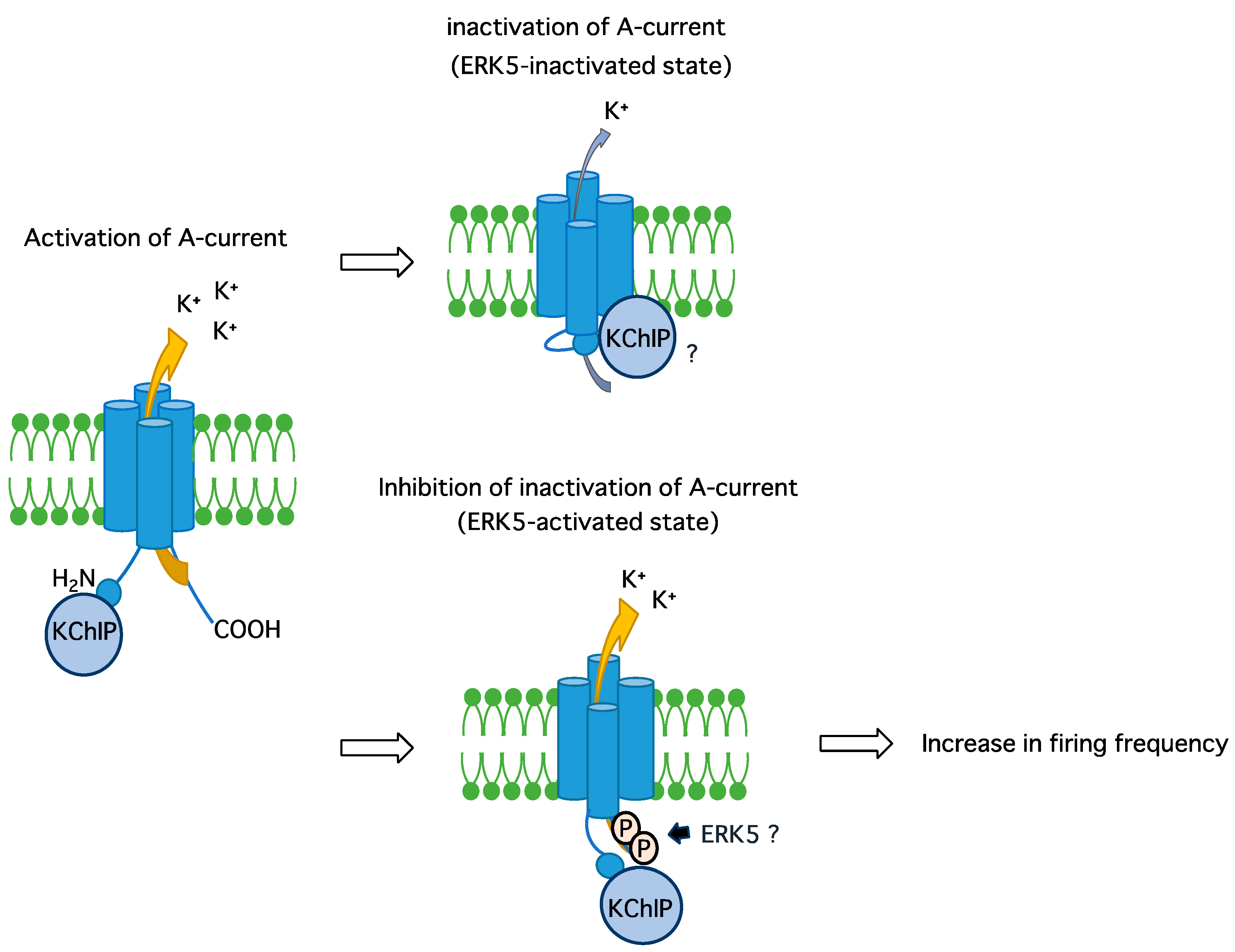
ERK5 Phosphorylates Kv4.2 and Inhibits Inactivation of the A-Type Current in PC12 Cells

1
Department of Pharmacology, Yamagata University School of Medicine, Yamagata 990-9585, Japan
2
Department of Pharmacology, Kyoto University Graduate School of Medicine, Kyoto 606-8501, Japan
*
Author to whom correspondence should be addressed.
Int. J. Mol. Sci. 2018, 19(7), 2008; https://doi.org/10.3390/ijms19072008
Submission received: 14 June 2018 / Revised: 28 June 2018 / Accepted: 28 June 2018 / Published: 10 July 2018

Abstract

:
Extracellular signal-regulated kinase 5 (ERK5) regulates diverse physiological responses such as proliferation, differentiation, and gene expression. Previously, we demonstrated that ERK5 is essential for neurite outgrowth and catecholamine biosynthesis in PC12 cells and sympathetic neurons. However, it remains unclear how ERK5 regulates the activity of ion channels, which are important for membrane excitability. Thus, we examined the effect of ERK5 on the ion channel activity in the PC12 cells that overexpress both ERK5 and the constitutively active MEK5 mutant. The gene and protein expression levels of voltage-dependent Ca2+ and K+ channels were determined by RT-qPCR or Western blotting. The A-type K+ current was recorded using the whole-cell patch clamp method. In these ERK5-activated cells, the gene expression levels of voltage-dependent L- and P/Q-type Ca2+ channels did not alter, but the N-type Ca2+ channel was slightly reduced. In contrast, those of Kv4.2 and Kv4.3, which are components of the A-type current, were significantly enhanced. Unexpectedly, the protein levels of Kv4.2 were not elevated by ERK5 activation, but the phosphorylation levels were increased by ERK5 activation. By electrophysiological analysis, the inactivation time constant of the A-type current was prolonged by ERK5 activation, without changes in the peak current. Taken together, ERK5 inhibits an inactivation of the A-type current by phosphorylation of Kv4.2, which may contribute to the neuronal differentiation process.

1. Introduction

Conventional mitogen-activated protein kinases (MAPKs) involve extracellular signal-regulated kinases (ERKs) 1, 2, and 5, c-Jun N-terminal kinase and p38 MAPKs, and atypical MAPKs include ERK3, 4, and 7 and nemo-like kinase [1]. In response to growth factors or neurotrophic factors, ERKs are strongly activated and regulate diverse physiological responses, such as proliferation, differentiation, and gene expression. The signal transduction leading to ERK1/2 activation and the involvement of ERK1/2 in cellular responses are the best studied among the MAPK family members.
ERK5 shares homology in the amino acid sequence in the kinase-domain with ERK1/2, and possesses a unique long C-terminal domain [2,3]. In the past 10 years, specific inhibitors of ERK5 signaling, such as BIX02189 [4,5] and XMD8-92 [6], have been developed. Using these pharmacological inhibitors, the role of ERK5 in tumor genesis and metastatic progression has been especially well understood [7,8]. We have shown that the levels of ERK5 and tyrosine hydroxylase, a rate-limiting enzyme for catecholamine biosynthesis, are co-related in normal human adrenal medulla, but this correlation is disrupted in pheochromocytomas [9]. However, signaling pathways for ERK5 activation and physiological roles of ERK5 in neuronal development are relatively unclear. For example, involvements of small G-proteins in ERK5 activation are vague [10], whereas it has been established that ERK1/2 is activated through Ras and Rap1 [11,12]. Some limited studies suggest that ERK5 is necessary and sufficient for neuronal differentiation of progenitor cells [13], and is essential for adult hippocampal neurogenesis [14,15]. ERK5 promotes neuronal survival in sympathetic or sensory neurons [16,17]. We have shown that ERK5 is essential for neurite/axon outgrowth and catecholamine biosynthesis in PC12 cells and sympathetic neurons [5,9]. Thus, ERK5 plays important roles in neuronal survival, as well as morphological and functional differentiation. However, although it is well known that ERK1/2 regulates membrane excitability (i.e., neuronal activity) [18,19,20,21], ERK5 regulation of membrane excitability has been poorly understood. Therefore, in the present study, we attempted to clarify the effect of ERK5 signaling on ion channel activity, which is important for regulating membrane excitability.

2. Results

To examine the effect of ERK5 signaling, we attempted to activate ERK5 selectively by the overexpression of ERK5 wildtype and a constitutively active mutant of MAPK/ERK kinase (MEK) 5 (MEK5S311D/T315D, or MEK5D for short). To confirm that ERK signaling is activated by transfection with these DNA constructs, we measured the myocyte-enhancer factor (MEF) 2 transcriptional activity by reporter gene assay as an index of ERK5 activation. It has been well-established that ERK5 phosphorylates MEF2C directly and the transcriptional activity increases [22]. In human embryonic kidney 293 cells (HEK293 cells), overexpression of MEK5D and ERK5 resulted in a dramatic enhancement of MEF2C activity (Figure 1).
We previously demonstrated that overexpression of MEK5D and ERK5 strengthens ERK5 signaling, accompanied by the phosphorylation of the Thr-Glu-Tyr (TEY) activation motif and auto-phosphorylation sites on ERK5, but the ERK1/2 TEY phosphorylation site is not affected [23]. Next, PC12 cells were co-transfected with MEK5D and ERK5, and the messenger RNA (mRNA) expression levels of the major voltage-dependent Ca2+ and K+ channels were measured by RT-qPCR (Figure 2). There were no significant changes in the expression levels of Cav1.2 (L-type) and Cav2.1 (P/Q-type), but a significant reduction of Cav2.2 (N-type) was observed. In contrast, the Kv4.2 and Kv4.3 expression levels, which are responsible for the transient outward Ito current (A type-current), were significantly promoted by ERK5 signaling. K+ channel-interacting proteins (KChIPs) are β-subunit for Kv4.2, and the A-type current is influenced by the expression of KChiPs [24]. We previously performed RNA-sequencing to examine the gene expression levels comprehensively in PC12 cells [25]. The RPKM values for KChIPs 1, 2, 3, and 4 were 0.021284, 0, 1.21248, and 0.067198, respectively (n = 3). Because KChIP3 is a major β-subunit for Kv4.2 in PC12 cells, we examined the KChIP3 expression levels. But, there was no significant change in the expression levels. It has been shown that overexpression of the RasG12V (RasV12) oncogenic mutant can strongly activate ERK1/2 signaling without affecting the phosphorylation status of the ERK5 TEY motif [5,23]. Constitutive ERK1/2 activation by the overexpression of RasV12 did not elevate the significant expression of Kv4.2 and Kv4.3 in our condition (0.791-fold, n = 6, p = 0.603 for Kv4.2 and 0.522-fold, n = 3, p = 0.454 for Kv4.3). Because Kv4.2 mediates the majority of the A-type current and is a critical molecule for the modulation of neuronal excitability in many types of neurons, including the cornu ammonis (CA) 1 pyramidal neurons of the hippocampus and the dorsal horn neurons [20,24], we focused on ERK5 regulation of Kv4.2 for further study.
We next examined the protein levels of Kv4.2 after th eERK5 activation in PC12 cells. Surprisingly, although the mRNA levels were increased, the protein levels were not altered significantly (Figure 3a). In addition to expression levels, we investigated the phosphorylation status of Kv4.2, because it has been reported that ERK1/2 phosphorylates at least three Ser/Thr residues at the C-terminus of Kv4.2 and both ERK5 and ERK1/2 preferentially phosphorylate Ser/Thr residues that have a similar minimum consensus sequence (i.e., Ser/Thr-Pro) [24,26,27]. We used a Phos-tag reagent, which tightly binds phosphorylated amino acids in the presence of Mn2+ or Zn2+. In principle, the Phos-tag-mixed sodium dodecyl sulfate-polyacrylamide gel electrophoresis (SDS-PAGE) causes a band-shift of phosphorylated proteins, and they can be clearly distinguished from unphosphorylated proteins. The overexpression of MEK5D and ERK5 caused the band-shift of Kv4.2, which was significantly diminished by the dominant-negative ERK5 kinase-dead mutant (ERK5K83M, or ERK5KD for short), suggesting that ERK5 signaling promoted phosphorylation levels of Kv4.2 (Figure 3b).
We next examined the effect of ERK5 signaling on the A-type current in PC12 cells. The peak current was unchanged in the PC12 cells overexpressing MEK5D and ERK5, but there was a significant slowing of inactivation (Figure 4). The time constant (τ) at the fast and slow phases was 6.663 and 213.0 (ms), respectively, in the control cells, and 16.69 and 334.7 (ms), respectively, in the cells co-transfected with MEK5D and ERK5. These results suggest that the A-type current inactivation was inhibited by the ERK5 activation, regulating membrane excitability.

3. Discussion

In the present study, we found that ERK5 signaling promoted the mRNA expression of the Kv4.2 primary subunits that underlie the transient A-type current in PC12 cells. However, its protein levels were not reflected by this mRNA up-regulation. Instead, the phosphorylation of endogenous Kv4.2 proteins was promoted by ERK5 and the inactivation rate of the A-type current decreased in these cells. This putative mechanism is shown in Figure 5.
It has been demonstrated that the ERK5 knock-down by antisense oligonucleotides suppressed levels of transient receptor potential (TRP) V1 and A1 in dorsal root ganglion neurons [28]. In the study above, ERK5 regulated the TRPV1 and TRPA1 expression by an unknown mechanism. It has been shown that various transcription factors bind to the Kv4.2 promoter and regulate the transcription. For example, GATA4 and 6, as well as FOG2 enhance Kv4.2 transcription in PC12 cells, although there is a possibility that these transcription factors influence indirectly, as the GATA-binding consensus sequence is lacking in the minimum Kv4.2 promoter [29]. Another study shows that the calcineurin/nuclear factor of the activated T cells (NFAT) pathway increases the Kv4.2 mRNA and protein expression and promoter activity, without affecting the KChIP2 and Kv4.3 levels in rat neonatal ventricular myocytes [30]. Furthermore, neuritin increases the A-type current density accompanied by the up-regulation of Kv4.2 mRNA and protein via the Ca2+/calmodulin/calcineurin/NFATc4 and ERK/NFATc4 pathways in the central neurons, and affects neuronal excitability with increased dendritic spine formation [21]. Because there are NFAT binding sites in the Kv4.2 promoter [21], ERK5 may phosphorylate NFAT to promote Kv4.2 transcription, as ERK1/2 activation resulted in phosphorylation of Ser676 on NFATc4 [31]. However, the reason remains unknown as to why the Kv4.2 protein levels were not reflected by its mRNA expression in this study. In contrast, ERK5 phosphorylation of Kv4.2 was promoted without changes in the protein expression levels. This may reflect the results obtained by electrophysiological experiments that the peak current was not altered by the ERK5 activation. Furthermore, it is also reasonable that the change in the time constant of the A-type current was affected by the changes in Kv4.2 phosphorylation status, but not the protein levels.
It has been shown that ERK1/2 directly phosphorylates Thr602, Thr607, and Ser616 residues at the C-terminal cytoplasmic domain of Kv4.2 [26]. These amino acids are entirely preserved among human and rat Kv4.2. In this study, epidermal growth factor enhanced phosphorylation of Kv4.2 at these three sites in COS7 cells overexpressing Kv4.2. Although this phosphorylation was attenuated by U0126, which blocks ERK1/2 signaling, the remaining phosphorylated band was still observed. Because epidermal growth factor can activate both ERK1/2 and ERK5 [5,23], the remaining U0126-resistant Kv4.2 phosphorylation component may result from ERK5 activity. This group further examined the effects of these three phosphorylated amino acids on the A-type current [24]. The mutation of these three amino acids to Asp caused the activation curve to shift toward more depolarized membrane potentials, whereas the mutation of these three amino acids to Ala showed no effect. Interestingly, the site-directed T607D mutant caused a rightward shift of the activation curve only in the presence of KChiP3, as observed in the case of the triple D mutant, but the S616D mutant caused a leftward shift, which is the totally opposite effect. It has been shown that ERK1/2 also phosphorylates Kv4.2, reducing its conductance in neurons [18]. The minimum consensus sequence of ERK5 and ERK1/2 is similar, but ERK5 may preferentially phosphorylate the Ser616 residue, which results in rapid repolarization to increase the firing frequency, as described below. In contrast, the pituitary adenylate cyclase-activating polypeptide (PACAP) down-regulates the A-type current density without influencing the voltage-dependence of the Kv4.2 channel currents by ERK1/2 phosphorylation of Kv4.2 in rat hippocampal neurons [19]. However, the characteristics of the site-directed mutants of the three amino acids above (T602A, T607A, and S616A) are different from the results found by the group mentioned above. The Kv4.2 S616A mutant did not show any pituitary adenylate cyclase-activating polypeptide (PACAP) induced reduction in the channel current density, whereas the overexpression of T607A mutants partially blocked the inhibitory effect of PACAP. Additionally, the mutational analysis of Kv4.2 indicates that Ser616 is the functionally relevant ERK1/2 phosphorylation site for the modulation of the Kv4.2-mediated currents in neurons derived from spinal cord dorsal horns [20]. Therefore, the roles of the ERK phosphorylation site at the Kv4.2 C-terminus are still controversial. Further study is necessary to identify the ERK5 phosphorylation site on Kv4.2, and to examine the effect on the A-type current.
Adjusting the classical Hodgkin–Huxley models, Rush and Rinzel studied the effects of the A-current on the steady firing rate of neurons. They showed that the number of spikes per burst increases as the conductance of the A-current decreases and as inactivation decreases [32]. When ERK5 was activated, the A-current inactivation rate was reduced in our results (Figure 4). According to their model, we assume that ERK5 may contribute to more rapid repolarization toward the resting potential for responding to the next firing. Therefore, ERK5 may increase the firing frequency through the phosphorylation of Kv4.2.
In conclusion, this study revealed, for the first time, that ERK5 signaling promotes phosphorylation of Kv4.2 and inhibits the inactivation of the A-type current for the enhancement of membrane excitability in PC12 cells. ERK5 promotes neurite outgrowth and catecholamine biosynthesis. In addition to these roles, the regulation of membrane excitability may be essential for the differentiation process toward mature neurons. Future directions are examining the role of the ERK5-enhanced A-type current in neuronal morphological changes and functions in primary cultured neurons, using ERK5 conditional knockout mice.

4. Materials and Methods

Materials: HRP-conjugated anti-glyceraldehyde-3-phosphate dehydrogenase (GAPDH) antibody was purchased from Cell Signaling Technology (Beverly, MA, USA). Anti-Kv4.2 antibody was purchased from UC Davis/NIH NeuroMab (Davis CA, USA), and HRP-conjugated anti-mouse IgG secondary antibody was purchased from GE Healthcare (Buckinghamshire, UK). Enhanced chemiluminescence (ECL) assay kits were purchased from either GE Healthcare, PerkinElmer (Waltham, MA, USA) or Nacalai Tesque (Kyoto, Japan). Lipofectamine 2000 was purchased from Invitrogen (Grand Island, NY, USA). Mn2+-Phos-tag was purchased from Wako Pure Chemicals (Osaka, Japan). TriPure Isolation Reagent for the total RNA extraction, and the FastStart Essential DNA Green Master for real-time PCR were purchased from Roche (Indianapolis, IN, USA), and a Reverse Transcription kit was purchased from Toyobo (Osaka, Japan). A DNA plasmid encoding enhanced green fluorescent protein (EGFP) was purchased from Takara (Tokyo, Japan). The DNA plasmid encoding a tandem MRE-driven firefly luciferase was kindly given by Ron Prywes (Columbia University, NY, USA), and MEK5D (S311D/T315D) was kindly given by Eisuke Nishida (Kyoto University, Japan). DNA plasmid encoding oncogenic RasG12V mutant was used to activate ERK1/2. It was kindly given from Philip J.S. Stork (Vollum Institute, Oregon Health Sciences University, OR, USA). Because these DNA plasmids were kind gifts, as described above, there is restriction for the availability of these plasmids. ERK5KD (K83M) mutant was created from wildtype ERK5 as a template, as described previously [5].
Cell lines: The HEK293 cells and PC12 cells are provided by the Department of Cellular Signaling, Graduate School of Pharmaceutical Sciences, Tohoku University, Japan. Results using these cell lines have been published [9,23]. The HEK293 cells were grown in Dulbecco’s modified Eagle’s medium (DMEM), supplemented with 10% fetal bovine serum, penicillin (50 units/mL), and streptomycin (50 μg/mL), in a 5% CO2 incubator at 37 °C. The PC12 cells were grown in DMEM, supplemented with 10% FBS, 5% horse serum, penicillin (50 units/mL), and streptomycin (50 μg/mL) in a 5% CO2 incubator at 37 °C.
qRT-PCR: The total RNA from the PC12 cells was extracted using TriPure isolation reagent according to the manufacturer’s protocol. The RNA was then reverse transcribed using a RT-PCR kit, and real-time PCR was performed using a LightCycler Nano thermal cycler (Roche), as described previously [33]. The PCR primers used in the PC12 cell experiments were as follows: Cav1.2 (5′-TGT TTC CAG ATG AGA CCC GC-3′ and 5′-GAG GCC CTT CGA CCT AGA GA-3′), Cav2.1 (5′- CTG CTT TGA AGA GGG GAC AG-3′ and 5′-GGA AAA CAG TGA GCA CAG CA-3′), Cav2.2 (5′-TCA TTG TGG TCT TCG CTC TG-3′ and 5′-CCT TTG CTG ACT CCT CCT TG-3′), Kv4.2 (5′-TTG GCG ACT GCT GTT ATG AG-3′ and 5′-TGA CTG AGA CGG CAA TGA AG-3′), Kv4.3 (5′-GGC TAC ACC CTG AAG AG CTG-3′ and 5′-GCC AAA TAT CTT CCC AGC AA-3′), KChIP3 (5′-GCC TTC GAT GCT GAT GGG AA-3′ and 5′-AGA GGT GCG TCC TTT CGC AG-3′), and GAPDH (5′-ACC ACA GTC CAT GCC ATC AC-3′ and 5′-TCC ACC ACC CTG TTG CTG TA-3′). PCR products were quantified and normalized to the GAPDH control before finally being presented as a fold change.
Reporter gene assay: Reporter gene assays were performed similarly, as described previously [33]. MEK5D, ERK5, and MRE-luciferase reporter genes were co-transfected into HEK293 cells in 24-well plates using Lipofectamine 2000. Two days after transfection, the lysates were collected and the luciferase activity was measured using a luminometer (Lumat LB9507, Berthold Japan K.K., Tokyo, Japan).
SDS-PAGE with or without Phos-tag and Western blotting: The proteins were separated by electrophoresis using 10–11% polyacrylamide gels. The proteins were then transferred from the gel onto a polyvinylidene difluoride membrane (GE Healthcare), according to standard protocols. The membranes were blocked for 0.5 h at room temperature in 5% skim milk in Tris-buffered saline containing 0.1% tween-20 (TBST), then incubated with the indicated primary antibodies overnight at 4 °C. The antibodies were dissolved in the blocking buffer, and used at the following dilutions: anti-Kv4.2 (1:500 or 1:1000), and HRP-conjugated anti-GAPDH (1:1000). The membranes were washed several times with TBST before being incubated with HRP-conjugated anti-mouse IgG secondary antibodies (diluted 1:5000 in blocking buffer) at room temperature for 1–2 h. The membranes were then washed with TBST, developed using an ECL chemiluminescence assay kit, and visualized using a ChemiDoc XRS imaging system (BioRad, Hercules, CA, USA) or LAS1000 (Fuji Film, Tokyo, Japan). The relative intensities of the bands corresponding to Kv4.2 and the internal control GAPDH were determined using Image-J densitometry software (National Institute of Health, Bethesda, MD, USA).
For electrophoresis using Phos-tag, the proteins were separated with 5% polyacrylamide gels containing 30 μM Phos-tag and 60 μM MnCl2. After the gels were washed twice for 10 min with transfer buffer containing 10 mM EDTA to remove Mn2+, the proteins were then transferred from the gel onto a polyvinylidene difluoride membrane at 30 V for 16 h. The further procedure is performed similarly, as described above.
Electrophysiology by patch-clamping: The PC12 cells were co-transfected with EGFP, MEK5D, and ERK5. EGFP was used as a marker for the transfected cells. The whole-cell patch clamp method was used for recording the membrane currents (patch-clamp amplifier Axopatch 200B, Molecular Devices, Chicago, IL, USA), as described previously [34]. Borosilicate glass electrodes had tip resistances between 2.5 and 4.5 MΩ when filled with internal solution composed of (mM) KOH 120, aspartic acid 80, Mg-ATP 5, KCl 20, HEPES 5, EGTA 5, and GTP-Na2 0.1 (pH 7.2 with aspartic acid). The composition of the external solution (mM) was: NaCl 136.9, KCl 5.4, CaCl2 1.8, MgCl2 0.5, NaH2PO4 0.33, HEPES 5.0, and glucose 5.5 (pH 7.4 with NaOH). To evoke membrane currents, the cells were held at a potential of −80 mM and depolarized for 500 ms to various potentials, ranging from −30 to +50 mV in 20 mV increments at 37 ± 0.5 °C. The pulse protocol and data acquisition and storage were accomplished with Clampex 9.2 (Molecular Devices). The sampling frequency was 10 kHz and low-pass filtering was performed at 5 kHz. The cell membrane capacitance (Cm) was determined by integrating the area under the capacitive transient elicited, by applying a 50 ms hyperpolarizing voltage-step from a potential of −40 to −45 mV. All membrane currents (Im) were normalized by Cm, then analyzed using IGOR software (Wavemetrics, Portland, OR, USA). The time-course of inactivation at 50 mV was fitted with a first order biexponential function, as follows:
I m ( t ) = y 0 + y 1 { 1 exp ( t τ f a s t ) } + y 2 { 1 exp ( t τ s l o w ) }
where τfast and τslow are fast and slow time constants, respectively.
Statistics: Data are expressed as means ± S.E.M., and the statistical significance of the differences between groups was analyzed using the unpaired Student’s t-test or one-way ANOVA, with post hoc test using Tukey’s test for multiple comparisons.

Author Contributions

Y.O. (Yutaro Obara), Y.H., and K.I. conceived and designed the experiments; Y.K., Y.O. (Yutaro Obara), Y.O. (Yosuke Okamoto), and T.S. performed the experiments; Y.O. (Yutaro Obara), Y.O. (Yosuke Okamoto), and K.I. wrote the paper.

Funding

This work was supported in part by Grants-in-Aid from the Japan Society for the Promotion of Science (No. 15K07963 to Y.O. [Yutaro Obara]) and the Takeda Science Foundation (Y.O. [Yutaro Obara]). These grants cover the costs to publish in open access. This work was also supported by RIKEN, Grant-in-Aid for Scientific Research (A) and Grant-in-Aid for Scientific Research on Innovative Area “Principles of memory dynamism elucidated from a diversity of learning systems”, Human Frontier Science Foundation, The Naito Foundation, Research Foundation for Opto-Science and Technology, Novartis Foundation to Y.H. and Grant-in-Aid for Scientific Research (B) from the MEXT, Japan (T.S.). The founding sponsors had no role in the design of the study; in the collection, analyses, or interpretation of data; in the writing of the manuscript; and in the decision to publish the results.

Acknowledgments

We thank Philip J.S. Stork (Vollum Institute, Oregon Health Sciences University, Portland, OR, USA), Ron Prywes (Columbia University, New York, NY, USA), and Eisuke Nishida (Kyoto University, Kyoto, Japan) for kindly providing DNA plasmids.

Conflicts of Interest

Y.H. is partly supported by Fujitsu Laboratories and Dwango. The other authors declare no conflict of interest.

Abbreviations

MAPKMitogen-activated protein kinase
ERKExtracellular signal-regulated kinase
MEKMAPK/ERK kinase
MEFMyocyte-enhancer factor
MRE HEK293 cellsMEF2 response elementhuman embryonic kidney 293 cells
SDS-PAGESodium dodecyl sulfate-polyacrylamide gel electrophoresis
TRPTransient receptor potential
NFATNuclear factor of activated T cells
KChIPCAK+ channel-interacting proteincornu ammonis
PACAPPituitary adenylate cyclase-activating polypeptide
GAPDHGlyceraldehyde-3-phosphate dehydrogenase
ECLEnhanced chemiluminescence
EGFPEnhanced green fluorescent protein
DMEMDulbecco’s modified Eagle’s medium
TBSTTris-buffered saline containing 0.1% tween-20

References

  1. Coulombe, P.; Meloche, S. Atypical mitogen-activated protein kinase: Structure, regulation and functions. Biochim. Biophys. Acta 2007, 1773, 1376–1387. [Google Scholar] [CrossRef] [PubMed]
  2. Drew, B.A.; Burow, M.E.; Beckman, B.S. MEK5/ERK5 pathway: The first fifteen years. Biochim. Biophys. Acta 2012, 1825, 37–48. [Google Scholar] [CrossRef] [PubMed] [Green Version]
  3. Wang, X.; Tournier, C. Regulation of cellular functions by the ERK5 signalling pathway. Cell. Signal. 2006, 18, 753–760. [Google Scholar] [CrossRef] [PubMed]
  4. Tatake, R.J.; O’Neill, M.M.; Kennedy, C.A.; Wayne, A.L.; Jakes, S.; Wu, D.; Kugler, S.Z., Jr.; Kashem, M.A.; Kaplita, P.; Snow, R.J. Identification of pharmacological inhibitors of the MEK5/ERK5 pathway. Biochem. Biophys. Res. Commun. 2008, 377, 120–125. [Google Scholar] [CrossRef] [PubMed]
  5. Obara, Y.; Yamauchi, A.; Takehara, S.; Nemoto, W.; Takahashi, M.; Stork, P.J.; Nakahata, N. ERK5 Activity Is Required for Nerve Growth Factor-induced Neurite Outgrowth and Stabilization of Tyrosine Hydroxylase in PC12 Cells. J. Biol. Chem. 2009, 284, 23564–23573. [Google Scholar] [CrossRef] [PubMed] [Green Version]
  6. Yang, Q.; Deng, X.; Lu, B.; Cameron, M.; Fearns, C.; Patricelli, M.P.; Yates, J.R., 3rd; Gray, N.S.; Lee, J.D. Pharmacological inhibition of BMK1 suppresses tumor growth through promyelocytic leukemia protein. Cancer Cell. 2010, 18, 258–267. [Google Scholar] [CrossRef] [PubMed]
  7. Hoang, V.T.; Yan, T.J.; Cavanaugh, J.E.; Flaherty, P.T.; Beckman, B.S.; Burow, M.E. MEK5-ERK5 signaling in cancer: Implications for targeted therapy. Cancer Lett. 2017, 392, 51. [Google Scholar] [CrossRef] [PubMed]
  8. Simoes, A.E.; Rodrigues, C.M.; Borralho, P.M. The MEK5/ERK5 signalling pathway in cancer: A promising novel therapeutic target. Drug Discov. Today 2016, 21, 1654–1663. [Google Scholar] [CrossRef] [PubMed]
  9. Obara, Y.; Nagasawa, R.; Nemoto, W.; Pellegrino, M.J.; Takahashi, M.; Habecker, B.A.; Stork, P.J.; Ichiyanagi, O.; Ito, H.; Tomita, Y.; et al. ERK5 induces ankrd1 for catecholamine biosynthesis and homeostasis in adrenal medullary cells. Cell. Signal. 2016, 28, 177–189. [Google Scholar] [CrossRef] [PubMed]
  10. Obara, Y.; Nakahata, N. The signaling pathway leading to extracellular signal-regulated kinase 5 (ERK5) activation via G-proteins and ERK5-dependent neurotrophic effects. Mol. Pharmacol. 2010, 77, 10–16. [Google Scholar] [CrossRef] [PubMed]
  11. Obara, Y.; Labudda, K.; Dillon, T.J.; Stork, P.J. PKA phosphorylation of Src mediates Rap1 activation in NGF and cAMP signaling in PC12 cells. J. Cell. Sci. 2004, 117, 6085–6094. [Google Scholar] [CrossRef] [PubMed] [Green Version]
  12. York, R.D.; Yao, H.; Dillon, T.; Ellig, C.L.; Eckert, S.P.; McCleskey, E.W.; Stork, P.J. Rap1 mediates sustained MAP kinase activation induced by nerve growth factor. Nature 1998, 392, 622–626. [Google Scholar] [CrossRef] [PubMed]
  13. Liu, L.; Cundiff, P.; Abel, G.; Wang, Y.; Faigle, R.; Sakagami, H.; Xu, M.; Xia, Z. Extracellular signal-regulated kinase (ERK) 5 is necessary and sufficient to specify cortical neuronal fate. Proc. Natl. Acad. Sci. USA 2006, 103, 9697–9702. [Google Scholar] [CrossRef] [PubMed] [Green Version]
  14. Pan, Y.W.; Chan, G.C.; Kuo, C.T.; Storm, D.R.; Xia, Z. Inhibition of adult neurogenesis by inducible and targeted deletion of ERK5 mitogen-activated protein kinase specifically in adult neurogenic regions impairs contextual fear extinction and remote fear memory. J. Neurosci. 2012, 32, 6444–6455. [Google Scholar] [CrossRef] [PubMed]
  15. Pan, Y.W.; Zou, J.; Wang, W.; Sakagami, H.; Garelick, M.G.; Abel, G.; Kuo, C.T.; Storm, D.R.; Xia, Z. Inducible and conditional deletion of extracellular signal-regulated kinase 5 disrupts adult hippocampal neurogenesis. J. Biol. Chem. 2012, 287, 23306–23317. [Google Scholar] [CrossRef] [PubMed]
  16. Finegan, K.G.; Wang, X.; Lee, E.J.; Robinson, A.C.; Tournier, C. Regulation of neuronal survival by the extracellular signal-regulated protein kinase 5. Cell. Death Differ. 2009, 16, 674–683. [Google Scholar] [CrossRef] [PubMed] [Green Version]
  17. Watson, F.L.; Heerssen, H.M.; Bhattacharyya, A.; Klesse, L.; Lin, M.Z.; Segal, R.A. Neurotrophins use the Erk5 pathway to mediate a retrograde survival response. Nat. Neurosci. 2001, 4, 981–988. [Google Scholar] [CrossRef] [PubMed]
  18. Morozov, A.; Muzzio, I.A.; Bourtchouladze, R.; Van-Strien, N.; Lapidus, K.; Yin, D.; Winder, D.G.; Adams, J.P.; Sweatt, J.D.; Kandel, E.R. Rap1 couples cAMP signaling to a distinct pool of p42/44MAPK regulating excitability, synaptic plasticity, learning, and memory. Neuron 2003, 39, 309–325. [Google Scholar] [CrossRef]
  19. Gupte, R.P.; Kadunganattil, S.; Shepherd, A.J.; Merrill, R.; Planer, W.; Bruchas, M.R.; Strack, S.; Mohapatra, D.P. Convergent phosphomodulation of the major neuronal dendritic potassium channel Kv4.2 by pituitary adenylate cyclase-activating polypeptide. Neuropharmacology 2016, 101, 291–308. [Google Scholar] [CrossRef] [PubMed] [Green Version]
  20. Hu, H.J.; Carrasquillo, Y.; Karim, F.; Jung, W.E.; Nerbonne, J.M.; Schwarz, T.L.; Gereau, R.W.T. The kv4.2 potassium channel subunit is required for pain plasticity. Neuron 2006, 50, 89–100. [Google Scholar] [CrossRef] [PubMed]
  21. Yao, J.J.; Zhao, Q.R.; Liu, D.D.; Chow, C.W.; Mei, Y.A. Neuritin Up-regulates Kv4.2 alpha-Subunit of Potassium Channel Expression and Affects Neuronal Excitability by Regulating the Calcium-Calcineurin-NFATc4 Signaling Pathway. J. Biol. Chem. 2016, 291, 17369–17381. [Google Scholar] [CrossRef] [PubMed]
  22. Kato, Y.; Kravchenko, V.V.; Tapping, R.I.; Han, J.; Ulevitch, R.J.; Lee, J.D. BMK1/ERK5 regulates serum-induced early gene expression through transcription factor MEF2C. Embo. J. 1997, 16, 7054–7066. [Google Scholar] [CrossRef] [PubMed] [Green Version]
  23. Honda, T.; Obara, Y.; Yamauchi, A.; Couvillon, A.D.; Mason, J.J.; Ishii, K.; Nakahata, N. Phosphorylation of ERK5 on Thr732 is associated with ERK5 nuclear localization and ERK5-dependent transcription. PLoS ONE 2015, 10, e0117914. [Google Scholar] [CrossRef] [PubMed]
  24. Schrader, L.A.; Birnbaum, S.G.; Nadin, B.M.; Ren, Y.; Bui, D.; Anderson, A.E.; Sweatt, J.D. ERK/MAPK regulates the Kv4.2 potassium channel by direct phosphorylation of the pore-forming subunit. Am. J. Physiol. Cell. Physiol 2006, 290, C852-861. [Google Scholar] [CrossRef] [PubMed]
  25. Obara, Y.; Ishii, K. Transcriptome Analysis Reveals That Midnolin Regulates mRNA Expression Levels of Multiple Parkinson’s Disease Causative Genes. Biol. Pharm. Bull. 2018, 41, 20–23. [Google Scholar] [CrossRef] [PubMed] [Green Version]
  26. Adams, J.P.; Anderson, A.E.; Varga, A.W.; Dineley, K.T.; Cook, R.G.; Pfaffinger, P.J.; Sweatt, J.D. The A-type potassium channel Kv4.2 is a substrate for the mitogen-activated protein kinase ERK. J. Neurochem. 2000, 75, 2277–2287. [Google Scholar] [CrossRef] [PubMed]
  27. Lugo, J.N.; Barnwell, L.F.; Ren, Y.; Lee, W.L.; Johnston, L.D.; Kim, R.; Hrachovy, R.A.; Sweatt, J.D.; Anderson, A.E. Altered phosphorylation and localization of the A-type channel, Kv4.2 in status epilepticus. J. Neurochem. 2008, 106, 1929–1940. [Google Scholar] [CrossRef] [PubMed]
  28. Katsura, H.; Obata, K.; Mizushima, T.; Sakurai, J.; Kobayashi, K.; Yamanaka, H.; Dai, Y.; Fukuoka, T.; Sakagami, M.; Noguchi, K. Activation of extracellular signal-regulated protein kinases 5 in primary afferent neurons contributes to heat and cold hyperalgesia after inflammation. J. Neurochem. 2007, 102, 1614–1624. [Google Scholar] [CrossRef] [PubMed] [Green Version]
  29. Jia, Y.; Takimoto, K. GATA and FOG2 transcription factors differentially regulate the promoter for Kv4.2 K(+) channel gene in cardiac myocytes and PC12 cells. Cardiovasc. Res. 2003, 60, 278–287. [Google Scholar] [CrossRef]
  30. Gong, N.; Bodi, I.; Zobel, C.; Schwartz, A.; Molkentin, J.D.; Backx, P.H. Calcineurin increases cardiac transient outward K+ currents via transcriptional up-regulation of Kv4.2 channel subunits. J. Biol. Chem. 2006, 281, 38498–38506. [Google Scholar] [CrossRef] [PubMed]
  31. Yang, T.T.; Xiong, Q.; Graef, I.A.; Crabtree, G.R.; Chow, C.W. Recruitment of the extracellular signal-regulated kinase/ribosomal S6 kinase signaling pathway to the NFATc4 transcription activation complex. Mol. Cell. Biol. 2005, 25, 907–920. [Google Scholar] [CrossRef] [PubMed]
  32. Rush, M.E.; Rinzel, J. The potassium A-current, low firing rates and rebound excitation in Hodgkin-Huxley models. Bull. Math. Biol. 1995, 57, 899–929. [Google Scholar] [CrossRef] [PubMed]
  33. Obara, Y.; Imai, T.; Sato, H.; Takeda, Y.; Kato, T.; Ishii, K. Midnolin is a novel regulator of parkin expression and is associated with Parkinson’s Disease. Sci. Rep. 2017, 7, 5885. [Google Scholar] [CrossRef] [PubMed]
  34. Okamoto, Y.; Kawamura, K.; Nakamura, Y.; Ono, K. Pathological impact of hyperpolarization-activated chloride current peculiar to rat pulmonary vein cardiomyocytes. J. Mol. Cell. Cardiol. 2014, 66, 53–62. [Google Scholar] [CrossRef] [PubMed]
Figure 1. Overexpression of constitutively active mitogen-activated protein and extracellular signal-regulated (MAPK/ERK) kinase (MEK) 5 mutant and ERK5 causes activation of ERK5 signaling. Human embryonic kidney 293 cells (HEK293 cells) were transfected with tandem myocyte-enhancer factor (MEF) 2 response element (MRE)-luciferase reporter gene and empty vector (Vec) or MEK5D and ERK5. Two days after transfection, the luciferase activity resulting from MEF2 activation was measured. ERK5 significantly increased MEF2C activity (one experiment in triplicate (n = 3), * p <0.05, unpaired Student’s t-test).
Ijms 19 02008 g001
Figure 2. ERK5 promotes gene expression of Kv4.2 and Kv4.3 in PC12 cells. PC12 cells were transfected with empty vector (Vec) or MEK5 and ERK5. Two days after transfection, the total RNA was isolated from the cell lysates and RT-qPCR was performed using specific primers for Cav1.2, Cav2.1, Cav2.2, Kv4.2, Kv4.3, and KChIP3. ERK5 significantly promoted the gene expression of Kv4.2 and Kv4.3, and attenuated Cav2.2 expression (two independent experiments in triplicate (n = 6), * p <0.05, unpaired Student’s t-test).
Ijms 19 02008 g002
Figure 3. ERK5 did not alter the protein expression of Kv4.2, but did promote the phosphorylation of Kv4.2 in PC12 cells. (a) PC12 cells were transfected with empty vector (Vec) or MEK5D and ERK5. Two days after transfection, Western blotting using Kv4.2 and glyceraldehyde-3-phosphate dehydrogenase (GAPDH) antibodies was performed. The density of the Kv4.2 bands was expressed as a fold of control cells (Vec) (two independent experiments in triplicate (n = 6), unpaired Student’s t-test); (b) PC12 cells were transfected with empty vector (Vec), MEK5D and ERK5, or MEK5D and ERK5KD. Two days after transfection, Western blotting using Phos-tag-contained polyacrylamide gels or Phos-tag-free gels was carried out with a Kv4.2 antibody. The density of the phospho-Kv4.2 bands was expressed as a fold of the control cells (Vec). Overexpression of MEK5D and ERK5KD significantly attenuated the MEK5D and ERK5-induced Kv4.2 phosphorylation (three similar independent experiments in triplicate (n = 3), * p <0.05, Tukey’s method).
Ijms 19 02008 g003
Figure 4. ERK5 inhibits inactivation of the A-type current in PC12 cells. PC12 cells were co-transfected with EGFP, MEK5D, and ERK5. Two days after transfection, the A-type current was recorded. (a) Representative traces and step-pulse protocol are shown; (b) maximal peak current was measured at +50 mV. The ERK5 did not significantly change amplitude levels (data from three independent experiments (n = 4), unpaired Student’s t-test); (c,d) the time constant (τ) at fast (c) and slow (d) phases at +50 mV was calculated. ERK5 significantly changed the time constant (τ) at the fast phase (data from three independent experiments (n = 4), * p <0.05, unpaired Student’s t-test).
Ijms 19 02008 g004
Figure 5. Putative mechanism of regulation of Kv4.2 channels by ERK5. The ERK5 phosphorylates unidentified Ser/Thr residue(s) on Kv4.2, resulting in the inhibition of the A-type current inactivation. This mechanism may contribute to rapid repolarization toward resting potential, which is necessary for causing the next firing.
Ijms 19 02008 g005

