
Biomedical Applications of Sulfonylcalix[4]arene-Based Metal–Organic Supercontainers
 , and
, and
Abstract
:
1. Introduction
2. Structural Regulation and Cavity Engineering of MOSCs
3. Biomedical Applications of MOSCs
3.1. Host–Guest Chemistry and Drug Encapsulation
3.2. Ion/Molecule Recognition
3.3. Targeted Drug Delivery and Therapeutic Effects
4. Conclusions and Outlook
Author Contributions
Funding
Conflicts of Interest
Abbreviations
| AP | aspirin |
| BBB | 4,4′,4″-(benzene-1,3,5-trityl)-tris(benzene-4,1-dial)tribenzoate |
| BDC | 1,4-benzenedicarboxylate |
| 1,3-BDC | 1,3-benzenedicarboxylate |
| BTB | 4,4′,4″-benzene-1,3,5-trial-tribenzoate |
| BTC | 1,3,5-benzene-tricarboxylate |
| BTE | 4,4′,4″-benzene-1,3,5-trityl-tris-(ethyne-2,1-days)-tribenzoate |
| CPT | (+)-Camptothecin |
| CTC | cyclohexane-1,3,5-tricarboxylate |
| D1 | (R)-(+)-rabeprazole sodium |
| D2 | (S)-(−)-pantoprazole sodium |
| DCF | diclofenac sodium |
| DHIA | 4,6-dihydroxyisophthalate |
| DOSY | 2D diffusion ordered NMR spectroscopy |
| FA | folic acid |
| H4PMTC | 5,5′-(p-phenylene-bis(methanamino)-di-isophthalic acid |
| H4TBSC | p-tert-butylsulfonylcalix[4]arene |
| HPLC | high performance liquid chromatography |
| Ibu | Ibuprofen |
| MB | methylene blue |
| MOPs | metal–organic polyhedrons |
| MOSCs | metal–organic supercontainers |
| NPX | naproxen |
| 1-OHP | 1-hydroxypyrene |
| PAHs | Polycyclic aromatic hydrocarbons |
| PBS | phosphate-buffered saline |
| PCCs | porous coordination cages |
| RAPA | rapamycin |
| RB | Rhodamine B |
| TDC | 2,5-thiophenedicarboxylate |
References
- Li, X.-X.; Zhao, D.; Zheng, S.-T. Recent advances in POM-organic frameworks and POM-organic polyhedra. Coord. Chem. Rev. 2019, 397, 220–240. [Google Scholar] [CrossRef]
- Zhang, Y.-Y.; Gao, W.-X.; Lin, L.; Jin, G.-X. Recent advances in the construction and applications of heterometallic macrocycles and cages. Coord. Chem. Rev. 2017, 344, 323–344. [Google Scholar] [CrossRef]
- Cook, T.R.; Stang, P.J. Recent Developments in the Preparation and Chemistry of Metallacycles and Metallacages via Coordination. Chem. Rev. 2015, 115, 7001–7045. [Google Scholar] [CrossRef]
- Cook, T.R.; Zheng, Y.R.; Stang, P.J. Metal-Organic Frameworks and Self-Assembled Supramolecular Coordination Complexes: Comparing and Contrasting the Design, Synthesis, and Functionality of Metal-Organic Materials. Chem. Rev. 2013, 113, 734–777. [Google Scholar] [CrossRef]
- Chakrabarty, R.; Mukherjee, P.S.; Stang, P.J. Supramolecular Coordination: Self-Assembly of Finite Two- and Three-Dimensional Ensembles. Chem. Rev. 2011, 111, 6810–6918. [Google Scholar] [CrossRef]
- Leininger, S.; Olenyuk, B.; Stang, P.J. Self-assembly of discrete cyclic nanostructures mediated by transition metals. Chem. Rev. 2000, 100, 853–907. [Google Scholar] [CrossRef]
- Deegan, M.M.; Ahmed, T.S.; Yap, G.P.A.; Bloch, E.D. Structure and redox tuning of gas adsorption properties in calixarene-supported Fe(II)-based porous cages. Chem. Sci. 2020, 11, 5273–5279. [Google Scholar] [CrossRef]
- Argent, S.P.; da Silva, I.; Greenaway, A.; Savage, M.; Humby, J.; Davies, A.J.; Nowell, H.; Lewis, W.; Manuel, P.; Tang, C.C.; et al. Porous Metal-Organic Polyhedra: Morphology, Porosity, and Guest Binding. Inorg. Chem. 2020, 59, 15646–15658. [Google Scholar] [CrossRef]
- Li, J.R.; Zhou, H.C. Bridging-ligand-substitution strategy for the preparation of metal-organic polyhedra. Nat. Chem. 2010, 2, 893–898. [Google Scholar] [CrossRef]
- Xue, W.; Wu, K.; Ouyang, N.; Brotin, T.; Nitschke, J.R. Allosterically Regulated Guest Binding Determines Framework Symmetry for an FeII4L4 Cage. Angew. Chem. Int. Ed. Engl. 2023, 62, e202301319. [Google Scholar] [CrossRef]
- Tamura, Y.; Takezawa, H.; Fujita, M. A Double-Walled Knotted Cage for Guest-Adaptive Molecular Recognition. J. Am. Chem. Soc. 2020, 142, 5504–5508. [Google Scholar] [CrossRef]
- Yan, K.; Dubey, R.; Arai, T.; Inokuma, Y.; Fujita, M. Chiral Crystalline Sponges for the Absolute Structure Determination of Chiral Guests. J. Am. Chem. Soc. 2017, 139, 11341–11344. [Google Scholar] [CrossRef]
- Mal, P.; Breiner, B.; Rissanen, K.; Nitschke, J.R. White Phosphorus Is Air-Stable Within a Self-Assembled Tetrahedral Capsule. Science 2009, 324, 1697–1699. [Google Scholar] [CrossRef]
- Li, K.; Zhang, L.Y.; Yan, C.; Wei, S.C.; Pan, M.; Zhang, L.; Su, C.Y. Stepwise assembly of Pd6(RuL3)8 nanoscale rhombododecahedral metal-organic cages via metalloligand strategy for guest trapping and protection. J. Am. Chem. Soc. 2014, 136, 4456–4459. [Google Scholar] [CrossRef]
- Jiao, T.; Chen, L.; Yang, D.; Li, X.; Wu, G.; Zeng, P.; Zhou, A.; Yin, Q.; Pan, Y.; Wu, B.; et al. Trapping White Phosphorus within a Purely Organic Molecular Container Produced by Imine Condensation. Angew. Chem. Int. Ed. 2017, 56, 14545–14550. [Google Scholar] [CrossRef]
- Takezawa, H.; Kanda, T.; Nanjo, H.; Fujita, M. Site-Selective Functionalization of Linear Diterpenoids through U-Shaped Folding in a Confined Artificial Cavity. J. Am. Chem. Soc. 2019, 141, 5112–5115. [Google Scholar] [CrossRef]
- Fang, Y.; Powell, J.A.; Li, E.; Wang, Q.; Perry, Z.; Kirchon, A.; Yang, X.; Xiao, Z.; Zhu, C.; Zhang, L.; et al. Catalytic reactions within the cavity of coordination cages. Chem. Soc. Rev. 2019, 48, 4707–4730. [Google Scholar] [CrossRef]
- Guo, J.; Xu, Y.-W.; Li, K.; Xiao, L.-M.; Chen, S.; Wu, K.; Chen, X.-D.; Fan, Y.-Z.; Liu, J.-M.; Su, C.-Y. Regio- and Enantioselective Photodimerization within the Confined Space of a Homochiral Ruthenium/Palladium Heterometallic Coordination Cage. Angew. Chem. Int. Ed. 2017, 56, 3852–3856. [Google Scholar] [CrossRef]
- Wang, C.; Wang, W.; Tan, J.; Zhang, X.; Yuan, D.; Zhou, H.-C. Coordination-based molecular nanomaterials for biomedically relevant applications. Coord. Chem. Rev. 2021, 438, 213752. [Google Scholar] [CrossRef]
- Sepehrpour, H.; Fu, W.; Sun, Y.; Stang, P.J. Biomedically Relevant Self-Assembled Metallacycles and Metallacages. J. Am. Chem. Soc. 2019, 141, 14005–14020. [Google Scholar] [CrossRef] [PubMed]
- Xu, W.-Q.; Fan, Y.-Z.; Wang, H.-P.; Teng, J.; Li, Y.-H.; Chen, C.-X.; Fenske, D.; Jiang, J.-J.; Su, C.-Y. Investigation of Binding Behavior between Drug Molecule 5-Fluoracil and M4L4-Type Tetrahedral Cages: Selectivity, Capture, and Release. Chem. Eur. J. 2017, 23, 3542–3547. [Google Scholar] [CrossRef]
- Castilla, A.M.; Ramsay, W.J.; Nitschke, J.R. Stereochemistry in subcomponent self-assembly. Acc. Chem. Res. 2014, 47, 2063–2073. [Google Scholar] [CrossRef] [PubMed]
- Smulders, M.M.J.; Riddell, I.A.; Browne, C.; Nitschke, J.R. Building on architectural principles for three-dimensional metallosupramolecular construction. Chem. Soc. Rev. 2013, 42, 1728–1754. [Google Scholar] [CrossRef] [PubMed]
- He, C.; Sheng, T.-P.; Dai, F.-R.; Chen, Z.-N. Sulfonylcalix[4]arene-based Coordination Supercontainers. Chin. J. Struct. Chem. 2020, 39, 2077–2084. [Google Scholar] [CrossRef]
- Bi, Y.; Du, S.; Liao, W. Thiacalixarene-Based Nanoscale Polyhedral Coordination Cages. Coord. Chem. Rev. 2014, 276, 61–72. [Google Scholar] [CrossRef]
- Kumagai, H.; Hasegawa, M.; Miyanari, S.; Sugawa, Y.; Sato, Y.; Hori, T.; Ueda, S.; Kamiyama, H.; Miyano, S. Facile synthesis of p-tert-butylthiacalix[4]arene by the reaction of p-tert-butylphenol with elemental sulfur in the presence of a base. Tetrahedron Lett. 1997, 38, 3971–3972. [Google Scholar] [CrossRef]
- Iki, N.; Kumagai, H.; Morohashi, N.; Ejima, K.; Hasegawa, M.; Miyanari, S.; Miyano, S. Selective oxidation of thiacalix[4]arenes to the sulfinyl- and sulfonylcalix[4]arenes and their coordination ability to metal ions. Tetrahedron Lett. 1998, 39, 7559–7562. [Google Scholar] [CrossRef]
- Morohashi, N.; Narumi, F.; Iki, N.; Hattori, T.; Miyano, S. Thiacalixarenes. Chem. Rev. 2006, 106, 5291–5316. [Google Scholar] [CrossRef]
- Casini, A.; Woods, B.; Wenzel, M. The Promise of Self-Assembled 3D Supramolecular Coordination Complexes for Biomedical Applications. Inorg. Chem. 2017, 56, 14715–14729. [Google Scholar] [CrossRef]
- Schmidt, A.; Casini, A.; Kühn, F.E. Self-assembled M2L4 coordination cages: Synthesis and potential applications. Coord. Chem. Rev. 2014, 275, 19–36. [Google Scholar] [CrossRef]
- Moreno-Alcántar, G.; Casini, A. Bioinorganic supramolecular coordination complexes and their biomedical applications. FEBS Lett. 2022, 597, 191–202. [Google Scholar] [CrossRef] [PubMed]
- Wang, Y.-P.; Zhang, Y.; Duan, X.-H.; Mao, J.-J.; Pan, M.; Shen, J.; Su, C.-Y. Recent progress in metal-organic cages for biomedical application: Highlighted research during 2018–2023. Coord. Chem. Rev. 2024, 501, 215570. [Google Scholar] [CrossRef]
- Hang, X.; Bi, Y. Thiacalix[4]arene-supported molecular clusters for catalytic applications. Dalton Trans. 2021, 50, 3749–3758. [Google Scholar] [CrossRef]
- Dai, F.-R.; Wang, Z. Modular Assembly of Metal–Organic Supercontainers Incorporating Sulfonylcalixarenes. J. Am. Chem. Soc. 2012, 134, 8002–8005. [Google Scholar] [CrossRef] [PubMed]
- Liu, M.; Liao, W.P.; Hu, C.H.; Du, S.C.; Zhang, H.J. Calixarene-Based Nanoscale Coordination Cages. Angew. Chem. Int. Ed. 2012, 51, 1585–1588. [Google Scholar] [CrossRef] [PubMed]
- Du, S.; Hu, C.; Xiao, J.-C.; Tan, H.; Liao, W. A giant coordination cage based on sulfonylcalix[4]arenes. Chem. Commun. 2012, 48, 9177–9179. [Google Scholar] [CrossRef] [PubMed]
- Du, S.; Yu, T.-Q.; Liao, W.; Hu, C. Structure Modeling, Synthesis and X-ray Diffraction Determination of an Extra-Large Calixarene-Based Coordination Cage and Its Application in Drug Delivery. Dalton Trans. 2015, 44, 14394–14402. [Google Scholar] [CrossRef]
- Fang, Y.; Lian, X.; Huang, Y.; Fu, G.; Xiao, Z.; Wang, Q.; Nan, B.; Pellois, J.P.; Zhou, H.C. Investigating Subcellular Compartment Targeting Effect of Porous Coordination Cages for Enhancing Cancer Nanotherapy. Small 2018, 14, e1802709. [Google Scholar] [CrossRef]
- Fang, Y.; Li, J.; Togo, T.; Jin, F.; Xiao, Z.; Liu, L.; Drake, H.; Lian, X.; Zhou, H.-C. Ultra-Small Face-Centered-Cubic Ru Nanoparticles Confined within a Porous Coordination Cage for Dehydrogenation. Chem 2018, 4, 555–563. [Google Scholar] [CrossRef]
- Fang, Y.; Xiao, Z.; Li, J.; Lollar, C.; Liu, L.; Lian, X.; Yuan, S.; Banerjee, S.; Zhang, P.; Zhou, H.-C. Formation of a Highly Reactive Cobalt Nanocluster Crystal within a Highly Negatively Charged Porous Coordination Cage. Angew. Chem. Int. Ed. 2018, 57, 5283–5287. [Google Scholar] [CrossRef]
- Sheng, T.-P.; He, C.; Wang, Z.; Zheng, G.-Z.; Dai, F.-R.; Chen, Z.-N. Precise Assembly and Supramolecular Catalysis of Tetragonal- and Trigonal-Elongated Octahedral Coordination Containers. CCS Chem. 2022, 4, 1098–1107. [Google Scholar] [CrossRef]
- Dai, F.-R.; Sambasivam, U.; Hammerstrom, A.J.; Wang, Z. Synthetic Supercontainers Exhibit Distinct Solution versus Solid State Guest-Binding Behavior. J. Am. Chem. Soc. 2014, 136, 7480–7491. [Google Scholar] [CrossRef]
- Tan, C.; Jiao, J.; Li, Z.; Liu, Y.; Han, X.; Cui, Y. Design and Assembly of a Chiral Metallosalen-Based Octahedral Coordination Cage for Supramolecular Asymmetric Catalysis. Angew. Chem. Int. Ed. 2018, 57, 2085–2090. [Google Scholar] [CrossRef]
- Xiong, K.; Jiang, F.; Gai, Y.; Yuan, D.; Chen, L.; Wu, M.; Su, K.; Hong, M. Truncated Octahedral Coordination Cage Incorporating Six Tetranuclear-Metal Building Blocks and Twelve Linear Edges. Chem. Sci. 2012, 3, 2321–2325. [Google Scholar] [CrossRef]
- Dai, F.-R.; Becht, D.C.; Wang, Z. Modulating Guest Binding in Sulfonylcalixarene-Based Metal-Organic Supercontainers. Chem. Commun. 2014, 50, 5385–5387. [Google Scholar] [CrossRef] [PubMed]
- Sun, C.Z.; Cheng, L.J.; Qiao, Y.; Zhang, L.Y.; Chen, Z.N.; Dai, F.R.; Lin, W.; Wang, Z. Stimuli-responsive Metal-Organic Supercontainers as Synthetic Proton Receptors. Dalton Trans. 2018, 47, 10256–10263. [Google Scholar] [CrossRef] [PubMed]
- Zhang, G.; Zhu, X.; Liu, M.; Liao, W. A window frame-like square constructed by bridging Co4-(TC4A-SO2) SBUs with 1,3-bis(2H-tertazol-5-yl)benzene. J. Mol. Struct. 2018, 1151, 29–33. [Google Scholar] [CrossRef]
- Dai, F.-R.; Qiao, Y.; Wang, Z. Designing structurally tunable and functionally versatile synthetic supercontainers. Inorg. Chem. Front. 2016, 3, 243–249. [Google Scholar] [CrossRef]
- Sheng, T.-P.; Wei, Y.; Jampani, P.; Li, C.; Dai, F.-R.; Huang, S.; Wang, Z.; Chen, Z.-N. Coordination cage with structural “defects” and open metal sites catalyzes selective oxidation of primary alcohols. Sci. China Chem. 2023, 66, 1714–1721. [Google Scholar] [CrossRef]
- Wang, S.; Gao, X.; Hang, X.; Zhu, X.; Han, H.; Li, X.; Liao, W.; Chen, W. Calixarene-Based {Ni18} Coordination Wheel: Highly Efficient Electrocatalyst for the Glucose Oxidation and Template for the Homogenous Cluster Fabrication. J. Am. Chem. Soc. 2018, 140, 6271–6277. [Google Scholar] [CrossRef] [PubMed]
- Hang, X.; Liu, B.; Zhu, X.; Wang, S.; Han, H.; Liao, W.; Liu, Y.; Hu, C. Discrete {Ni40} Coordination Cage: A Calixarene-Based Johnson-Type (J17) Hexadecahedron. J. Am. Chem. Soc. 2016, 138, 2969–2972. [Google Scholar] [CrossRef]
- Geng, D.; Han, X.; Bi, Y.; Qin, Y.; Li, Q.; Huang, L.; Zhou, K.; Song, L.; Zheng, Z. Merohedral icosahedral M48 (M = CoII, NiII) cage clusters supported by thiacalix[4]arene. Chem. Sci. 2018, 9, 8535–8541. [Google Scholar] [CrossRef]
- Wang, S.; Gao, X.; Hang, X.; Zhu, X.; Han, H.; Liao, W.; Chen, W. Ultrafine Pt Nanoclusters Confined in a Calixarene-Based {Ni24} Coordination Cage for High-Efficient Hydrogen Evolution Reaction. J. Am. Chem. Soc. 2016, 138, 16236–16239. [Google Scholar] [CrossRef]
- Cheng, L.-J.; Fan, X.-X.; Li, Y.-P.; Wei, Q.-H.; Dai, F.-R.; Chen, Z.-N.; Wang, Z. Engineering solid-state porosity of synthetic supercontainers via modification of exo-cavities. Inorg. Chem. Commun. 2017, 78, 61–64. [Google Scholar] [CrossRef]
- Sheng, T.-P.; Fan, X.-X.; Zheng, G.-Z.; Dai, F.-R.; Chen, Z.-N. Cooperative Binding and Stepwise Encapsulation of Drug Molecules by Sulfonylcalixarene-Based Metal-Organic Supercontainers. Molecules 2020, 25, 2656. [Google Scholar] [CrossRef]
- Chen, X.; He, C.; Sheng, T.; Wang, Z.; Xu, W.; Dai, F.; Zhang, S. A Magnesium-Based Coordination Container as A Multi-Drugs Co-Loaded System for Boosting Anti-Inflammatory Therapy in Joints. Chem. Eng. J. 2021, 415, 128939. [Google Scholar] [CrossRef]
- He, C.; Chen, X.; Sun, C.Z.; Zhang, L.Y.; Xu, W.; Zhang, S.; Wang, Z.; Dai, F.R. Decahexanuclear Zinc(II) Coordination Container Featuring a Flexible Tetracarboxylate Ligand: A Self-Assembly Supermolecule for Highly Efficient Drug Delivery of Anti-Inflammatory Agents. ACS Appl. Mater. Interfaces 2021, 13, 33812–33820. [Google Scholar] [CrossRef] [PubMed]
- Chen, X.; Li, C.; Cao, X.; Jia, X.; Chen, X.; Wang, Z.; Xu, W.; Dai, F.; Zhang, S. Mitochondria-targeted supramolecular coordination container encapsulated with exogenous itaconate for synergistic therapy of joint inflammation. Theranostics 2022, 12, 3251–3272. [Google Scholar] [CrossRef] [PubMed]
- Sun, C.-Z.; Zhang, L.-Y.; Wang, J.-Y.; Chen, Z.-N.; Dai, F.-R. Sensitive and selective urinary 1-hydroxypyrene detection by dinuclear terbium-sulfonylcalixarene complex. Dalton Trans. 2018, 47, 8301–8306. [Google Scholar] [CrossRef] [PubMed]
- Liang, Y.; Fang, Y.; Cui, Y.; Zhou, H. A stable biocompatible porous coordination cage promotes in vivo liver tumor inhibition. Nano Res. 2021, 14, 3407–3415. [Google Scholar] [CrossRef]


| MOSCs | Carboxylate Linker | Metal Ion | Sulfonylcalix[4]arene | Molecular Topology | Drug | Application | Ref |
|---|---|---|---|---|---|---|---|
| MOSC-1-Co |  | Co2+ |  | Face-directed octahedron |  | Drug encapsulation | [34] |
| CIAC-105 | BTC | Co2+ | TBSC | Face-directed octahedron |  | Drug loading and releasing | [37] |
| CIAC-106 |  | Co2+ | TBSC | Face-directed octahedron | Ibu | Drug loading and releasing | [37] |
| CIAC-107 |  | Co2+ | TBSC | Face-directed octahedron | Ibu | Drug loading and releasing | [37] |
| CIAC-114 |  | Co2+ | TBSC | Face-directed octahedron | Ibu | Drug loading and releasing | [37] |
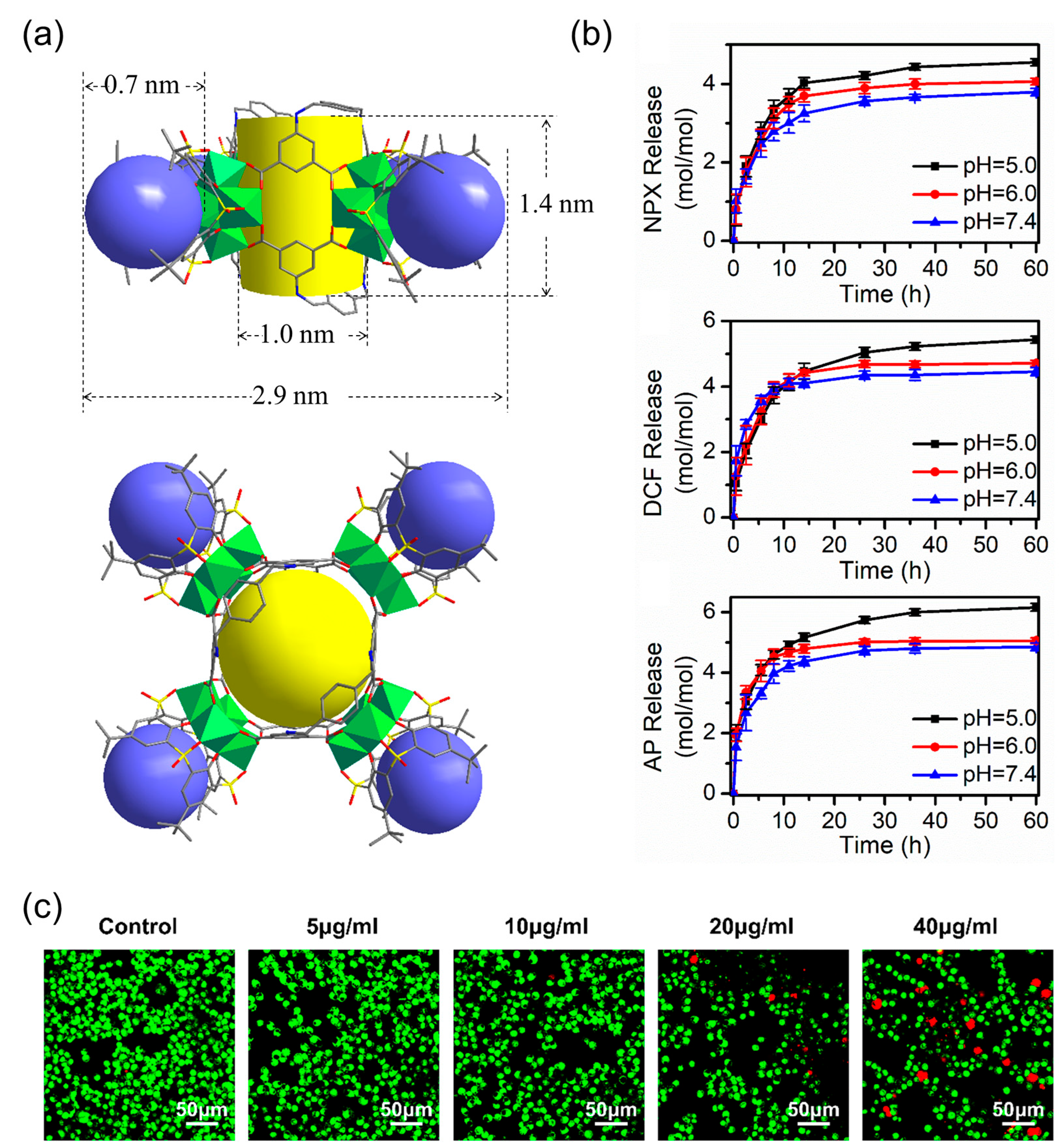
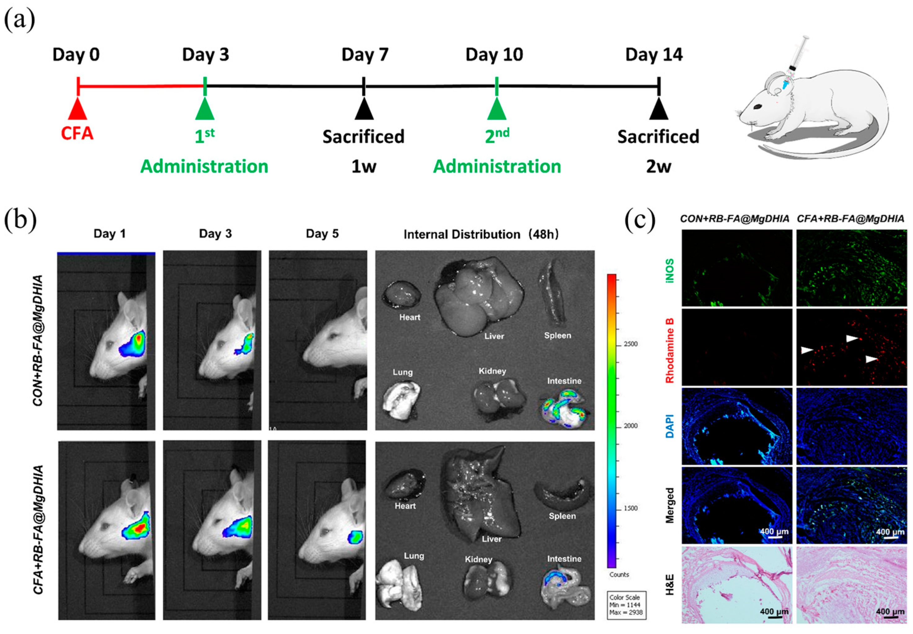
| MgDHIA |  | Mg2+ | TBSC | Type-IIIbarrel-shaped box |   | Anti-inflammatory therapy in temporomandibular joint | [56] |
| ZnPMTC |  | Zn2+ | TBSC | Type-IIIbarrel-shaped box |  | Drug loading and releasing | [57] |
| Co-NH-pyr |  | Co2+ | TBSC | Type-IIIbarrel-shaped box | Proton sponge | [46] | |
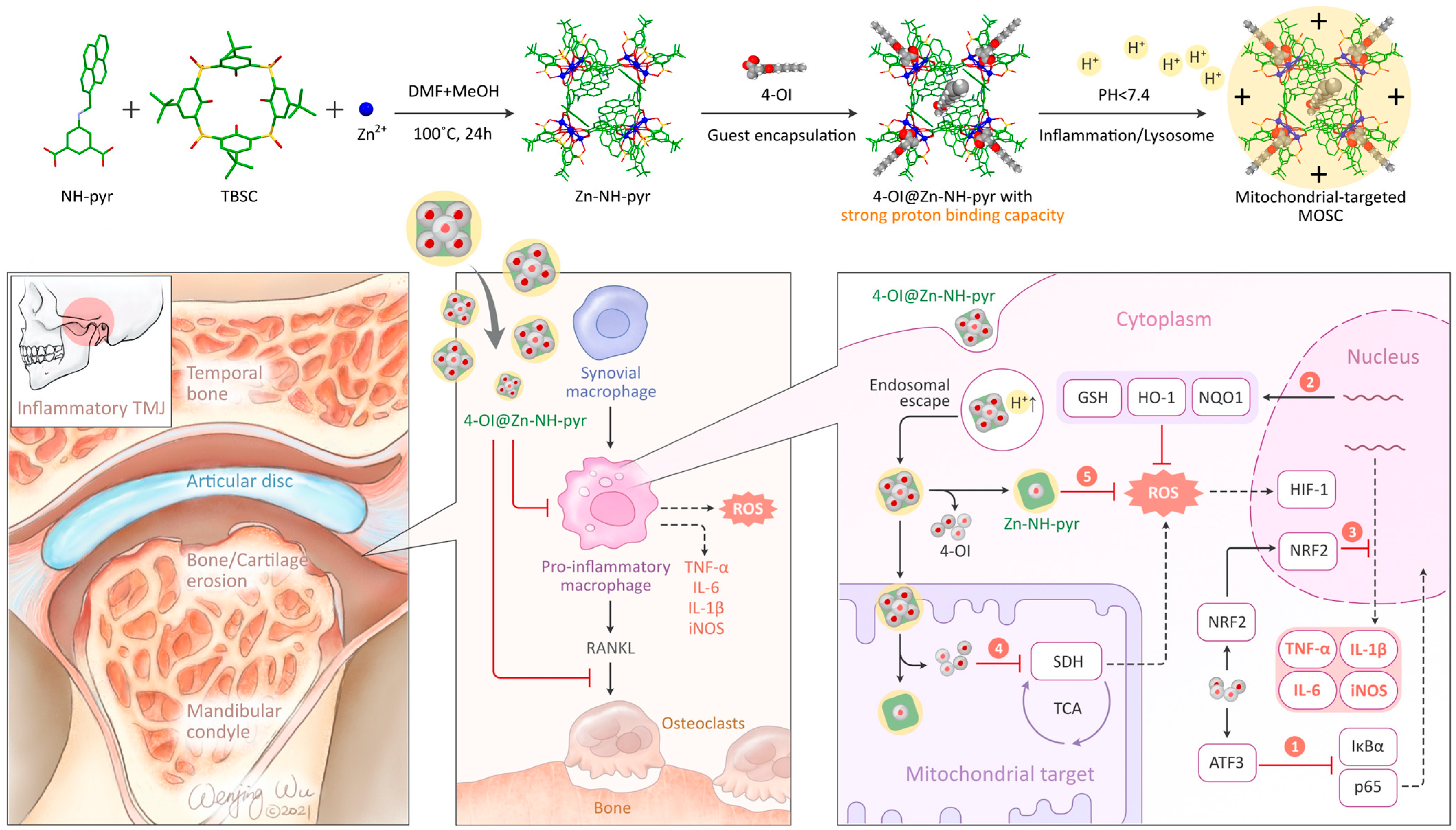
| Zn-NH-pyr | Zn2+ | TBSC | Type-IIIbarrel-shaped box |  | Synergistic therapy of joint inflammation | [58] | |
| Tb-TBSC | - | Tb3+ | TBSC | Dinuclear terbium(III) complex | - | Urinary 1-hydroxypyrene (1-OHP) detection | [59] |
| PCC-1 |  | Zn2+ | TBSC | Face-directed octahedron |  | Cancer nano-therapy | [38] |
| PCC-2 | BTB | Co2+ |  | Face-directed octahedron | CPT | Cancer nano-therapy | [38] |
Disclaimer/Publisher’s Note: The statements, opinions and data contained in all publications are solely those of the individual author(s) and contributor(s) and not of MDPI and/or the editor(s). MDPI and/or the editor(s) disclaim responsibility for any injury to people or property resulting from any ideas, methods, instructions or products referred to in the content. |
© 2024 by the authors. Licensee MDPI, Basel, Switzerland. This article is an open access article distributed under the terms and conditions of the Creative Commons Attribution (CC BY) license (https://creativecommons.org/licenses/by/4.0/).
Share and Cite
Fan, Y.-W.; Shi, M.-X.; Wang, Z.; Dai, F.-R.; Chen, Z.-N. Biomedical Applications of Sulfonylcalix[4]arene-Based Metal–Organic Supercontainers. Molecules 2024, 29, 1220. https://doi.org/10.3390/molecules29061220
Fan Y-W, Shi M-X, Wang Z, Dai F-R, Chen Z-N. Biomedical Applications of Sulfonylcalix[4]arene-Based Metal–Organic Supercontainers. Molecules. 2024; 29(6):1220. https://doi.org/10.3390/molecules29061220
Chicago/Turabian StyleFan, Ya-Wen, Meng-Xue Shi, Zhenqiang Wang, Feng-Rong Dai, and Zhong-Ning Chen. 2024. "Biomedical Applications of Sulfonylcalix[4]arene-Based Metal–Organic Supercontainers" Molecules 29, no. 6: 1220. https://doi.org/10.3390/molecules29061220