Article

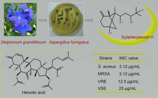
Chemical Constituents and Bioactivities of the Plant-Derived Fungus Aspergillus fumigatus

1
Hunan Key Laboratory of Diagnostic and Therapeutic Drug Rsearch for Chronic Diseases, Xiangya School of Pharmaceutical Sciences, Central South University, Changsha 410013, China
2
National Engineering Research Center of Navel Orange, Gannan Normal University, Ganzhou 341000, China
3
Key Laboratory of South China Agricultural Plant Molecular Analysis and Genetic Improvement, Guangdong Provincial Key Laboratory of Applied Botany, South China Botanical Garden, Chinese Academy of Sciences, Guangzhou 510650, China
*
Authors to whom correspondence should be addressed.
These authors contributed equally to this work.
Molecules 2024, 29(3), 649; https://doi.org/10.3390/molecules29030649
Submission received: 8 December 2023 / Revised: 11 January 2024 / Accepted: 27 January 2024 / Published: 30 January 2024

Abstract

:
A new bergamotane sesquiterpenoid, named xylariterpenoid H (1), along with fourteen known compounds (215), were isolated from the crude extract of Aspergillus fumigatus, an endophytic fungus isolated from Delphinium grandiflorum L. Their structures were elucidated mainly by extensive analyses of NMR and MS spectroscopic data. In addition, the screening results of antibacterial and cytotoxic activities of compounds 115 showed that compound 4 displayed antibacterial activities against Staphylococcus aureus and MRSA (methicillin-resistant S. aureus) with an MIC value of 3.12 µg/mL.

Graphical Abstract

1. Introduction

Delphinium grandiflorum L., a tremendously important species of the genus Delphinium belonging to the family Ranunculaceae, is widely distributed in China and India [1]. Previous studies have reported the isolation and identification of a variety of biologically meaningful natural compounds from D. grandiflorum L., including diterpene alkaloids, flavonoids, and phenolic acids [2,3]. However, there are no related reports on the chemical constituents of endophytic fungi of D. grandiflorum L., which, thus, successfully aroused our research interest. Therefore, our research group conducted phytochemical and biological activity screenings on endophytic fungi isolated from D. grandiflorum L., and finally selected Aspergillus fumigatus as the targeted research strain.
The previous excellent phytochemical studies on endophytic fungi of A. fumigatus have established the presence of pyranones [4,5], terpenes [6,7], alkaloids [8], and thiophenols [9], most of which showed considerable biological activities such as antibacterial [10], anticancer [11], anti-inflammatory [12], and antioxidant activities [9]. In this study, a previously undescribed compound with the name of xylariterpenoid H (1), together with fourteen known compounds (215), was successfully isolated and identified from the endophytic fungus A. fumigatus by our research group (Figure 1). Moreover, the antibacterial and cytotoxic activities of these isolated compounds 115 were assayed, wherein compound 4 had been disclosed to display very significant antibacterial activities against Staphylococcus aureus and MRSA (methicillin-resistant S. aureus). Herein, the details of the extraction, purification, structure elucidation, and their biological evaluation are described.

2. Results and Discussion

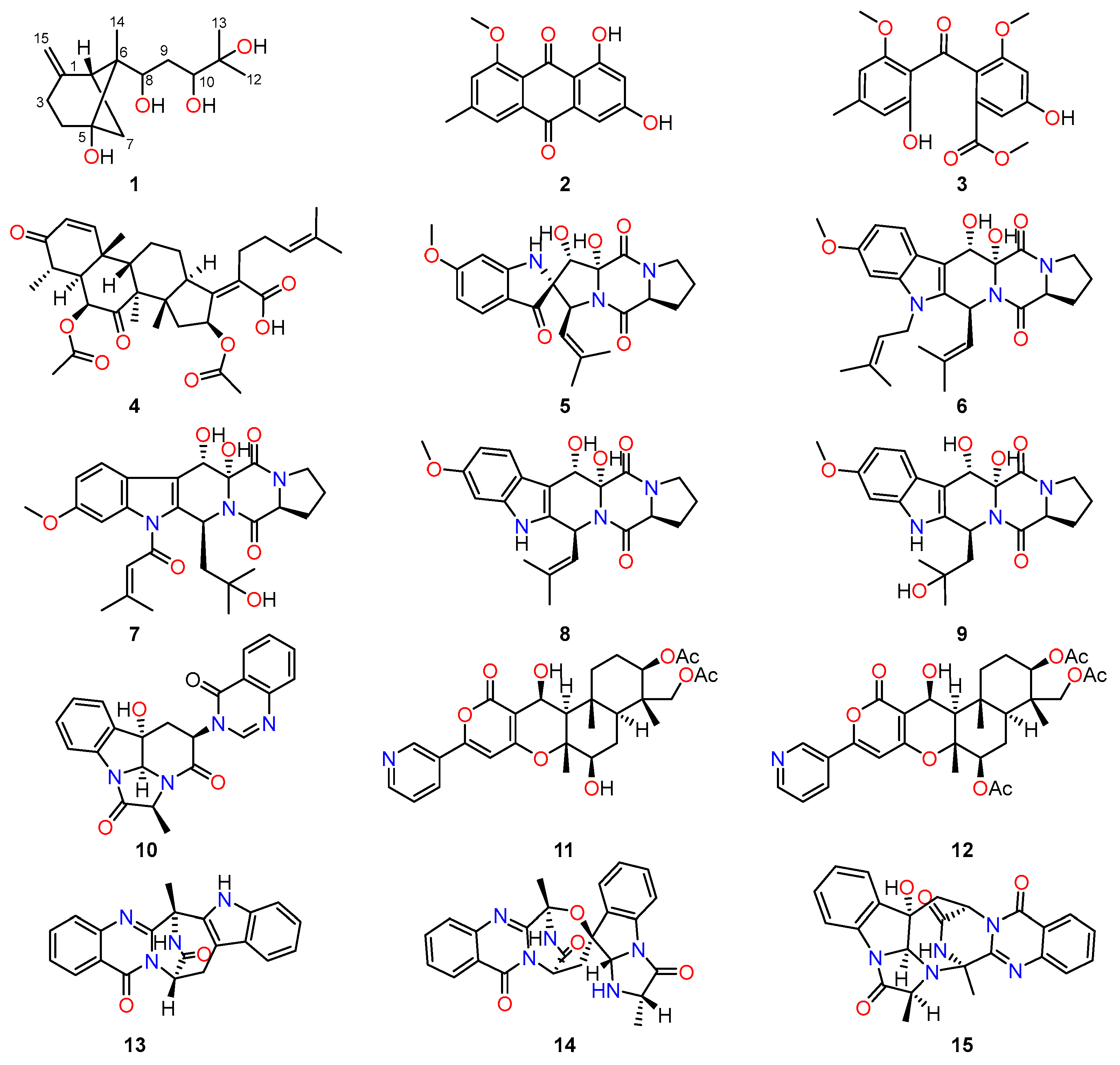
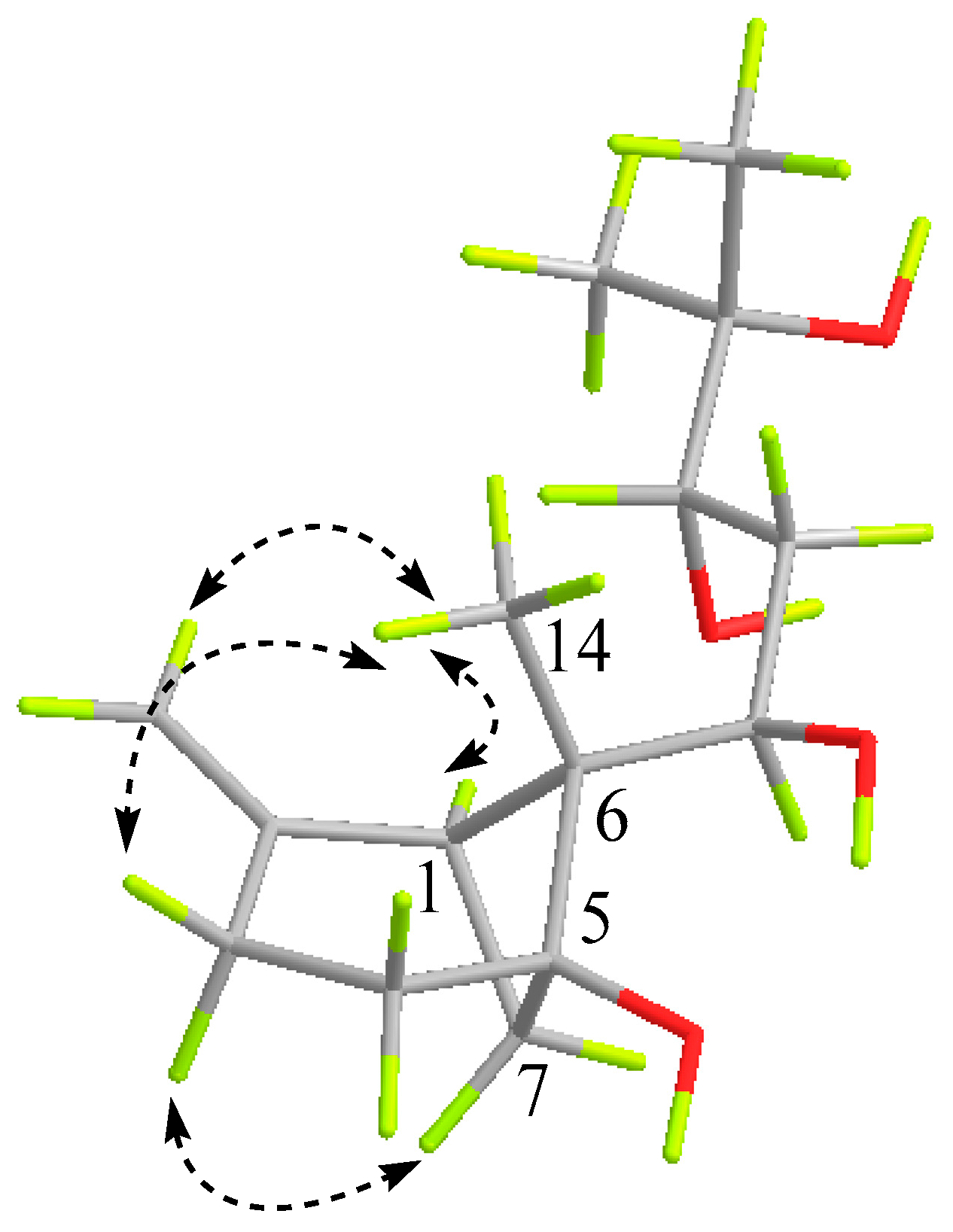
2.1. Structure Characterization of Isolated Compounds

Compound 1 was isolated as a colorless oil with the chemical molecular formula of C15H26O4 deduced by HRESIMS (Figure S1) at m/z 293.1740, [M + Na]+ (calculated for 293.1729), accounting for three degrees of unsaturation. Its IR (KBr) spectrum (Figure S4) exhibited absorptions at 3375 cm−1 (hydroxy) and 1637 cm−1 (double bond). Analyses of the 1H NMR data (Figure S5 and Table 1) revealed the presence of three singlet methyl groups (δH 0.81 (3H, s, H-14), 1.20 (3H, s, H-12), and 1.25 (3H, s, H-13)), two terminal olefin proton signals (δH 4.63 (1H, brs, H-14a), 4.69 (1H, brs, H-14b)). Furthermore, its 13C-NMR and HSQC spectra (Figures S6 and S8) exhibited the signals of 15 carbon resonances, including three methyls (δC 10.5, 23.9, and 26.4), four methylenes (δC 25.2, 31.7, 33.5, and 36.1), three methines involving an olefinic carbon (δC 70.4, 74.9, and 107.9), and four nonprotonated carbons at δC 52.3, 73.2, 76.7, and 147.7. These results together with the molecular formula, suggested that compound 1 was most likely a sesquiterpenoid. Considering the three degrees of unsaturation in the molecule and the terminal olefin double bond accounting for one of the degrees of unsaturation, the remaining two degrees of hydrogen deficiency necessitated compound 1 should possess a bicyclic ring system.
In order to construct the bicyclic skeleton of compound 1, the 2 D NMR spectra involving both to the HMBC and 1H-1H COSY (Figure 2) spectra were performed and elucidated. The HMBC spectrum (Figure S9) showed the cross peak from the terminal olefin proton H-15 (δH 4.69 and 4.63) to C-1 (δC 42.0), C-2 (δC 147.7), and C-3 (δC 25.2), from H-3 (δH 2.36 and 2.63) and H-7 (δH 1.91 and 2.47) to C-5 (δC 76.7). Along with the COSY correlations (Figure S7) of H-1 (δH 2.35) with H-7 and of H-3 with H-4 (δH 1.79 and 1.98) indicated the presence of a 4-methylene cyclohexanol ring in the molecule. Additionally, the HMBC correlations of H-14 (δH 0.82) with C-1 (δC 42.0), and C-5 (δC 76.7), of H-7 with C-4 and C-6 (δC 52.3) suggested that the presence of a 6-methylbicyclo [3.1.1] heptane skeleton. In addition, based on the 1H-1H COSY correlations from H-8 to H-10, the HMBC correlations of the methylene proton H-9 (δH 1.35) with the oxygenated carbon C-11 (δC 73.2) and of the methyl protons H-12 (δH 1.24) and H-13 (δH 0.82) with C-10 (δC 74.9) and C-11 suggested the presence of a 1,3,4-trihydroxyl-4-methylpent side chain in 1. Finally, the linkage of the two moieties was secured by the HMBC correlations of H-9 (δH 1.35) with C-6 (δC 52.3) as well as of H-14 to C-8. On the basis of the above evidence, the planar structure of compound 1 was thus established, which suggested that compound 1 should be a new bergamotane sesquiterpene. This type of compound was once isolated from a deep-sea-derived fungus [13]. Following the naming of this type of compound by Niu et al., the name of compound 1 was determined to be xyloterpene H.
The partial relative configuration of 1 was confirmed by the NOESY experiment (Figure S10 and Figure 3), based on the informative NOE correlations observed between H-3α/H-7α, which suggested that the two protons were cofacial and were arbitrarily assigned as α-orientation. The critical NOE interactions observed between H-3β/H3-14, H-3β/H2-15, and H-1/H3-14 indicated H-1 and H3-14 were oriented in the same direction. Then, the relative configuration of the cyclohexane ring and bridged cyclobutane ring were established.
However, the relative configuration of 1,3-dihydroxyl functionality for C-8 and C-10 positions in compound 1 was a failure to be determined. Although the mosher ester strategy towards the determination of the absolute configuration of this 1,3-dihydroxyl moiety was conducted, it provided a complex mixture, probably attributing to the presence of four free hydroxyl groups. The acetonide derivation of the 1,3-dihydroxyl moiety with acetone was also performed to establish the relative configuration, whereas it generated the acetonide product of C-10 and C-11 hydroxyls with low yield. Moreover, the ECD and 13C NMR calculations were also evidenced to be inefficient due to the existence of too many probable configurations caused by the two unestablished chiral centers. Therefore, the relative and absolute configurations of compound 1 had not been completely determined because of its limited amount and intractable structure characteristic in this study.
Notably, fourteen known compounds were also successfully isolated from the endophytic fungus A. fumigatus, and their structures were then identified as 1-methyl emodin (2) [14], monomethylsulochrin (3) [15], helvolic acid (4) [16], spiro-[5H,10H-dipyrrolo[1,2-a:1′,2′-d]pyrazine-2-(3H),2′-[2H]indole]-3′,5,10(1′H)-trione (5) [17], fumitremorgin B (6) [18], asperfumigatin (7) [10], 12,13-dihydroxyfumitremorgin C (8) [19], verruculogen TR-2 (9) [20], chaetominine (10) [21], 7-deacetylpyripyropene A (11) [22], pyripyropene A (12) [22,23], fumiquinazoline J (13) [24], fumiquinazoline C (14) [25], and fumiquinazoline D (15) [25] by comparing their spectroscopic data (Figures S11–S38) with those of the reported literatures. The structures of these known compounds are shown in Figure 1.

2.2. Antibacterial Activity

All of the isolated compounds were evaluated for their antibacterial activities against the Gram-positive bacteria S. aureus and MRSA by the microbroth dilution method [26]. As a result (Table S1 and Figure S39), among these tested compounds, helvolic acid (4) exhibited potent antibacterial activity against S. aureus and MRSA with MIC values of 3.12 μg/mL. Moreover, compound 3 exhibited modest antibacterial activity against S. aureus and MRSA with MIC values of 20 μg/mL. Unfortunately, the MIC values of compound 1 for all tested strains exceeded 100 μg/mL, and other compounds did not show any significant antibacterial activities. In order to evaluate the effect of helvolic acid on other strains, vancomycin-resistant Enterococci (VRE), vancomycin-sensitive Enterococci (VSE), and Gram-negative bacterium Shigella dysenteriae were chosen to perform the antibacterial experiments, and the biological screening results illustrated that the MIC values for helvolic acid towards these tested strains were 12.5, 25, and 100 μg/mL, respectively (Table S2). The results collectively pointed to helvolic acid (4) showing broad antibacterial spectrum with significant activities for the development of antibacterial innovative drugs.

2.3. Cytotoxic Activity

In addition, the antiproliferative effects of the isolated compounds 115 were further evaluated by a panel of human cancer cell lines, including Hela, HepG2, and A549. However, none of them showed any noticeable cytotoxic activity, even at the concentration of 50 μM. Among them, the inhibitory rates of compound 1 against A549, Hela, and HepG2 at 50 μM were 25.46%, 31.90%, and 28.55%, respectively; the inhibitory rates of compound 4 against A549, Hela, and HepG2 at 50 μM were 64.81%, 30.54%, and 69.12%, respectively. The intriguing result of neglectable cytotoxicity for helvolic acid (4) tentatively suggested that helvolic acid (4) could exhibit significant biological activities against a broad panel of bacteria with potent selectivity, which thus strongly indicated that helvolic acid might serve as a promising lead compound for the further development of anti-infective innovative drugs with limited cytotoxicity in future.

2.4. Discussion

The microbial community can be described as a “bio-diversified tropical rainforest”. It contains a large number of biologically active substances, which are a series of tremendously important sources of new drugs and active leads [27]. The helvolic acid isolated from A. fumigatus belongs to the fusidane-type antibiotics, and it has remarkable antibacterial activity against Gram-positive bacteria, especially S. aureus. Fusidane-type antibiotics belong to the only type of fungal triterpene with proterpene alcohol as the mother core, representing the only triterpene-derived antibiotic class [28], and they have been known for nearly 80 years [29]. The two representative drugs are cephalosporin P1 and fusidic acid, of which fusidic acid has been widely used in clinical therapeutics [30,31].
Currently, commercially available antibiotics with different mechanisms of action are experiencing resistance crises to varying degrees. However, the rate of development of bacterial resistance is much faster than the rate of antibiotic development, and resistant strains towards all of the usually-used antibiotics have been clinically detected. Therefore, the continuation of exploring new drug targets to meet the challenge of the antibiotic crisis is still extremely appealing. To our surprise, fusidane-type antibiotic helvolic acid (4) exhibited potent antibacterial activity against MRSA with a MIC value of 3.12 μg/mL. Notably, fusidane-type antibiotics are the only known antibiotics that selectively target bacteria elongation factor G (EF-G) [32] to show potent bacteriostatic and bactericidal effects [33]. The specific antibacterial mechanism of the fusidane-type antibiotics logically indicates that helvolic acid (4) might lead to little antibacterial cross-resistance in comparison with other commonly used antibiotics.
Helvolic acid (4) possesses intriguing structural features and excellent biological activity, which aroused an emerging new interest among chemists and biologists regarding the growing threat of antibiotic resistance. After the identification of the helvolic acid biosynthetic gene cluster (BGC) of A. fumigatus Af 293 in 2009, biosynthetic research on fusidane-type antibiotics has developed vigorously [34,35], and the biosynthetic pathway of helvolic acid has been fully proposed so far [36]. In this study, helvolic acid (4) demonstrated potent activity against bacterial pathogens, suggesting it was responsible for the antimicrobial activity initially observed in the crude extract of A. fumigatus. In future, the biosynthetic synthesis with epigenetic regulation of A. fumigatus towards the abundant generation of helvolic acid (4) is also appealed for the devolvement of A. fumigatus as a promising antibacterial biological agent.
Alkaloid molecules contain an N atom and have great structural diversity. Depending on the function of the amine, alkaloids can act as either a hydrogen-receptor or a hydrogen-donor for hydrogen bonding, which is crucial for the drug to exert its function [37]. It is worth mentioning that we have isolated multiple different types of alkaloids from A. fumigatus, including indole diketopiperazine alkaloids, quinazoline alkaloids, and pyridine alkaloids. According to literature reports, indole diketopiperazine alkaloids (IDAs) have significant pharmacological activities such as antimicrobial [38,39,40,41,42], antiviral [43,44,45,46], anticancer [47,48,49], immunomodulatory [50], antioxidant [51], and insecticidal activities [52]. Therefore, they may have promising potential to be used in drugs and/or serve as lead structures for drug development. Meanwhile, quinazoline alkaloids (QAs) as a series of heterocyclic compounds with nitrogen are one of the most significant heterocyclic motifs with diverse chemical reactivities and biological applications [53,54]. Especially, their derivatives play a crucial role in medicinal chemistry, evident in the chemical makeup of a wide range of FDA approved medications, clinical candidates, and bioactive compounds [55]. Unfortunately, none of the isolated alkaloids did not show any obvious antibacterial or cytotoxic activity in our preliminary pharmacological activity experiments. In future, the research efforts on the structural and pharmacological diversities of IDAs and QAs from the endophytic fungi A. fumigatus were still required to disclose their potent pharmacological applications.
During the isolation of A. fumigatus, we isolated one new compound and fourteen old compounds. By reviewing the literature, we found that changing some experimental conditions may be able to obtain more novel secondary metabolites. For example, adding 3-hydroxytyrosol, a new signaling molecule in fungi that can regulate biofilm growth, to the culture medium promoted the biotransformation process [56]. Inoculating medicinal plants with arbuscular mycorrhizal fungi (AMF) represents an alternative approach to enhance the quality and quantity of secondary metabolites. AMF can form endophytes or symbiotic relationships with numerous microorganisms in different parts of the plant. Subsequently, they influence the production of secondary metabolites by indirectly stimulating the biosynthetic pathways of these compounds [57].
Moreover, the strain of Aspergillus used in this experiment possesses enzymes such as cytochrome P450s (CYPs) with broad substrate specificity. A. fumigatus and its enzymes exhibit significant potential in biotransformation, bioremediation of environmental contaminants, and the biocatalytic production of essential compounds [58].

3. Materials and Methods

3.1. General Experimental Procedures

Optical rotations were measured on an MCP-500 spectropolarimeter (Anton Paar, Graz, Austria). UV spectra and ECD spectra were acquired on a UV-2600 spectrophotometer (Shimadzu, Kyoto, Japan). IR spectra were obtained on an Affinity-1 spectrometer (Shimadzu, Kyoto, Japan) using KBr discs. NMR spectra were recorded on a Bruker Avance-500 spectrometer (Bruker, Fällanden, Switzerland) using residual solvent signals as references. HRESIMS data were acquired by the Thermo MAT95XP spectrometer (Thermo Fisher Scientific, Bremen, Germany). Silica gel (80–100, 100–200, and 200–300 mesh, Qingdao Puke Parting Materials Co., Ltd., Qingdao, China) used for flash column chromatography was purchased commercially. The TLC analysis was carried out using commercially available silica gel plates (Qingdao Puke Parting Materials Co., Ltd., Qingdao, China). A Hitachi Primaide (Hitachi Instruments (Dalian) Co., Ltd., Dalian, China) equipped with a diode array detector (DAD) using a preparative YMC ODS C18 column (20 mm × 250 mm, 5 μm) was used for semipreparative HPLC separation. All solvents were analytical grade and used without further purification (Guangzhou Chemical Regents Company, Ltd., Guangzhou, China).

3.2. Fungal Material and Fermentation

The fungus A. fumigatus was isolated from Delphinium grandiflorum L., which was collected from the Aba Tibetan Autonomous Prefecture in July 2021. The plant species was authenticated on the basis of morphological characteristics and comparison with specimens, and the fungal species was authenticated on the basis of morphological characteristics and ITS DNA sequence data (GenBank: No. MT529485.1). The strain was preserved at Xiangya School of Pharmaceutical Sciences, Central South University, Changsha. The strain was cultured on potato dextrose agar (PDA) at 28 °C for 5 days containing 200 g/L of potato, 20 g/L of glucose, 3 g/L of KH2PO4, 1.5 g/L of MgSO4•7H2O, and 10 mg/L of vitamin B1 in distilled water. Then, a quarter of the agar with fungal colony was added to a conical flask (500 mL) with 250 mL of potato dextrose liquid medium, and the flask was incubated on a rotary shaker at 28 °C and 140 rpm for 5 days to prepare seed culture. Agar plugs were inoculated into 80 Erlenmeyer flasks (1 L) that were previously sterilized by autoclaving, with each containing 250 g of rice and 200 mL of distilled water. All flasks were incubated at 28 °C for 30 days.

3.3. Extraction and Isolation

The fermented rice substrate was extracted 3 times with EtOAc at room temperature, and the solvent was evaporated under vacuum to yield a total extract (50.9 g). The crude extract was subjected to silica gel column chromatography eluting with petroleum ether and EtOAc (100:1 to 1:1, v/v) as well as EtOAc and MeOH (1:1 to 1:5, v/v) to afford six main fractions (Fr. 1–Fr. 6).
Fraction 3 (7.5 g) was fractionated by an ODS column chromatography eluted with a gradient of MeOH-H2O (v/v, 40:60 → 100:0) to obtain seven subfractions (Fr.3-1 to Fr.3-7). Fr.3-2 (1.5 g) was separated by Sephadex LH-20 CC and eluted with CH2Cl2-MeOH (v/v, 1:3) to afford five subfractions (Fr.3-2-1 to Fr.3-2-5). Fr.3-2-3 was further fractionated by using semipreparative HPLC (MeCN-H2O, 50:50, v = 2.0 mL/min) to give compound 9 (21.0 mg, tR = 8.0 min) and compound 10 (6.7 mg, tR = 10.0 min). Fr.3-2-4 was isolated on silica gel and eluted with petroleum ether-EtOAc gradient (v/v, 100:1 → 1:2) to obtain six sub-fractions (Fr.3-2-4-1 to Fr.3-2-4-6). Fr.3-2-4-3 was further purified by silica gel, eluting with CH2Cl2-MeOH (v/v, 1:0 → 20:1) to afford compound 5 (4.2 mg). Fr.3-2-4-6 was further purified by silica gel, eluting with ether-EtOAc (v/v, 1:2 → 1:5) to afford compound 1 (4.9 mg).
Fr.3-3 (1.1 g) was separated by Sephadex LH-20 CC and eluted with CH2Cl2-MeOH (v/v, 1:3) to afford six subfractions (Fr.3-3-1 to Fr.3-3-6). Fr.3-3-3 was further fractionated by using semipreparative HPLC (MeCN-H2O, 60:40, v = 2.0 mL/min) to give compound 11 (4.2 mg, tR = 12.0 min). Fr.3-3-5 was further fractionated by using semipreparative HPLC (MeCN-H2O, 65:35, v = 2.0 mL/min) to give compound 8 (3.7 mg, tR = 22.0 min). Fr.3-4 (0.7 g) was separated by Sephadex LH-20 CC and eluted with CH2Cl2-MeOH (v/v, 1:3) to afford four subfractions (Fr.3-4-1 to Fr.3-4-4). Fr.3-4-1 was further fractionated by using silica gel, eluting with CH2Cl2-MeOH (v/v, 1:0 → 50:1) to afford compound 12 (11.2 mg). Fr.3-4-2 was purified by silica gel and eluted with CH2Cl2-MeOH gradient (v/v, 100:1 → 10:1) to obtain compound 7 (7.5 mg). Fr.3-5 (1.6 g) was separated by Sephadex LH-20 CC and eluted with CH2Cl2-MeOH (v/v, 1:3) to afford five subfractions (Fr.3-5-1 to Fr.3-5-5). Fr.3-5-4 was isolated on silica gel and eluted with petroleum ether-EtOAc gradient (v/v, 100:1 → 1:2) to obtain compound 6 (25.5 mg).
Fraction 2 (8.2 g) was fractionated by an ODS column chromatography eluted with a gradient of MeOH-H2O (v/v, 40:60 → 100:0) to obtain six subfractions (Fr.2-1 to Fr.2-6). Fr.2-2 (1.5 g) was separated by Sephadex LH-20 CC and eluted with CH2Cl2-MeOH (v/v, 1:3) to afford four subfractions (Fr.2-2-1 to Fr.2-2-4). Fr.2-2-2 was purified by silica gel and eluted with CH2Cl2-MeOH gradient (v/v, 100:1 → 10:1) to obtain compound 14 (79.0 mg). Fr.2-2-6 was further fractionated by using semipreparative HPLC (MeCN-H2O, 60:40, v = 2.0 mL/min) to give compound 15 (2.8 mg, tR = 14.0 min). Fr.2-3 (0.6 g) was separated by Sephadex LH-20 CC and eluted with CH2Cl2-MeOH (v/v, 1:3) to afford five subfractions (Fr.2-3-1 to Fr.2-3-5). Fr.2-3-5 was purified by silica gel and eluted with ether-EtOAc (v/v, 10:1 → 1:1) to obtain compounds 2 (4.4 mg) and 13 (11.8 mg). In addition, a white solid was precipitated in Fr.2-3 to give compound 3 (55.0 mg), and a large amount of white solid was precipitated in Fr.2-5 to give compound 4 (808.0 mg).
Xylariterpenoid H (1): colorless oil; [α]25D −0.15 (c 0.1, MeOH); ECD (MeOH) λmax (Δε): 200 (+11.84) nm; UV (MeOH) λmax (log ε): 200 (1.92) nm; IR (KBr): 3853, 3375, 2922, 2852, 1734, 1637, 1465, 1186, 962, 721, 518 cm−1, 1H (500 MHz) and 13C (125 MHz) NMR data see Table 1. HRESIMS: m/z 293.1740 [M + Na]+ (calculated for C15H26O4Na, 293.1729).

3.4. Cytotoxic Activity Assay

Cytotoxic viability was determined by using the SRB method [59]. The cell lines (Hela, HepG2, and A549) were cultured in RPMI-1640 medium with 10% fetal bovine serum at 37 °C. The suspended cells were seeded in 96-well plates at a density of 3 × 10⁴ cells/mL in an incubator under an atmosphere of 5% CO2 at 37 °C for 24 h. Then, 20 μL of various concentrations of compounds were added and further incubated for 72 h. After that, the cell monolayers were fixed by 50% (wt/v) trichloroacetic acid (50 μL) and stained for 30 min by 0.4% (wt/v) SRB, which was dissolved in 1% acetic acid. The unbound dye was removed by washing repeatedly with 1% acetic acid, and the resulting cells were then dissolved the protein-bound dye in 10 mM Tris base solution (200 μL), and the absorbance was measured at 570 nm. Cisplatin was used as a positive control possessing potent cytotoxic activity. All data were obtained in triplicate and are presented as means ± S.D.

3.5. Antibacterial Assay

All isolated compounds were evaluated against bacteria strains embodying S. aureus (CMCC 26003), MRSA (NCTC 10442), Escherichia coli (ATCC 8739), VRE (No. 151458137), and VSE (No. 160119481), all of which were obtained from Guangdong Microbiology Culture Center (Guangzhou, China). MIC values were determined by the methodology of microbroth dilution in Mueller–Hinton broth medium (MHB) according to CLSI guidelines; the positive control was vancomycin or polymyxin B. Briefly, 20 μL tested compounds with a concentration of 1 mg/mL was added to 180 μL bacterial liquid, and the method of double dilution was adopted in 96-well plates. The lowest concentration of the drug preventing visible growth of the pathogen was taken as the MIC.

4. Conclusions

In summary, this study performed a comprehensive chemical investigation on the bioactive natural product of the endophytic fungi A. fumigatus isolated from Delphinium grandiflorum L., and it has resulted in the successful isolation and structure identification of an undescribed compound xylariterpenoid H (1) together with fourteen known compounds (215). The biological activity screening of these isolates revealed that helvolic acid (4) exhibited significantly potent antibacterial activities against S. aureus and MRSA, which were comparable to the positive control vancomycin without any significant cytotoxicity, revealing tremendous promise in the development of innovative anti-infective drugs. These findings not only disclose the biological chemical constitute of A. fumigatus but also note the further development potential of Delphinium grandiflorum L. Furthermore, the antibacterial mechanism experiments towards the bioactive lead compound helvolic acid (4) are now underway and will be revealed in due course.

Supplementary Materials

The following supporting information can be downloaded at: https://www.mdpi.com/article/10.3390/molecules29030649/s1, Figure S1: HRESIMS spectrum of compound 1; Figure S2: UV spectrum of compound 1; Figure S3: ECD spectrum of compound 1; Figure S4: IR spectrum of compound 1; Figure S5: 1H NMR spectrum (500 MHz, CDCl3) of compound 1; Figure S6: 13C NMR spectrum (125 MHz, CDCl3) of compound 1; Figure S7: 1H-1H COSY spectrum of compound 1; Figure S8: HSQC spectrum of compound 1; Figure S9: HMBC spectrum of compound 1; Figure S10: NOESY spectrum of compound 1; Figure S11: 1H NMR spectrum (500 MHz, CD3OD) of compound 2; Figure S12: 13C NMR spectrum (125 MHz, CD3OD) of compound 2; Figure S13: 1H NMR spectrum (500 MHz, DMSO-d6) of compound 3; Figure S14: 13C NMR spectrum (125 MHz, DMSO-d6) of compound 3; Figure S15: 1H NMR spectrum (500 MHz, CDCl3) of compound 4; Figure S16: 13C NMR spectrum (125 MHz, CDCl3) of compound 4; Figure S17: 1H NMR spectrum (500 MHz, CDCl3) of compound 5; Figure S18: 13C NMR spectrum (125 MHz, CDCl3) of compound 5; Figure S19: 1H NMR spectrum (500 MHz, CDCl3) of compound 6; Figure S20: 13C NMR spectrum (125 MHz, CDCl3) of compound 6; Figure S21: 1H NMR spectrum (500 MHz, CDCl3) of compound 7; Figure S22: 13C NMR spectrum (125 MHz, CDCl3) of compound 7; Figure S23: 1H NMR spectrum (500 MHz, CDCl3) of compound 8; Figure S24: 13C NMR spectrum (125 MHz, CDCl3) of compound 8; Figure S25: 1H NMR spectrum (500 MHz, DMSO-d6) of compound 9; Figure S26: 13C NMR spectrum (125 MHz, DMSO-d6) of compound 9; Figure S27: 1H NMR spectrum (500 MHz, CD3OD) of compound 10; Figure S28: 13C NMR spectrum (125 MHz, CD3OD) of compound 10; Figure S29: 1H NMR spectrum (500 MHz, CDCl3) of compound 11; Figure S30: 13C NMR spectrum (125 MHz, CDCl3) of compound 11; Figure S31: 1H NMR spectrum (500 MHz, CDCl3) of compound 12; Figure S32: 13C NMR spectrum (125 MHz, CDCl3) of compound 12; Figure S33: 1H NMR spectrum (500 MHz, CDCl3) of compound 13; Figure S34: 13C NMR spectrum (125 MHz, CDCl3) of compound 13; Figure S35: 1H NMR spectrum (500 MHz, CDCl3) of compound 14; Figure S36: 13C NMR spectrum (125 MHz, CDCl3) of compound 14; Figure S37: 1H NMR spectrum (500 MHz, CDCl3) of compound 15; Figure S38: 13C NMR spectrum (125 MHz, CDCl3) of compound 15; Table S1: Evaluation of antibacterial activity of compounds 115; Figure S39: 96-well plate antibacterial results; Table S2: Evaluation of antibacterial activity of compounds 4; Excel: preliminary screening data of cytotoxic activity of compounds 115.

Author Contributions

Conceptualization, H.T. and Z.Z.; methodology, Z.S. and Y.Z.(Yanjiang Zhang); software, K.Q.; validation, H.T., Z.Z. and Y.Z. (Yuting Zheng); formal analysis, Z.S.; investigation, C.C.; resources, L.X.; data curation, Z.S.; writing—original draft preparation, Z.S.; writing—review and editing, Z.S., Y.Z. (Yanjiang Zhang) and H.T.; visualization, J.L.; supervision, H.T. and Z.Z. All authors have read and agreed to the published version of the manuscript.

Funding

Financial support for this research was provided by the South China Botanical Garden, Chinese Academy of Sciences (Granted No: QNXM-02), the Key Research and Development Project of Hainan Province (No. ZDYF2022SHFZ048), the Natural Science Foundation of Hunan Province (No. 2021JJ30917), the Youth Innovation Promotion Association of CAS (2020342), the National Natural Science Foundation of China (No. 82173711), and the Open Sharing Fund for the Large-Scale Instruments and Equipment of Central South University.

Institutional Review Board Statement

Not applicable.

Informed Consent Statement

Not applicable.

Data Availability Statement

All the data, including HRESIMS, IR, UV, 1D/2D NMR, and CD spectra, are available in this publication and the Supplementary Materials.

Conflicts of Interest

The authors declare no conflicts of interest.

References

  1. Chen, F.Z.; Chen, D.L.; Chen, Q.H.; Wang, F.P. Diterpenoid alkaloids from Delphinium majus. J. Nat. Prod. 2009, 72, 18–23. [Google Scholar] [CrossRef]
  2. Marin, C.; Ramirez-Macias, I.; Lopez-Cespedes, A.; Olmo, F.; Villegas, N.; Diaz, J.G.; Rosales, M.J.; Gutierrez-Sanchez, R.; Sanchez-Moreno, M. In vitro and in vivo trypanocidal activity of flavonoids from Delphinium staphisagria against chagas disease. J. Nat. Prod. 2011, 74, 744–750. [Google Scholar] [CrossRef]
  3. Shen, Y.; Liang, W.J.; Shi, Y.N.; Kennelly, E.J.; Zhao, D.K. Structural diversity, bioactivities, and biosynthesis of natural diterpenoid alkaloids. Nat. Prod. Rep. 2020, 37, 763–796. [Google Scholar] [CrossRef]
  4. Li, T.X.; Meng, D.D.; Wang, Y.; An, J.L.; Bai, J.F.; Jia, X.W.; Xu, C.P. Antioxidant coumarin and pyrone derivatives from the insect-associated fungus Aspergillus versicolor. Nat. Prod. Res. 2020, 34, 1360–1365. [Google Scholar] [CrossRef] [PubMed]
  5. Wu, C.Z.; Peng, X.P.; Li, G.; Wang, Q.; Lou, H.X. Naphtho-gamma-pyrones (NγPs) with obvious cholesterol absorption inhibitory activity from the marine-derived fungus Aspergillus niger S-48. Molecules 2022, 27, 2514. [Google Scholar] [CrossRef] [PubMed]
  6. Yin, G.P.; Gong, M.; Li, Y.J.; Zhang, X.; Zhu, J.J.; Hu, C.H. 14-Membered resorcylic acid lactone derivatives with their anti-inflammatory from the fungus Aspergillus sp. ZJ-65. Fitoterapia 2021, 151, 104884. [Google Scholar] [CrossRef] [PubMed]
  7. Chaiyosang, B.; Kanokmedhakul, K.; Yodsing, N.; Boonlue, S.; Yang, J.X.; Wang, Y.A.; Andersen, R.J.; Yahuafai, J.; Kanokmedhakul, S. Three new indole diterpenoids from Aspergillus aculeatus KKU-CT2. Nat. Prod. Res. 2022, 36, 4973–4981. [Google Scholar] [CrossRef] [PubMed]
  8. Liang, Z.; Zhang, T.; Zhang, X.; Zhang, J.; Zhao, C. An alkaloid and a steroid from the endophytic fungus Aspergillus fumigatus. Molecules 2015, 20, 1424–1433. [Google Scholar] [CrossRef] [PubMed]
  9. Xu, Y.; Liu, W.; Wu, D.; He, W.; Zuo, M.; Wang, D.; Fu, P.; Wang, L.; Zhu, W. Sulfur-containing phenolic compounds from the cave soil-derived Aspergillus fumigatus GZWMJZ-152. J. Nat. Prod. 2022, 85, 433–440. [Google Scholar] [CrossRef]
  10. Zhang, R.; Wang, H.; Chen, B.; Dai, H.; Sun, J.; Han, J.; Liu, H. Discovery of anti-MRSA secondary metabolites from a marine-derived fungus Aspergillus fumigatus. Mar. Drugs 2022, 20, 302. [Google Scholar] [CrossRef]
  11. Li, S.; Chen, J.F.; Qin, L.L.; Li, X.H.; Cao, Z.X.; Gu, Y.C.; Guo, D.L.; Deng, Y. Two new sesquiterpenes produced by the endophytic fungus Aspergillus fumigatus from Ligusticum wallichii. J. Asian Nat. Prod. Res. 2020, 22, 138–143. [Google Scholar] [CrossRef] [PubMed]
  12. El-hawary, S.S.; Moawad, A.S.; Bahr, H.S.; Abdelmohsen, U.R.; Mohammed, R. Natural product diversity from the endophytic fungi of the genus Aspergillus. RSC Adv. 2020, 10, 22058–22079. [Google Scholar] [CrossRef] [PubMed]
  13. Niu, S.; Xie, C.L.; Zhong, T.; Xu, W.; Luo, Z.H.; Shao, Z.; Yang, X.W. Sesquiterpenes from a deep-sea-derived fungus Graphostroma sp. MCCC 3A00421. Tetrahedron 2017, 73, 7267–7273. [Google Scholar] [CrossRef]
  14. Hawas, U.W.; El-Beih, A.A.; El-Halawany, A.M. Bioactive anthraquinones from endophytic fungus Aspergillus versicolor isolated from red sea algae. Arch. Pharm. Res. 2012, 35, 1749–1756. [Google Scholar] [CrossRef]
  15. Han, J.; Liu, M.; Jenkins, I.D.; Liu, X.; Zhang, L.; Quinn, R.J.; Feng, Y. Genome-inspired chemical exploration of marine fungus Aspergillus fumigatus MF071. Mar. Drugs 2020, 18, 352. [Google Scholar] [CrossRef]
  16. Fujimoto, H.; Negishi, E.; Yamaguchi, K.; Nishi, N.; Yamazaki, M. Isolation of new tremorgenic metabolites from an ascomycete, Corynascus setosus. Chem. Pharm. Bull. 1996, 44, 1843–1848. [Google Scholar] [CrossRef]
  17. Li, X.J.; Zhang, Q.; Zhang, A.L.; Gao, J.M. Metabolites from Aspergillus fumigatus, an endophytic fungus associated with Melia azedarach, and their antifungal, antifeedant, and toxic activities. J. Agric. Food Chem. 2012, 60, 3424–3431. [Google Scholar] [CrossRef]
  18. Yamazaki, M.; Fujimoto, H.; Kawasaki, T. Chemistry of tremorogenic metabolites. I. Fumitremorgin A from Aspergillus fumigatus. Chem. Pharm. Bull. 1980, 28, 245–254. [Google Scholar] [CrossRef] [PubMed]
  19. Afiyatullov, S.S.; Kalinovskii, A.I.; Pivkin, M.V.; Dmitrenok, P.S.; Kuznetsova, T.A. Fumitremorgins from the marine isolate of the fungus Aspergillus fumigatus. Chem. Nat. Compd. 2004, 40, 615–617. [Google Scholar] [CrossRef]
  20. Fill, T.P.; Rodrigues Asenha, H.B.; Marques, A.S.; Ferreira, A.G.; Rodrigues-Fo, E. Time course production of indole alkaloids by an endophytic strain of Penicillium brasilianum cultivated in rice. Nat. Prod. Res. 2013, 27, 967–974. [Google Scholar] [CrossRef] [PubMed]
  21. Jiao, R.H.; Xu, S.; Liu, J.Y.; Ge, H.M.; Ding, H.; Xu, C.; Zhu, H.L.; Tan, R.X. Chaetominine, a cytotoxic alkaloid produced by endophytic Chaetomium sp. IFB-E015. Org. Lett. 2006, 8, 5709–5712. [Google Scholar] [CrossRef]
  22. Lan, W.J.; Fu, S.J.; Xu, M.Y.; Liang, W.L.; Lam, C.K.; Zhong, G.H.; Xu, J.; Yang, D.P.; Li, H.J. Five new cytotoxic metabolites from the marine fungus Neosartorya pseudofischeri. Mar. Drugs 2016, 14, 18. [Google Scholar] [CrossRef]
  23. Odani, A.; Ishihara, K.; Ohtawa, M.; Tomoda, H.; Omura, S.; Nagamitsu, T. Total synthesis of pyripyropene A. Tetrahedron 2011, 67, 8195–8203. [Google Scholar] [CrossRef]
  24. Zhang, H.; Liu, R.; Yang, J.; Li, H.; Zhou, F. Bioactive alkaloids of Aspergillus fumigatus, an endophytic fungus from Astragalus membranaceus. Chem. Nat. Compd. 2017, 53, 802–805. [Google Scholar] [CrossRef]
  25. Afiyatullov, S.S.; Kalinovskii, A.I.; Pivkin, M.V.; Dmitrenok, P.S.; Kuznetsova, T.A. Alkaloids from the marine isolate of the fungus Aspergillus fumigatus. Chem. Nat. Compd. 2005, 41, 236–238. [Google Scholar] [CrossRef]
  26. Wang, M.; Huo, L.; Liu, H.; Zhao, L.; Xu, Z.; Tan, H.; Qiu, S.X. Thujasutchins N and O, two new compounds from the stems and roots of Thuja sutchuenensis. Nat. Prod. Res. 2022, 36, 2356–2362. [Google Scholar] [CrossRef]
  27. Katz, L.; Baltz, R.H. Natural product discovery: Past, present, and future. J. Ind. Microbiol. Biotechnol. 2016, 43, 155–176. [Google Scholar] [CrossRef] [PubMed]
  28. Durand, G.A.; Raoult, D.; Dubourg, G. Antibiotic discovery: History, methods and perspectives. Int. J. Antimicrob. Agents 2019, 53, 371–382. [Google Scholar] [CrossRef] [PubMed]
  29. Zhao, M.; Goedecke, T.; Gunn, J.; Duan, J.A.; Che, C.T. Protostane and fusidane triterpenes: A mini-review. Molecules 2013, 18, 4054–4080. [Google Scholar] [CrossRef]
  30. Siala, W.; Rodriguez-Villalobos, H.; Fernandes, P.; Tulkens, P.M.; Van Bambeke, F. Activities of combinations of antistaphylococcal antibiotics with fusidic acid against staphylococcal biofilms in in vitro static and dynamic models. J. Antimicrob. Chemother. 2018, 62, e00598-18. [Google Scholar] [CrossRef]
  31. Long, J.; Ji, W.; Zhang, D.; Zhu, Y.; Bi, Y. Bioactivities and structure-activity relationships of fusidic acid derivatives: A review. Front. Pharmacol. 2021, 12, 759220. [Google Scholar] [CrossRef]
  32. Zhou, J.; Lancaster, L.; Donohue, J.P.; Noller, H.F. Crystal structures of EF-G-Ribosome complexes trapped in intermediate states of translocation. Science 2013, 340, 1236086. [Google Scholar] [CrossRef] [PubMed]
  33. Tomlinson, J.H.; Kalverda, A.P.; Calabrese, A.N. Fusidic acid resistance through changes in the dynamics of the drug target. Proc. Natl. Acad. Sci. USA 2020, 117, 25523–25531. [Google Scholar] [CrossRef]
  34. Lodeiro, S.; Xiong, Q.; Wilson, W.K.; Ivanova, Y.; Smith, M.L.; May, G.S.; Matsuda, S.P.T. Protostadienol biosynthesis and metabolism in the pathogenic fungus Aspergillus fumigatus. Org. Lett. 2009, 11, 1241–1244. [Google Scholar] [CrossRef] [PubMed]
  35. Mitsuguchi, H.; Seshime, Y.; Fujii, I.; Shibuya, M.; Ebizuka, Y.; Kushiro, T. Biosynthesis of steroidal antibiotic fusidanes: Functional analysis of oxidosqualene cyclase and subsequent tailoring enzymes from Aspergillus fumigatus. J. Am. Chem. Soc. 2009, 131, 6402–6411. [Google Scholar] [CrossRef]
  36. Lv, J.M.; Hu, D.; Gao, H.; Kushiro, T.; Awakawa, T.; Chen, G.D.; Wang, C.X.; Abe, I.; Yao, X.S. Biosynthesis of helvolic acid and identification of an unusual C-4-demethylation process distinct from sterol biosynthesis. Nat. Commun. 2017, 8, 1644. [Google Scholar] [CrossRef]
  37. Silva, J.; Garcia, J.; Guimarães, R.; Palito, C.; Lemos, A.; Barros, L.; Alves, M.J. Alkaloids from Fungi. In Natural Secondary Metabolites: From Nature, Through Science, to Industry; Carocho, M., Heleno, S.A., Barros, L., Eds.; Springer: Berlin/Heidelberg, Germany, 2023; pp. 529–554. [Google Scholar]
  38. Willems, T.; De Mol, M.L.; De Bruycker, A.; De Maeseneire, S.L.; Soetaert, W.K. Alkaloids from marine fungi: Promising antimicrobials. Antibiotics 2020, 9, 340. [Google Scholar] [CrossRef]
  39. Du, F.Y.; Li, X.; Li, X.M.; Zhu, L.W.; Wang, B.G. Indolediketopiperazine alkaloids from Eurotium cristatum EN-220, an endophytic fungus isolated from the marine alga Sargassum thunbergii. Mar. Drugs 2017, 15, 24. [Google Scholar] [CrossRef]
  40. Yan, L.-H.; Li, X.-M.; Chi, L.-P.; Li, X.; Wang, B.-G. Six new antimicrobial metabolites from the deep-sea sediment-derived fungus Aspergillus fumigatus SD-406. Mar. Drugs 2022, 20, 4. [Google Scholar] [CrossRef] [PubMed]
  41. Yang, J.; Gong, L.; Guo, M.; Jiang, Y.; Ding, Y.; Wang, Z.; Xin, X.; An, F. Bioactive indole diketopiperazine alkaloids from the marine endophytic fungus Aspergillus sp. YJ191021. Mar. Drugs 2021, 19, 157. [Google Scholar] [CrossRef] [PubMed]
  42. Zhou, L.N.; Zhu, T.J.; Cai, S.X.; Gu, Q.Q.; Li, D.H. Three new indole-containing diketopiperazine alkaloids from a deep-ocean sediment derived fungus Penicillium griseofulvum. Helv. Chim. Acta 2010, 93, 1758–1763. [Google Scholar] [CrossRef]
  43. Li, J.; Hu, Y.; Hao, X.; Tan, J.; Li, F.; Qiao, X.; Chen, S.; Xiao, C.; Chen, M.; Peng, Z.; et al. Raistrickindole A, an anti-HCV oxazinoindole alkaloid from Penicillium raistrickii IMB17-034. J. Nat. Prod. 2019, 82, 1391–1395. [Google Scholar] [CrossRef] [PubMed]
  44. Nishiuchi, K.; Ohashi, H.; Nishioka, K.; Yamasaki, M.; Furuta, M.; Mashiko, T.; Tomoshige, S.; Ohgane, K.; Kamisuki, S.; Watashi, K.; et al. Synthesis and antiviral activities of neoechinulin B and its derivatives. J. Nat. Prod. 2022, 85, 284–291. [Google Scholar] [CrossRef]
  45. Peng, J.; Lin, T.; Wang, W.; Xin, Z.; Zhu, T.; Gu, Q.; Li, D. Antiviral alkaloids produced by the mangrove-derived fungus Cladosporium sp. PJX-41. J. Nat. Prod. 2013, 76, 1133–1140. [Google Scholar] [CrossRef]
  46. Alhadrami, H.A.; Burgio, G.; Thissera, B.; Orfali, R.; Jiffri, S.E.; Yaseen, M.; Sayed, A.M.; Rateb, M.E. Neoechinulin A as a promising SARS-CoV-2 Mpro inhibitor: In vitro and in silico study showing the ability of simulations in discerning active from inactive enzyme inhibitors. Mar. Drugs 2022, 20, 163. [Google Scholar] [CrossRef] [PubMed]
  47. Sharifi-Rad, J.; Bahukhandi, A.; Dhyani, P.; Sati, P.; Capanoglu, E.; Docea, A.O.; Al-Harrasi, A.; Dey, A.; Calina, D. Therapeutic potential of neoechinulins and their derivatives: An overview of the molecular mechanisms behind pharmacological activities. Front. Nutr. 2021, 8, 664197. [Google Scholar] [CrossRef] [PubMed]
  48. Ma, A.; Jiang, K.; Chen, B.; Chen, S.; Qi, X.; Lu, H.; Liu, J.; Zhou, X.; Gao, T.; Li, J.; et al. Evaluation of the anticarcinogenic potential of the endophyte, Streptomyces sp. LRE541 isolated from Lilium davidii var. unicolor (Hoog) Cotton. Microb. Cell Fact. 2021, 20, 217. [Google Scholar] [CrossRef] [PubMed]
  49. Jia, B.; Ma, Y.; Chen, D.; Chen, P.; Hu, Y. Studies on structure and biological activity of indole diketopiperazine alkaloids. Prog. Chem. 2018, 30, 1067–1081. [Google Scholar]
  50. Fujimoto, H.; Sumino, M.; Okuyama, E.; Ishibashi, M. Immunomodulatory constituents from an ascomycete, Chaetomium seminudum. J. Nat. Prod. 2004, 67, 98–102. [Google Scholar] [CrossRef]
  51. Kuramochi, K.; Ohnishi, K.; Fujieda, S.; Nakajima, M.; Saitoh, Y.; Watanabe, N.; Takeuchi, T.; Nakazaki, A.; Sugawara, F.; Arai, T.; et al. Synthesis and biological activities of neoechinulin A derivatives: New aspects of structure-activity relationships for neoechinulin A. Chem. Pharm. Bull. 2008, 56, 1738–1743. [Google Scholar] [CrossRef]
  52. De Guzman, F.S.; Gloer, J.B.; Wicklow, D.T.; Dowd, P.F. New diketopiperazine metabolites from the sclerotia of Aspergillus ochraceus. J. Nat. Prod. 1992, 55, 931–939. [Google Scholar] [CrossRef]
  53. Shang, X.F.; Morris-Natschke, S.L.; Liu, Y.Q.; Guo, X.; Xu, X.S.; Goto, M.; Li, J.C.; Yang, G.Z.; Lee, K.H. Biologically active quinoline and quinazoline alkaloids part I. Med. Res. Rev. 2018, 38, 775–828. [Google Scholar] [CrossRef]
  54. Shang, X.F.; Morris-Natschke, S.L.; Yang, G.Z.; Liu, Y.Q.; Guo, X.; Xu, X.S.; Goto, M.; Li, J.C.; Zhang, J.Y.; Lee, K.H. Biologically active quinoline and quinazoline alkaloids part II. Med. Res. Rev. 2018, 38, 1614–1660. [Google Scholar] [CrossRef]
  55. Boddapati, S.N.M.; Bollikolla, H.B.; Bhavani, K.G.; Saini, H.S.; Ramesh, N.; Jonnalagadda, S.B. Advances in synthesis and biological activities of quinazoline scaffold analogues: A review. Arab. J. Chem. 2023, 16, 105190. [Google Scholar] [CrossRef]
  56. Khan, M.F.; Murphy, C.D. 3-Hydroxytyrosol regulates biofilm growth in Cunninghamella elegans. Fungal Biol. 2021, 125, 211–217. [Google Scholar] [CrossRef]
  57. Zhao, Y.; Cartabia, A.; Lalaymia, I.; Declerck, S. Arbuscular mycorrhizal fungi and production of secondary metabolites in medicinal plants. Mycorrhiza 2022, 32, 221–256. [Google Scholar] [CrossRef] [PubMed]
  58. Khan, M.F.; Hof, C.; Niemcova, P.; Murphy, C.D. Recent advances in fungal xenobiotic metabolism: Enzymes and applications. World J. Microbiol. Biotechnol. 2023, 39, 296. [Google Scholar] [CrossRef] [PubMed]
  59. Liu, H.; Tan, H.; Chen, Y.; Guo, X.; Wang, W.; Guo, H.; Liu, Z.; Zhang, W. Cytorhizins A-D, four highly structure-combined benzophenones from the endophytic fungus Cytospora rhizophorae. Org. Lett. 2019, 21, 1063–1067. [Google Scholar] [CrossRef]
Figure 1. The chemical structures of compounds 115.
Molecules 29 00649 g001
Figure 2. Key 1H-1H COSY and HMBC correlations of compound 1.
Molecules 29 00649 g002
Figure 3. Key NOESY correlations of compound 1.
Molecules 29 00649 g003
Table 1. 1H (500 MHz) and 13C (125 MHz) NMR data for compound 1 in CDCl3.
PositionδCδH (J in Hz)
142.0, CH2.35, d (6.9)
2147.7, C
325.2, CH22.35, m
2.63, m
431.7, CH21.79, m
1.98, m
577.2, C
652.3, C
736.1, CH21.89, d (10.0)
2.51, dd (10.0, 6.9)
870.4, CH4.66, m
933.5, CH21.35, m
1.60, ddd (13.4, 10.4, 2.7)
1074.9, CH3.72, m
1173.2, C
1223.9, CH31.20, s
1326.4, CH31.24, s
1410.5, CH30.82, s
15107.9, CH24.62, brs
4.68, brs
Disclaimer/Publisher’s Note: The statements, opinions and data contained in all publications are solely those of the individual author(s) and contributor(s) and not of MDPI and/or the editor(s). MDPI and/or the editor(s) disclaim responsibility for any injury to people or property resulting from any ideas, methods, instructions or products referred to in the content.

