Article

An Imine-Based Porous 3D Covalent Organic Polymer as a New Sorbent for the Solid-Phase Extraction of Amphenicols from Water Sample

1
Key Laboratory of Molecular and Nano Probes, Collaborative Innovation Center of Functionalized Probes for Chemical Imaging in Universities of Shandong, College of Chemistry, Chemical Engineering and Materials Science, Ministry of Education, Shandong Normal University, Jinan 250014, China
2
Key Laboratory for Applied Technology of Sophisticated Analytical Instruments of Shandong Province, Shandong Analysis and Test Center, Qilu University of Technology (Shandong Academy of Sciences), Jinan 250014, China
*
Authors to whom correspondence should be addressed.
These authors contributed equally to this work.
Molecules 2023, 28(8), 3301; https://doi.org/10.3390/molecules28083301
Submission received: 4 March 2023 / Revised: 25 March 2023 / Accepted: 3 April 2023 / Published: 7 April 2023

Abstract

:
In this paper, an imine-based porous 3D covalent organic polymer (COP) was synthesized via solvothermal condensation. The structure of the 3D COP was fully characterized by Fourier transform infrared spectroscopy, scanning electron microscopy, transmission electron microscopy, and powder X-ray diffractometry, thermogravimetric analysis, and Brunauer–Emmer–Teller (BET) nitrogen adsorption. This porous 3D COP was used as a new sorbent for the solid-phase extraction (SPE) of amphenicol drugs, including chloramphenicol (CAP), thiamphenicol (TAP), and florfenicol (FF) in aqueous solution. Factors were investigated for their effects on the SPE efficiency, including the types and volume of eluent, washing speed, pH, and salinity of water. Under the optimized conditions, this method gave a wide linear range (0.1–200 ng/mL) with a high correlation coefficient value (R2 > 0.99), low limits of detection (LODs, 0.01–0.03 ng/mL), and low limits of quantification (LOQs, 0.04–0.10 ng/mL). The recoveries ranged from 83.98% to 110.7% with RSDs ≤ 7.02%. The good enrichment performance for this porous 3D COP might contribute to the hydrophobic and π–π interactions, the size-matching effect, hydrogen bonding, and the good chemical stability of 3D COP. This 3D COP-SPE method provides a promising approach to selectively extract trace amounts of CAP, TAP, and FF in environmental water samples in ng quantities.

Graphical Abstract

1. Introduction

Amphenicols, including chloramphenicol (CAP), thiamphenicol (TAP), and florfenicol (FF), are bacteriostatic antibiotics with broad-spectrum activity [1]. Due to their high efficacies and good bacterial inhibition, they are widely used in animal aquaculture and animal husbandry to cure and control infectious diseases. However, CAP, TAP, and FF have various severe side effects in humans such as leukemia, aplastic anemia, and gray baby syndrome [2,3]. This suggests that antibiotic residues in the food chain can potentially be harmful to human health. As a result, many countries have strictly regulated or banned the usage of CAP, TAP, and FF in animals to prevent them from entering the food chain [4,5,6]. The maximum residue limits of CAP, TAP, the total quantity of FF and its major metabolite florfenicol amine (FFA) are regulated to 0.3 μg kg−1, 50 μg kg−1, and 100 μg kg−1, respectively, in poultry tissues by EU and China. However, monitoring the trace of residue amphenicols in the environment to protect humans’ health and safety remains an enormous challenge because of the limited and efficient analytical protocols. Therefore, it is important to establish a simple, sensitive, and accurate method to detect residual CAP, TAP, and FF in vivo and in vitro.
For this, several analytical methods, such as subcritical water extraction (SWE) [7], liquid chromatography/tandem mass spectrometry (LC-MS/MS) [8,9], gas chromatography-mass spectrometry (GC-MS) [10,11], enzyme-linked immunosorbent assay (ELISA) [12,13], and immunochemical techniques [14], have been developed to detect amphenicols in fish [15,16], urine [17], chicken [18], honey [17,19,20,21], milk [17,20], and shrimp [10]. However, a limited number of methods to detect trace amounts of CAP, TAP, and FF in water samples were reported [22,23,24]. The fundamental research to detect CAP, TAP, and FF in water can be used to extend analytical methodologies for real environmental water samples. Solid-phase extraction (SPE) is a classic extraction technique that has some merits over other extraction protocols such as a stable extraction efficiency, short extraction time, high enrichment coefficient, and low solvent usage [25]. During SPE, the selection of a suitable sorbent is pivotal to obtain a good enrichment and high recovery results [26]. Previously, some sorbents such as molecular-imprinted polymers (MIPs) [27,28], carbon nanomaterials [29,30], metallic nanomaterials [31,32], and metal-organic frameworks (MOFs) [33,34] have been investigated. Even some materials, as mentioned above for SPE, were reported; the development and characterization of novel sorbents with good adsorption capabilities, high selectivity, and excellent chemical and thermal stabilities are still hot topics in SPE research [26].
In contrast to MOF materials with metal counterparts, light elements constituting porous organic polymers (POPs) have attracted broad interest in the past years on account of their wide applications [35,36]. The surface area and porosity of POPs could be well controlled by the polymerization between the specifically designed monomers. The POPs were generally classified into amorphous structures such as porous aromatic frameworks [37,38], conjugated microporous polymers [39,40], hyper-crosslinked polymers [41,42], and crystalline covalent organic frameworks (COFs) [43,44,45]. Among these, COFs showed various merits such as high crystallinity, highly-ordered mesoporous structures, low density, high surface area, and easy functionalization [43]. Up to now, various two-dimensional (2D) and some three-dimensional (3D) COFs constructed by various building blocks and functional groups have been reported [46,47,48,49]. However, it is generally difficult to efficiently control the crystallization process and high experimental techniques are needed, thus hindering their practical applications. On the other hand, covalent organic polymers (COPs) also exhibited COF-like characteristics such as controllable pore size, highly crosslinked porous structure, and functionality [50]. In contrast to COFs, COPs generally have an amorphous to semi-crystalline nature, and their fabrication procedures are simpler than those of COFs [51]. Therefore, COPs showed widespread applications in separation [51], therapy [52], and catalysis [53]. However, previous reports have been mainly based on 2D COPs [51,52,53,54], while only a limited number of studies have explored the construction of 3D COPs [55,56]. Few applications of 3D COPs as new sorbents for the SPE of amphenicol drugs in water have been reported.
In this paper, we report the construction of an imine-based porous 3D COP, which was used for the SPE and enrichment of CAP, TAP, and FF in water in ng quantities. Different factors including the type and volume of eluent, washing speed, pH, and salinity of water were investigated and optimized. Furthermore, the working curve equation, linear range, correlation coefficient, limit of detection, and quantification under the optimal conditions were obtained. The possible enrichment mechanism was initially discussed according to the structural relationship between analytes and 3D COPs. The reusability of the 3D COP was finally evaluated.

2. Results and Discussion

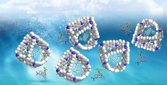
2.1. Synthesis and Characterization of Imine-Based 3D COP

An imine-based porous 3D covalent organic polymer (COP) was synthesized via solvothermal condensation (Scheme 1). Tetrakis(4-formylphenyl)-methane (TFPM) and 4,4′-diaminobiphenyl (DABP) were suspended in a mixture of 1,4-dioxane and mesitylene in the existence of acetic acid with subsequently heating at 120 °C for 3 days. The Fourier-transform infrared (FT IR) spectra of the synthesized polymer (Figure 1a) show the almost disappearance of the –N–H stretching vibration from 3100 to 3400 cm−1 and the appearance of a new stretching vibration band at 1619 cm−1 for a –C=N bond, indicating the occurrence of a condensation reaction. The stretching signal at 1695 cm−1 corresponded to the unreacted residue –CHO group at the edges/termini of the 3D COP. Powder X-ray diffraction (PXRD) data showed that the peak at 7.12° of the PXRD patterns (Figure S1) was not corresponding to the Bragg peaks (5.19°) of space group I41/a for the earlier reported COF [48]. In addition, other reflection peaks at 8.49, 12.15, and 13.46° were not observed in the present PXRD data. Furthermore, the intensity of the reflection peak at 7.12° was weak, indicating that the synthesized polymer was not well crystalized, at least not the same 3D COF as previous report [48]. Thermogravimetric analysis (TGA) data (Figure 1b) showed that the 3D COP was thermally stable below 400 °C, revealing the good thermal stability of the 3D COP. N2 adsorption–desorption isotherms were obtained at 77 K to evaluate the porosity of 3D COPs. Brunauer–Emmer–Teller (BET) nitrogen adsorption data (Figure 1c) displayed a sharp gas uptake at low pressure (<0.1 P/P0), indicating that the 3D COP exhibited a microporous structure. The literature revealed that the inclination of isotherms in the 0.8–1.0 P/P0 range showed the existence of textural mesopores due to the agglomeration of polymers [48]. According to the data in Figure 1c, the calculated BET surface area (169 m2/g) is smaller than that (653 m2/g) of the reported COF material constructed by the same monomers [48]. The poor crystallinity of 3D COPs might induce the lower experimental N2 adsorption or calculated surface area than those of the well-crystalized COF. According to the size distribution in Figure 1d, the calculated average pore width was 1.03 nm. Based on the above data, this polymer was defined as a porous 3D COP. Scanning electron microscopy (SEM) (Figure 1e) and transmission electron microscopy (TEM) (Figure 1f) revealed that the 3D COP exhibited rod-like aggregations.

2.2. Optimization of SPE

For the SPE process, the types and volume of eluent, washing speed, pH, and salinity of water significantly influenced the extraction efficiency. As described in a previous report [57], the optimized parameters were applied to optimize subsequent parameters. To ensure data reproducibility, every optimization step was repeated three times.

2.2.1. Effect of Eluent

The purpose of elution is to flush the analytes adsorbed on the solid phase as much as possible without destroying the original properties of the filler. As seen in Figure 2a, six solvent system, including acetonitrile, acetone, methanol, methanol/acetic acid (Vmethanol/Vacetic acid, 99:1), methanol/formic acid (Vmethanol/Vformic acid, 97:3), and methanol/trifluoroacetic acid (Vmethanol/Vtrifluoroacetic acid, 95:5) could desorb the three analytes from the solid phase. For the single solvent system, methanol provided a better recovery compared with acetonitrile and acetone. The addition of formic acid in methanol (Vmethanol/Vformic acid, 97:3) obviously improved the recovery of FF, TAP, and CAP, among which the recovery of FF reached 80%. Therefore, the methanol/formic acid solution (Vmethanol/Vformic acid, 97:3) was used to evaluate the influence of elution volume on recovery. From the data in Figure 2b, when the volume of elution increased from 5 to 15 mL, the recovery of the three analytes increased to 90%. Further increasing the volume of elution to 20 mL did not obviously change the recovery; therefore, an elution volume of 15 mL was chosen for subsequent investigations.

2.2.2. Effect of Flow Velocity and pH of Loading Sample Solution

The flow velocity of sample solutions was related to the extraction equilibrium and kinetics; thus, it directly influenced the adsorption speed of target molecules on the porous 3D COP. The data in Figure 3a clearly show that the recovery of three analytes decreased dramatically by reaching the higher flow speed (larger than 0.5 mL/min). On the other hand, when the speed was lower than 0.5 mL/min, a slight change in the recovery was observed, indicating that extraction equilibrium was achieved. Thus, 0.5 mL/min was chosen as the optimized flow velocity.
The pH of the water sample may influence the charge state of analytes in solution, which can further influence the extraction efficiency. Figure 3b showed that the recovery of CAP, TAP, and FF was relatively high when the pH of water ranged from 3–6. However, as the pH increased from 7 to 10 upon the addition of NaOH solution, the recovery obviously decreased. This was probably caused by increasing the solubility of analytes in water through protonation under acidic conditions, which weakened the hydrophobic interactions between the analytes and 3D COP. Thus, a pH of 6 was chosen as the optimal condition for the following investigations.

2.2.3. Effect of Salinity of Water Sample

Previous reports revealed that the addition of salt in water samples increases the ion concentration and ionic strength of a solution, which positively influences the adsorption efficiency of hydrophilic compounds. As shown in Figure 4, when the salinity increased from 0 to 15, the recovery of the three analytes increased. As the salinity increased beyond 15, the recovery decreased dramatically. This was probably due to the competitive adsorption of excess Na+ or Cl on the surface or pores of the 3D COP, which exceeded the salt-out effect according to a previous investigation [58]. Hence, a salinity of 15 was chosen for further investigations.
Briefly, the effects of the five factors—eluent type, eluent volume, loading flow rate, pH, and salinity—on the extraction process were optimized and listed as follows: a methanol formic acid solution (Vmethanol/Vformic acid, 97:3); an eluent volume of 15 mL; a loading flow rate, 0.5 mL min−1; a water sample pH of 6; and a salinity of 15.

2.3. Methodological Investigation

Under the optimized conditions mentioned above, method validation including the linear range, correlation coefficient (R2), limits of detection (LODs), limits of quantification (LOQs), precision, and relative standard deviation (RSD) are listed in Table 1. CAP, TAP, and FF were added to water samples to increase the analyte concentrations to 0.1–200 ng/mL for CAP and those of FF and TAP were 0.3–200 ng/mL. The LODs obtained from three replicates were 0.01 ng/mL for CAP, and 0.03 ng/mL for FF and TAP, respectively, with a signal-to-noise ratio (S/N) of 3. The LOQs were 0.04 ng/mL for CAP and 0.10 ng/mL for FF and TAP, with a baseline S/N of 10. This indicated the high sensitivity of this method and demonstrated that it was sufficiently sensitive to detect trace amounts of CAP, TAP, and FF in real water samples. Furthermore, the linear correlation coefficients (R2) of working curves for CAP were larger than 0.99, while R2 of FF and TAP was larger than 0.999. This demonstrated that the concentration of substances in water samples showed excellent linear relationship with the peak area when the concentration of CAP was 0.1–200 ng/mL and when FF and TAP ranged from 0.3–200 ng/mL. As shown in Table 1, the relative standard deviation for intra-day and inter-day precisions ranged from 1.72 to 3.15% and 3.70 to 4.87% respectively. The RSDs (n = 3) of the extraction performance ranged from 3.6% to 5.3%, indicating the good reproducibility of the 3D COP-SPE method. These data demonstrate that the 3D COP-SPE method was accurate and could reliably and reproducibly extract CAP, FF, and FF from water samples.
The standard recovery was further investigated to evaluate the precision and accuracy of the method. Table 2 and Table S1 list the recoveries and RSD%, which demonstrate that the 3D COP-SPE method can be applied to determinate CAP, TAP, and FF in water matrices with good precision and accuracy.

2.4. Reusability of 3D COP

The extraction recovery over 10 continuous cycles was carried out to evaluate the reusability of the 3D COP. From the data in Figure 5a, the recovery showed almost no significant change as the number of extraction cycles increased, demonstrating that the 3D COP used in this research has good recycling performance. A comparison of the FTIR spectra (Figure 5b) after 100 cycles under different conditions further indicates the good stability of the 3D COP after extraction. The corresponding calculated BET surface area was 120 m2/g, slightly lower than that (169 m2/g) before extraction. However, this change of surface area did not obviously affect the extraction efficiency.

2.5. Proposed Enrichment Mechanism

From the structural perspective of CAP, TAP and FF, phenyl groups with hydrophobic properties and π donors and hydroxyl groups with hydrophilic characteristics and hydrogen bond doners are included (Figure 6). The imine-based 3D COPs with aromatic skeletons are highly hydrophobic and bearing strong π–π interaction binding sites. The above discussion shows that the imine-based 3D COP has a strong adsorption and good enrichment for each of the three analytes. The unique pore width of the 3D COP is 1.03 nm as discussed above, and the length of adsorb analytes is calculated to be 0.98 nm by Chem 3D. This indicated that the analytes could sufficiently adsorb on the pores of 3D COPs due to the size-matching effect. In addition, this process was mainly driven by the combination of the hydrophobic attraction between the hydrophobic groups of the 3D COP and the analytes and their π–π interactions. In addition to these interactions, hydrogen bonding between the lone electron pair of the imine and the hydrogen of the hydroxyl group may have also contributed to the enrichment. Based on the above discussion, the good enrichment performance was ascribed to hydrophobic and π–π interactions, the size-matching effect, hydrogen bonding, and the good chemical stability of 3D COP.

2.6. Comparison with Previous Reports

The 3D COP-SPE method was compared with previous reports of the determination of residual amphenicol drugs in water samples. The linearity, LODs, and recoveries are summarized in Table 3. The 3D COP-SPE method exhibited a wider linearity and comparable recoveries to previously-reported methods. The LODs of 3D COP-SPE were obviously lower than those obtained from other pretreatment methods, including direct measure without treatment [24], SPME using fiber, [25] and SPE using resin [26]. Based on the above discussions, combining the 3D COP-SPE with HPLC-DAD offers a simple, accurate, and sensitive method for determining amphenicol drugs in water samples.

3. Materials and Methods

3.1. Chemicals

Standard samples of CAP, TAP, and FF were obtained from Tianjin Guangcheng Chemical Reagent Co., Ltd. (Tianjin, China). Tetrakis(4-formylphenyl)-methane was obtained from Aladdin Reagent Co., Ltd. (Shanghai, China) 1,4-Dioxane, mesitylene, tetrahydrofuran (THF), acetone, formic acid, acetic acid, and methanol were obtained from Tianjin Guangcheng Chemical Reagent Co., Ltd. (Tianjin, China). Ethyl acetate, anhydrous ethanol, hydrochloric acid (37%), trifluoroacetic acid, Wahaha pure water, and acetonitrile were obtained from Tianjin Fuyu Chemical Reagent Co., Ltd. (Tianjin, China).
Standard solutions were prepared by dissolving CAP, TAP, or FF (1.000 g) in acetonitrile (100 mL) to obtain a final concentration of 10 mg/mL. The working standard solution was obtained by diluting the stock solution with acetonitrile to the required concentrations for methodological investigation. The above solutions were stored in the fridge under 4 °C.

3.2. Instruments and Chromatographic Conditions

FT IR spectra were acquired using a Bruker FT IR spectrometer (Berlin, Germany). The morphology of the 3D COP was imaged using a transmission electron microscope (Hitachi, HT7700, Tokyo, Japan), an 80 kV accelerating voltage and scanning electron microscopy (Hitachi, SU8010, Tokyo, Japan), and a 5 kV accelerating voltage. The PXRD data were obtained using a Rigaku D/Max 2500 PC diffractometer (Tokyo, Japan) with Cu Kα radiation. TGA was conducted with a heating rate of 10 °C·min−1 and under nitrogen flow on a TA Instrument Q5 analyzer. BET data were collected by a Micromeritics ASAP 2000 sorption/desorption analyzer (Norcross, GA, USA) using N2 adsorption at 77 K. The solid-phase extractions were performed using an extraction instrument (SPR-SPE12), and samples were analyzed by the HPLC (Agilent 1260, Palo Alto, CA, USA), which was equipped with a diode-array detector (DAD) for sample analysis using a detection wavelength of 224 nm and a C18 reversed-phase chromatographic column (5 μm, 250 mm × 4.6 mm). Eluent A (acetonitrile) and eluent B (water) (20% acetonitrile in water) were used as the mobile phase to separate CAP, TAP, and FF using isometric elution with a 1.0 mL/min flow speed. The running time for HPLC was 20 min, and the volume of injection was 20 μL.

3.3. Synthesis of Imine-Based 3D COP

Tetrakis(4-formylphenyl)-methane (TFPM, 64.8 mg, 0.15 mmol) and 4,4′-diaminobiphenyl (DABP, 55.2 mg, 0.3 mmol) in a mixed solvent of 1,4-dioxane (1.8 mL), mesitylene (0.6 mL), and acetic acid (3 M, 0.3 mL) were put in A Pyrex tube. The above mixture was sonicated for 1 min to obtain the homogeneous dispersion of solids in the solvent. Subsequently, the mixture was sealed, and heated at 120 °C for 3 days. After cooling down to the room temperature, a solid was obtained by filtration, and then purified by Soxhlet extraction in THF by heating to reflux overnight. The solvent was removed under the reduced pressure to afford an imine-based 3D COP (80 mg) as a yellow solid with a yield of 87%.

3.4. SPE Procedure

The 3D COP powder was transferred into the SPE cartridge and compacted with a height of 2 cm, which was used as a column. The cartridge was pretreated with acetonitrile and methanol under vacuum until the effluent was colorless to activate the column. The optimized parameter of 3D COP-SPE protocol was listed as follows:
The pH of sample solution was adjusted using hydrochloric acid (0.02 mol/L). Then, the sample in water (20.0 mL) was charged onto the SPE cartridge with a 5.0 mL/min flow speed. The cartridge was dried under vacuum for 3 min. After that, ammonia methanol solution (Vammonia/Vmethanol = 8:92, 2.0 mL) with a flow speed of 3.0 mL/min was used to elute the cartridge. The eluent was completely removed under vacuum and redispersed with methanol (0.05 mL) for HPLC-DAD analysis.
The mixed standard solution (10 mg/mL) was diluted to 3 mg/mL using acetonitrile and then passed through the SPE column filled with 3D COPs at a 0.5 mL/min flow rate. The filtrates were collected by passing through a Millipore filter (0.2 μm) which was obtained from Tianjin Branch billion Lung Experimtental Equipment Co., Ltd. (Tianjin, China) before HPLC analysis.

4. Conclusions

An imine-based porous 3D COP was synthesized by solvothermal condensation and was successfully used as a new solid-phase sorbent for SPE. The 3D COP efficiently enriched and detected trace amounts of CAP, TAP, and FF in water samples with ng quantities. The low LODs and LOQs for CAP, TAP, and FF were located in the ng/mL range, indicating the high sensitivity of this method. Hydrophobic effects, π–π interactions, size-matching effects, hydrogen bonding, and excellent chemical stability likely contributed to the excellent enrichment of three analytes in this study. These results indicate that using the 3D COP as a sorbent for SPE can provide an accurate, reliable, and reproducible method to detect trace amounts of three substances often present in environmental water samples.

Supplementary Materials

The following supporting information can be downloaded at: https://www.mdpi.com/article/10.3390/molecules28083301/s1, Figure S1: PXRD for imine-based 3D COP; Table S1: RSD of analytical data for determination of amphenicols in water samples.

Author Contributions

Conceptualization, J.W., Y.C. and Z.Z.; methodology, J.W. and W.J; software, L.C. and Y.Y.; validation, formal analysis, investigation, L.C., R.Z. and Y.Y.; writing—original draft preparation, L.C.; writing—review and editing, J.W., W.J., Z.H., Y.C. and Z.Z.; supervision, J.W. and Z.Z.; funding acquisition, J.W., W.J. and Z.Z.; J.W. and L.C. contributed equally. The manuscript was written through contributions of all authors. All authors have read and agreed to the published version of the manuscript.

Funding

The authors are grateful for the financial support from the Scientific and Technological Project of Shandong Province (Grant Nos. 2012GGX10705 and 2006GG2203001), the Scientific and Technological Project of Shandong Province Colleges and Universities (Grant No. J02C01), Education and Industry Integration Innovation Pilot Project from Qilu University of Technology (Shandong Academy of Sciences) (2022PYI012), Shandong Province Science and Technology of Small and Medium-Sized Enterprises Innovation Ability Upgrading (Grant No. 2021TSGC1293), Shandong Provincial Natural Science Foundation (Grant No. ZR2022MB031), and the National Nature Science Foundation of China (Grant No. 21708022).

Institutional Review Board Statement

Not applicable.

Informed Consent Statement

Not applicable.

Data Availability Statement

Data is contained within the article.

Conflicts of Interest

The authors declare no conflict of interest.

Sample Availability

Samples of the compounds CAP, TAP and FF are available from Wenhua Ji.

References

  1. Pongs, O. Mechanism of Action of Antibacterial Agents; Springer: Verlag, Germany, 1979; pp. 26–42. [Google Scholar]
  2. Festing, M.F.W.; Diamanti, P.; Turton, J.A. Strain differences in haematological response to chloramphenicol succinate in mice: Implications for toxicological research. Food Chem. Toxicol. 2001, 39, 375–383. [Google Scholar] [CrossRef]
  3. Holt, D.E.; Andrews, C.M.; Payne, J.P.; Williams, T.C.; Turton, J.A. The myelotoxicity of chloramphenicol: In vitro and in vivo studies: II: In vivo myelotoxicity in the B6C3F1 mouse. Hum. Exp. Toxicol. 1998, 17, 8–17. [Google Scholar] [CrossRef]
  4. Epstein, R.L.; Henry, C.; Holland, K.P.; Dreas, J. International validation study for the determination of chloramphenicol in bovine muscle. J. AOAC Int. 1994, 77, 570–576. [Google Scholar] [CrossRef] [PubMed]
  5. European Commission. European Commission Regulation (EC) no. 1430/94 of 22 June 1994 amending Annexes I, II, III and IV of Council Regulation (EEC) No. 2377/90 laying down a Community procedure for the establishment of maximum residue limits of veterinary medicinal products in foodstuffs of animal origin. Off. J. Eur. Commun. 1994, 156, 6–8. [Google Scholar]
  6. European Commission. Commission decision 2003/181/EC of 13 March 2003 amending decision 2002/657/EC as regards the setting of minimum required performance limits (MRPLs) for certain residues in food of animal origin. Off. J. Eur. Commun. 2003, 71, 17–18. [Google Scholar]
  7. Xiao, Z.; Song, R.; Rao, Z.; Wei, S.; Jia, Z.; Suo, D.; Fan, X. Development of a subcritical water extraction approach for trace analysis of chloramphenicol, thiamphenicol, florfenicol, and florfenicol amine in poultry tissues. J. Chromatogr. A 2015, 1418, 29–35. [Google Scholar] [CrossRef] [PubMed]
  8. Xie, X.; Wang, B.; Pang, M.; Zhao, X.; Xie, K.; Zhang, Y.; Wang, Y.; Guo, Y.; Liu, C.; Bu, X.; et al. Quantitative analysis of chloramphenicol, thiamphenicol, florfenicol and florfenicol amine in eggs via liquid chromatography-electrospray ionization tandem mass spectrometry. Food Chem. 2018, 269, 542–548. [Google Scholar] [CrossRef]
  9. Aldeek, F.; Hsieh, K.C.; Ugochukwu, O.N.; Gerard, G.; Hammack, W. Accurate quantitation and analysis of nitrofuran metabolites, chloramphenicol, and florfenicol in seafood by ultrahigh-performance liquid chromatography-tandem mass spectrometry: Method validation and regulatory samples. J. Agric. Food Chem. 2018, 66, 5018–5030. [Google Scholar] [CrossRef]
  10. Liu, W.L.; Lee, R.J.; Lee, M.R. Supercritical fluid extraction in situ derivatization for simultaneous determination of chloramphenicol, florfenicol and thiamphenicol in shrimp. Food Chem. 2010, 121, 797–802. [Google Scholar] [CrossRef]
  11. Li, P.; Qiu, Y.; Cai, H.; Kong, Y.; Tang, Y.; Wang, D.; Xie, M. Simultaneous determination of chloramphenicol, thiamphenicol, and florfenicol residues in animal tissues by gas chromatography/mass spectrometry. Chin. J. Chromatogr. 2006, 24, 14–18. [Google Scholar] [CrossRef] [PubMed]
  12. Shen, H.Y.; Jiang, H.L. Screening, determination and confirmation of chloramphenicol in seafood, meat and honey using ELISA, HPLC-UVD, GC-ECD, GC-MS-EI-SIM and GCMS-NCI-SIM methods. Anal. Chim. Acta 2005, 535, 33–41. [Google Scholar] [CrossRef]
  13. Tao, X.; He, Z.; Cao, X.; Jiang, H.; Li, H. Approaches for the determination of florfenicol and thiamphenicol in pork using a chemiluminescent ELISA. Anal. Methods 2015, 7, 8386–8392. [Google Scholar] [CrossRef]
  14. Fodey, T.L.; George, S.E.; Traynor, I.M.; Delahaut, P.; Kennedy, D.G.; Elliott, C.T.; Crooks, S.R.H. Approaches for the simultaneous detection of thiamphenicol, florfenicol and florfenicol amine using immunochemical techniques. J. Immunol. Methods 2013, 393, 30–37. [Google Scholar] [CrossRef] [PubMed]
  15. Sadeghi, S.; Jahani, M. Selective solid-phase extraction using molecular imprinted polymer sorbent for the analysis of Florfenicol in food samples. Food Chem. 2013, 141, 1242–1251. [Google Scholar] [CrossRef]
  16. Pan, X.; Wu, P.; Jiang, W.; Ma, B. Determination of chloramphenicol, thiamphenicol, and florfenicol in fish muscle by matrix solid-phase dispersion extraction (MSPD) and ultra-high pressure liquid chromatography tandem mass spectrometry. Food Control 2015, 52, 34–38. [Google Scholar] [CrossRef]
  17. Rejtharová, M.; Rejthar, L. Determination of chloramphenicol in urine, feed water, milk and honey samples using molecular imprinted polymer clean-up. J. Chromatogr. A 2009, 1216, 8246–8253. [Google Scholar] [CrossRef] [PubMed]
  18. Shen, J.; Xia, X.; Jiang, H.; Li, C.; Li, J.; Li, X.; Ding, S. Determination of chloramphenicol, thiamphenicol, florfenicol, and florfenicol amine in poultry and porcine muscle and liver by gas chromatography-negative chemical ionization mass spectrometry. J. Chromatogr. B 2009, 877, 1523–1529. [Google Scholar] [CrossRef]
  19. Chen, L.; Li, B. Magnetic molecularly imprinted polymer extraction of chloramphenicol from honey. Food Chem. 2013, 141, 23–28. [Google Scholar] [CrossRef]
  20. Liu, H.Y.; Lin, S.L.; Fuh, M.R. Determination of chloramphenicol, thiamphenicol and florfenicol in milk and honey using modified QuEChERS extraction coupled with polymeric monolith-based capillary liquid chromatography tandem mass pectrometry. Talanta 2016, 150, 233–239. [Google Scholar] [CrossRef]
  21. Veach, B.T.; Anglin, R.; Mudalige, T.K.; Barnes, P.J. Quantitation and confirmation of chloramphenicol, florfenicol, and nitrofuran metabolites in honey using LC-MS/MS. J. AOAC Int. 2018, 101, 897–903. [Google Scholar] [CrossRef]
  22. Teixeira, S.; Delerue-Matos, C.; Alves, A.; Santos, L. Fast screening procedure for antibiotics in waste waters by direct HPLC-DAD analysis. J. Sep. Sci. 2008, 31, 2924–2931. [Google Scholar] [CrossRef] [PubMed]
  23. Aresta, A.; Bianchi, D.; Calvano, C.D.; Zambonin, C.G. Solid phase microextraction-Liquid chromatography (SPME-LC) determination of chloramphenicol in urine and environmental water samples. J. Pharm. Biomed. Anal. 2010, 53, 440–444. [Google Scholar] [CrossRef]
  24. Peng, X.; Tan, J.; Tang, C.; Yu, Y.; Wang, Z. Multiresidue determination of fluoroquinolone, sulfonamide, trimethoprim, and chloramphenicol antibiotics in urban waters in China. Environ. Toxicol. Chem. 2008, 27, 73–79. [Google Scholar] [CrossRef]
  25. Poole, C.F. New trends in solid-phase extraction. Trends Anal. Chem. 2003, 22, 362–373. [Google Scholar] [CrossRef]
  26. Wen, Y.; Chen, L.; Li, J.; Liu, D.; Chen, L. Recent advances in solid-phase sorbents for sample preparation prior to chromatographic analysis. Trends Anal. Chem. 2014, 59, 26–41. [Google Scholar] [CrossRef]
  27. Chen, L.; Xu, S.; Li, J. Recent advances in molecular imprinting technology: Current status, challenges and highlighted applications. Chem. Soc. Rev. 2011, 40, 2922–2942. [Google Scholar] [CrossRef]
  28. Liu, J.; Chen, H.; Lin, Z.; Lin, J.M. Preparation of surface imprinting polymer capped Mn-doped ZnS quantum dots and their application for chemiluminescence detection of 4-nitrophenol in tap water. Anal. Chem. 2010, 82, 7380–7386. [Google Scholar] [CrossRef] [PubMed]
  29. Lucena, R.; Simonet, B.M.; Cárdenas, S.; Valcárcel, M. Potential of nanoparticles in sample preparation. J. Chromatogr. A 2011, 1218, 620–637. [Google Scholar] [CrossRef]
  30. Jiménez-Soto, M.J.; Cárdenas, S.; Valcárcel, M. Evaluation of carbon nanocones/disks as sorbent material for solid-phase extraction. J. Chromatogr. A 2009, 1216, 5626–5633. [Google Scholar] [CrossRef]
  31. Chen, L.; Wang, T.; Tong, J. Application of derivatized magnetic materials to the separation and the preconcentration of pollutants in water samples. Trends Anal. Chem. 2011, 30, 1095–1108. [Google Scholar] [CrossRef]
  32. Jiang, C.; Sun, Y.; Yu, X.; Gao, Y.; Zhang, L.; Wang, Y.; Zhang, H.; Song, D. Liquid-solid extraction coupled with magnetic solid-phase extraction for determination of pyrethroid residues in vegetable samples by ultrafast liquid chromatography. Talanta 2013, 114, 167–175. [Google Scholar] [CrossRef]
  33. Chang, N.; Yan, X.P. Exploring reverse shape selectivity and molecular sieving effect of metal-organic framework UIO-66 coated capillary column for gas chromatographic separation. J. Chromatogr. A 2012, 1257, 116–124. [Google Scholar] [CrossRef]
  34. Huang, H.; Lin, C.; Wu, C.; Cheng, Y.; Lin, C. Metal organic framework-organic polymer monolith stationary phases for capillary electrochromatography and nano-liquid chromatography. Anal. Chim. Acta 2013, 779, 96–103. [Google Scholar] [CrossRef]
  35. Yang, D.; Tao, Y.; Ding, X.; Han, B. Porous organic polymers for electrocatalysis. Chem. Soc. Rev. 2022, 51, 761–791. [Google Scholar] [CrossRef]
  36. Hao, Q.; Tao, Y.; Ding, X.; Yang, Y.; Feng, J.; Wang, R.-L.; Chen, X.-M.; Chen, G.-L.; Li, X.; OuYang, H.; et al. Porous organic polymers: A progress report in China. Sci. China Chem. 2023, 66, 620–682. [Google Scholar] [CrossRef]
  37. Tian, Y.; Zhu, G. Porous aromatic frameworks (PAFs). Chem. Rev. 2020, 120, 8934–8986. [Google Scholar] [CrossRef] [PubMed]
  38. Yuan, Y.; Yang, Y.; Zhu, G. Multifunctional porous aromatic frameworks: State of the art and opportunities. EnergyChem 2020, 2, 100037. [Google Scholar] [CrossRef]
  39. Lee, J.; Cooper, A. Advances in conjugated microporous polymers. Chem. Rev. 2020, 120, 2171–2214. [Google Scholar] [CrossRef] [PubMed] [Green Version]
  40. Xu, Y.; Jin, S.; Xu, H.; Nagai, A.; Jiang, D. Conjugated microporous polymers: Design, synthesis and application. Chem. Soc. Rev. 2013, 42, 8012–8031. [Google Scholar] [CrossRef] [PubMed]
  41. Castaldo, R.; Gentile, G.; Avella, M.; Carfagna, C.; Ambrogi, V. Microporous hyper-crosslinked polystyrenes and nanocomposites with high adsorption properties: A review. Polymers 2017, 9, 651. [Google Scholar] [CrossRef] [PubMed] [Green Version]
  42. Fu, Z.; Jia, J.; Li, J.; Liu, C. Transforming waste expanded polystyrene foam into hyper-crosslinked polymers for carbon dioxide capture and separation. Chem. Eng. J. 2017, 323, 557–564. [Google Scholar] [CrossRef]
  43. Côté, A.P.; Benin, A.I.; Ockwig, N.W.; O’Keeffe, M.; Matzger, A.J.; Yaghi, O.M. Porous, crystalline, covalent organic frameworks. Science 2005, 310, 1166–1170. [Google Scholar] [CrossRef] [Green Version]
  44. Dalapati, S.; Jin, E.; Addicoat, M.; Heine, T.; Jiang, D. Highly emissive covalent organic frameworks. J. Am. Chem. Soc. 2016, 138, 5797–5800. [Google Scholar] [CrossRef] [PubMed]
  45. Xu, H.; Gao, J.; Jiang, D. Stable, crystalline, porous, covalent organic frameworks as a platform for chiral organocatalysts. Nat. Chem. 2015, 7, 905–912. [Google Scholar] [CrossRef] [PubMed]
  46. Huang, N.; Wang, P.; Jiang, D. Covalent organic frameworks: A materials platform for structural and functional designs. Nat. Rev. Mater. 2016, 1, 16068. [Google Scholar] [CrossRef]
  47. Li, Z.; Li, H.; Guan, X.; Tang, J.; Yusran, Y.; Li, Z.; Xue, M.; Fang, Q.; Yan, Y.; Valtchev, V.; et al. Three-dimensional ionic covalent organic frameworks for rapid, reversible, and selective ion exchange. J. Am. Chem. Soc. 2017, 139, 17771–17774. [Google Scholar] [CrossRef]
  48. Guan, X.; Ma, Y.; Li, H.; Yusran, Y.; Xue, M.; Fang, Q.; Yan, Y.; Valtchev, V.; Qiu, S. Fast, Ambient temperature and pressure ionothermal synthesis of three-dimensional covalent organic frameworks. J. Am. Chem. Soc. 2016, 138, 14783–14788. [Google Scholar] [CrossRef]
  49. Uribe-Romo, F.J.; Hunt, J.R.; Furukawa, H.; Klöck, C.; O’Keeffe, M.; Yaghi, O.M. A Crystalline imine-linked 3D porous covalent organic framework. J. Am. Chem. Soc. 2009, 131, 4570–4571. [Google Scholar] [CrossRef]
  50. Dawson, R.; Cooper, A.I.; Adams, D.J. Nanoporous organic polymer networks. Prog. Polym. Sci. 2012, 37, 530–563. [Google Scholar] [CrossRef]
  51. Asadi Tashvigh, A.; Benes, N.E. Covalent organic polymers for aqueous and organic solvent nanofiltration. Sep. Purif. Technol. 2022, 298, 121589. [Google Scholar] [CrossRef]
  52. Shi, Y.; Fu, Q.; Li, J.; Liu, H.; Zhang, Z.; Liu, T.; Liu, Z. Covalent organic polymer as a carborane carrier for imaging-facilitated boron neutron capture therapy. ACS Appl. Mater. Interfaces 2020, 12, 55564–55573. [Google Scholar] [CrossRef]
  53. Subodh; Prakash, K.; Chaudhary, K.; Masram, D. A new triazine-cored covalent organic polymer for catalytic applications. Appl. Catal. A Gen. 2020, 593, 117411. [Google Scholar] [CrossRef]
  54. Hong, Y.; Rozyyev, V.; Yavuz, C. Alkyl-linked porphyrin porous polymers for gas capture and precious metal adsorption. Small Sci. 2021, 1, 2000078. [Google Scholar] [CrossRef]
  55. Preet, K.; Gupta, G.; Kotal, M.; Kansal, S.K.; Salunke, D.B.; Sharma, H.K.; Sahoo, S.C.; Voort, P.V.D.; Roy, S. Mechanochemical synthesis of a new triptycene-based imine-linked covalent organic polymer for degradation of organic dye. Cryst. Growth Des. 2019, 19, 2525–2530. [Google Scholar] [CrossRef]
  56. Li, W.; Zhao, Z.; Hu, W.; Cheng, Q.; Yang, L.; Hu, Z.; Liu, Y.A.; Wen, K.; Yang, H. Design of thiazolo[5,4-d]thiazole-bridged ionic covalent organic polymer for highly selective oxygen reduction to H2O2. Chem. Mater. 2020, 32, 8553–8560. [Google Scholar] [CrossRef]
  57. Zhou, T.; Ding, J.; He, Z.; Li, J.; Liang, Z.; Li, C.; Li, Y.; Chen, Y.; Ding, L. Preparation of magnetic superhydrophilic molecularly imprinted composite resin based on multi-walled carbon nanotubes to detect triazines in environmental water. Chem. Eng. J. 2018, 334, 2293–2302. [Google Scholar] [CrossRef]
  58. Liu, L.; Meng, W.; Zhou, Y.; Wang, X.; Xu, G.; Wang, M.; Lin, J.; Zhao, R. β-Ketoenamine-linked covalent organic framework coating for ultra-high performance solid-phase microextraction of polybrominated diphenyl ethers from environmental samples. Chem. Eng. J. 2019, 356, 926–933. [Google Scholar] [CrossRef]
Scheme 1. Synthetic route of imine-based 3D COP.
Molecules 28 03301 sch001
Figure 1. Characterization of the imine-based porous 3D COP. (a) FTIR spectra comparison of 3D COP (blue), TFPM (black), and DABP (red); (b) TGA analysis; (c) N2 absorption–desorption isotherms; (d) pore width distribution; SEM (e) and TEM (f) images of 3D COP. This indicates that 3D COP exhibits rod-like aggregations.
Molecules 28 03301 g001
Figure 2. Optimization of SPE conditions. ACT: Acetone; ACN: Acetonitrile; MeOH: Methanol; HAc: Acetic acid; FA: Formic acid; TFA: Trifluoroacetic acid. (a) Effect of types of eluent on recovery (n = 3). Conditions: Concentration of analytes, 5 ng/mL; Sample volume, 10 mL; Flow velocity, 1 mL/min; Sample pH, 6; Sample salinity, 0. (b) Effect of volume of eluent on recovery (n = 3). Conditions: Concentration of analytes, 5 ng/mL; VMeOH/VFA, 97:3; Flow velocity, 1 mL/min; Sample pH, 6; Sample salinity, 0.
Molecules 28 03301 g002
Figure 3. (a) Effect of flow velocity on recovery (n = 3). Conditions: Concentration of analytes, 5 ng/mL; VMeOH/VFA, 97:3; Volume of eluent, 15 mL; Sample pH, 6; Sample salinity, 0; (b) Effect of pH on recovery (n = 3). Conditions: Concentration of analytes, 5 ng/mL; VMeOH/VFA, 97:3; Volume of eluent, 15 mL; Flow velocity, 0.5 mL/min; Sample salinity, 0.
Molecules 28 03301 g003
Figure 4. Effect of salinity on recovery (n = 3). Conditions: Concentration of analytes, 5 ng/mL; VMeOH/VFA, 97:3; Volume of eluent, 15 mL; Flow velocity, 0.5 mL/min; Sample pH, 6.
Molecules 28 03301 g004
Figure 5. (a) Effect of time of use on recovery; (b) FTIR spectra comparison without the SPE process (blue), 10 cycles (black), and after 100 cycles (red).
Molecules 28 03301 g005
Figure 6. The chemical (ac) and 3D (df) structure of the three analytes. (a,d) CAP, (b,e) TAP, and (c,f) FF. The red and blue circle (ac) indicated the binding sites of analytes with the 3D COP by π–π interaction and H-bonding, respectively. The 3D structure was obtained by optimizing the minimized energy using ChemDraw (20.0.0.41).
Molecules 28 03301 g006
Table 1. Analytical data for 3D COP-SPE method.
AnalytesLiner Range (ng/mL)R2LOD
(ng/mL)
LOQ
(ng/mL)
Precision (%, n = 3)Reproducibility
(RSD, n = 3, %)
Intra DayInter Day
CAP0.1–2000.99390.010.041.724.383.6
FF0.3–2000.99910.030.103.153.704.1
TAP0.3–2000.99930.030.102.954.875.3
Table 2. The standard recovery data for the determination of amphenicols in water samples. Three tests were repeated for each experiment.
Added
(ng mL−1)
Recovery (%)
CAPFFTAP
0.5101.73 ± 3.185.92 ± 5.286.14 ± 3.4
1098.9 ± 2.9100.02 ± 4.783.98 ± 5.2
100104.3 ± 4.6109.12 ± 6.8110.7 ± 3.6
15088.65 ± 6.394.23 ± 2.6106.45 ± 3.9
Table 3. Method comparison for the analysis of amphenicol drugs in water samples.
Pretreatment MethodAnalytical MethodNumber of AnalytesLinearity
(ng/mL)
LOD
(ng/mL)
Recovery
(%)
Ref.
Without pretreatmentHPLC-DAD540–4001390–109[24]
Fiber-SPMELC-UV10.1–100.1Not report[25]
Resin-SPEHPLC-UV40.2–500.2563–126[26]
3D COP-SPEHPLC-DAD30.1–2000.01–0.0383.98–110.7This work
Disclaimer/Publisher’s Note: The statements, opinions and data contained in all publications are solely those of the individual author(s) and contributor(s) and not of MDPI and/or the editor(s). MDPI and/or the editor(s) disclaim responsibility for any injury to people or property resulting from any ideas, methods, instructions or products referred to in the content.

