Article

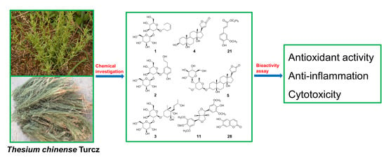
Chemical Constituents of Thesium chinense Turcz and Their In Vitro Antioxidant, Anti-Inflammatory and Cytotoxic Activities

Inflammation and Immune Mediated Diseases Laboratory of Anhui Province, School of Pharmacy, Anhui Medical University, Hefei 230032, China
*
Authors to whom correspondence should be addressed.
Molecules 2023, 28(6), 2685; https://doi.org/10.3390/molecules28062685
Submission received: 8 February 2023 / Revised: 7 March 2023 / Accepted: 14 March 2023 / Published: 16 March 2023

Abstract

:
Three novel compounds (1–3) along with twenty-six known compounds, two known steroids (4–5) and twenty-four known phenylpropanoids (6–29) were isolated from the whole plant of Thesium chinense Turcz. The structures of the three new compounds were elucidated on the basis of ESI-MS, HR-ESIMS, 1D and 2D NMR, IR, UV spectroscopic data. The absolute stereochemistry of compound 1 was determined by the Gauge-Including Atomic Orbitals (GIAO) method. The in vitro antioxidant, anti-inflammatory and cytotoxic activities of the isolated compounds were evaluated by DPPH radical-scavenging assay, LPS-activated RAW 264.7 cells model and CCK-8 kit, respectively. Compound 11 showed high antioxidant activity with an SC50 value of 16.2 ± 1.6 μM. Compound 21 showed considerable anti-inflammatory activity with an IC50 value of 28.6 ± 3.0 μM. Compounds 4 and 5 displayed potent cytotoxic activity against human NCI-H292, SiHa, A549, and MKN45 cell lines, with the compound 4 having IC50 values of 17.4 ± 2.4, 22.2 ± 1.1, 9.7 ± 0.9, 9.5 ±0.7 μM, and the compound 5 having all IC50 values less than 0.1 μM in vitro.

Graphical Abstract

1. Introduction

There are approximately 350 species of the genus Thesium (Santalaceae) occurring worldwide in Africa, Europe, Asia, South America and North America [1]. Thesium chinense Turcz, a small perennial and hemi-parasitic plant belonging to the family Santalaceae, distributes in East Asia (China, Japan, Korea, and Mongolia) [2]. The whole plant of T. chinense, commonly called “Bai-Rui-Cao” in China, was first recorded in the ancient medicinal monograph “Tu Jing Ben Cao” in the North Song Dynasty 1000 years ago. In traditional Chinese medicine (TCM), Bai-Rui-Cao is entitled “Botanical Antibiotics” and is mainly used to treat different diseases including mastitis, pulmonitis, tonsillitis, laryngopharyngitis and upper respiratory tract infections. [3]. Previous studies have reported the presence of polysaccharides, flavonoids, alkaloids, terpenoids, D-mannitol, aromatic compounds and aliphatic acids in the plant of T. chinense [4,5]. Modern pharmacological studies have found that T. chinense has diverse activities including anti-inflammation [6], antimicrobial effect [7], analgesic activity [8], antioxidant activity [9] and anti-nephropathy [10].
The chemical and pharmacological investigations into T. chinense are carried out as part of our ongoing work on the discovery of the bioactive compounds from Chinese medicinal herbs. From EtOAc and n-BuOH extracts of T. chinense, three novel compounds (1–3), along with twenty-six known compounds, two known steroids (4–5) and twenty-four known phenylpropanoids (6–29) (Figure 1), are isolated. The structures of the new compounds (1–3) are identified on the basis of ESI-MS, HR-ESI-MS, 1D and 2D NMR, IR, UV spectroscopic evidence. To determine the absolute stereochemistry, compound 1 was subjected to the Gauge-Including Atomic Orbitals (GIAO) method. The isolated compounds are evaluated for their antioxidant, anti-inflammatory activities and in vitro cytotoxic activities against four human NCI-H292, SiHa, A549, and MKN45 cancer cell lines by using DPPH radical-scavenging assay, LPS-activated RAW 264.7 cells model and CCK-8 kit, respectively. Herein, we report the isolation, structure determination and activity evaluation, and we preliminarily discuss the structure–activity relationship of the isolates.

2. Results and Discussion

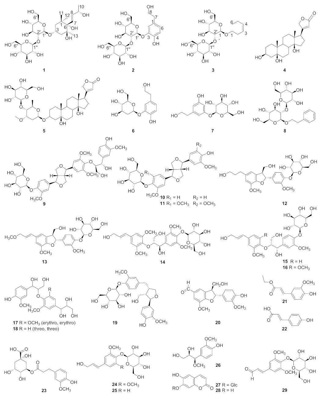
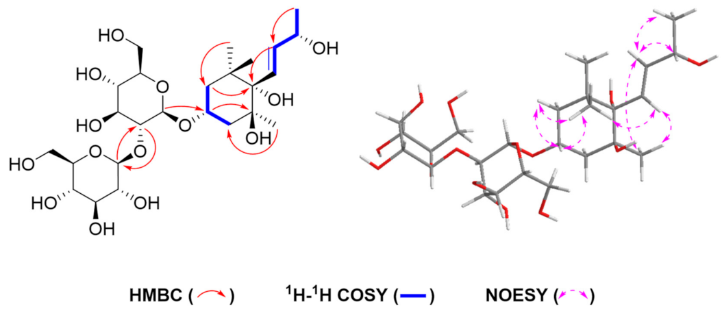
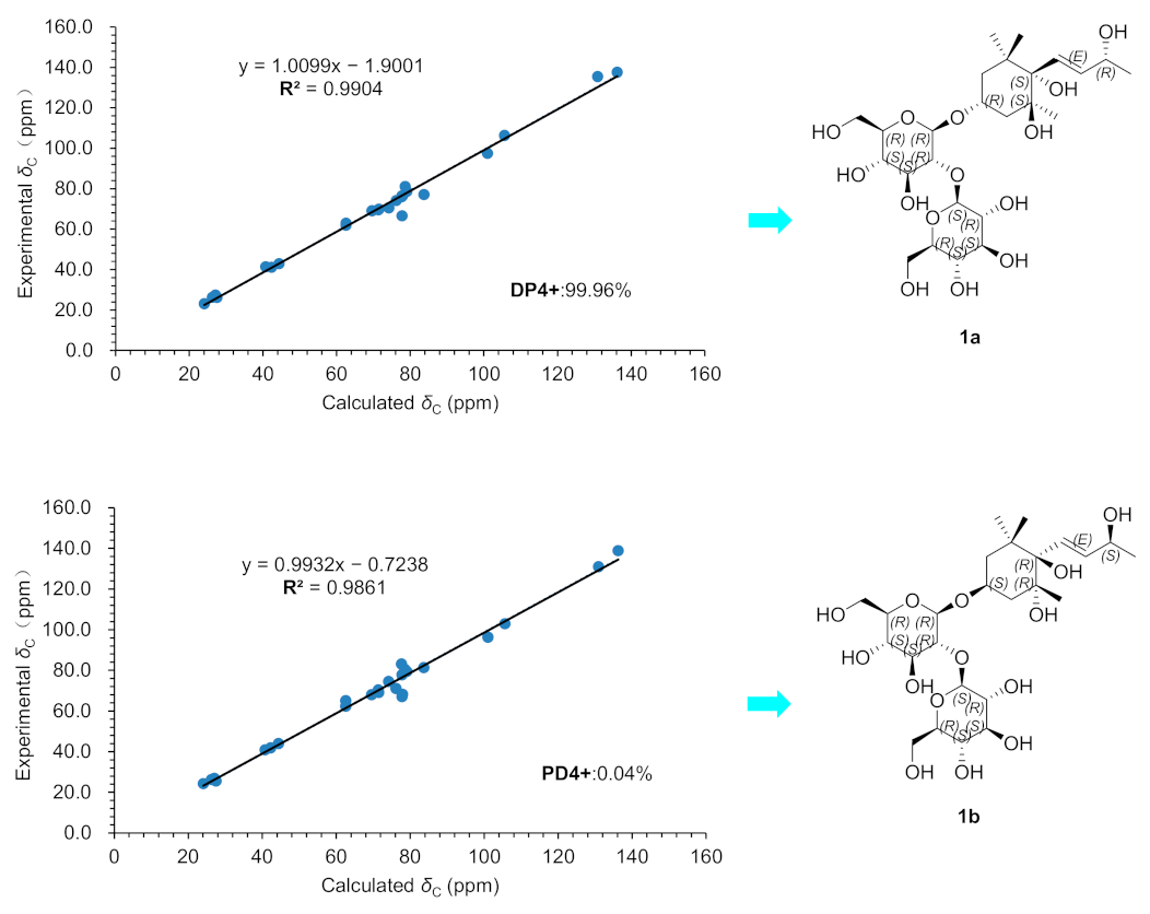
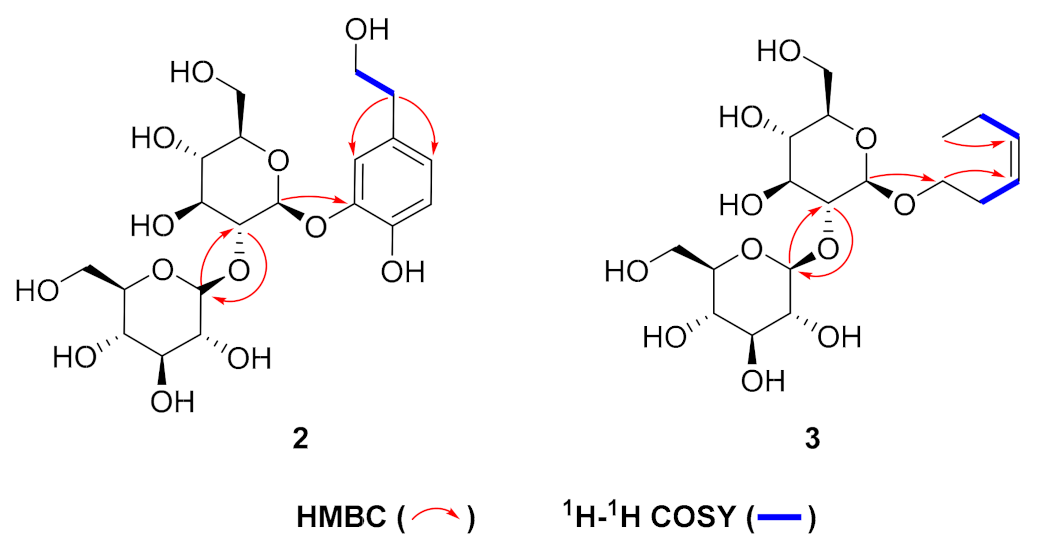
2.1. Structure Identification

Compound 1 was obtained as a white amorphous powder, and the molecular formula was confirmed as C25H44O14 by HR-ESI-MS (negative) (m/z 567.2654 [M−H], calcd. for C25H43O14, 567.2658). The 1H and 13C NMR (Table 1) spectrum showed a secondary methyl signal at δH 1.26 (3H, d, J = 6.4 Hz, H-10), three tertiary methyl signals at δH 0.87 (3H, s, H-12), δH 1.11 (3H, s, H-13) and δH 1.21 (3H, s, H-11), two methylene signals at δH 1.59 (1H, ddd, J = 12.3, 4.3, 2.1 Hz, H-2), δH 1.75 (1H, tt, J = 12.2, H-2) and δH 1.87 (1H, m, H-4), δH 1.96 (1H, ddd, J = 13.3, 4.3, 2.2 Hz, H-4), two secondary carbinyl proton signals at δH 6.04 (1H, dd, J = 15.8, 1.3 Hz, H-7) and δH 5.78 (1H, dd, J = 15.8, 6.4 Hz, H-8), two set glucosyl anomeric protons [δH 4.54 (1H, d, J = 7.5 Hz, H-1′) and δH 4.54 (1H, d, J = 7.5 Hz, H-1″)]. In addition to the signals due to the above functional groups, the spectrum showed signals due to a quaternary carbon atom at 40.8 and two quaternary carbon atoms with an oxygen atom at δC 77.8, 79.1. The side chain structure was confirmed by the 1H-1H COSY correlations of H-10/H-9, H-9/H-8, and H-8/H-7. Furthermore, according to the HMBC correlations of H-11/C-1, C-2, H-12/C-1, C-6, H-2/C-4, C-6, H-4/C-5, H-13/C-4, and C-6, the 1H-1H COSY correlations of H-2/H-3, H3/H-4 and the NOESY correlations of H-2 (δH 1.59)/H-3 (δH 4.21), H-11 (δH 1.21)/H-2 (δH 1.59), H-3 (δH 4.21), H-7 (δH 6.04), H-12 (δH 0.87)/H-8 (δH 5.78) and H-13 (δH 1.11)/H-7 (δH 6.04), compound 1 was presumed to have a structure with a megastigm-7-ene carbon skeleton to which four hydroxyl groups are introduced on C-3, 5, 6 and 9. Further in HMBC (Figure 2), the correlation between H-1′ [δH 4.54 (1H, d, J = 7.5 Hz)] with C-3 (δC 74.2) suggested that one β-glucosyl moiety was located at the C-3 position of the cycle. Correlations of HMBC from H-1″ [δH 4.54 (1H, d, J = 7.5 Hz)] with C-2′ (δC 83.7) and H-2′ [δH 3.36 (1H, m)] with C-1″ (δC 105.6) indicated that another β-glucosyl moiety was attached at the C-2′ position. To determine the absolute stereochemistry, compound 1 was subjected to the Gauge-Including Atomic Orbitals (GIAO) method [11,12,13]. Compound 1 was represented at the mPW1PW91/6-31G(d) level two isomers 1a (3R, 5S, 6S, 9R) and 1b (3S, 5R, 6R, 9S) using Gaussian quantum chemistry NMR calculations and DP4+ probabilistic analysis. The results show that the configuration of 1a (3R, 5S, 6S, 9R) is more consistent with the experimental value of 1 (Figure 3) and has 99.96% DP4+ probability (Show in Supplementary Materials). Thus, the absolute configuration of compound 1 is confirmed and named as thesiumin A.
Compound 2 was obtained as a white amorphous powder, and the molecular formula was confirmed as C20H30O13 by HR-ESI-MS (negative) (m/z 477.1610 [M−H], calcd. for C20H29O13, 477.1614). The 1H NMR spectrum of compound 2 showed the signals of two sets of methylene protons [δH 2.71(2H, t, J = 7.1 Hz, H-7) and δH 3.69 (2H, t, J = 7.0 Hz, H-8)], an ABX aromatic system proton [δH 7.15(1H, d, J = 2.0 Hz, H-2), δH 6.76 (1H, d, J = 8.1 Hz, H-5) and δH 6.80 (1H, d, J = 2.0, 8.1 Hz, H-6)], and two sets of glucosyl anomeric protons [δH 4.78 (1H, d, J = 7.8 Hz, H-1′) and δH 4.74 (1H, d, J = 7.8 Hz, H-1″)], which indicated the presence of 3,4- dihydroxyphenyl alcohol and two glucose units. Further comparison of the 1H and 13C NMR spectra of compound 2 with 3, 4-dihydroxyphenyl alcohol 3-O-β-D-glucopyranoside (6) [14] suggested that compound 2 contained one more glucose unit than 6. In HMBC (Figure 4), the correlation between H-1′ [δH 4.78 (1H, d, J = 7.8 Hz)] with C-3 (δC 146.7) suggested one β-glucosyl moiety was located at the C-3 position of the aromatic cycle. Correlations of HMBC from H-1″ [δH 4.74 (1H, d, J = 7.8 Hz)] with C-2′ (δC 83.3) and H-2″ [δH 3.75(1H, m)] with C-1″(δC 105.7) indicated that another β-glucosyl moiety was attached at the C-2′ position. On the basis of the above evidence, a new phenylethanoid glucoside was structurally determined as 3, 4-dihydroxyphenethyl alcohol 3-O-β-D-glucopyranosyl (1→2)-β-D-glucopyranoside (2), named thesiumin B.
Compound 3 was obtained as a white amorphous powder, and the molecular formula was confirmed as C18H32O13 by HR-ESI-MS (negative) (m/z 423.1871 [M−H], calcd. for C18H31O13, 423.1874). The 1H and 13C NMR (Table 2) together with the HSQC spectrum showed three sets of methylenes [δH 2.00 (2H, m, J = 7.3 Hz), δC 20.3, C-5; δH 3.44 (2H, m), δC 27.6, C-2; δH 3.74 (2H, m), δC 68.3, C-1], one methyl group [δH 0.91 (3H, t, J = 7.5 Hz), δC 14.3, C-6], one cis-configuration double bond [δH 5.34 (1H, dt, J = 11.3, 6.9 Hz), δC 125.4, C-3; δH 5.39 (1H, dt, J = 11.3, 6.9 Hz), δC 132.9, C-4], and two sets of glucosyl anomeric protons [δH 4.29 (1H, d, J = 7.7 Hz,), δC 101.4, C-1′; δH 4.37 (1H, d, J = 7.7 Hz), δC 104.2, C-1″], which indicated the presence of a carbon chain and two glucose units. In HMBC (Figure 4), the correlation between H-1′ [δH 4.29 (1H, d, J = 7.7 Hz)] with C-1 (δC 68.3) suggested that one β-glucosyl moiety was located at the C-1 position of the carbon chain. Correlations of HMBC from H-1″ [δH 4.29 (1H, d, J = 7.7 Hz)] with C-2′ (δC 82.5) and H-2″ [δH 2.98(1H, t, J = 8.2 Hz)] with C-1″(δC 104.2) indicated that another β-glucosyl moiety was attached at the C-2′ position. Furthermore, in HMBC, there was correlation between H-1 with C-3, and the 1H-1H COSY correlations of H-2/H-3, and H-4/H-5. Thus, compound 3 was identified as shown and named as (Z)-hex-3-ene 3-O-β-D- glucopyranosyl (1→2)-β-D- glucopyranoside, named thesiumin C.
Beside the three novel compounds (1–3), twenty-six known compounds (4–29), including two steroids (4–5) and twenty-four phenylpropanoids (6–29), were also identified from the whole plants of T. chinense. Their strctures were determined as: perlplogemn [15] (4), periplocin [16] (5), 2-hydroxy-5-(2-hydroxy-ethyl) phenyl-β-D-glucopyranoside (6) [11], 3, 5-dihydroxyphenethyl alcohol 3-O-β-D-glucopyranoside (7) [17], phenethyl alcohol β-sophoroside (8) [18], samsesquinoside (9) [19], pinoresinol-4-O-D- glucoside (10) [20], syringaresinol-4′-O-β-D-glucopyranoside (11) [21], dihydrodehydro diconiferyl alcohol 4-O-β-D-glucoside (12) [22], (7S, 8R) 9′- methoxy- dehydrodiconiferyl alcohol 4-O-β-D-glucopyranoside (13) [23], picraquassioside C (14) [24], citrusin A (15) [25], citrusin B (16) [25], (erythro, erythro)-1-[4-[2-hydroxy-2-(4-hydroxy-3-methoxy-phenyl)-1-(hydroxy methyl) ethoxy]-3, 5-dimethoxy phenyl]-1, 2, 3-propanetriol (17) [26], 1-(4-hydroxy-3-methoxy)-phenyl -2-[4-(1, 2, 3-trihydroxy propyl)-2-methoxy]-phenoxy-1, 3-propan-diol (18) [27], lariciresinol-4-O-β-D-glucoside (19) [28], ficusal (20) [29], 3-O-ethyl ferulate (21) [30], p-coumaric acid (22) [31], feruloylquinic acid (23) [32], syringin (24) [33], coniferin (25) [34], alatusol D (26) [35], scopolin (27) [36], scopoletin (28) [36] and sinapaldehyde glucoside (29) [37]. All the known compounds (4–29) were isolated from Thesium chinense Turcz for the first time.

2.2. Bio-Activities of Compounds 2–29

2.2.1. Antioxidant Activity

The antioxidant activities of all the isolated compounds except compound 1 were evaluated using DPPH radical scavenging assay (Table 3). Comparing with positive control (ascorbic acid, SC50 = 15.5 ± 0.8 μM), compound 11 showed high antioxidant activity with an SC50 value of 16.2 ± 1.6 μM. Compounds 9, 10, 14, 17, 21 and 23 exhibited moderate activities with SC50 values ranging from 30.5 to 75.6 μM. Compounds 6, 7, 18 and 26 displayed weak antioxidant activities on radical scavenging.

2.2.2. Anti-Inflammatory Activity

The in vitro anti-inflammatory activities of compounds 2–29 were evaluated by their inhibitory effects on NO production in LPS-activated RAW 264.7 cell models, and the results showed that most compounds had no anti-inflammatory activities except for ethyl ferulate (21) (Table 4). Compared with the positive control quercetin (IC50 = 11.1 ± 1.4 μM), compound 21 displayed considerable anti-inflammatory activity with an IC50 value of 28.6 ± 3.0 μM. Recent in vivo studies reported that ethyl ferulate (21) also displayed obvious anti-inflammatory effects against LPS-induced acute lung injury in mice [38,39].

2.2.3. Cytotoxic Activity

In the CCK-8 assay, many of the isolated compounds from T. chinense showed moderate or considerable cytotoxic activities against four human cancer cell lines of A549, NCI-H292, SiHa and MKN45 (Table 5). Among them, Scopoletin (28) showed moderate and selective cytotoxic activity against MKN45 with the IC50 value of 52.8 ± 5.3 μM. Perlplogemn (4) displayed potent cytotoxic activity against NCI-H292, SiHa, A549, and MKN45 with IC50 values of 17.4 ± 2.4, 22.2 ± 1.1, 9.7 ± 0.9, and 9.5 ±0.7 μM, respectively. It is worth noting that periplocin (5) demonstrated promising cytotoxic activity against the four human cancer cell lines with all the IC50 values less than 0.1 μM compared to that of the positive control cisplatin (IC50 = 7.0 ± 0.3 μM). Both perlplogemn (4) and periplocin (5) are known for their cytotoxicity towards different cancer cells, and are now considered as potent leading compounds for several tumors with high drug resistance [40,41,42].

3. Materials and Methods

3.1. General Experimental Procedures

A Shimadzu UV-2401PC spectrophotometer was used to obtain the UV spectra. A Thermo NICOLET Is10 FT-IR spectrometer was used for IR spectra with KBr pellets. 1D and 2D NMR spectra were recorded on an Avance III-600 spectrometer with TMS as internal standard, and chemical shifts (δ) are expressed in ppm. MS and HR-MS were performed on an Agilent 1290 UPLC/6540 Q-TOF spectrometer. Column chromatography was carried out on Sephadex LH-20 gel (25–100 μm, Pharmacia Fine Chemical Co. Ltd., Stockholm, Sweden), MCI-gel (75–150 µm, Mitsubishi Chemical Corporation, Tokyo, Japan), ODS silica gel (50 µm, YMC Ltd., Kyoto, Japan) and silica gel (200–300 mesh, Qingdao Haiyang Chemical Co. Ltd., Qingdao, China). Thin layer chromatography (TLC) was carried out on silica gel GF254 precoated plates (Qingdao Haiyang Chemical Co. Ltd., Qingdao, China), and spots were detected by spraying with 5% H2SO4 in EtOH followed by heating.

3.2. Plant Material

The whole dry herb of T. chinense was collected from Xiangyang city, Hubei Province, China in May 2019. The species was identified by Prof. Kai-Jin Wang at the School of Life Sciences, Anhui University, and a voucher specimen (No. 20190927) was deposited in the School of Pharmacy, Anhui Medical University.

3.3. Extraction and Isolation

The air-dried whole plant of T. chinense (10 kg) was extracted with 85% EtOH (3 × 100 L, each 4 h) at 60 °C. The combined EtOH extracts were evaporated at 60 °C using a rotatory evaporator with a vacuum pump to obtain suspended water (9 L), the suspension was successively extracted with petroleum ether, EtOAc, n-BuOH (1:2, v/v, three times each).
The EtOAc fraction (369 g) was partitioned into six fractions (Fr. E1→E6) by MCI gel column chromatography (CC) eluted with EtOH-H2O (50:50 to 100:0, v/v). Fr. E1 was separated by MCI gel CC eluted with MeOH-H2O (10:90 to 100:0, v/v) to afford five subfractions (Fr. E1-1→Fr. E1-5). Fr. E1-4 was fractioned over Sephadex LH-20 gel CC eluted with MeOH-H2O (10:90 to 100:0, v/v) and further purified by silica gel CC eluted with CH2Cl2-MeOH (40:1) to obtain compounds 28 (25.0 mg) and 29 (5.0 mg). Fr. E2 was chromatographed on silica gel CC eluted with a gradient of CH2Cl2-MeOH (100:1 to 50:1, v/v) and then purified by ODS gel CC eluted with MeOH-H2O (5:95 to 100:0, v/v) to yield compound 20 (4.0 mg). Fr. E3 was subjected to Sephadex LH-20 gel CC eluted with EtOH-H2O (50:50, v/v) and then purified by ODS gel CC to obtain compound 21(4.3 mg).
The n-BuOH fraction (950 g) was separated by silica gel CC eluted with CH2Cl2-MeOH-H2O (12.25:3:0.1, v/v) to afford four fractions (Fr.1→4). Fr.1 was further separated by silica gel CC eluted with a gradient of CH2Cl2-MeOH (25:1 to 1:1, v/v) to produce three fractions (Fr.1-1→Fr.1-3). Fr.1-1 was subjected to MCI gel CC eluted with MeOH-H2O (10:90 to 100:0, v/v) to afford six subfractions (Fr.1-1-1→Fr.1-1-6). Fr.1-1-3 was separated by Sephadex LH-20 gel CC and then purified by silica gel CC eluted with a gradient of CH2Cl2-MeOH (50:1 to 18:1, v/v) to give compound 3 (63 mg) and 26 (7.8 mg), and further purified by ODS gel CC eluted with MeOH-H2O (5:95 to 100:0, v/v) to obtain compounds 17 (10.3 mg), 27 (2.2 mg) and 23 (15.0 mg). Fr.1-1-5 was chromatographed on Sephadex LH-20 gel CC eluted with MeOH (10:90, v/v) and further purified by ODS gel CC eluted with MeOH-H2O (5:95, v/v) to yield compounds 1 (1.8 mg), 9 (10.0 mg), 10 (25.0 mg) and 11 (25.0 mg). Fr.1-2 was fractioned on MCI gel CC eluted with a gradient of MeOH-H2O (10:90 to 100:0, v/v) to produce eleven fractions (Fr.1-2-1→Fr.1-2-5). Fr.1-2-2 was subjected to Sephadex LH-20 gel CC and further purified by ODS gel CC eluted with MeOH-H2O (5:95, v/v) to obtain compounds 18 (10.0 mg), 24 (2.3 mg) and 25 (6.0 mg), respectively. Fr.1-2-5 was chromatographed on Sephadex LH-20 gel CC eluted with MeOH-H2O (10:90, v/v) and further purified by ODS gel CC eluted with MeOH-H2O (5:95, v/v) to give compounds 13 (77.0 mg), 19 (120.0 mg), 16 (20.0 mg), 14 (2.3 mg) and 22 (15.5 mg). Fr.1-3 was fractioned over Sephadex LH-20 gel CC eluted with MeOH-H2O (10:90, v/v) and further purified by silica gel CC eluted with CH2Cl2-MeOH (15:1, v/v) to obtain compounds 4 (86 mg), 12 (220.0 mg) and 7 (80.0 mg). Fr.2 was subjected to silica gel CC eluted with CH2Cl2-MeOH (5:1, v/v) to give three fractions (Fr.2-1→Fr.2-3). Fr.2-3 was fractioned over Sephadex LH-20 gel CC eluted with a gradient of MeOH-H2O (10:90 to 100:0, v/v) and then purified by recrystallization to obtain compounds 15 (40.0 mg) and 8 (14.0 mg). Fr.3 was chromatographed on silica gel CC eluted with CH2Cl2-MeOH (16:1 to 2:1, v/v) and separated by Sephadex LH-20 gel CC eluted with MeOH-H2O (10:90, v/v) to yield compounds 5 (96 mg) and 6 (210.0 mg), and further purified by ODS gel CC eluted with MeOH-H2O (10:90, v/v) to obtain compound 2 (66.0 mg).

Characterization of the Isolated Compounds 1, 2, 3:

Thesiumin A (1): White amorphous powder; [α] D 25 = −26.70 (c 0.10, MeOH); UV (MeOH) λmax (log ε): 204 (3.4) nm; IR (KBr) νmax: 3391, 2960, 2923, 2876, 2854, 1735, 1606, 1508, 1456, 1383, 1317, 1262, 1229, 1171, 1074, 1030, 938, 895, 862, 803, 774, 747, 718, 623, 579 cm−1; ESI-MS (negative) m/z: 568 [M−H], HR-ESI-MS (negative) m/z 567.2654 [M−H] (calcd. for C25H43O14, 567.2658).
Thesiumin B (2): White amorphous powder; [α] D 25 = −1.20 (c 0.30, MeOH); UV (MeOH) λmax (log ε): 202.5 (4.28), 217.5 (3.95), 279.0 (3.50) nm; IR (KBr) νmax 3456, 3390, 2927, 2882, 1608, 1512, 1436, 1382, 1374, 1274, 1233, 1134, 1077, 885, 825, 804, 565 cm−1; ESI-MS (negative) m/z: 477 [M−H], HR-ESI-MS (negative) m/z 477.1610 [M−H] (calcd. for C20H29O13, 477.1614).
Thesiumin C (3): White amorphous powder; [α] D 25 = −14.22 (c 1.00, DMSO); UV (DMSO) λmax (log ε): 252.5 (1.1), 279.5 (1.4) nm; IR (KBr) νmax: 3371, 3008, 2963, 2964, 2881, 1409, 1371, 1231, 1161, 1077, 895 cm−1; ESI-MS (negative) m/z: 424 [M−H], HR-ESI-MS (negative) m/z 423.1871 [M−H] (calcd. for C18H31O13, 423.1874).

3.4. DPPH Radical Scavenging Assay

The 2, 2-diphenyl-1-picrylhydrazyl (Aldrich Chem. Co. Ltd, Shanghai, China) radical scavenging activity assay was performed according to the previously published method [43] with some modification. Test samples were dissolved in MeOH to six different concentrations ranging from 3.125 to 100 μM. Then, 100 μL of 100 μM DPPH in MeOH were mixed with 100 μL of samples at different concentrations in the wells of 96-well plates. The reaction mixtures were incubated in the dark at 37 °C for 30 min and then measured at 517 nm. Ascorbic acid was used as the positive control. The scavenging activity (SC) was estimated as follows: % SC = [1 − (Asample − Ablank)/Acontrol] ×100. Asample was the equivalent mixture of test sample and DPPH solution. Ablank was the equivalent mixture of test sample and MeOH solution. Acontrol was the equivalent mixture of DPPH and MeOH solution. The SC50 values were used to evaluate the antioxidant activities of isolated compounds. The experiments were performed in triplicate, and the data were expressed as the means ± SD of three independent experiments.

3.5. Anti-Inflammatory Activity

The macrophage RAW 264.7 cells were obtained from the Cell Bank of the Chinese Academy of Sciences (Shanghai, China). A total of 180 μL of RAW 264.7 cells (1 × 10⁴) were plated respectively in each well of 96-well plate and cultured for 24 h, then the supernatant of the culture was discarded and 180 μL complete medium with 1 μg/mL LPS and different concentrations of test samples were added into triplicate wells for 24 h. The inhibition effects on LPS-stimulated NO production was evaluated by the Griess reaction assay. The experiments were performed in triplicate, and the data were expressed as the means ± SD of three independent experiments.
The cell viability was determined by the CCK-8 assay method. The cytotoxicity was calculated from the plotted results using untreated cells at 100%.

3.6. Cytotoxic Assay (CCK-8 Assay)

Four human cancer cell lines, A549, NCI-H292, SiHa and MKN45, were purchased from the Cell Bank of the Chinese Academy of Sciences (Shanghai, China). All cancer cells were cultured in 1640 medium (Thermo Fisher Scientific, Waltham, MA, USA) supplemented with 10% FBS (Sijiqing, Huzhou, China) at 37 °C in a humidified atmosphere with 5% CO2. Cisplatin (Abmole Bioscience, Shanghai, China) was used as positive control.
A total of 180 μL of cancer cells (5 × 10³) were plated in each well of 96-well plates and cultured for 24 h, then the supernatant of the culture was discarded and 180 μL complete medium with different concentrations of test samples were added into triplicate wells of each cell line, respectively. Thereafter, A549, NCI-H292, SiHa and MKN45 were incubated for 48 h, respectively. The cell viability was evaluated by Cell Counting Kit-8 (CCK-8, Best Bio., Beijing, China), respectively. The experiments were performed in triplicate, and the data were expressed as the means ± SD of three independent experiments.

4. Conclusions

In summary, three new compounds (1–3) along with two known steroids (4–5) and twenty-four known phenylpropanoids (6–29) were isolated from the EtOAc and n-BuOH extracts from the whole plant of T. chinense. All the compounds were isolated from this species for the first time. The isolates except for compound 1 are evaluated for their antioxidant and anti-inflammatory activities as well as their in vitro cytotoxic activities against four human cancer cell lines. The two new biglycosides (2 and 3) have no activity. Only compound 21 displayed considerable anti-inflammatory activity with an IC50 value of 28.6 ± 3.0 μM by inhibited LPS-induced NO production in RAW 264.7 cells. Most phenylpropanoids with a phenolic hydroxyl group displayed significant or moderate scavenging activity on DPPH radicals (Table 3). Compared to the positive control ascorbic acid, the three furofuran lignan glycosides (9–11) and tetrahydrofuran lignan glycoside (19) displayed significant scavenging activity on DPPH radicals. The two oxyneoligan 17 and 18 showed moderate activity, and compound 17 displayed much stronger activity than that of 18, which indicated that the methoxy substitution on the benzene ring could enhance the activity. Two phenylethyl derivatives 6 and 7 exhibited weak activity. The phenylpropanoids 8, 12–16 and 29 without phenolic hydroxyl group did not show activity, which suggests that the phenolic hydroxyl group may be one of the key factors in the enhancement of the antioxidant activity.
Two steroid compounds 4 and 5 with α,β-unsaturated lactone ring showed extremely strong cytotoxicity against all four tested cancer cell lines, with IC50 values ranging from < 0.1 to 22.2 ± 1.1 μM, relative to the positive control, cisplatin (IC50 values from 7.0 ± 0.3 to 52.8 ± 5.3 μM). In particular, compound 5 showed the strongest cytotoxicity, and its four IC50 values were less than 0.1 μM. Compound 5 was formed by O-glycosylation at C-3 of 4, which allowed us to reach a preliminary deduction that O-glycosylation at C-3 caused a more positive effect on cytotoxic activity. Compound 28 showed highly selective cytotoxic activity against MKN45 with the IC50 value of 52.8 ± 5.3 μM among the four human cancer cell lines. Of all the compounds tested, only compounds 4, 5 and 28, with α, β-unsaturated lactone ring, showed certain cytotoxicity, which suggested that α, β-unsaturated lactone ring was an active functional group of anti-tumor, and this research result is consistent with the literature report [44].
This study not only revealed the structural diversity of the chemical components of T. chinense, but also provided lead compounds for the development of antioxidants, anti-inflammatory and antitumor agents, and promoted reasonable use of this herb.

Supplementary Materials

The following supporting information can be downloaded at: https://www.mdpi.com/article/10.3390/molecules28062685/s1, Figures S1–S34 showing 1D, 2D NMR, MS-ESI, HRMS-ESI, IR, UV, rotation spectrum of 1–3 and Tables S1–S3 showing “GIAO” method results and Energies of the calculated configuration of 1a, 1b.

Author Contributions

Z.-Z.L.: conceptualization, investigation, data curation and writing—original draft preparation. J.-C.M.: investigation and formal analysis. P.D.: Research literature and relevant information collection. F.-C.R. and N.L.: supervision, project administration, writing-reviewing and editing. All authors have read and agreed to the published version of the manuscript.

Funding

This research was funded by [the National Science Foundation of China] grant number (32270424), [Major Project of Science and Technology of Anhui Province, China] grant number (202203a07020014), [Scientific Research Platform Improvement Project of Anhui Medical University] grant number (2022xkjT045), and the APC was funded by [the Cell Bank of the Chinese Academy of Sciences].

Institutional Review Board Statement

Not applicable.

Informed Consent Statement

Not applicable.

Data Availability Statement

The data presented in this study are available on request from the corresponding authors.

Conflicts of Interest

All authors declare that they have no conflict of interest.

References

  1. Nickrent, D.L.; García, M.A. Lacomucinaea, a new monotypic genus in Thesiaceae (Santalales). Phytotaxa 2015, 224, 173–184. [Google Scholar] [CrossRef]
  2. Editorial Board of Flora of China. Flora of China; Science Press: Beijing, China, 1988; pp. 76–80. [Google Scholar]
  3. Li, G.H.; Fang, K.L.; Yang, K.; Cheng, X.P.; Wang, X.N.; Shen, T.; Lou, H.X. Thesium chinense Turcz.: An ethnomedical, phytochemical and pharmacological review. J. Ethnopharmacol. 2021, 273, 113950. [Google Scholar] [CrossRef]
  4. Lombard, N.; Stander, M.A.; Redelinghuys, H.; Le-Roux, M.M.; Van-Wyk, B.E. A Study of Phenolic Compounds and Their Chemophenetic Value in the Genus Thesium (Santalaceae). Diversity 2022, 14, 590. [Google Scholar] [CrossRef]
  5. Lombard, N.; Van-Wyk, B.E.; Le-Roux, M.M. A review of the ethnobotany, contemporary uses, chemistry and pharmacology of the genus Thesium (Santalaceae). J. Ethnopharmacol. 2020, 256, 112745. [Google Scholar] [CrossRef]
  6. Parveen, Z.; Deng, Y.; Saeed, M.K.; Dai, R.; Ahamad, W.; Yu, Y.H. Antiinflammatory and analgesic activities of Thesium chinense Turcz extracts and its major flavonoids, kaempferol and kaempferol-3-O-glucoside. Yakugaku Zasshi 2007, 127, 1275–1279. [Google Scholar] [CrossRef] [PubMed] [Green Version]
  7. Yuan, Y.; Long, Z.; Xu, X.; Wang, L.; Ying, M. Comparison of wild and cultured Thesium chinense Turcz on bacteriostasis and anti-inflammation. Chin. J. Pharm. Biotec. 2006, 13, 219–222. [Google Scholar]
  8. Ding, X.; Zhang, S.; Ming, L. Analgesic effect of Bairui buccal tablet on mice. J. Huaihai Med. 2001, 1, 17–18. [Google Scholar]
  9. Shao, L.; Sun, Y.; Liang, J.; Li, M.; Li, X. Decolorization affects the structural characteristics and antioxidant activity of polysaccharides from Thesium chinense Turcz: Comparison of activated carbon and hydrogen peroxide decolorization. Int. J. Biol. Macromol. 2020, 155, 1084–1091. [Google Scholar] [CrossRef]
  10. Xuan, W.; Tang, D.; Bian, J.; Hu, S.; Hu, J.; Fan, Z. Experimental study on therapeutic effect of Thesium chinense on adriamycin induced nephropathy in rats. J. Pharm. Pract. 2012, 30, 443–446. [Google Scholar]
  11. Frisch, M.J.; Trucks, G.W.; Schlegel, H.B.; Scuseria, G.E.; Robb, M.A.; Cheeseman, J.R.; Scalmani, G.; Barone, V.; Mennucci, B.; Petersson, G.A.; et al. Gaussian 16, Version C.01. Software for NMR Computation, Gaussian Inc.: Wallingford, CT, USA, 2022.
  12. Lodewyk, M.W.; Siebert, M.R.; Tantillo, D.J. Computational Prediction of 1H and 13C Chemical Shifts: A Useful Tool for Natural Product, Mechanistic, and Synthetic Organic Chemistry. Chem. Rev. 2012, 112, 1839–1862. [Google Scholar] [CrossRef] [PubMed]
  13. Grimblat, N.; Zanardi, M.M.; Sarotti, A.M. Beyond DP4: An improved probability for the stereochemical assignment of isomeric compounds using quantum chemical calculations of NMR shifts. J. Org. Chem. 2015, 80, 12526–12534. [Google Scholar] [CrossRef]
  14. Sugiyama, M.; Kikuchi, M. Studies on the Constituents of Osmanthus Species. X. Structures of phenolic glucosides from the leaves of Osmanthus asiaticus Nakai. Chem. Pharm. Bull. 1992, 40, 325–326. [Google Scholar] [CrossRef] [Green Version]
  15. Kiichiro, K.; Masao, H.; Tsutomu, F. Biotransformation of digitoxigenin by cell suspension cultures of Strophanthus amboensis. Phytochemistry 1988, 27, 3475–3479. [Google Scholar]
  16. Seo, S.; Tomita, Y.; Tori, K. Biosynthesis of oleanene- and ursene-type triterpenes from [4-13C] mevalonic acid in tissue cultures of Isodon japonicus Hara. J. Chem. Soc. Chem. Commun. 1875, 6, 270–271. [Google Scholar] [CrossRef]
  17. Saracoglu, I.; Varel, M.; Harput, U.S.; Nagatsu, A. Acylated flavonoids and phenol glycosides from Veronica thymoides subsp. Pseudocinerea. Phytochemistry 2004, 65, 2379–2385. [Google Scholar] [CrossRef]
  18. Yin, J.G.; Yuan, C.S.; Jia, Z.J. A new iridoid and other chemical constituents from Pedicularis kansuensis forma albiflora Li. Arch. Pharm. Res. 2007, 30, 431–435. [Google Scholar] [CrossRef] [PubMed]
  19. Xiao, H.H.; Dai, Y.; Wong, M.S.; Yao, X.S. New lignans from the bioactive fraction of Sambucus williamsii Hance and proliferation activities on osteoblastic-like UMR106 cells. Fitoterapia 2014, 94, 29–35. [Google Scholar] [CrossRef]
  20. Tsukamoto, H.; Hisada, S.; Nishide, S. Lignans from bark of Fraxinus mandshurica var. japonica and F. japonica. Chem. Pharm. Bull. 1984, 32, 4482–4489. [Google Scholar] [CrossRef] [Green Version]
  21. Yang, D.; Wu, W.; Gan, G.; Wang, D.; Gong, J.; Fang, K.; Lu, F. (-)-Syringaresinol-4-O-beta-D-glucopyranoside from Cortex Albizziae inhibits corticosterone-induced PC12 cell apoptosis and relieves the associated dysfunction. Food Chem. Toxicol. 2020, 141, 111394. [Google Scholar] [CrossRef]
  22. Matsuda, N.; Sato, H.; Yaoita, Y.; Kikuchi, M. Isolation and absolute structures of the neolignan glycosides with the enantiometric aglycones from the leaves of Viburnum awabuki K Koch. Chem. Pharm. Bull. 1996, 44, 1122–1123. [Google Scholar] [CrossRef] [Green Version]
  23. Iizuka, M.; Warashina, T.; Noro, T. Bufadienolides and a new lignan from the bulbs of Urginea maritima. Chem. Pharm. Bull. 2001, 49, 282–286. [Google Scholar] [CrossRef] [Green Version]
  24. Qin, Y.; Yin, C.; Cheng, Z. A new tetrahydrofuran lignan diglycoside from Viola tianshanica Maxim. Molecules 2013, 18, 13636–13644. [Google Scholar] [CrossRef] [Green Version]
  25. Wu, T.; He, F.; Ma, Q.L.; Chen, J.; Aisa, H.A. Chemical constituents of Artemisia rupestris. Chem. Nat. Comp. 2017, 53, 991–993. [Google Scholar] [CrossRef]
  26. Li, L.; Seeram, N.P. Further investigation into maple syrup yields 3 new lignans, a new phenylpropanoid, and 26 other phytochemicals. J. Agric. Food. Chem. 2011, 59, 7708–7716. [Google Scholar] [CrossRef] [PubMed] [Green Version]
  27. Greca, M.D.; Ferrara, M.; Fiorentino, A.; Monaco, P.; Previtera, L. Antialgal compounds from Zantedeschia aethiopica. Phytochemistry 1998, 49, 1299–1304. [Google Scholar] [CrossRef]
  28. Sugiyama, M.; Kikuchi, M. Characterization of lariciresinol glucosides from Osmanthus asiaticus. Heterocycles 1993, 36, 117–121. [Google Scholar]
  29. Li, Y.C.; Kuo, Y.H. Four new compounds, ficusal, ficusesquilignan A, B, and ficusolide diacetate from the heartwood of Ficus microcarpa. Chem. Pharm. Bull. 2000, 48, 1862–1865. [Google Scholar] [CrossRef] [Green Version]
  30. Suárez-Escobedo, L.; Gotor-Fernández, V. Solvent role in the lipase-catalysed esterification of cinnamic acid and derivatives, optimisation of the biotransformation conditions. Tetrahedron 2021, 81, 131873. [Google Scholar] [CrossRef]
  31. Alavi, S.H.R.; Yassa, N.; Hajiaghaee, R.; Yekta, M.M.; Ashtiani, N.R.; Ajani, Y.; Hadjiakhondi, A. Phenolic compounds from Peucedanum ruthenicum M. Bieb, Iran. J. Pharm. Res. 2009, 8, 71–75. [Google Scholar]
  32. Kamto, E.L.D.; Ngono, D.S.B.; Mbing, J.N.; Atchadé, A.T.; Pegnyemb, D.E.; Westhuizen, J.H. An aromatic amide C-glycoside and a cyclitol derivative from stem barks of Piper guineense Schum and Thonn (Piperaceae). Phytochem. Lett. 2014, 10, 76–81. [Google Scholar] [CrossRef]
  33. Liu, L.; Zou, M.; Yin, Q.; Zhang, Z.; Zhang, X. Phenylpropanoids from Liparis nervosa and their in vitro antioxidant and α-glucosidase inhibitory activities. Med. Chem. Res. 2021, 30, 1005–1010. [Google Scholar] [CrossRef]
  34. Sticher, O.; Lahloub, M.F. Phenolic glycosides of Paulownia tomentosa bark. Planta Med. 1982, 46, 145–148. [Google Scholar] [CrossRef] [PubMed]
  35. Kim, K.H.; Ha, S.K.; Choi, S.U.; Kim, S.Y.; Lee, K.R. Phenolic Constituents from the twigs of Euonymus alatus and their cytotoxic and anti-inflammatory activity. Planta Med. 2013, 79, 361–364. [Google Scholar] [CrossRef] [Green Version]
  36. Zang, E.H.; Chen, Z.W.; Zhang, C.H.; Li, M.H. Chemical constituents of Physochlaina physaloides (L.) G. Don (Solanaceae). Biochem. Syst. Ecol. 2021, 98, 104332. [Google Scholar] [CrossRef]
  37. Wang, C.; Chao, Z.; Sun, W.; Wu, X.; Ito, Y. Isolation of glycosides from the barks of Ilex Rotunda by high-speed counter-current chromatography. J. Liq. Chromatogr. Relat. Technol. 2014, 37, 2363–2376. [Google Scholar] [CrossRef] [Green Version]
  38. Wu, X.; Wang, Y.Y.; Gao, Z.Q.; Chen, D.; Liu, G.; Wan, B.B.; Jiang, F.J.; Wei, M.X.; Zuo, J.; Zhu, J.; et al. Ethyl ferulate protects against lipopolysaccharide-induced acute lung injury by activating AMPK/Nrf2 signaling pathway. Acta Pharmacol. Sin. 2021, 42, 2069–2081. [Google Scholar] [CrossRef]
  39. Wang, Y.; Zhang, X.; Li, L.; Zhang, Z.; Wei, C.X.; Gong, G.H. Ethyl ferulate contributes to the inhibition of the inflammatory responses in murine RAW 264.7 macrophage cells and acute lung injury in mice. PLoS ONE 2021, 16, e0251578. [Google Scholar] [CrossRef]
  40. Sharma, K.; Kumar, H.; Priyanka. Formation of nitrogen-containing six-membered heterocycles on steroidal ring system: A review. Steroids 2022, 191, 109171. [Google Scholar] [CrossRef] [PubMed]
  41. Jayatunge, N.; Duncan, T.; Knapp, S.; Oligbo, N.; Thirunavukkarasu, N.; Mazibrada, J. Different clinocopathological presentations of steroid cell tumour—Report of three rare cases. Int. J. Surg. Case Rep. 2023, 102, 107842. [Google Scholar] [CrossRef] [PubMed]
  42. Ishii, N.; Hatakeyama, S.; Yoneyama. Humoral response after SARS-CoV-2 mRNA vaccination in patients with prostate cancer using steroids. Urol. Oncol. 2022, 40, 451.e1–451.e8. [Google Scholar] [CrossRef]
  43. Herald, T.J.; Gadgil, P.; Tilley, M. High-throughput micro plate assays for screening flavonoid content and DPPH-scavenging activity in sorghum bran and flour. J. Sci. Food Agric. 2012, 92, 2326–2331. [Google Scholar] [CrossRef] [PubMed]
  44. Ngoc, N.T.; Hanh, T.T.H.; Quang, T.H.; Cuong, N.X.; Nam, N.H.; Thao, D.T.; Thung, D.C.; Kiem, P.V.; Minh, C.V. Polyhydroxylated steroids from the Vietnamese soft coral Sarcophyton ehrenbergi. Steroids 2021, 176, 108932. [Google Scholar] [CrossRef] [PubMed]
Figure 1. Chemical structures of compounds 1–29.
Molecules 28 02685 g001
Figure 2. Selected HMBC, 1H-1H COSY and NOSY correlations of 1.
Molecules 28 02685 g002
Figure 3. The 13C NMR calculations of 1a and 1b.
Molecules 28 02685 g003
Figure 4. Selected HMBC and 1H-1H COSY correlations of 2 and 3.
Molecules 28 02685 g004
Table 1. 1H (800 MHz) and 13C (200 MHz) NMR data of compound 1 in CD3OD, (δ in ppm, J in Hz).
PositionδHδCPositionδHδC
1-40.81′4.54 (d, J = 7.5 Hz)101.0
21.59 (ddd, J = 12.3, 4.3, 2.1 Hz)
1.75 (t, J = 12.2 Hz)
44.42′3.36 (m)83.7
34.21 (tt, J = 11.6, 4.3 Hz)74.23′3.54 (m)77.8
41.87 (m)
1.96 (ddd, J = 13.3, 4.3, 2.2 Hz)
42.34′3.33 (m)71.4
5-77.85′3.28 (m)78.6
6-79.16′3.68 (m)
3.85 (dd, J = 12.0, 2.3 Hz)
62.6
76.04 (dd, J = 15.8, 1.3 Hz)130.91″4.54 (d, J = 7.5 Hz)105.6
85.78 (dd, J = 15.8, 6.4 Hz)136.22″3.24 (m)76.2
94.33 (d, J = 6.4 Hz)69.63″3.27 (m)77.9
101.26 (d, J = 6.4 Hz)24.14″3.28 (m)71.5
111.21 (s)26.35″3.37 (m)77.7
120.87 (s)27.56″3.68 (m)
3.92 (dd, J = 12.1, 1.6 Hz)
62.6
131.11 (s)27.1
Table 2. 1H (600 MHz) and 13C (150 MHz) NMR data of compounds 2 in CD3OD, 3 in DMSO (δ in ppm, J in Hz).
Position23
δHδCδHδC
1-132.13.74 (m)
3.44 (m)
68.3
27.15 (d, J = 2.0 Hz)119.92.26 (m)27.6
3-146.95.34 (dt, J = 11.3, 6.9 Hz)125.4
4-146.75.39 (dt, J = 11.1, 6.9 Hz)132.9
56.76 (d, J = 8.1 Hz)116.42.00 (m, J = 7.3 Hz)20.3
66.80 (dd, J = 2.0, 8.1 Hz)125.60.91 (t, J = 7.5 Hz)14.3
72.72 (t, J = 7.1 Hz)39.5--
83.69 (t, J = 7.0 Hz)64.3--
1′4.78 (d, J = 7.8 Hz)103.64.29 (d, J = 7.7 Hz)101.4
2′3.75 (m)83.33.20 (t, J = 8.4 Hz)82.5
3′3.40 (m)78.23.07 (m)77.1
4′3.43 (m)71.03.10 (m)69.9
5′3.64 (t, J = 8.9 Hz)77.83.35 (t, J = 8.5 Hz)76.1
6′3.77 (m)
3.71 (m)
62.33.62 (m)
3.50 (m)
60.9
1″4.74 (d, J = 7.8 Hz)105.74.37 (d, J = 8.1 Hz)104.2
2″3.28 (dd, J = 9.2, 7.9 Hz)75.82.98 (t, J = 8.2 Hz)75.0
3″3.36 (m)78.53.12 (m)76.7
4″3.35 (m)71.33.10 (m)69.8
5″3.74 (m)77.73.13 (d, J = 2.6 Hz)76.2
6″3.93 (m)
3.70 (m)
64.33.66 (m)
3.43 (m)
61.0
Table 3. Antioxidant activities of compounds and positive control in DPPH radical scavenging assay.
CompoundSC50 (μM)CompoundSC50 (μM)
1nd *16nc **
2nc **1775.6 ± 6.1
3nc **18127.9 ± 15.4
4nc **1948.8 ± 2.9
5nc **20nc **
6194.9 ± 12.62150.8 ± 1.4
7255.2 ± 12.622nc **
8nc **2336.4 ± 5.0
947.3 ± 2.32450.8 ± 1.4
1030.5 ± 6.525nc **
1116.2 ± 1.626284.0 ± 16.5
12nc **27nc **
13nc **28nc **
14nc **29nc **
15nc **Ascorbic acid15.6 ± 0.8
* nd, the weight was too small to conduct related detect. ** nc, SC50 cannot be calculated by GraphPad Prism 9.0 software due to weak or no related activities. SC50: radical-scavenging activity (concentration in μM required for 50% reduction of DPPH radicals).
Table 4. NO inhibitory activities of compounds and positive control in RAW 264.7 cell line.
CompoundsIC50 (μM)Cytotoxicity (IC50)CompoundsIC50 (μM)Cytotoxicity (IC50)
1nd *nd *2–20, 22–29>200>200
2128.6 ± 3.0>200Quercetin11.1 ± 1.4>50
* nd, the amount of compound 1 was too small for activity detection. IC50 values were expressed as mean ± SD (n = 3).
Table 5. Human Cancer Cell Proliferation Inhibition of Compounds and Positive Control.
CompoundsIC50 (μM)
A549NCI-H292SiHaMKN45
1nd *nd *nd *nd *
417.4 ± 2.422.2 ± 1.19.7 ± 0.99.5 ±0.7
50.1>0.1>0.1>0.1>
28>200>200>20052.8 ± 5.3
2, 3, 6–27, 29>200>200>200>200
cisplatin19.5 ± 6.752.8 ± 5.37.0 ± 0.77.0 ± 0.3
* nd, the amount of compound 1 was too small for activity detection. IC50 values were expressed as mean ± SD (n = 3).
Disclaimer/Publisher’s Note: The statements, opinions and data contained in all publications are solely those of the individual author(s) and contributor(s) and not of MDPI and/or the editor(s). MDPI and/or the editor(s) disclaim responsibility for any injury to people or property resulting from any ideas, methods, instructions or products referred to in the content.

