
Controllable Preparation of Gold Nanocrystals with Different Porous Structures for SERS Sensing
Abstract
:1. Introduction
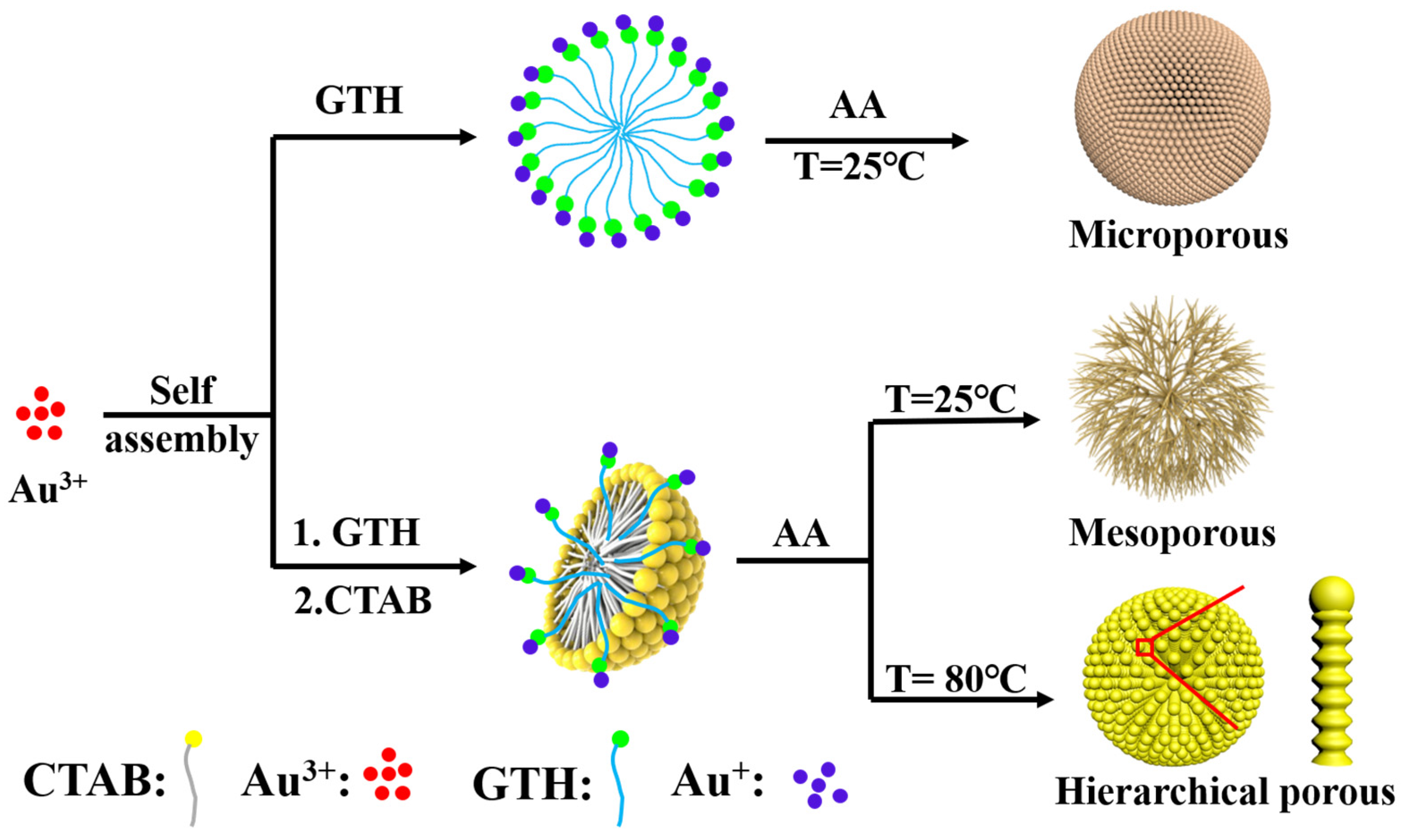
2. Results
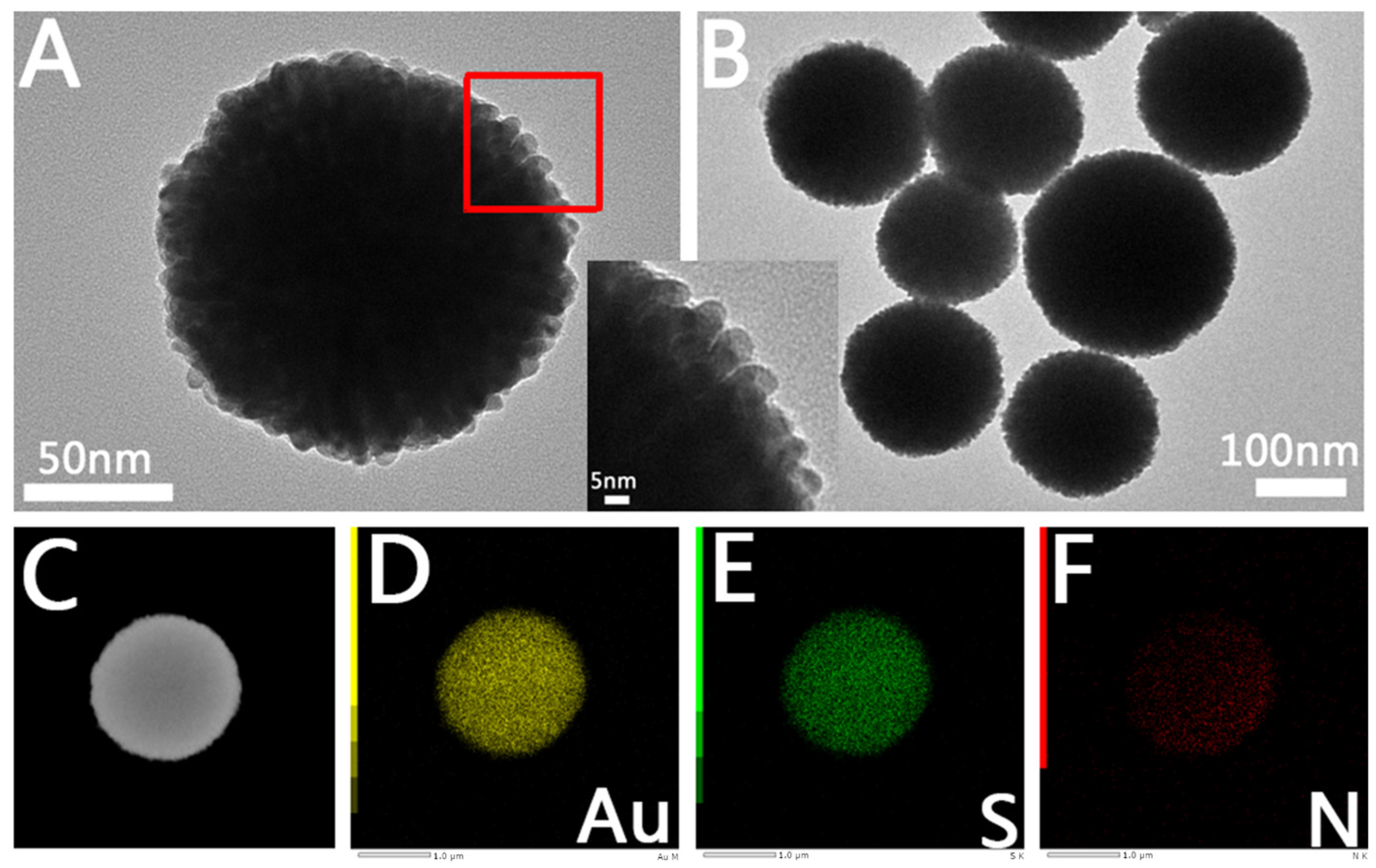
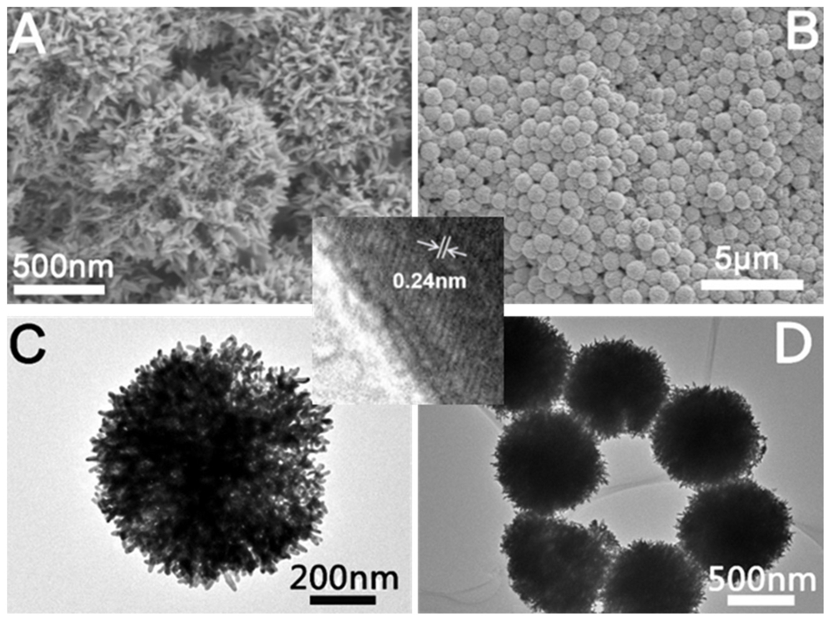
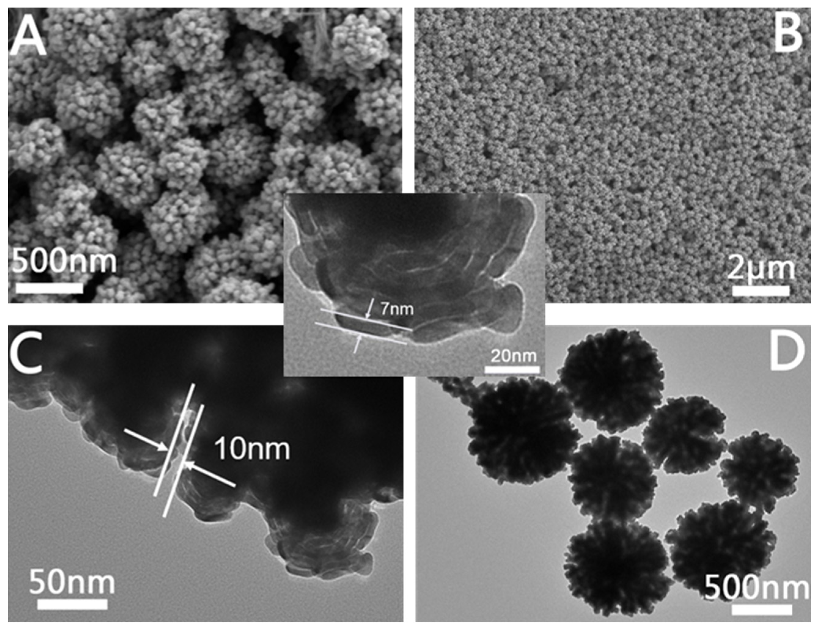
2.1. Structural Characterization
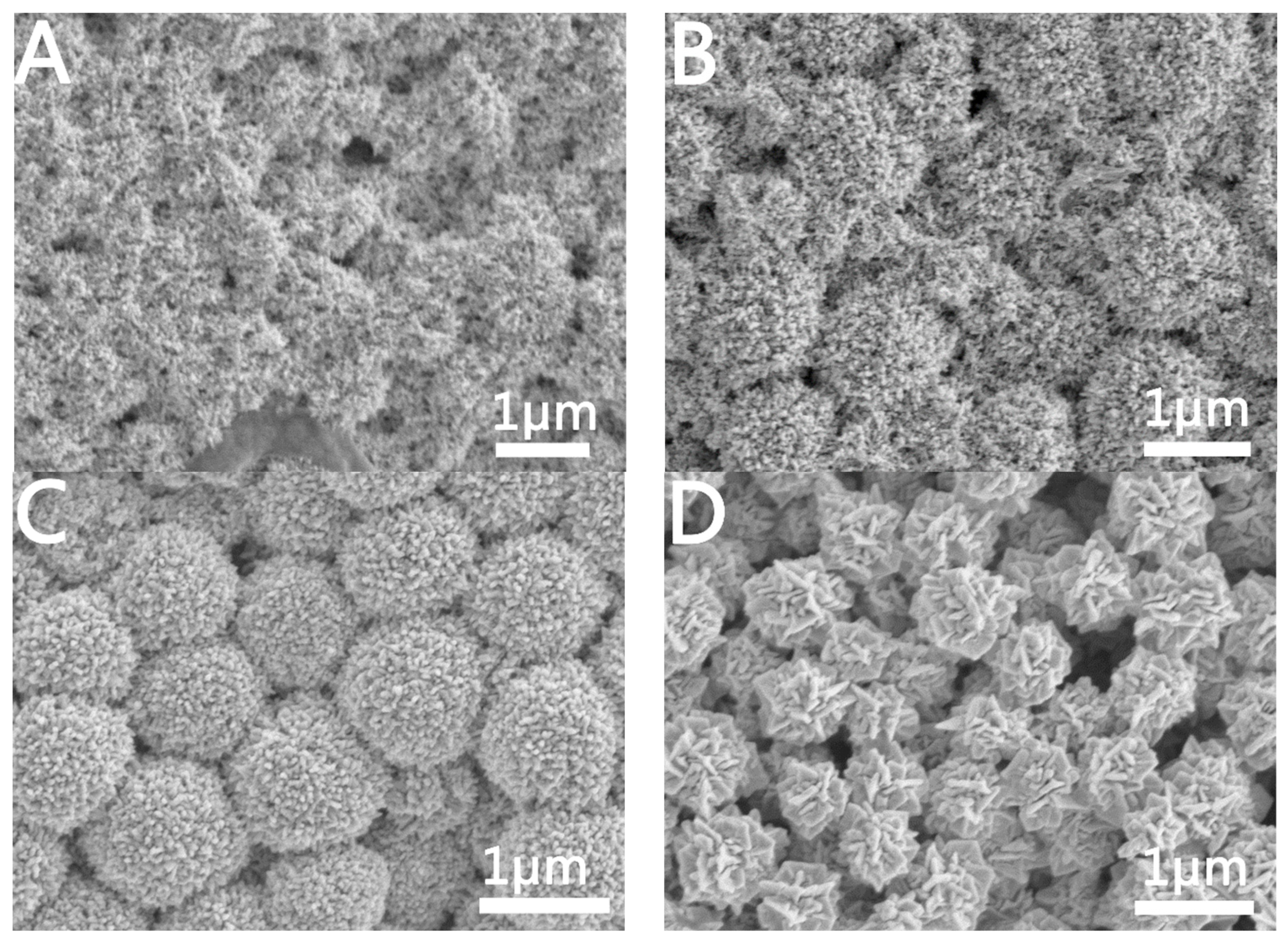
2.1.1. Characterization of Microporous Au NCs
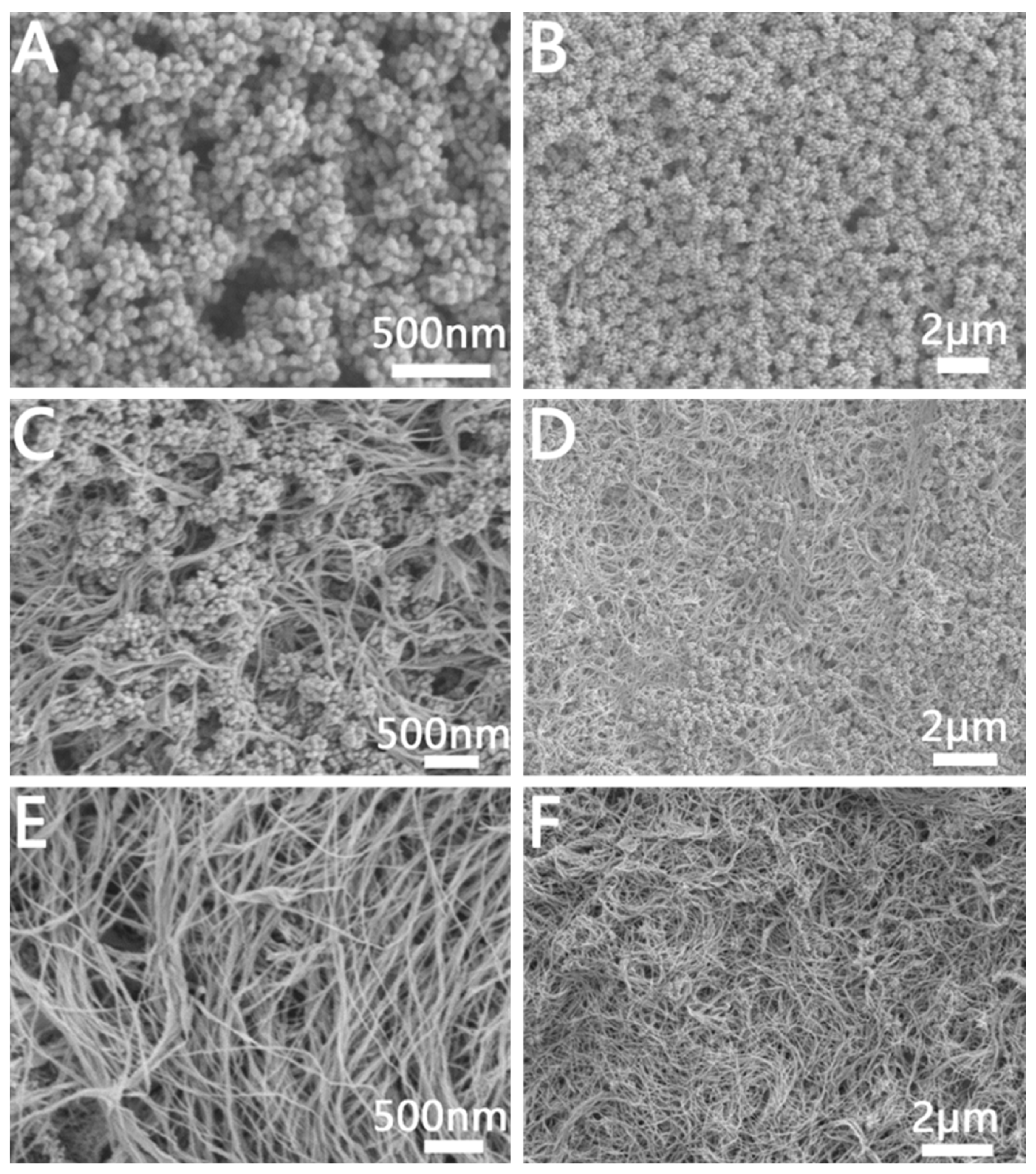
2.1.2. Characterization of Mesoporous Au NCs
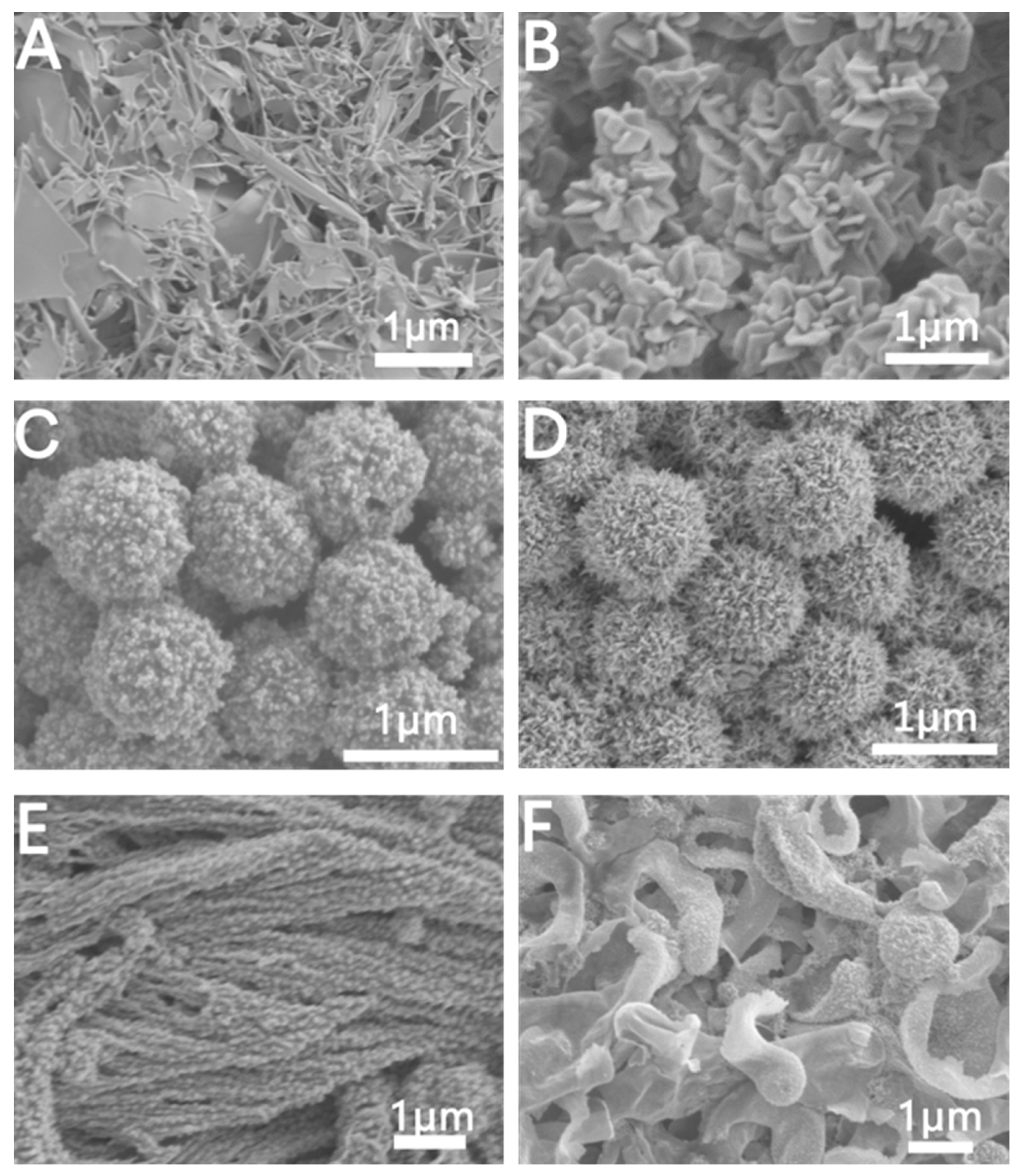
2.1.3. Characterization of Hierarchical Porous Au NCs
2.2. Effects of Reaction Parameters on Au NCs
2.2.1. Effects of GTH on the Au NCs
2.2.2. Effects of Temperature on the Au NCs
2.2.3. Effect of C16TAB Concentration and Alkyl Chain Length on Au NCs
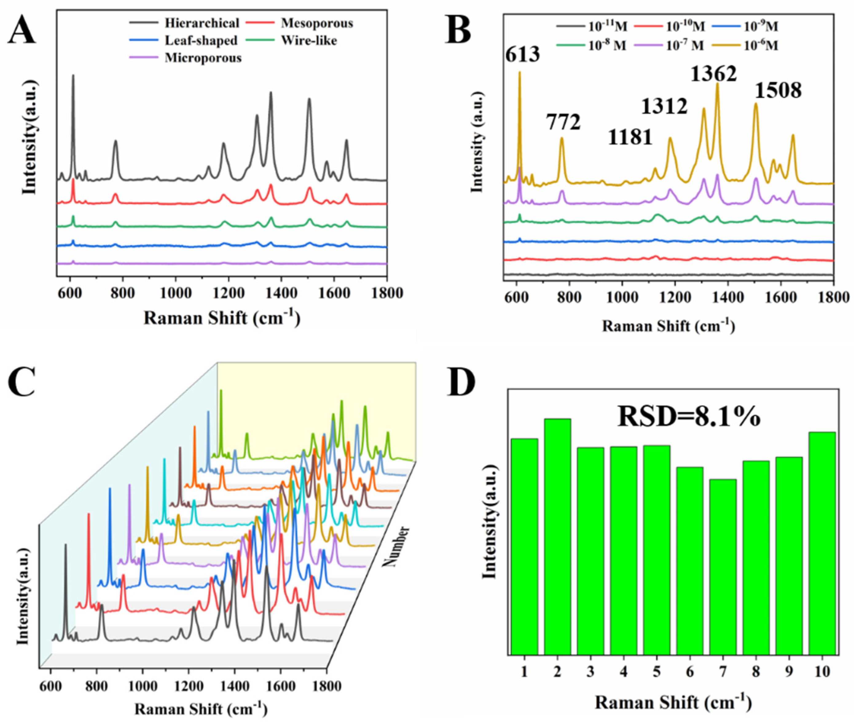
2.3. 3 SERS Performance
3. Materials and Methods
3.1. Materials
3.2. Instruments
3.3. Synthesis of Microporous Au NCs
3.4. Synthesis of Mesoporous Au NCs
3.5. Synthesis of Hierarchical Porous Au NCs
3.6. SERS Measurements in Colloids
4. Conclusions
Supplementary Materials
Author Contributions
Funding
Institutional Review Board Statement
Informed Consent Statement
Data Availability Statement
Conflicts of Interest
Sample Availability
References
- Jin, R.; Cao, Y.; Mirkin, C.A.; Kelly, K.L.; Schatz, G.C.; Zheng, J.G. Photoinduced conversion of silver nanospheres to nano prisms. Science 2001, 294, 1901–1903. [Google Scholar]
- Jin, R.; Cao, Y.C.; Hao, E.; Me, G.S.; Schatz, G.C.; Mirkin, C.A. Controlling anisotropic nanoparticle growth through plasmon excitation. Nature 2003, 425, 487–490. [Google Scholar]
- Tian, N.; Zhou, Z.Y.; Sun, S.G.; Ding, Y.; Wang, Z.L. Synthesis of Tetrahexahedral Platinum Nanocrystals with High-Index Facets and High Electro-Oxidation Activity. Science 2007, 316, 732–735. [Google Scholar]
- Langille, M.R.; Zhang, J.; Mirkin, C.A. Plasmon-mediated synthesis of heterometallic nanorods and icosahedra. Angew. Chem. Int. Ed. 2011, 50, 3543–3547. [Google Scholar]
- Zhai, Y.; Du-Chene, J.S.; Wang, Y.C.; Qiu, J.; Johnston-Peck, A.C.; You, B.; Guo, W.; DiCiaccio, B.; Qian, K.; Zhao, E.W.; et al. Polyvinylpyrro-lidone-induced anisotropic growth of gold nanoprisms in plasmon-driven synthesis. Nat. Mater. 2016, 15, 889–895. [Google Scholar]
- Qin, Y.Z.; Lu, Y.X.; Pan, W.F.; Yu, D.D.; Zhou, J.G. One-pot synthesis of hollow hydrangea Au nanoparticles as a dual catalyst with SERS activity for in situ monitoring of a reduction reaction. RSC Adv. 2019, 9, 10314–10319. [Google Scholar]
- Zhang, Q.F.; Hernandez, T.; Smith, K.W.; Jebeli, S.A.; Dai, A.X.; Warning, L.; Baiyasi, R.; McCarthy, L.A.; Guo, H.; Chen, D.H.; et al. Unraveling the Origin of Chirality from Plasmonic Nanoparticle-Protein Complexes. Science 2020, 365, 1475–1478. [Google Scholar]
- Lee, H.-E.; Ahn, H.-Y.; Mun, J.; Lee, Y.Y.; Kim, M.; Cho, N.H.; Chang, K.; Kim, W.S.; Rho, J.; Nam, K.T. Amino-Acid- and Peptide-Directed Synthesis of Chiral Plasmonic Gold Nanoparticles. Nature 2018, 556, 360–365. [Google Scholar]
- Cho, N.H.; Byun, G.H.; Lim, Y.C.; Im, S.W.; Kim, H.; Lee, H.E.; Ahn, H.Y.; Nam, K.T. Uniform Chiral Gap Synthesis for High Dissymmetry Factor in Single Plasmonic Gold Nanoparticle. ACS Nano 2020, 14, 3595–3602. [Google Scholar]
- Qin, Y.Z.; Yin, S.; Chen, M.; Yao, W.X.; He, Y.S. Surface-enhanced Raman spectroscopy for detection of fentanyl and its analogs by using Ag-Au nanoparticles. Spectrochim. Acta Part A Mol. Biomol. Spectrosc. 2023, 285, 121923–121933. [Google Scholar]
- Kim, F.; Connor, S.; Song, H.; Kuykendall, T.; Yang, P.D. Platonic Gold Nanocrystals. Angew. Chem. Int. Ed. 2004, 43, 3673–3677. [Google Scholar]
- Park, J.E.; Lee, Y.; Nam, J.M.; Shaped, P. Uniformly Formed Gold Nanocubes with Ultrahigh Reproducibility in Single-Particle Scattering and Surface-Enhanced Raman Scattering. Nano Lett. 2018, 18, 6475–6482. [Google Scholar]
- Lu, Y.; Zhang, H.; Wu, F.; Liu, H.; Fang, J.Z. Size-tunable uniform gold octahedra: Fast synthesis, characterization, and plasmonic properties. RSC Adv. 2017, 7, 18601–18608. [Google Scholar]
- Li, C.; Shuford, K.L.; Chen, M.; Lee, E.J.; Cho, S.O. A facile polyol route to uniform gold octahedra with tailorable size and their optical properties. ACS Nano 2008, 2, 1760–1769. [Google Scholar]
- Madasu, M.; Hsieh, P.L.; Chen, Y.J.; Huang, M.H. Formation of Silver Rhombic Dodecahedra, Octahedra, and Cubes through Pseudomorphic Conversion of Ag2O Crystals with Nitroarene Reduction Activity. ACS Appl. Mater. Interfaces 2019, 11, 38039–38045. [Google Scholar]
- Qin, Y.Z.; Pan, W.F.; Yu, D.D.; Lu, Y.X.; Wu, W.; Zhou, J.G. Stepwise evolution of Au micro/nanocrystals from an octahedron into a truncated ditetragonal prism. Chem. Commun. 2018, 54, 3411–3414. [Google Scholar]
- Kuo, B.H.; Hsia, C.F.; Chen, T.N.; Huang, M.H. Systematic Shape Evolution of Gold Nanocrystals Achieved through Adjustment in the Amount of HAuCl4 Solution Used. J. Phys. Chem. C 2018, 122, 25118–25126. [Google Scholar]
- Singh, P.; Ko, T.A.F.; Jaiswal, A. NIR active Plasmonic Gold Nanocapsules Synthesized using Thermally Induced Seed Twinning for Surface Enhanced Raman Scattering Applications. ACS Appl. Mater. Interfaces 2018, 10, 39380–39390. [Google Scholar]
- Brown, M.; Wiley, B.J. Bromide Causes Facet-Selective Atomic Addition in Gold Nanorod Syntheses. Chem. Mater. 2020, 32, 6410–6415. [Google Scholar]
- Khanal, B.P.; Zubarev, E.R. Chemical Transformation of Nanorods to Nanowires: Reversible Growth and Dissolution of Anisotropic Gold Nanostructures. ACS Nano 2019, 13, 2370–2378. [Google Scholar]
- Lin, G.; Xian, L.X.; Wang, S.; Shah, Z.H.; Edwards, S.A.; Gao, Y. Design and One-Pot Synthesis of Capsid-like Gold Colloids with Tunable Surface Roughness and Their Enhanced Sensing and Catalytic Performances. ACS Appl. Mater. Interfaces 2020, 12, 50152–50160. [Google Scholar]
- Liu, C.; Xu, X.; Hu, W.X.; Yang, X.; Zhou, P.; Qiu, G.; Ye, W.; Li, Y.; Jiang, C. Synthesis of Clean Cabbagelike (111) Faceted Silver Crystals for Efficient Surface-Enhanced Raman Scattering Sensing of Papaverine. Anal. Chem. 2018, 90, 9805–9812. [Google Scholar]
- Qin, Y.Z.; Mo, F.; Yao, S.; Wu, Y.Z.; He, Y.S.; Yao, W.X. Facile Synthesis of Porous Ag Crystals as SERS Sensor for Detection of Five Methamphetamine Analogs. Molecules 2022, 27, 3939. [Google Scholar]
- Lee, E.; Ryu, S. Nucleation and Growth-Controlled Facile Fabrication of Gold Nanoporous Structures for Highly Sensitive Surface-Enhanced Raman Spectroscopy Applications. Nanomaterials 2021, 11, 1463–1470. [Google Scholar]
- Kim, D.; Kim, J.; Henzie, J.; Ko, Y.; Lim, H.; Kwon, G.; Na, J.; Kim, H.J.; Yamauchi, Y.; You, J. Mesoporous Au films assembled on flexible cellulose nanopaper as high-performance SERS substrates. Chem. Eng. J. 2021, 419, 129445–129454. [Google Scholar]
- Lee, M.J.; Lim, S.H.; Ha, J.M.; Choi, S.M. Green Synthesis of High-Purity Mesoporous Gold Sponges Using Self-Assembly of Gold Nanoparticles Induced by Thiolated Poly (ethylene glycol). Langmuir 2016, 32, 5937–5945. [Google Scholar]
- Wei, X.Y.; Fan, Q.K.; Liu, H.P.; Bai, Y.C.; Zhang, L.; Zheng, H.Q.; Yin, Y.D.; Gao, C.B. Holey Au–Ag alloy nanoplates with built-in hotspots for surface-enhanced Raman scattering. Nanoscale 2016, 8, 15689–15695. [Google Scholar]
- Zhang, T.; Sun, Y.Q.; Hang, L.F.; Li, H.L.; Liu, G.Q.; Zhang, X.M.; Lyu, X.J.; Cai, W.P.; Li, Y. Periodic Porous Alloyed Au−Ag Nanosphere Arrays and Their Highly Sensitive SERS Performance with Good Reproducibility and High Density of Hotspots. ACS Appl. Mater. Interfaces 2018, 10, 9792–9801. [Google Scholar]
- Sattayasamitsathit, S.; Gu, Y.; Kaufmann, K.; Minteer, S.; Polsky, R.; Wang, J. Tunable hierarchical macro/mesoporous gold microwires fabricated by dual-templating and dealloying processes. Nanoscale 2013, 5, 7849–7854. [Google Scholar]
- Nguyen, N.T.; Altomare, M.; Yoo, J.; Schmuki, P. Efficient Photocatalytic H2 Evolution: Controlled Dewetting-Dealloying to Fabricate Site-Selective High Activity Nanoporous Au Particles on Highly Ordered TiO2 Nanotube Arrays. Adv. Mater. 2015, 27, 3208–3215. [Google Scholar]
- Li, X.Q.; Chen, Q.; McCue, I.; Snyder, J.D.; Crozier, P.; Erlebacher, J.; Sieradzki, K. Dealloying of Noble-Metal Alloy Nanoparticles. Nano Lett. 2014, 14, 2569–2577. [Google Scholar]
- Vidal, C.; Wang, D.; Schaaf, P.; Hrelescu, C.; Klar, T.A. Optical Plasmons of Individual Au Nanosponges. ACS Photonics 2015, 2, 1436–1442. [Google Scholar]
- Li, J.; Zhang, G.; Wang, J.; Maksymov, I.S.; Greentree, A.D.; Hu, J.; Shen, A.; Wang, Y.; Trau, M. Facile One-Pot Synthesis of Nanodot-Decorated Au-Silver Alloy Nanoboxes for Single-Particle Surface-Enhanced Raman Scattering Activity. ACS Appl. Mater. Interfaces 2018, 10, 32526–32535. [Google Scholar]
- Xu, S.; Joseph, S.; Zhang, H.; Lou, J.; Lu, Y. Controllable high-throughput fabrication of porous Au nanorods driven by Rayleigh instability. RSC Adv. 2016, 6, 66484–66489. [Google Scholar]
- Chauvin, A.; Delacôte, C.; Molina, L.L.; Duerrschnabel, M.; Boujtita, M.; Thiry, D.; Du, K.; Ding, J.; Choi, C.H.; Tessier, P.Y.; et al. Planar Arrays of Nanoporous Au Nanowires: When Electrochemical Dealloying Meets Nanopatterning. ACS Appl. Mater. Interfaces 2016, 8, 6611–6620. [Google Scholar]
- Li, C.; Dag, Ö.; Dao, T.D.; Nagao, T.; Sakamoto, Y.; Kimura, T.; Terasaki, O.; Yamauchi, Y. Electrochemical Synthesis of Mesoporous Au Films toward Mesospace-Stimulated Optical Properties. Nat. Commun. 2015, 6, 6608. [Google Scholar]
- Lv, H.; Xu, D.; Henzie, J.; Feng, J.; Lopes, A.; Yamauchi, Y.; Liu, B. Mesoporous Au Nanospheres via Thiolate−Au(I) Intermediates. Chem. Sci. 2019, 10, 6423–6430. [Google Scholar]
- Malgras, V.; Ataee-Esfahani, H.; Wang, H.; Jiang, B.; Li, C.; Wu, K.C.W.; Kim, J.H.; Yamauchi, Y. Nanoarchitectures for Mesoporous Metals. Adv. Mater. 2016, 28, 993–1010. [Google Scholar]
- Jiang, B.; Li, C.; Dag, Ö.; Abe, H.; Takei, T.; Imai, T.; Hossain, M.S.A.; Islam, M.T.; Wood, K.; Henzie, J.; et al. Mesoporous Metallic Rhodium Nanoparticles. Nat. Commun. 2017, 8, 15581. [Google Scholar]
- Lohse, S.E.; Burrows, N.D.; Scarabelli, L.; Marzán, L.L.; Murphy, C. Anisotropic Noble Metal Nanocrystal Growth: The Role of Halides. Chem. Mater. 2014, 26, 34–43. [Google Scholar]
- Lv, H.; Xu, D.D.; Sun, L.; Liu, B. Surfactant Design Strategy for one-pot Seedless Synthesis of Hollow Mesoporous AuAg Alloy Nanospheres. J. Phys. Chem. Lett. 2020, 11, 5777–5784. [Google Scholar]
- Requejo, K.I.; Liopo, A.V.; Zubarev, E.R. Au Nanorod Synthesis with Small Thiolated Molecules. Langmuir 2020, 36, 3758–3769. [Google Scholar]
- Lu, J.; Chang, Y.; Zhang, N.; Wei, Y.; Li, A.; Tai, J.; Xue, Y.; Wang, Z.; Yang, Y.; Zhao, L.; et al. Chiral Plasmonic Nanochains via the Self-Assembly of Au Nanorods and Helical Glutathione Oligomers Facilitated by Cetyltrimethylammonium Bromide Micelles. ACS Nano 2017, 11, 3463–3475. [Google Scholar]
- Basu, A.; Vaskevich, A.; Chuntonov, L. Glutathione Self-Assembles into a Shell of Hydrogen-Bonded Intermolecular Aggregates on “Naked” Silver Nanoparticles. J. Phys. Chem. B 2021, 125, 895–906. [Google Scholar]
- Wang, Y.Q.; Ma, S.; Yang, Q.Q.; Li, X.J. Size-dependent SERS detection of R6G by silver nanoparticles immersion-plated on silicon nanoporous pillar array. Appl. Surf. Sci. 2012, 258, 5881–5885. [Google Scholar]
- Pham, T.B.; Hoang, T.H.C.; Nguyen, V.C.; Vu, D.C.; Bui, H.; Pham, V.H. Improved versatile SERS spheroid end-facet optical fiber substrate based on silver nano-dendrites directly planted with gold nanoparticles using dual-laser assisted for pesticides detection. Opt. Mater. 2022, 126, 112196–112206. [Google Scholar]


| HAuCl4/10 mM | GTH/2 mM | C16TAB/mg | T/°C | AA/0.1 M | Pore Size | |
|---|---|---|---|---|---|---|
| Microporous Au NCs | 200 μL | 50 μL | 0 | 25 | 475 μL | 0.8 nm |
| Mesoporous Au NCs | 200 μL | 50 μL | 30 mg | 25 | 475 μL | 2 nm–50 nm |
| Leaf-shaped Au NCs | 200 μL | 50 μL | 30 mg (C6TAB) | 25 | 475 μL | None |
| Hierarchical porous Au NCs | 200 μL | 50 μL | 30 mg | 80 | 475 μL | 0.8 nm and 10 nm |
| Strip structure Au NCs | 200 μL | 250 μL | 30 mg | 80 | 475 μL | None |
Disclaimer/Publisher’s Note: The statements, opinions and data contained in all publications are solely those of the individual author(s) and contributor(s) and not of MDPI and/or the editor(s). MDPI and/or the editor(s) disclaim responsibility for any injury to people or property resulting from any ideas, methods, instructions or products referred to in the content. |
© 2023 by the authors. Licensee MDPI, Basel, Switzerland. This article is an open access article distributed under the terms and conditions of the Creative Commons Attribution (CC BY) license (https://creativecommons.org/licenses/by/4.0/).
Share and Cite
Qin, Y.; Fang, D.; Wu, Y.; Wu, Y.; Yao, W. Controllable Preparation of Gold Nanocrystals with Different Porous Structures for SERS Sensing. Molecules 2023, 28, 2316. https://doi.org/10.3390/molecules28052316
Qin Y, Fang D, Wu Y, Wu Y, Yao W. Controllable Preparation of Gold Nanocrystals with Different Porous Structures for SERS Sensing. Molecules. 2023; 28(5):2316. https://doi.org/10.3390/molecules28052316
Chicago/Turabian StyleQin, Yazhou, Dewang Fang, Yulun Wu, Yuanzhao Wu, and Weixuan Yao. 2023. "Controllable Preparation of Gold Nanocrystals with Different Porous Structures for SERS Sensing" Molecules 28, no. 5: 2316. https://doi.org/10.3390/molecules28052316