
Phytocannabinoids: Chromatographic Screening of Cannabinoids and Loading into Lipid Nanoparticles
 , , , , ,
Abstract
:1. Introduction
2. Results and Discussion
2.1. Chromatography Analysis
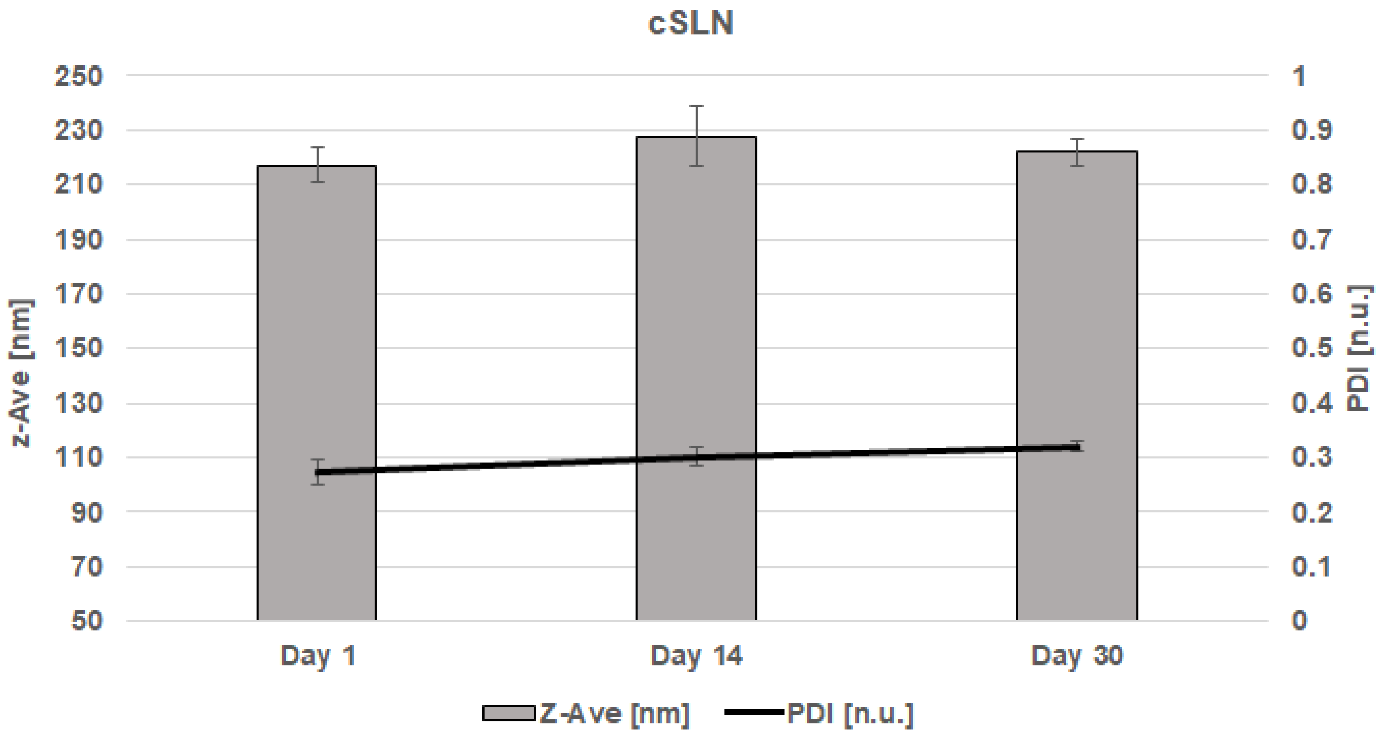
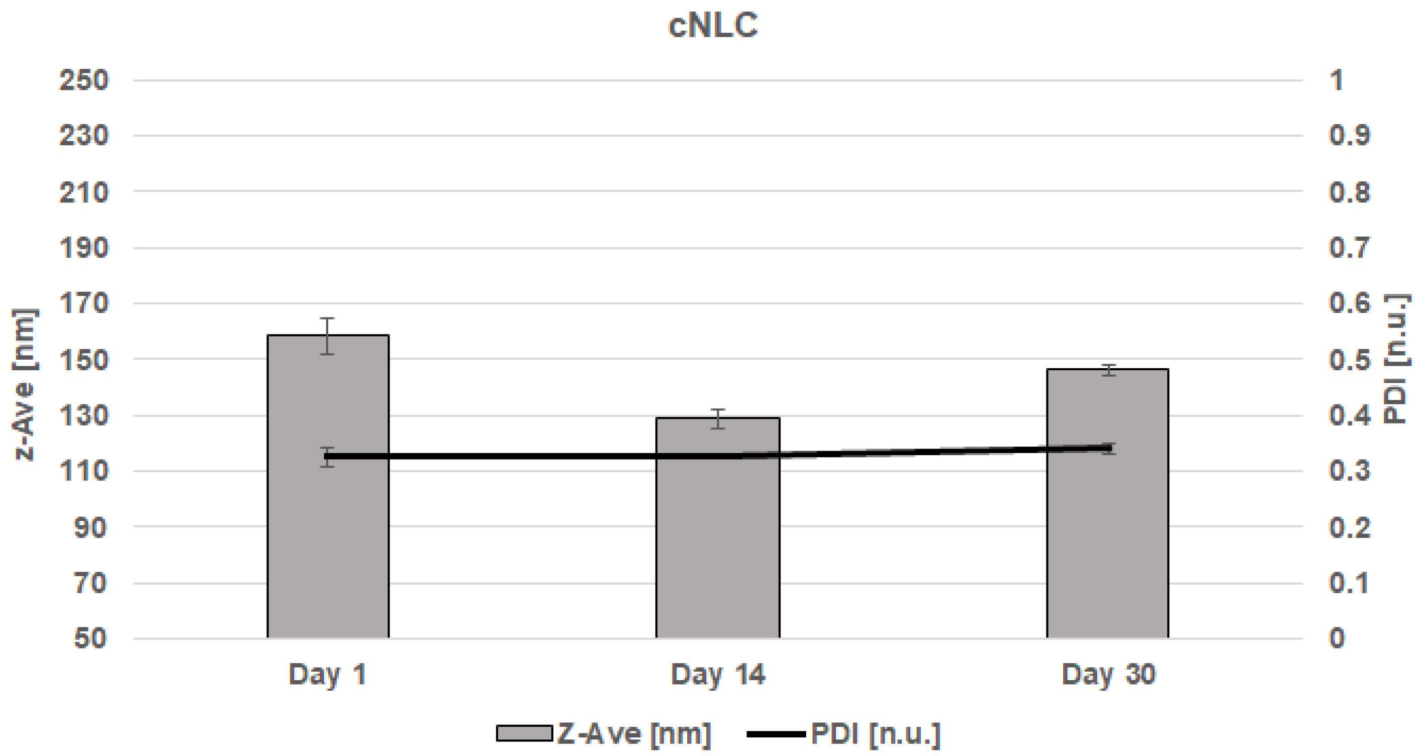
2.2. Determination of Particle Size, Polydispersity Index, and Zeta Potential
2.3. Encapsulation Efficiency and Loading Capacity
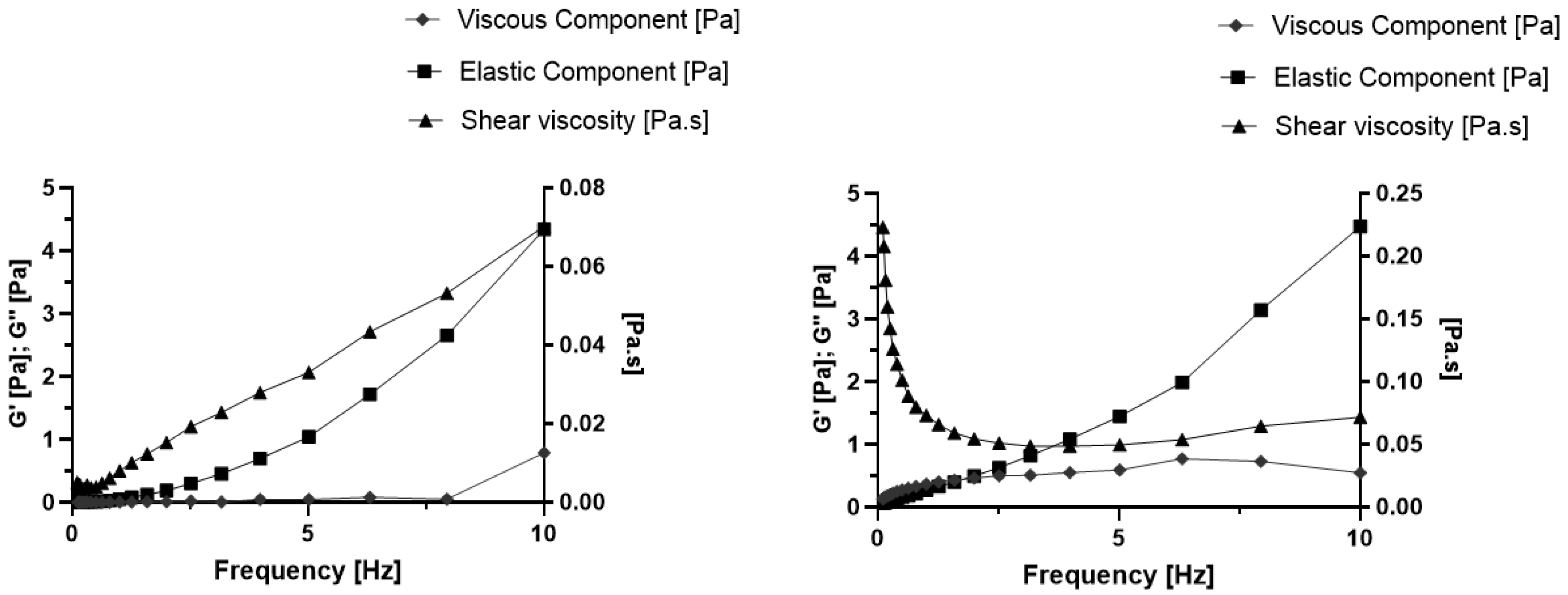
2.4. Rheology Study
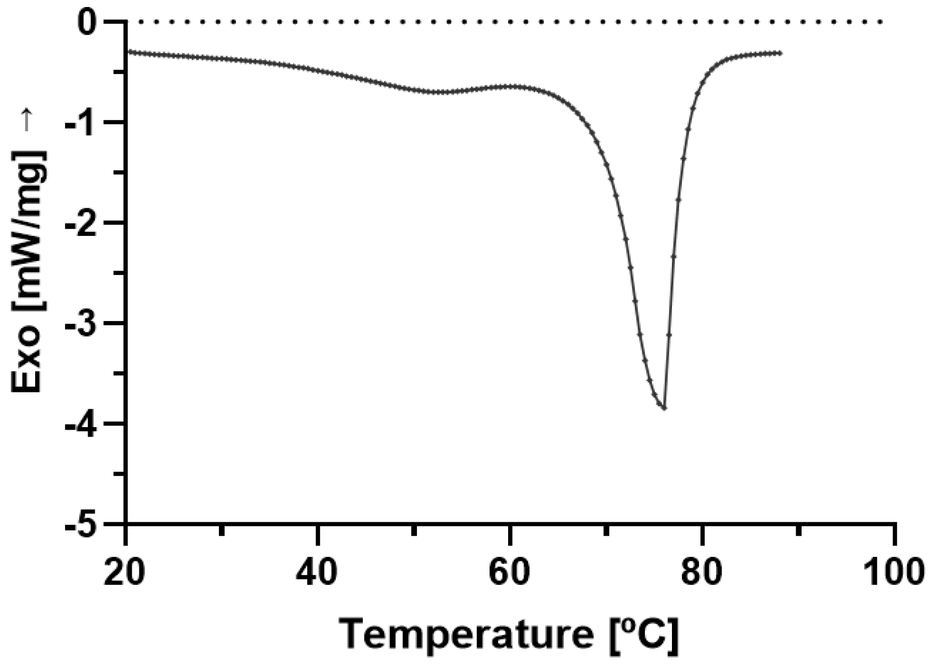
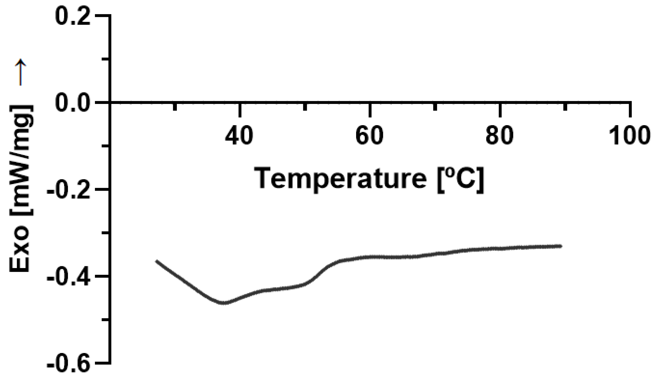
2.5. DSC
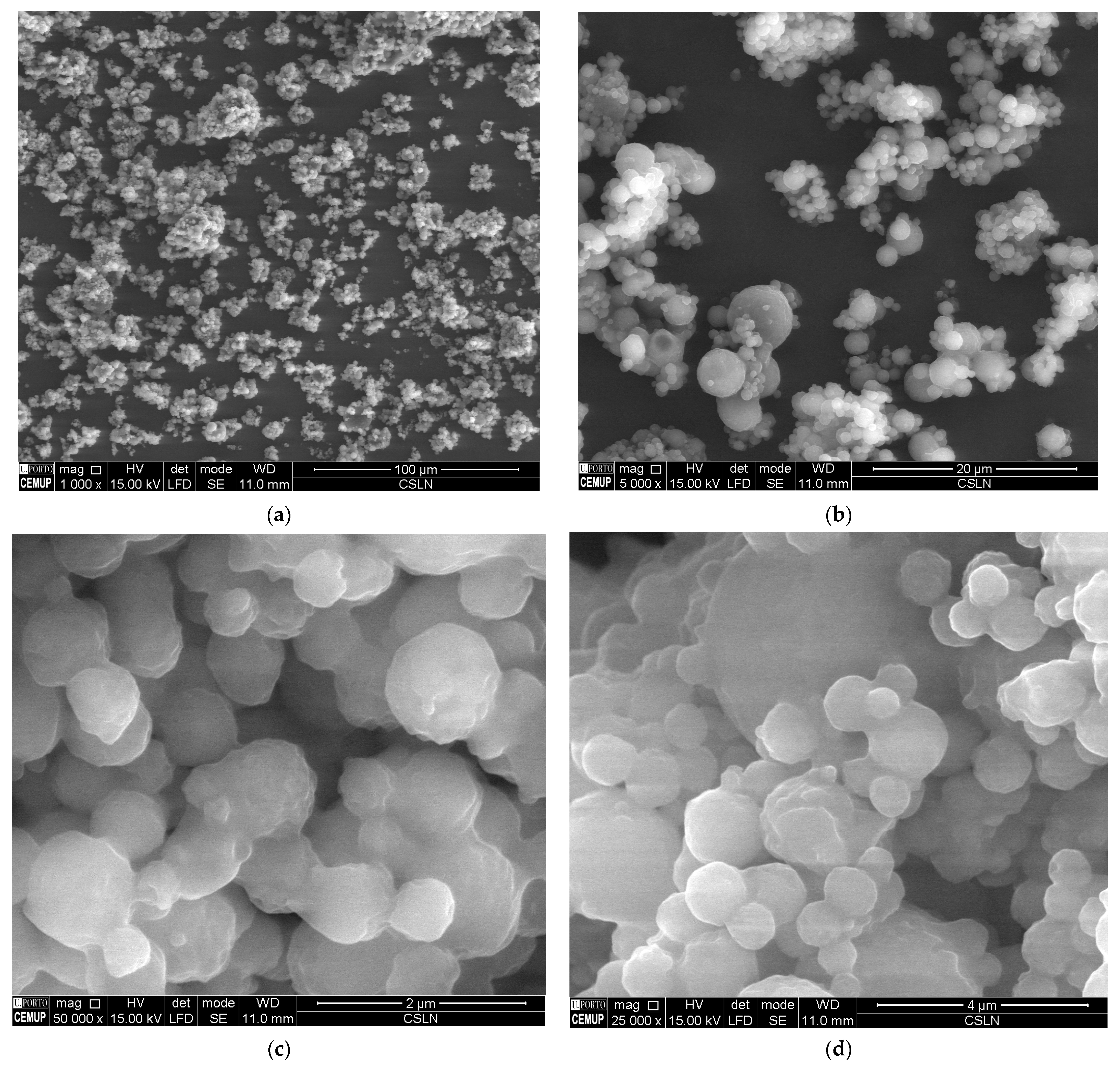
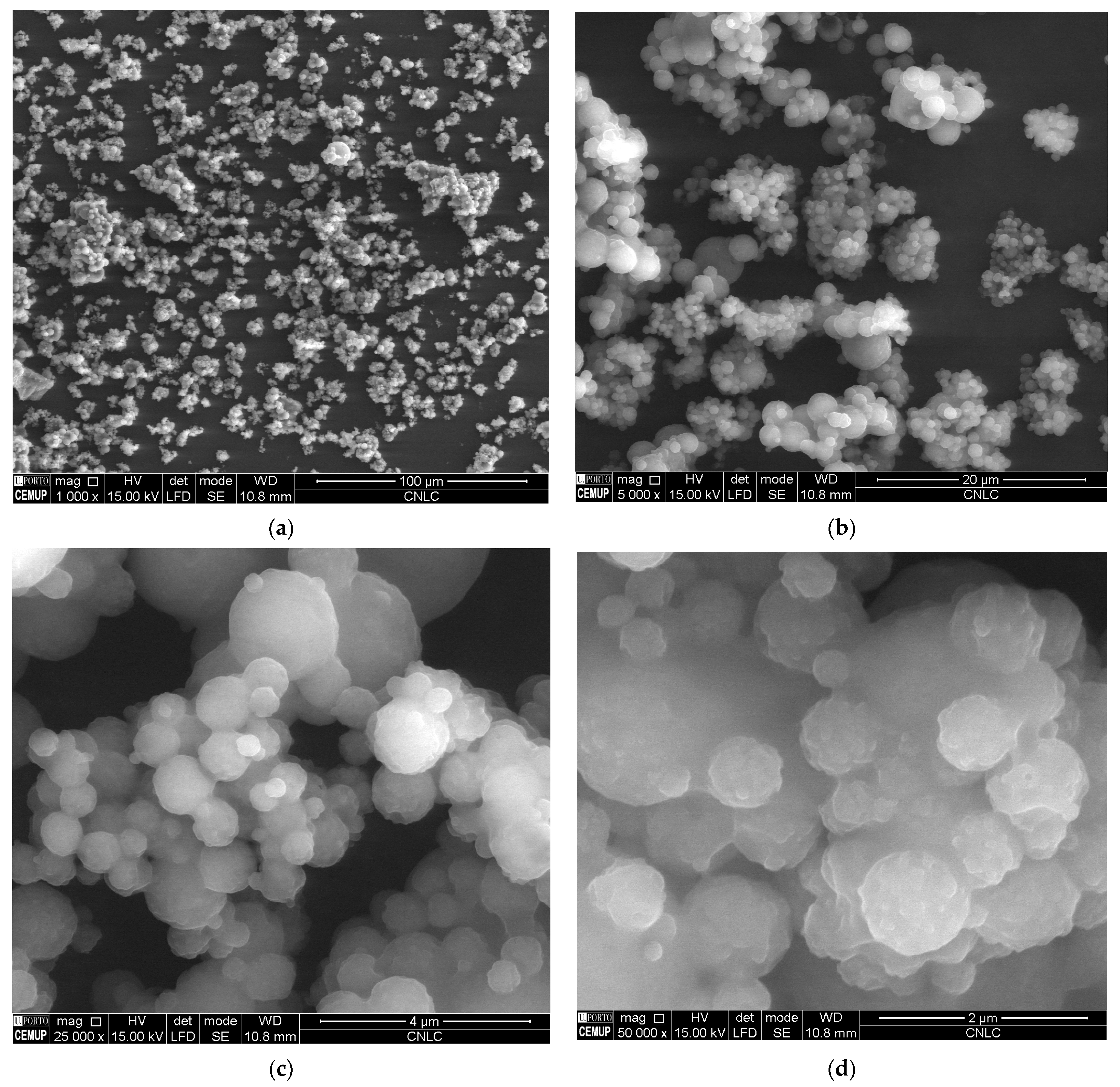
2.6. SEM
3. Materials and Methods
3.1. Plant Extract Preparation
3.2. Chromatography of Plant Extract
3.3. Production of SLN and NLC
3.4. Formulation Characterization
3.4.1. Particle Size, Polydispersity, and Zeta Potential
3.4.2. Encapsulation Efficiency and Loading Capacity
3.4.3. Rheology
3.4.4. Differential Scanning Calorimetry (DSC)
3.4.5. Scanning Electron Microscopy
4. Conclusions
Supplementary Materials
Author Contributions
Funding
Institutional Review Board Statement
Informed Consent Statement
Data Availability Statement
Acknowledgments
Conflicts of Interest
References
- Souto, E.B.; Baldim, I.; Oliveira, W.P.; Rao, R.; Yadav, N.; Gama, F.M.; Mahant, S. SLN and NLC for topical, dermal, and transdermal drug delivery. Expert Opin. Drug Deliv. 2020, 17, 357–377. [Google Scholar] [CrossRef] [PubMed]
- Mahant, S.; Rao, R.; Souto, E.B.; Nanda, S. Analytical tools and evaluation strategies for nanostructured lipid carrier-based topical delivery systems. Expert Opin. Drug Deliv. 2020, 17, 963–992. [Google Scholar] [CrossRef] [PubMed]
- Doktorovova, S.; Kovacevic, A.B.; Garcia, M.L.; Souto, E.B. Preclinical safety of solid lipid nanoparticles and nanostructured lipid carriers: Current evidence from in vitro and in vivo evaluation. Eur. J. Pharm. Biopharm. 2016, 108, 235–252. [Google Scholar] [CrossRef] [PubMed]
- Doktorovova, S.; Souto, E.B.; Silva, A.M. Nanotoxicology applied to solid lipid nanoparticles and nanostructured lipid carriers—A systematic review of in vitro data. Eur. J. Pharm. Biopharm. 2014, 87, 1–18. [Google Scholar] [CrossRef]
- Müller, R.; Petersen, R.; Hommoss, A.; Pardeike, J. Nanostructured lipid carriers (NLC) in cosmetic dermal products. Adv. Drug Deliv. Rev. 2007, 59, 522–530. [Google Scholar] [CrossRef]
- Lippacher, A.; Müller, R.; Mäder, K. Liquid and semisolid SLN™ dispersions for topical application: Rheological characterization. Eur. J. Pharm. Biopharm. 2004, 58, 561–567. [Google Scholar] [CrossRef]
- Rashidi, L. Different nano-delivery systems for delivery of nutraceuticals. Food Biosci. 2021, 43, 101258. [Google Scholar] [CrossRef]
- Mohammadi, M.; Assadpour, E.; Jafari, S.M. Encapsulation of food ingredients by nanostructured lipid carriers (NLCs). In Lipid-based Nanostructures for Food Encapsulation Purposes; Elsevier: Amsterdam, The Netherlands, 2019; pp. 217–270. [Google Scholar]
- Haider, M.; Abdin, S.M.; Kamal, L.; Orive, G. Nanostructured lipid carriers for delivery of chemotherapeutics: A review. Pharmaceutics 2020, 12, 288. [Google Scholar] [CrossRef] [Green Version]
- Salvi, V.R.; Pawar, P. Nanostructured lipid carriers (NLC) system: A novel drug targeting carrier. J. Drug Deliv. Sci. Technol. 2019, 51, 255–267. [Google Scholar] [CrossRef]
- Oliveira, D.R.B.; Michelon, M.; de Figueiredo Furtado, G.; Sinigaglia-Coimbra, R.; Cunha, R.L. β-Carotene-loaded nanostructured lipid carriers produced by solvent displacement method. Food Res. Int. 2016, 90, 139–146. [Google Scholar] [CrossRef]
- Bagherpour, S.; Alizadeh, A.; Ghanbarzadeh, S.; Mohammadi, M.; Hamishehkar, H. Preparation and characterization of Betasitosterol-loaded nanostructured lipid carriers for butter enrichment. Food Biosci. 2017, 20, 51–55. [Google Scholar] [CrossRef]
- Zheng, M.; Falkeborg, M.; Zheng, Y.; Yang, T.; Xu, X. Formulation and characterization of nanostructured lipid carriers containing a mixed lipids core. Colloids Surf. A Physicochem. Eng. Asp. 2013, 430, 76–84. [Google Scholar] [CrossRef]
- Cui, S.; Zhang, S.; Ge, S.; Xiong, L.; Sun, Q. Green preparation and characterization of size-controlled nanocrystalline cellulose via ultrasonic-assisted enzymatic hydrolysis. Ind. Crops Prod. 2016, 83, 346–352. [Google Scholar] [CrossRef]
- Artiga-Artigas, M.; Odriozola-Serrano, I.; Oms-Oliu, G.; Martín-Belloso, O. Nanostructured systems to increase bioavailability of food ingredients. In Nanomaterials for Food Applications; Elsevier: Amsterdam, The Netherlands, 2019; pp. 13–33. [Google Scholar]
- Lacatusu, I.; Mitrea, E.; Badea, N.; Stan, R.; Oprea, O.; Meghea, A. Lipid nanoparticles based on omega-3 fatty acids as effective carriers for lutein delivery. Preparation and in vitro characterization studies. J. Funct. Foods 2013, 5, 1260–1269. [Google Scholar] [CrossRef]
- Ali, A.; Ahmad, U.; Akhtar, J.; Khan, M.M. Engineered nano scale formulation strategies to augment efficiency of nutraceuticals. J. Funct. Foods 2019, 62, 103554. [Google Scholar] [CrossRef]
- Maretti, E.; Pavan, B.; Rustichelli, C.; Montanari, M.; Dalpiaz, A.; Iannuccelli, V.; Leo, E. Chitosan/heparin polyelectrolyte complexes as ion-paring approach to encapsulate heparin in orally administrable SLN: In vitro evaluation. Colloids Surf. A Physicochem. Eng. Asp. 2021, 608, 125606. [Google Scholar] [CrossRef]
- Babazadeh, A.; Ghanbarzadeh, B.; Hamishehkar, H. Formulation of food grade nanostructured lipid carrier (NLC) for potential applications in medicinal-functional foods. J. Drug Deliv. Sci. Technol. 2017, 39, 50–58. [Google Scholar] [CrossRef]
- Gasa-Falcon, A.; Odriozola-Serrano, I.; Oms-Oliu, G.; Martín-Belloso, O. Nanostructured lipid-based delivery systems as a strategy to increase functionality of bioactive compounds. Foods 2020, 9, 325. [Google Scholar] [CrossRef] [Green Version]
- Luan, J.; Zhang, D.; Hao, L.; Li, C.; Qi, L.; Guo, H.; Liu, X.; Zhang, Q. Design and characterization of Amoitone B-loaded nanostructured lipid carriers for controlled drug release. Drug Deliv. 2013, 20, 324–330. [Google Scholar] [CrossRef] [Green Version]
- Bashiri, S.; Ghanbarzadeh, B.; Ayaseh, A.; Dehghannya, J.; Ehsani, A. Preparation and characterization of chitosan-coated nanostructured lipid carriers (CH-NLC) containing cinnamon essential oil for enriching milk and anti-oxidant activity. Lwt 2020, 119, 108836. [Google Scholar] [CrossRef]
- Kovacevic, A.; Savic, S.; Vuleta, G.; Mueller, R.H.; Keck, C.M. Polyhydroxy surfactants for the formulation of lipid nanoparticles (SLN and NLC): Effects on size, physical stability and particle matrix structure. Int. J. Pharm. 2011, 406, 163–172. [Google Scholar] [CrossRef] [PubMed] [Green Version]
- Kiss, E.; Bertóti, I.; Vargha-Butler, E. XPS and wettability characterization of modified poly (lactic acid) and poly (lactic/glycolic acid) films. J. Colloid Interface Sci. 2002, 245, 91–98. [Google Scholar] [CrossRef] [PubMed]
- Amasya, G.; Ergin, A.D.; Cakirci, O.E.; Ozçelikay, A.T.; Bayindir, Z.S.; Yuksel, N. A study to enhance the oral bioavailability of s-adenosyl-l-methionine (SAMe): SLN and SLN nanocomposite particles. Chem. Phys. Lipids 2021, 237, 105086. [Google Scholar] [CrossRef] [PubMed]
- Aburahma, M.H.; Badr-Eldin, S.M. Compritol 888 ATO: A multifunctional lipid excipient in drug delivery systems and nanopharmaceuticals. Expert Opin. Drug Deliv. 2014, 11, 1865–1883. [Google Scholar] [CrossRef]
- Real, D.A.; Hoffmann, S.; Leonardi, D.; Goycoolea, F.M.; Salomon, C.J. A quality by design approach for optimization of Lecithin/Span® 80 based nanoemulsions loaded with hydrophobic drugs. J. Mol. Liq. 2021, 321, 114743. [Google Scholar] [CrossRef]
- Chun, J.Y.; Kang, H.K.; Jeong, L.; Kang, Y.O.; Oh, J.-E.; Yeo, I.-S.; Jung, S.Y.; Park, W.H.; Min, B.-M. Epidermal cellular response to poly (vinyl alcohol) nanofibers containing silver nanoparticles. Colloids Surf. B Biointerfaces 2010, 78, 334–342. [Google Scholar] [CrossRef]
- Fangueiro, J.F.; Andreani, T.; Egea, M.A.; Garcia, M.L.; Souto, S.B.; Souto, E.B. Experimental factorial design applied to mucoadhesive lipid nanoparticles via multiple emulsion process. Colloids Surf. B Biointerfaces 2012, 100, 84–89. [Google Scholar] [CrossRef]
- Gonzalez-Mira, E.; Egea, M.; Souto, E.; Calpena, A.; García, M. Optimizing flurbiprofen-loaded NLC by central composite factorial design for ocular delivery. Nanotechnology 2010, 22, 045101. [Google Scholar] [CrossRef]
- Andrzejewska, A.; Staszak, K.; Kaczmarek-Ryś, M.; Słomski, R.; Hryhorowicz, S. Understanding cannabinoid receptors: Structure and function. Acta Univ. Lodz. Folia Biol. Oecologica 2018, 14, 1–13. [Google Scholar] [CrossRef] [Green Version]
- Hryhorowicz, S.; Kaczmarek-Ryś, M.; Zielińska, A.; Scott, R.J.; Słomski, R.; Pławski, A. Endocannabinoid System as a Promising Therapeutic Target in Inflammatory Bowel Disease–A Systematic Review. Front. Immunol. 2021, 12, 790803. [Google Scholar] [CrossRef]
- Brighenti, V.; Pellati, F.; Steinbach, M.; Maran, D.; Benvenuti, S. Development of a new extraction technique and HPLC method for the analysis of non-psychoactive cannabinoids in fibre-type Cannabis sativa L.(hemp). J. Pharm. Biomed. Anal. 2017, 143, 228–236. [Google Scholar] [CrossRef]
- Shah, M.; Agrawal, Y. Ciprofloxacin hydrochloride-loaded glyceryl monostearate nanoparticle: Factorial design of Lutrol F68 and Phospholipon 90G. J. Microencapsul. 2012, 29, 331–343. [Google Scholar] [CrossRef]
- Le Bars, G.; Dion, S.; Gauthier, B.; Mhedhbi, S.; Pohlmeyer-Esch, G.; Comby, P.; Vivan, N.; Ruty, B. Oral toxicity of Miglyol 812® in the Göttingen® minipig. Regul. Toxicol. Pharmacol. 2015, 73, 930–937. [Google Scholar] [CrossRef]
- Brubach, J.B.; Jannin, V.; Mahler, B.; Bourgaux, C.; Lessieur, P.; Roy, P.; Ollivon, M. Structural and thermal characterization of glyceryl behenate by X-ray diffraction coupled to differential calorimetry and infrared spectroscopy. Int. J. Pharm. 2007, 336, 248–256. [Google Scholar] [CrossRef]
- Chawla, V.; Saraf, S.A. Glyceryl Behenate and Its Suitability for Production of Aceclofenac Solid Lipid Nanoparticles. J. Am. Oil Chem. Soc. 2011, 88, 119–126. [Google Scholar] [CrossRef]
- Campos, J.R.; Fernandes, A.R.; Sousa, R.; Fangueiro, J.F.; Boonme, P.; Garcia, M.L.; Silva, A.M.; Naveros, B.C.; Souto, E.B. Optimization of nimesulide-loaded solid lipid nanoparticles (SLN) by factorial design, release profile and cytotoxicity in human Colon adenocarcinoma cell line. Pharm. Dev. Technol. 2019, 24, 616–622. [Google Scholar] [CrossRef]
- Doktorovova, S.; Silva, A.M.; Gaivao, I.; Souto, E.B.; Teixeira, J.P.; Martins-Lopes, P. Comet assay reveals no genotoxicity risk of cationic solid lipid nanoparticles. J. Appl. Toxicol. 2014, 34, 395–403. [Google Scholar] [CrossRef]
- Souto, E.B.; Doktorovova, S.; Zielinska, A.; Silva, A.M. Key production parameters for the development of solid lipid nanoparticles by high shear homogenization. Pharm. Dev. Technol. 2019, 24, 1181–1185. [Google Scholar] [CrossRef]
- Souto, E.B.; Zielinska, A.; Souto, S.B.; Durazzo, A.; Lucarini, M.; Santini, A.; Silva, A.M.; Atanasov, A.G.; Marques, C.; Andrade, L.N.; et al. (+)-Limonene 1,2-Epoxide-Loaded SLNs: Evaluation of Drug Release, Antioxidant Activity, and Cytotoxicity in an HaCaT Cell Line. Int. J. Mol. Sci. 2020, 21, 1449. [Google Scholar] [CrossRef] [Green Version]


| Lp. | 1. | 2. | 3. | 4. | 5. | 6. | 7. | 8. | 9. | 10. | 11. |
|---|---|---|---|---|---|---|---|---|---|---|---|
| Cannabinoid * | CBDV [%] | CBDA [%] | CBGA [%] | CBG [%] | CBD [%] | CBN [%] | Δ9-THC [%] | CBNA [%] | CBC [%] | THCA [%] | CBCA [%] |
| Sample 1 | 0.07 | 7.41 | 0.09 | 0.06 | 3.07 | 0.08 | 0.25 | 0.04 | 0.22 | 0.38 | 0.39 |
| Sample 2 | 0.06 | 7.09 | 0.04 | 0.06 | 2.97 | 0.08 | 0.25 | 0.04 | 0.22 | 0.37 | 0.38 |
| Sample Name | Measurement Time (Days after Production) | ZP [mV] ± SD |
|---|---|---|
| empty-cSLN | 0 | −18.09 ± 0.81 |
| CBD-cSLN | 1 | −12.99 ± 0.90 |
| 30 | −12.42 ± 2.14 | |
| empty-cNLC | 0 | −13.63 ± 0.89 |
| CBD-cNLC | 1 | −5.51 ± 1.58 |
| 30 | −12.06 ± 1.50 |
| CBD-cSLN | CBD-cNLC | |
|---|---|---|
| EE [%] | 74.23 | 70.44 |
| LC [%] | 15.77 | 15.11 |
| DSC Parameters | CBD-cSLN | CBD-cNLC |
|---|---|---|
| Peak Maximum (°C) | 71.00 | 63.60 |
| Enthalpy (J/g) | 3.960 | 1.934 |
| Crystallinity Index (%) | 90.41 | 40.18 |
| Formulation | ||
|---|---|---|
| Ingredients | CBD-cSLN | CBD-cNLC |
| CBD | 1 | 1 |
| Compritol® 888 ATO | 4 | 3 |
| Miglyol® 812 | - | 1 |
| Poloxamer® 188 | 1.5 | 1.5 |
| Water | 93.5 | 93.5 |
Disclaimer/Publisher’s Note: The statements, opinions and data contained in all publications are solely those of the individual author(s) and contributor(s) and not of MDPI and/or the editor(s). MDPI and/or the editor(s) disclaim responsibility for any injury to people or property resulting from any ideas, methods, instructions or products referred to in the content. |
© 2023 by the authors. Licensee MDPI, Basel, Switzerland. This article is an open access article distributed under the terms and conditions of the Creative Commons Attribution (CC BY) license (https://creativecommons.org/licenses/by/4.0/).
Share and Cite
Zielińska, A.; da Ana, R.; Fonseca, J.; Szalata, M.; Wielgus, K.; Fathi, F.; Oliveira, M.B.P.P.; Staszewski, R.; Karczewski, J.; Souto, E.B. Phytocannabinoids: Chromatographic Screening of Cannabinoids and Loading into Lipid Nanoparticles. Molecules 2023, 28, 2875. https://doi.org/10.3390/molecules28062875
Zielińska A, da Ana R, Fonseca J, Szalata M, Wielgus K, Fathi F, Oliveira MBPP, Staszewski R, Karczewski J, Souto EB. Phytocannabinoids: Chromatographic Screening of Cannabinoids and Loading into Lipid Nanoparticles. Molecules. 2023; 28(6):2875. https://doi.org/10.3390/molecules28062875
Chicago/Turabian StyleZielińska, Aleksandra, Raquel da Ana, Joel Fonseca, Milena Szalata, Karolina Wielgus, Faezeh Fathi, M. Beatriz P. P. Oliveira, Rafał Staszewski, Jacek Karczewski, and Eliana B. Souto. 2023. "Phytocannabinoids: Chromatographic Screening of Cannabinoids and Loading into Lipid Nanoparticles" Molecules 28, no. 6: 2875. https://doi.org/10.3390/molecules28062875