Article

Nucleotide Imbalance, Provoked by Downregulation of Aspartate Transcarbamoylase Impairs Cold Acclimation in Arabidopsis

1
Faculty of Biology, Plant Physiology, University of Kaiserslautern, Erwin-Schrödinger-Straße, D-67663 Kaiserslautern, Germany
2
Institute for Biosciences, Physiology of Plant Metabolism, University of Rostock, Albert-Einstein-Strasse 3, D-18059 Rostock, Germany
*
Author to whom correspondence should be addressed.
Molecules 2023, 28(4), 1585; https://doi.org/10.3390/molecules28041585
Submission received: 27 December 2022 / Revised: 1 February 2023 / Accepted: 1 February 2023 / Published: 7 February 2023

Abstract

:
Aspartate transcarbamoylase (ATC) catalyzes the first committed step in pyrimidine de novo synthesis. As shown before, mutants with 80% reduced transcript and protein levels exhibit reduced levels of pyrimidine metabolites and thus nucleotide limitation and imbalance. Consequently, reduced photosynthetic capacity and growth, accompanied by massive transcriptional changes, were observed. Here, we show that nucleotide de novo synthesis was upregulated during cold acclimation of Arabidopsis thaliana (ecotype Columbia, Col-0) plants, but ATC knockdown mutants failed to acclimate to this condition as they did not accumulate neutral sugars and anthocyanins. A global transcriptome analysis revealed that most of the transcriptional changes observed in Col-0 plants upon cold exposure were also evident in ATC knockdown plants. However, several responses observed in cold-treated Col-0 plants could already be detected in knockdown plants when grown under standard conditions, suggesting that these mutants exhibited typical cold responses without prior cold stimulation. We believe that nucleotide signaling is involved in “cold-like priming” and “cold acclimation” in general. The observed transcript levels of genes involved in central carbon metabolism and respiration were an exception to these findings. These were upregulated in the cold but downregulated in warm-grown ATC mutants.

1. Introduction

Due to their sessile lifestyle, plants are forced to acclimate changing environmental conditions. Over the last decades, extensive research on various plant species has already led to the discovery of various metabolic pathways important for acclimation of the plant’s metabolism to low temperatures. This complex process of acclimation to low but non-freezing temperatures has been described in plants as “cold acclimation” (CA) and can be divided into three stages involving a variety of molecular, biochemical, and physiological changes [1,2]. In general, CA involves the rearrangement of membrane composition, adjustment of water contents in different tissues, global modification of gene expression, and regulation of protein activity [2,3,4,5].
The first phase of CA involves the direct cellular response to cold, including responses to an energy imbalance between photochemistry, electron transport, and metabolism. State transitions and enhanced non-photochemical quenching can minimize excitation energy at photosystem II (PSII) and therefore reduce the risk of damage [6].
To further counteract the effects of low temperatures, in the second phase of cold acclimation, extensive reprogramming of the transcriptome, proteome, and metabolome results in alterations in primary and specialized metabolism [7]. Further, cold treatment often goes along with increased production of reactive oxygen species (ROS) in the plant’s metabolism. Thereby, ROS emerge at various sites in the plant’s metabolism, e.g., at photosystem I, as NADP+, the final acceptor of the photosynthetic electron transport chain, is limited under cold conditions (acceptor-site limitation). To ensure ROS homeostasis, the accumulation of ROS-scavenging compounds is induced, and photosynthetic efficiency is reduced under cold stress [8].
In the third and final phase of CA, expression changes that occurred during the second phase of acclimation are established under persistently altered environmental conditions [3].
Overall, there is no unique metabolite, transcript, or pathway that can be assigned as responsible for cold tolerance; rather, a multifaceted reorganization of metabolic homeostasis appears to be required. However, different metabolites were shown to have protective effects against different forms of cold stress.
The accumulation of flavonoids, for example, is of particular importance for the protection against the high reducing ability of the photosynthetic electron transport chain under cold conditions [9]. In the first step of the flavonoid-biosynthesis phenylalanine, derived from the chloroplast, is converted to p-coumaroyl-CoA in the phenylpropanoid pathway in the cytosol. Further reaction steps, which include early (e.g., CHI) and late (e.g., LDOX) biosynthetic enzymes, end with the accumulation of colored anthocyanin pigments in the vacuole. In plants, the regulation of flavonoids is still not completely understood, but several signals have been reported to trigger the induction of the early biosynthetic genes that provide flavonols as well as the late biosynthetic genes leading to anthocyanins as their product [10]. Such signals include sugars, phytohormones, ROS, and transcription factors (e.g., MYB111, TT8, and PAP1) [10,11,12,13,14]. Moreover, transcription factors in general have dominant roles in cold acclimation [15].
An interesting phenomenon in CA is known as “cold priming”, where a short cold stimulus induces the induction of a core set of stress response genes, thereby affecting the cold response of plants upon longer exposition to cold [16,17].
As cold acclimation requires extensive restructuring of metabolic processes, ribosome biogenesis and translation have to be affected by cold treatment. Indeed, transcriptomic analysis of Arabidopsis during a time course of cold acclimation and deacclimation revealed ribosome biogenesis and translation as key processes involved in cold acclimation [7]. Given that up to 50% of the cellular nucleotides are bound in ribosomal RNA, it becomes clear that sufficient nucleotide provision is a prerequisite for full functionality of the cold acclimation response [18].
The enzyme aspartate transcarbamoylase (ATC) catalyzes the first committed step in pyrimidine de novo synthesis, and the amount of ATC protein is a limiting factor of plant growth [19,20]. It was shown that ATC knockdown mutants harbor massively reduced levels of pyrimidine nucleotides (UMP, UDP) and the corresponding nucleotide sugars (e.g., UDP-Glc). This nucleotide limitation and imbalance affected gene expression and photosynthetic performance in ATC mutants. Genes involved in central carbon metabolism, intracellular transport, and respiration were mainly downregulated in ATC mutants, thereby being putatively responsible for the observed impaired growth. In this work, we aim to unravel and link phenotypical, physiological, and transcriptomic changes in ATC knockdown mutants grown in cold temperatures.

2. Results

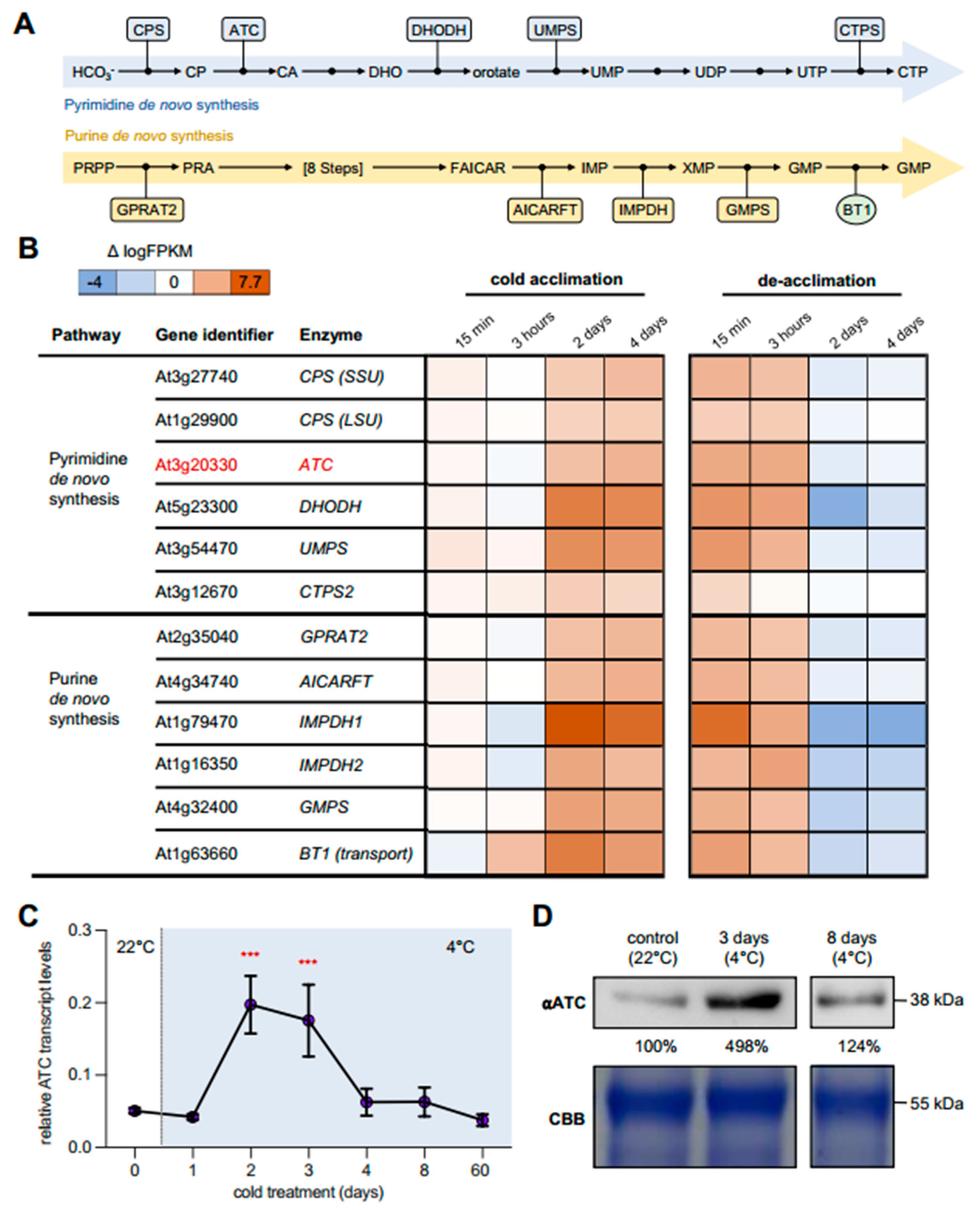
2.1. Cold Leads to Increased Expression of Genes Involved in Nucleotide De Novo Synthesis

Although the precise balancing of nucleotides and de novo biosynthesis play an important role in plant development, little research on the role of this metabolic pathway in the abiotic stress response has been performed in the past. Nevertheless, a recent study showed that the biosynthesis of pyrimidines and purines was greatly increased during cold acclimation, resulting in significantly larger nucleotide pools [21]. In-depth analysis of a previously published transcriptome dataset obtained from cold-treated wild-type (Col-0) plants revealed that major transcriptional changes in pyrimidine and purine biosynthesis occur during the early phases of cold acclimation (1–3 days) (Figure 1A,B) [7]. Among all analyzed pyrimidine de novo synthesis genes, dihydroorotate dehydrogenase (DHODH) exhibited the greatest increase in expression (1.8-fold) during cold treatment. Further transcriptional changes could be observed for the plastid-localized enzymes carbamoyl phosphate synthase (CPS SSU/LSU) and aspartate transcarbamoylase (ATC, red) (Figure 1B). In line with this, genes of purine biosynthesis also showed an increase in transcript levels. The most pronounced change was seen in inosine monophosphate dehydrogenase 1 (IMPDH1) (Figure 1A,B). During the de-acclimation phase, transcript levels for both pyrimidine and purine de novo synthesis genes decreased to or even below the initial levels before cold acclimation (Figure 1B).

2.2. ATC Abundancy Is Strongly Increased Up on Cold Acclimation and Adaptation

To confirm the increased expression of ATC in response to cold in our laboratory, corresponding analyses were carried out using targeted gene expression analysis (qRT-PCR). Plants were grown under control conditions (22 °C, 14 h light/10 h dark) for 21 days and subsequently transferred to 4 °C for defined timespans. The greatest changes in ATC transcript levels were seen after 2 and 3 days of cold treatment, with increases up to 3.5-fold compared to the onset of cold treatment (Figure 1C). During prolonged cold exposure (days 8 and 63), the transcript levels again decreased significantly to the initial level before the cold treatment (Figure 1C). Comparable results could be obtained by quantifying protein levels using an ATC antiserum raised against the recombinant protein [19]. Thereby, Arabidopsis wild type (Col-0) plants revealed an almost 5-fold increase in ATC protein levels after 3 days of cold treatment, which declined to initial levels at 8 days after transfer to 4 °C (Figure 1D).
After seven days at 4 °C, Col-0 plants showed 40.8% less fresh weight compared to their growth at ambient temperature. In contrast, the fresh weight of the ATC mutants was not impaired after 7 days of incubation in the cold, compared to untreated plants of the same genotype (Figure 2A). However, it became obvious that atc#1 and atc#2 failed to accumulate anthocyanins (Figure 2B,D). While Col-0 plants accumulated 147-fold more anthocyanins in the cold, the atc#2 line only showed a 3.6-fold increase compared to the control plants (Figure 2D). Col-0 plants that were exposed to 4 °C for 63 days showed a significant reduction in their maximum growth height, but no differences in plant height could be observed in ATC mutants between the control and cold conditions (Figure 2C). It was also found that the chlorophyll content in the ATC mutants was reduced, thereby explaining the slightly pale green appearance of the mutant leaves after cold treatment (Figure 2E).

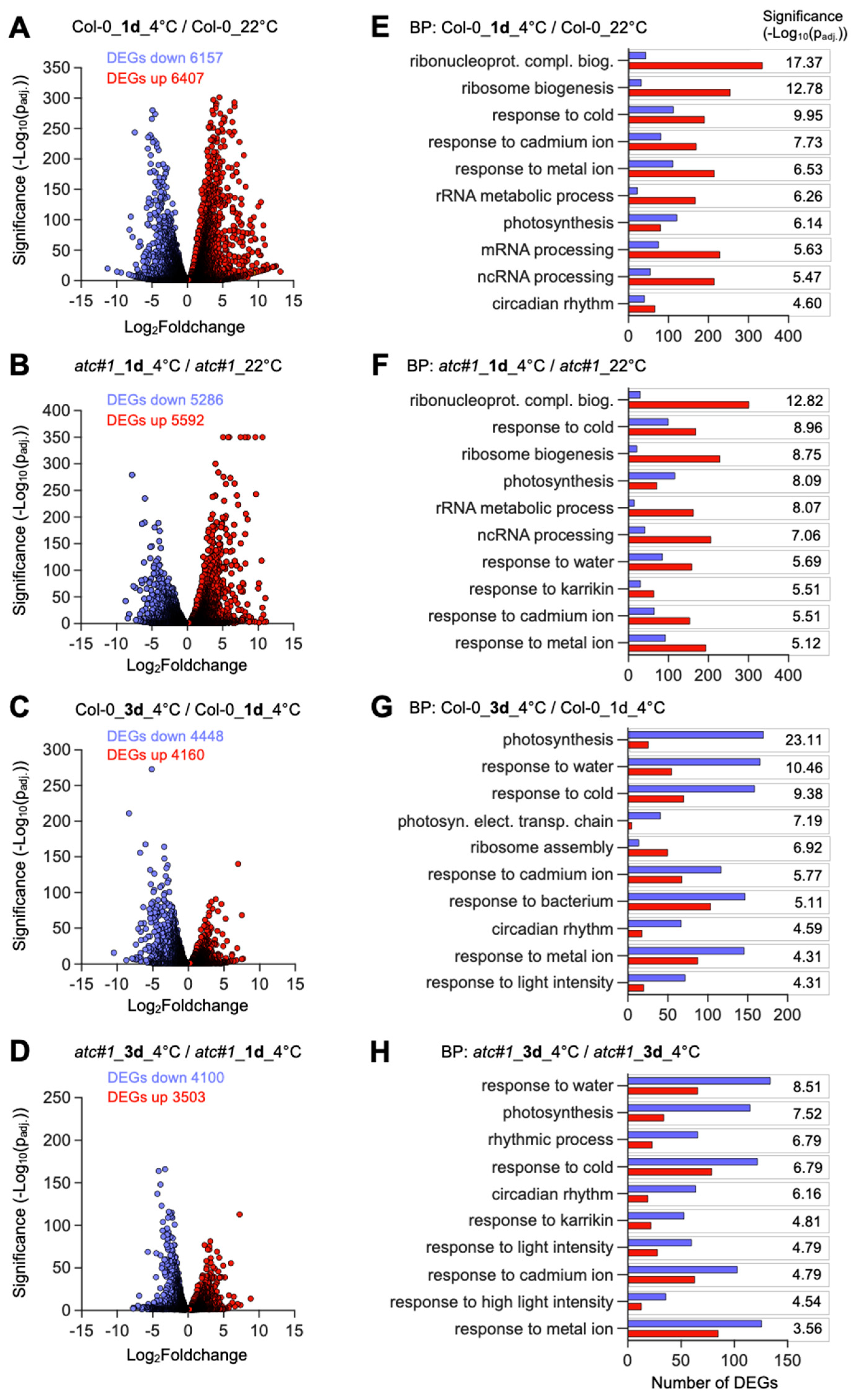
2.3. Cold Acclimation Imposes Global Adaptations of the Transcriptome

To investigate the impact of an altered nucleotide metabolism on the transcriptional changes induced during cold acclimation, RNA-Seq analysis was performed. To this end Col-0 and atc#1 grown at ambient temperature for six weeks (control) were transferred to cold (4 °C) for one and three days, before harvesting the aboveground tissue for RNA isolation. A comparison of transcriptional changes between Col-0 and atc#1 (grown under ambient temperature) was shown previously [20]. Where necessary for a better understanding, we will also refer to these results in the following.
Overall, when analyzing the differentially expressed genes (DEGs) that were significantly (padj. < 0.05) altered by cold treatment and not filtered by a certain fold change threshold, massive changes in the global transcriptome could be observed in both genotypes.
Compared to the control conditions, one day of cold treatment resulted in 12,564 DEGs in Col-0 (Figure 3A, Supplemental Data S1), and the number of DEGs decreased to 8608 transcripts after 3 days compared to day 1 at 4 °C (Figure 3B, Supplemental Data S1). For atc#1, 10,878 genes with altered expression were observed after 1 day of cold compared to the control at 22 °C (Figure 3C), and 7603 after three days of cold compared to the same genotype after one day of cold (Figure 3D). Though there was a significant overlap of DEGs between Col-0 and atc#1 after one to three days of cold treatment, approximately 13% of the DEGs observed for Col-0 were not altered in expression in atc#1.
Gene ontology analysis showed that similar groups of transcripts were cold-responsive in both genotypes. After one day of cold, functional groups of DEGs exhibiting the highest significance values were “ribonucleotide-complex biogenesis,” “response to cold,” “response to cadmium ions,” and “photosynthesis” (Figure 3E,F). These are also typical categories found previously in the analysis of plant cold acclimation [7]. When comparing atc#1 to Col-0 grown at 22 °C, most GO terms differ, with only “photosynthesis” and “response to metal ions” overlapping in both lines [20].
After three days of cold, new categories appear among the ten most significantly regulated functional groups, with “response to water,” ”response to bacterium,” and “response to light intensity” in both genotypes except for “response to bacterium,” which only appeared in atc#1 (Figure 3G,H). To sum up, for atc#1, we found a lower number of DEGs, but in general, changes were part of the same pathways when comparing both genotypes. After 1 day of cold, both genotypes exhibited more upregulated and less downregulated genes, whereas after 3 days of cold, the majority of transcripts were downregulated compared to 1 day of cold in both genotypes.
A category only appearing in the top 10 list in atc#1 (one day and three days of cold) is “response to Karrikin,” a compound synthesized during wildfire. Upregulated genes in this category belong to sugar metabolism, photosynthesis, chaperones, and transcription factors.
When comparing Col-0 to atc#1 at 22 °C, a total of 2757 DEGs were found. After 1 day of cold, 9303 genes showed differential expression in both Col-0 and atc#1, whereas 3261 were only found in Col-0 and 1575 only in atc#1 (Figure 4A, middle panel). Up- and downregulated genes (red and blue arrows) were similar in numbers, with slightly more upregulation in the intercept but slightly more downregulation in the individual groups (Figure 4A, middle panel). After 3 days of cold, the number of DEGs in the intercept was reduced to 59%, whereas the number of DEGs in the atc#1 group increased by 37% and was constant in Col-0 (Figure 4A, right panel). When focusing on those DEGs found in both comparisons (1 day of cold and 3 days of cold), the higher number of upregulated genes after one day of cold is reversed after 3 days of cold. In fact, as can be seen from the analysis of the functional groups, genes upregulated after 1 day of cold were often downregulated after 3 days of cold and vice versa. In other words, the global expression after 3 days of a cold is more like the situation before the onset of a cold.

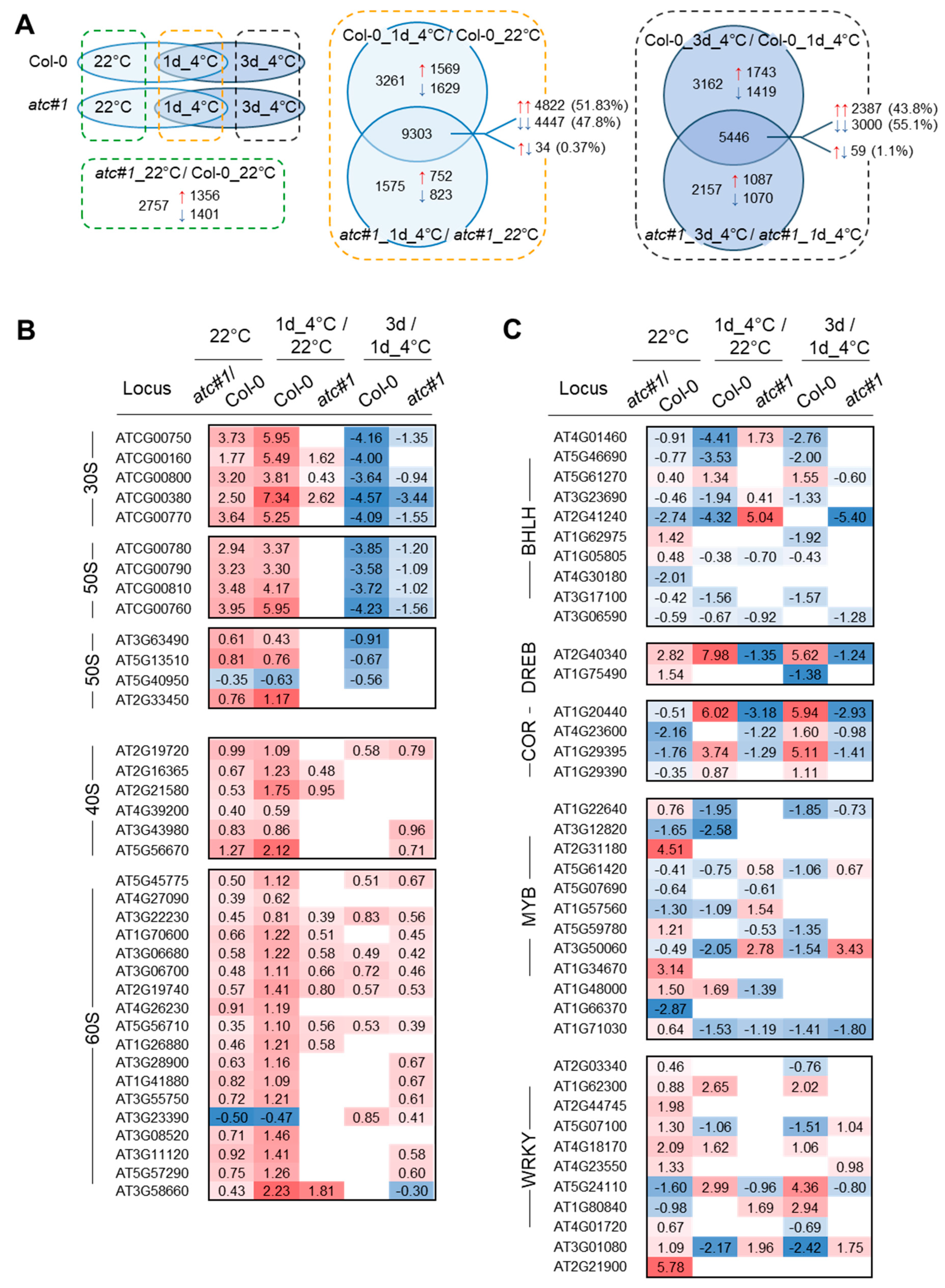
2.4. Transcriptional Response in Functional Groups “Ribosomal Protein” and “Transcription Factor”

A new hub in acclimation to cold was identified with “Translation”. In this category, a high number of ribosomal proteins dominate, and most were found to be upregulated upon cold exposure [7]. From 207 genes encoding ribosomal proteins altered in expression, we identified 154 in Col-0 after one day in the cold but only 135 genes in atc#1 (Supplemental Data S2) [7]. Among these, 13 genes were only altered (upregulated) in atc#1, whereas 32 genes were found altered (upregulated) exclusively in Col-0. This could indicate a reduced cold responsiveness of the atc#1. In several cases, we observed that the cold response was already seen in atc#1 grown at ambient temperature, compared to Col-0 (Figure 4B). This phenomenon is similar to “cold priming” [17], but without the requirement of a brief cold stimulus. We also observe this phenomenon in genes involved in nucleotide metabolism (Table 1) and, to a lesser extent, intracellular transport (Table 2), whereas in genes involved in carbohydrate metabolism and respiration for atc#1, contrasting expression in ambiently grown plants compared to cold-grown plants was observed (Table 3).
Because of the clearly defined role of transcription factors (TF) in cold acclimation [15], we inspected the expression of corresponding TF in our experimental setup. (Supplemental Data S3). With a focus on those TF showing changes between atc#1 and Col-0 already at 22 °C (Figure 4C), several TF with massively altered expression upon cold were identified. Among these are BHLH100 (At2g41240) involved in iron homeostasis and leaf growth [22,23], DREB2C (at2g40340) of the DREBA-2 subfamily, involved in drought response [24]; COR47 (At1g20440) involved in cryoprotection [25]; COR314 (At1g29395), cold regulated protein of the chloroplast inner envelope [26]; MYB77 (At3g50060) involved in light dependent auxin signaling [27] and WRKY30 (At5g24110), and WRKY58 (At3g01080) [28].

2.5. Transcriptional Response in Functional Groups “Nucleotide Metabolism”, Intracellular Transport” and “Central Carbohydrate Metabolism and Respiration”

For ribosomal proteins and TF, we observed that changes after 1 day of cold were reversed almost completely after 3 days of cold (Figure 4B,C). This was also observed in the next category, “nucleotide metabolism”.
Nucleotide metabolism is directly targeted by knocking down ATC (encoded by PYRB). Most genes in de novo synthesis and salvage were upregulated after 1 day cold and partially this is already seen in the comparison of atc#1 against Col-0 at 22 °C (AIR carb., GMPS, IMPDH1, DNK) (Table 1). Nucleotide catabolic genes (UAH, ALN, PYD2, PYD3) show downregulation, as already seen in atc#1 grown at ambient temperature compared to Col-0. Furthermore, the pastidic uracil importer PLUTO is upregulated in cold conditions, whereas the nucleotide transporters PMANT1 and ENT3 are downregulated (Table 1).
When inspecting further, intracellular transporters [29] and tonoplast transporters marked the top five downregulated genes (Table 2). Tip1.2 and Tip2.1 function as aquaporins; CLCa is a nitrate transporter; TST1 is a monosaccharide transporter; and CAT2 is a basic amino acid transporter. Furthermore, the tonoplast-located transporters ALMT9 and TDT, both responsible for malate transport, and TST2, a sucrose/monosaccharide transporter, were upregulated in Col-0 and in atc#1 upon cold (Table 2). In contrast, two plastid-localized carriers were down-regulated in both genotypes (pGlcT, a glucose transporter, and FOLT1, a folate transporter). Strongest induction after 1 day of cold exposure was found for the glucose-6-phosphate-phosphate transporter GPT2 (1d 4 °C/22 °C = log2fc of 5.9 and 7.5 in Col-0 and act#1, respectively) [30].
In a comparison of DEG between atc#1 and Col-0 in the warm, a downregulation of glycolysis, tricarboxyclic acid (TCA) cycle and respiration was observed [20]. When inspecting the same set of genes for DEGs in cold-grown plants, 11 genes were found upregulated and only four downregulated in Col-0 and atc#1 (Table 3). Among those upregulated were AOX1A (alternative oxidase) and NDA1 (alternative NAD(P)H dehydrogenase) both already upregulated in atc#1 at 22 °C and less responsive in atc#1 in the cold. Both corresponding gene products function by avoiding mitochondrial respiration’s energy-saving reactions. AOX1A has a function in acclimation to low temperatures and can attenuate ROS production [31,32], a limitation Of ROS production was also observed in NDA1 overexpressor lines [33]. Cytosolic fumarase 2 (FUM2) was downregulated in Col-0 after 1 day of cold exposure and further repressed after 3 days of cold exposure (Table 3). Interestingly, atc#1 already shows downregulation of FUM2 at 22 °C, and most likely as a consequence, less downregulation after 1 day of cold and no further response after 3 days of cold. Again, this is another example of apparent cold priming at 22 °C.

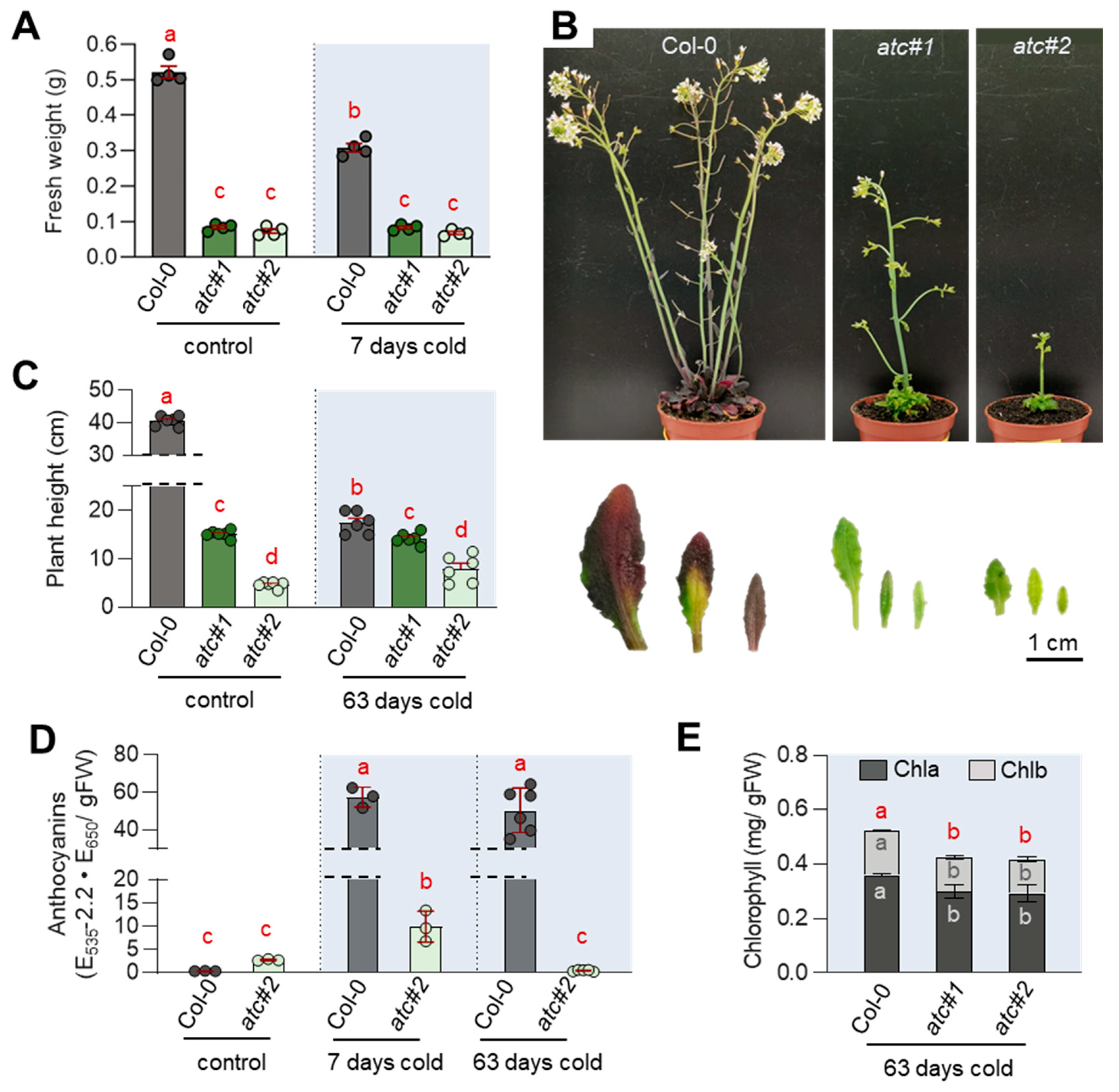
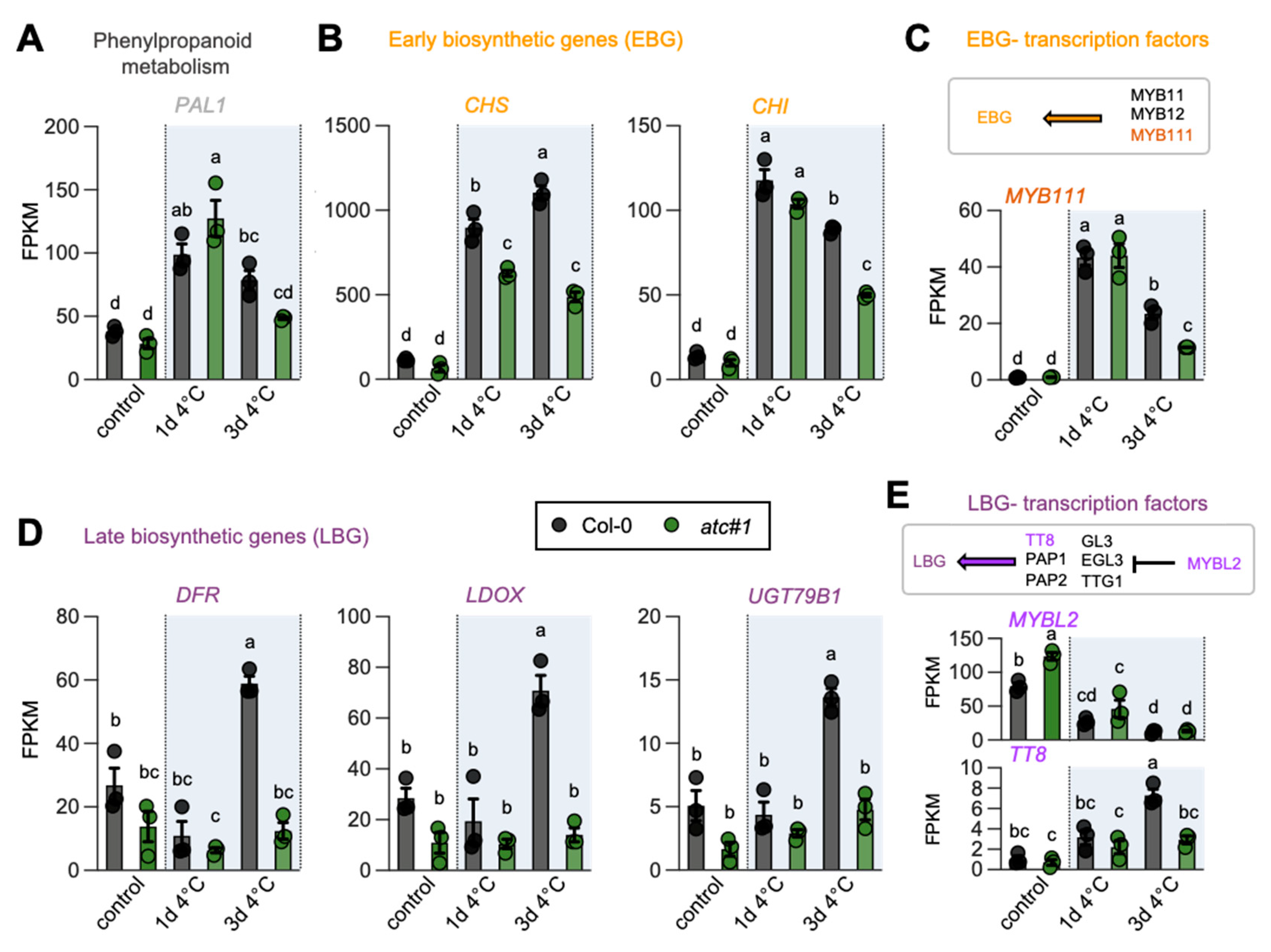
2.6. ATC Knockdown Showed Diminished Activation of Flavonoid Biosynthesis in the Cold

As we observed reduced anthocyanin levels in cold-grown ATC knockdown mutants (Figure 2), we had a closer look at the corresponding genes in the pathway. We observed reduced expression of early biosynthetic genes and a corresponding TF (e.g., MYB111) after 3 days of cold treatment (Figure 5A–C). However, a more pronounced effect was observed for the late biosynthetic genes involved in anthocyanin biosynthesis. While strongly induced in Col-0 at 4 °C, DFR, LDOX, UGT79B1, and the TF TT8 were markedly reduced in expression in atc#1 after 3 days of cold (Figure 5D,E).

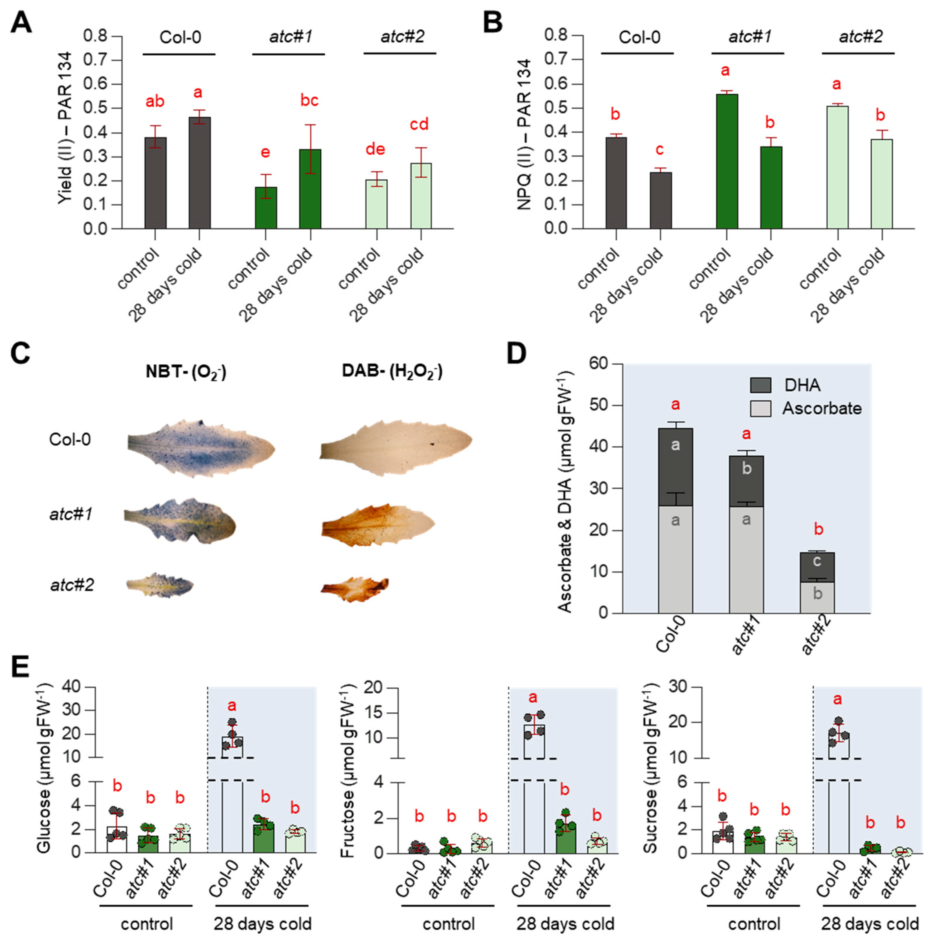
2.7. Alterations in Photosynthetic Rate Led to ROS Accumulation & Reduced Sugar Accumulation in atc#1 and 2

ATC knockdown mutants are characterized by impaired photosynthesis [20]. When plants were grown for 28 days in the cold, an increase in photosynthetic efficiency (YII) compared to 22 °C was observed in Col-0; however, this increase was more pronounced in atc#1 and atc#2 (Figure 6A). For easier comparison, we only show results for PAR 134; full light curves are given in Supplemental Figure S1. At the same time, reductions in NPQ levels were detected in all genotypes, again more pronounced in both ATC mutants (Figure 6B, Supplemental Figure S1). Reduced photosynthetic efficiency was accompanied by increased levels of reactive oxygen species, as revealed by NBT and DAB staining (Figure 6C). Surprisingly, levels of ascorbate and dehydro-ascorbate were massively reduced in atc#2 (Figure 6D). Accumulation of the neutral sugars glucose, fructose, and sucrose is a typical response during cold acclimation, which was corroborated with our analysis of Col-0 exposed to the cold for 28 days (Figure 6E). However, both ATC knockdown mutants entirely failed to accumulate sugars in the cold and essentially showed the same carbohydrate level at 4 °C and 22 °C.

3. Discussion

Here we showed that upon cold exposure, transcripts encoding enzymes involved in purine and pyrimidine de novo synthesis were induced, while those involved in catabolism were repressed on a global scale (Figure 1). This finding was further corroborated by the transcriptional induction of aspartate transcarbamoylase (ATC) in qPCR analysis and the accumulation of ATC protein in Col-0 (wild type) plants after 3 days of growth at 4 °C (Figure 1). In line with these observations, upregulation of purine and pyrimidine de novo synthesis was observed by increased amounts of pathway intermediates in a cold-time course experiment as well [21].
These observations agree well with a general upregulation of components for translation (synthesis of ribosomal proteins and ribosome assembly) upon cold exposure, facilitating reprogramming of metabolism during cold acclimation [7]. As ribosomes consist of proteins and RNA, the latter of which is built from nucleotides, it is conceivable that increased translation, requiring ribosome biosynthesis, is demanding increased nucleotide provision. Interestingly, the target of rapamycin (TOR), a central growth regulator, was shown to activate nucleotide de novo synthesis upon growth signals, and vice versa, nucleotide limitation impairs TOR activity [23]. Together with the observation of markedly altered expression of transcription factors involved in the abiotic stress (including cold) response, it becomes likely that nucleotide signaling is at least partially responsible for the massive alterations in gene expression between Col-0 and atc#1 in warm and cold.
Downregulation of the first committed step in pyrimidine de novo synthesis in plants, catalyzed by ATC, leads to impaired growth and development [19,34,35]. Reduced chlorophyll contents, impaired photosynthetic activity, and accumulation of reactive oxygen species (ROS) are very likely causative of the growth phenotype [20]. ATC knockdown mutants atc#1 and atc#2, exhibiting a 3- and 20-fold reduction in ATC protein abundance compared to Col-0 [19], were unable to accumulate sugars and anthocyanins as usually observed in plants undergoing cold acclimation [13,36] (Figure 2 and Figure 6). Surprisingly, photosynthetic yield (YII) in atc#1 and 2 was improved in the cold, whereas this was hardly observed in Col-0. However, ATC knockdowns still perform worse than controls. When looking at central carbohydrate metabolism, genes upregulated upon cold in both genotypes were downregulated in atc#1 vs. Col-0 at ambient temperature (Table 3). This may hinder cold acclimation and explain why ATC mutants are not able to accumulate neutral sugars in the cold (Figure 6), which is most likely related to the inability of atc#1 to accumulate anthocyanins (Figure 2) [14]. Similar to carbohydrate metabolism, reduced expression of genes active in anthocyanin metabolism was also observed. A downregulation of early biosynthetic genes CHS and CHI as well as MYB111 transcription factor but even more pronounced all late biosynthetic genes and the TT8 transcription factor was observed in the mutants (Figure 5), explaining the low anthocyanin content in cold treated ATC knockdown plants.
In contrast, increased expression of genes encoding ribosomal proteins was observed in atc#1 plants grown at ambient temperature. When these plants were cold treated, the same set of genes was induced, however, at a lower level as compared to Col-0 at day 1 of cold treatment. For transcripts corresponding to “nucleotide metabolism,” the sum of changes in atc#1 vs. Col-0 and atc#1 4 °C vs. 22 °C equals the changes observed in Col-0 4 °C vs. 22 °C (i.e., the first and third columns of Table 1 with respect to the second columns (e.g., ADSL, AIRCAR, GMPS, and so on). This behavior in a way resembles a phenomenon known as “cold priming,” where a short cold treatment, normally too short to permit the full induction of the acclimation response, affects the expression of a certain set of genes essential to cope with low growth temperatures. Among these are the ROS markers ZAT10 and BAP1 [37]. These were also found upregulated in warm-grown atc#1 [19]. As a result, we conclude that atc#1 and the current nucleotide imbalance mimic cold priming via an as-yet unknown mechanism.
Two further putative scenarios arise from the transcriptomic data: 1. reprogramming to support chloroplast metabolism; and 2. actions to remove excess reducing power and thus limit ROS accumulation.
Upregulation of TST2 upon cold allows for increased import of sucrose into the vacuole, which represents the main site of sucrose cleavage by invertase [38]. Thus, sucrose and monosaccharides can accumulate in the vacuole, a typical response to cold acclimation also seen in Col-0 in our study (Figure 6). Alternatively, glucose might be exported from the vacuole by monosaccharide transporters [39], to allow for hexose-phosphate synthesis and subsequent reimport into the chloroplast by GPT2, which was highly upregulated in the cold in Col-0 but even more in atc#1 (Table 2). This apparent futile cycle of chloroplast triose phosphate export and reimport in the form of glucose-6-phosphate could prevent the depletion of Calvin cycle intermediates. Moreover, strongly impaired photosynthesis in atc#1 means less energy (ATP and reducing equivalents) for growth and development. In line with this observation, in addition to what was seen in Col-0, NTT was already upregulated at 22 °C in atc#1 and could allow for ATP import into the chloroplast to replenish the energy pool. Reimport of sugars is further supported by reduced expression of TCA cycle genes in atc#1, which may further explain the inability of atc#1 and 2 to accumulate sugars in the cold.
ROS accumulating in atc#1 and 2 under warm and cold conditions (Figure 6C) may drive the removal of reducing power by the malate valve, converting NADPH and oxaloacetate to malate in the chloroplast, and subsequently exporting this metabolite to the cytosol [31]. We observed higher malate contents in atc#2 at 22 °C [20], together with increased alternative oxidases AOXA1 and NADPH dehydrogenase ND1, and both can dissipate excess energy without producing more ATP [40]. Under cold conditions, these effects are more pronounced as DIC transporters mediating the exchange of dicarboxylates between mitochondria and cytosol are upregulated, as well as tonoplastidic TDT and ALMT carriers that might balance malate levels between the cytosol and vacuole (Table 2). In addition, AOX1a shows a further marked increase in expression upon exposure to cold. Downregulation of cytosolic fumarase 2 (FUM2) represents a typical cold response [41] and is seen in both genotypes (Table 3). In addition, this response of FUM2 is seen in atc#1 grown at ambient temperature, together with reduced levels of succinate and fumarate but wild-type-like levels of 2-oxoglutarate [20]. This may point to an export of oxoglutarate to the chloroplast to serve as the carbon skeleton for amino acid synthesis by refixing ammonia released by photorespiration [42,43]. High glyoxylate levels are indicative of high photorespiration, as glyoxylate is a key intermediate of this pathway [43]. In addition, glyoxylate is a product of purine catabolism [44]. Together with the observed accumulation of other intermediates in purine catabolism, this points to purine breakdown to balance purine to pyrimidine pools, latter being low in atc#1 [20].
When oxoglutarate is exported from mitochondria, resulting in low succinate and fumarate, as discussed above, this will lead to low malate generation in the TCA cycle. At the same time, this might support malate production from oxaloacetate in the cytosol, thereby reducing NADH levels.

4. Materials and Methods

4.1. Plant Growth

For RNA isolation, tissue collection, and phenotypic inspection, wild-type (Col-0) and transgenic Arabidopsis thaliana (L.) Heynh. plants (ecotype Columbia) were used throughout. Plants were grown on standardized ED73 soil (Einheitserde and Humuswerke Patzer) under a short day regime (10 h light and 14 h darkness). Light conditions were set to an intensity of 120 μmol quanta m−2s−1 Illumination was done with LED light (Valoya NS1, Valoya, Finland). The ambient temperature was either set to 22 °C or, for cold treatment, to 4 °C with a constant humidity of 60%. If not stated otherwise, plant material was harvested in the middle of the light period and frozen in liquid nitrogen for further use. The used ATC ami-RNA knockdown lines were described previously [19].

4.2. RNA Extraction

RNA was extracted from the entire leaf material of soil-grown plants, which was homogenized in liquid nitrogen prior to the extraction of RNA with the Nucleospin RNA Plant Kit (Macherey-Nagel, Düren, Germany) according to the manufacturer’s advice. RNA purity and concentration were quantified using a NanoPhotometer (Implen, Munich, Germany).

4.3. cDNA Synthesis and Gene Expression Analyses

Total RNA was transcribed into cDNA using the qScript cDNA Synthesis Kit (Quantabio, Beverly, MA, USA). QPCR was performed using the quantabio SYBR green quantification kit (Quantabio) on the PFX96 system (BioRad, Hercules, CA, USA) using specific primers. Actin (At3g18780) was used as a reference gene for transcript normalization. All used primers are listed in Supplementary Table S1.

4.4. RNA Seq and Transcriptome Analyses

All leaf material from at least three plants was sampled for one biological replicate. All conditions were analyzed with three biological replicates. RNA library preparation and transcriptome sequencing were performed by Novogene (Beijing, China). Before library preparation, the isolated RNA was first analyzed for quality by agarose gel electrophoresis and the Agilent 2100 Bioanalyzer (Agilent, Palo Alto, CA, USA). To remove ribosomal RNA and generate the cDNA libraries, poly(A)+ RNA enrichment and mRNA fragmentation followed by random-prime cDNA synthesis was performed. This was followed by paired-end sequencing of the reads of 150 base pairs in length, which were captured using an Illumina sequencer (NovaSeq 6000). To verify and quantify the quality of the reads, the program Hisat2 was used in bioinformatic analyses, and the reads were mapped to the Arabidopsis reference genome. The further bioinformatic evaluation was also performed by Novogene. To investigate which biological processes were highly altered in expression, DESeq2 [45] was used. Therefore, all genes with an adjusted p-value < 0.05 are included. We compared the following groups: Col-0 1d_4 °C vs. Col-0_22 °C; Col-0 3d_4 °C vs. Col-0_1d_4 °C; atc#1 1d_4 °C vs. atc#1 _22 °C; atc#1 3d_4 °C vs. atc#1 _1d_4 °C. In the publication Bellin et al. (2021b), we compared atc#1_22 °C vs. Col-0_22 °C, and these data were also shown in Figure 4B,C, and Table 1, Table 2 and Table 3. RNA-Seq reads have been uploaded to the Sequence Read Archive (SRA) at NCBI (National Center for Biotechnology Information), BioProject ID: PRJNA925318. FPKM values for all samples are included in Supplemental Data S4.

4.5. Protein Extraction

Leaf extract of wild-type and knockdown mutants was prepared by homogenizing leaf material in an extraction buffer (50 mM HEPES-KOH, pH 7.2, 5 mM MgCl2, 2 mM DTT, 0.01% DDM, 1 mM EDTA, and 2 mM phenylmethylsulfonyl fluoride (PMSF)) on ice. This homogenous extract was centrifuged for 10 min, 20,000× g and 4 °C. The supernatant was collected and stored on ice until use.

4.6. Immunoblotting and Western Blot Detection

For immunoblotting 15 µg of a protein extract were separated in a 15% SDS-PAGE gel were transferred onto a nitrocellulose membrane (Whatman, Germany) by blotting. The membrane was blocked in phosphate-buffered saline plus 0.1% [v/v] Tween 20 (PBS-T) with 3% milk powder for 1 h at room temperature, followed by three washes of 10 min in PBS-T. Then, the membrane was incubated with a rabbit polyclonal antiserum raised against recombinant ATC (Eurogentec, Belgium) for 1 h, followed by three washes with PBS-T. Next, the membrane was incubated for 1 h with a horseradish peroxidase (HPR)-conjugated anti-rabbit antibody (Promega, Walldorf, Germany) diluted in PBS-T with 3% milk powder. The result was visualized by chemiluminescence using the ECL Prime Western blotting reagent (GE Healthcare) and a Fusion Solo S6 (Vilber-Lourmat) imager.

4.7. Chlorophyll and Anthocyanin Extraction and Quantification

Chlorophyll pigments were extracted from 50 mg of ground leaf tissue in 80% ethanol. After boiling at 95 °C for 10 min and subsequent sedimentation of insoluble contents (5 min, 12,500 rpm), chlorophyll was measured by the absorbance of the supernatant at 652 nm. The calculation was performed as described by [46].
Anthocyanin pigments were extracted from 50 mg in an extraction buffer (H2O: 1-propanol: HCL (18:1:81)) on ice. After boiling at 95 °C for 3 min while being shaken at 650 rpm, samples were incubated on RT in darkness for 24 h. After centrifugation (15 min, 12,500 rpm), the extinction of the supernatant was measured at 535 nm and 650 nm. Extinction was corrected by Rayleigh’s formula (E535-2.2E650).

4.8. Pulse-Amplitude-Modulation (PAM) Fluorometry Measurements

A MINI-IMAGING-PAM fluorometer (Walz Instruments, Effeltrich, Germany) was used for in vivo chlorophyll measurements. A light curve assays intact, six-week-old plants that were grown for three weeks under standard conditions (22 °C) before being transferred to cold conditions (4 °C). After 10 min of dark adaptation, measurements were performed in a standard light curve setting [47]. Therefore, incrementally increasing light pulses with intensities from PAR (µmol photons m−2 s−1) 0 to PAR 726 in 14 steps were applied to the plants.

4.9. NBT (O2) and DAB (H2O2) Staining

Superoxide (O2) was detected by nitroblue tetrazolium (NBT) staining [48]. For this, leaves from six weeks old plants (three weeks 22 °C and three weeks 4 °C) were used. In brief, leaves were vacuum-infiltrated with 0.1% NBT. 50 mM potassium phosphate buffer (pH 7.8) and 10 mM sodium azide for 20 min and incubated for 1 h at room temperature. Stained leaves were boiled in 95% ethanol for 15 min and photographed. The detection of H2O2 was performed by treating plants with DAB-HCl. Leaves were vacuum-infiltrated with 5 mM DAB-HCl, pH 3, for 20 min and incubated in the same solution for at least 8 h overnight. Stained leaves were boiled in an ethanol: acetic acid: glycerol (3:1:1) solution under the hood until they turned transparent and were later documented.

4.10. Whole-Leaf Ascorbate Determination

Whole-leaf samples from six-week-old plants were harvested six hours after the onset of light. These plants were grown for three weeks under standard conditions (22 °C) and three weeks in cold (4 °C). Ascorbate and dehydroascorbate levels were determined calorimetrically as described previously [49].

4.11. Sugar Extraction

For sugar extraction 50 mg of frozen, grounded leaf sample were boiled in 500 µL deionized water for 15 min at 95 °C. After centrifugation (20,000× g for 10 min at 4 °C), the supernatant was used for further sugar analysis. Extracted sugars were quantified using a NAD+-coupled enzymatic assay [50].

4.12. Quantification of Aminoacids

For the quantification of amino acids, the supernatant derived from sugar extraction was used. 20 µL of the probe were mixed with 60 µL 0.2 M pH 8.8 (NaOH) borat and 20 µL 1.28 mM AQC (6-aminoquinolyl-N-hydroxysuccinimidyl carbamate, Synchem UG & Co., KG-S041, dissolved in acetonitrile), and the probe was directly vortexed for 10 s. The measurement was done using high-pressure liquid chromatography (HPLC).

Supplementary Materials

The following supporting information can be downloaded at: https://www.mdpi.com/article/10.3390/molecules28041585/s1, Figure S1. Photosynthetic parameters of Col-0 and ATC knockdown mutants grown in warm and cold. Plants were grown for three weeks at ambient temperature und then transferred to 4 °C for 28 days. Yield (upper panel) and NPQ (lower panel) were recorded by imaging PAM. Error bars show the standard deviation of the mean. n = 5. Supplemental Data S1, DEG after 1 and 3 days of cold, for Table 1, Table 2 and Table 3. Supplemental Data S2, DEG encoding ribosomal proteins. Supplemental Data S3, DEG encoding transcription factors. Supplemental Data S4, FPKM values of all samples.

Author Contributions

L.B. and T.M. designed the research; L.B., D.L.G.A., A.J., V.S. and T.P. performed the experiments; L.B., V.S. and T.M. analyzed the data; and L.B., V.S., A.R. and T.M. wrote the manuscript. All authors have read and agreed to the published version of the manuscript.

Funding

This work was funded by the Deutsche Forschungsgemeinschaft (DFG) TRR175, project B08 to T.M. and project C06 to A.R.

Institutional Review Board Statement

Not applicable.

Informed Consent Statement

Not applicable.

Data Availability Statement

RNA-Seq reads have been uploaded to the Sequence Read Archive (SRA) at NCBI (National Center for Biotechnology Information; BioProject ID: PRJNA925318).

Acknowledgments

We thank Ekkehard Neuhaus and Benjamin Pommerrenig for fruitful discussions.

Conflicts of Interest

The authors declare that the research was conducted in the absence of any commercial or financial relationships that could be construed as potential conflict of interest.

References

  1. Guy, C. Molecular responses of plants to cold shock and cold acclimation. J. Mol. Microbiol. Biotechnol. 1999, 1, 231–242. [Google Scholar] [PubMed]
  2. Thomashow, M.F. Plant cold acclimation: Freezing Tolerance Genes and Regulatory Mechanisms. Annu. Rev. Plant Physiol. Plant Mol. Biol. 1999, 50, 571–599. [Google Scholar] [CrossRef] [PubMed]
  3. Kaplan, F.; Kopka, J.; Sung, D.Y.; Zhao, W.; Popp, M.; Porat, R.; Guy, C.L. Transcript and metabolite profiling during cold acclimation of Arabidopsis reveals an intricate relationship of cold-regulated gene expression with modifications in metabolite content. Plant J. 2007, 50, 967–981. [Google Scholar] [CrossRef]
  4. Kleine, T.; Nägele, T.; Neuhaus, H.E.; Schmitz-Linneweber, C.; Fernie, A.R.; Geigenberger, P.; Grimm, B.; Kaufmann, K.; Klipp, E.; Meurer, J.; et al. Acclimation in plants—The Green Hub consortium. Plant J. 2021, 106, 23–40. [Google Scholar] [CrossRef] [PubMed]
  5. Schwenkert, S.; Fernie, A.R.; Geigenberger, P.; Leister, D.; Möhlmann, T.; Naranjo, B.; Neuhaus, H.E. Chloroplasts are key players to cope with light and temperature stress. Trends Plant Sci 2022, 27, 577–587. [Google Scholar] [CrossRef] [PubMed]
  6. Huner, N.P.; Oquist, G.; Hurry, V.M.; Krol, M.; Falk, S.; Griffith, M. Photosynthesis, photoinhibition and low temperature acclimation in cold tolerant plants. Photosynth. Res. 1993, 37, 19–39. [Google Scholar] [CrossRef]
  7. Garcia-Molina, A.; Kleine, T.; Schneider, K.; Mühlhaus, T.; Lehmann, M.; Leister, D. Translational Components Contribute to Acclimation Responses to High Light, Heat, and Cold in Arabidopsis. iScience 2020, 23, 101331. [Google Scholar] [CrossRef]
  8. Foyer, C.H.; Noctor, G. Redox regulation in photosynthetic organisms: Signaling, acclimation, and practical implications. Antioxid. Redox Signal. 2009, 11, 861–905. [Google Scholar] [CrossRef]
  9. Schulz, E.; Tohge, T.; Zuther, E.; Fernie, A.R.; Hincha, D.K. Flavonoids are determinants of freezing tolerance and cold acclimation in Arabidopsis thaliana. Sci. Rep. 2016, 6, 34027. [Google Scholar] [CrossRef]
  10. Araguirang, G.E.; Richter, A.S. Activation of anthocyanin biosynthesis in high light—What is the initial signal? New Phytol. 2022, 236, 2037–2043. [Google Scholar] [CrossRef]
  11. Hörmiller, I.I.; Nägele, T.; Augustin, H.; Stutz, S.; Weckwerth, W.; Heyer, A.G. Subcellular reprogramming of metabolism during cold acclimation in Arabidopsis thaliana. Plant Cell Environ. 2017, 40, 602–610. [Google Scholar] [CrossRef]
  12. Wanner, L.A.; Junttila, O. Cold-induced freezing tolerance in Arabidopsis. Plant Physiol. 1999, 120, 391–400. [Google Scholar] [CrossRef] [PubMed]
  13. Xu, Z.; Mahmood, K.; Rothstein, S.J. ROS Induces Anthocyanin Production Via Late Biosynthetic Genes and Anthocyanin Deficiency Confers the Hypersensitivity to ROS-Generating Stresses in Arabidopsis. Plant Cell Physiol. 2017, 58, 1364–1377. [Google Scholar] [CrossRef] [PubMed]
  14. Zirngibl, M.E.; Araguirang, G.E.; Kitashova, A.; Jahnke, K.; Rolka, T.; Kuhn, C.; Nägele, T.; Richter, A.S. Triose phosphate export from chloroplasts and cellular sugar content regulate anthocyanin biosynthesis during high light acclimation. Plant Commun. 2023, 4, 100423. [Google Scholar] [CrossRef] [PubMed]
  15. Van Buskirk, H.A.; Thomashow, M.F. Arabidopsis transcription factors regulating cold acclimation. Physiol. Plant. 2006, 126, 72–80. [Google Scholar] [CrossRef]
  16. Bittner, A.; Hause, B.; Baier, M. Cold-priming causes dampening of oxylipin biosynthesis and signalling during the early cold- and light-triggering response of Arabidopsis thaliana. J. Exp. Bot. 2021, 72, 7163–7179. [Google Scholar] [CrossRef]
  17. Bittner, A.; van Buer, J.; Baier, M. Cold priming uncouples light- and cold-regulation of gene expression in Arabidopsis thaliana. BMC Plant Biol. 2020, 20, 281. [Google Scholar] [CrossRef] [PubMed]
  18. Busche, M.; Scarpin, M.R.; Hnasko, R.; Brunkard, J.O. TOR coordinates nucleotide availability with ribosome biogenesis in plants. Plant Cell 2021, 33, 1615–1632. [Google Scholar] [CrossRef]
  19. Bellin, L.; Del Cano-Ochoa, F.; Velazquez-Campoy, A.; Möhlmann, T.; Ramon-Maiques, S. Mechanisms of feedback inhibition and sequential firing of active sites in plant aspartate transcarbamoylase. Nat. Commun. 2021, 12, 947. [Google Scholar] [CrossRef]
  20. Bellin, L.; Melzer, M.; Hilo, A.; Amaya, D.L.G.; Keller, I.; Meurer, J.; Möhlmann, T. Pyrimidine nucleotide availability is essential for efficient photosynthesis, ROS scavenging, and organelle development. bioRxiv 2021. [Google Scholar] [CrossRef]
  21. Chen, X.; Kim, S.H.; Rhee, S.; Witte, C.P. A plastid nucleoside kinase is involved in inosine salvage and control of purine nucleotide biosynthesis. Plant Cell 2023, 35, 510–528. [Google Scholar] [CrossRef] [PubMed]
  22. Kurt, F.; Filiz, E. Genome-wide and comparative analysis of bHLH38, bHLH39, bHLH100 and bHLH101 genes in Arabidopsis, tomato, rice, soybean and maize: Insights into iron (Fe) homeostasis. Biometals 2018, 31, 489–504. [Google Scholar] [CrossRef]
  23. Van Dingenen, J.; Antoniou, C.; Filippou, P.; Pollier, J.; Gonzalez, N.; Dhondt, S.; Goossens, A.; Fotopoulos, V.; Inze, D. Strobilurins as growth-promoting compounds: How Stroby regulates Arabidopsis leaf growth. Plant Cell Environ. 2017, 40, 1748–1760. [Google Scholar] [CrossRef] [PubMed]
  24. Feng, J.X.; Liu, D.; Pan, Y.; Gong, W.; Ma, L.G.; Luo, J.C.; Deng, X.W.; Zhu, Y.X. An annotation update via cDNA sequence analysis and comprehensive profiling of developmental, hormonal or environmental responsiveness of the Arabidopsis AP2/EREBP transcription factor gene family. Plant Mol. Biol. 2005, 59, 853–868. [Google Scholar] [CrossRef]
  25. Ohkubo, T.; Kameyama, A.; Kamiya, K.; Kondo, M.; Hara, M. F-segments of Arabidopsis dehydrins show cryoprotective activities for lactate dehydrogenase depending on the hydrophobic residues. Phytochemistry 2020, 173, 112300. [Google Scholar] [CrossRef] [PubMed]
  26. Okawa, K.; Nakayama, K.; Kakizaki, T.; Yamashita, T.; Inaba, T. Identification and characterization of Cor413im proteins as novel components of the chloroplast inner envelope. Plant Cell Environ. 2008, 31, 1470–1483. [Google Scholar] [CrossRef]
  27. Yang, Y.; Liu, H. Coordinated Shoot and Root Responses to Light Signaling in Arabidopsis. Plant Commun. 2020, 1, 100026. [Google Scholar] [CrossRef]
  28. Scarpeci, T.E.; Zanor, M.I.; Müller-Röber, B.; Valle, E.M. Overexpression of AtWRKY30 enhances abiotic stress tolerance during early growth stages in Arabidopsis thaliana. Plant Mol. Biol. 2013, 83, 265–277. [Google Scholar] [CrossRef] [PubMed]
  29. Linka, N.; Weber, A.P. Intracellular metabolite transporters in plants. Mol. Plant 2010, 3, 21–53. [Google Scholar] [CrossRef]
  30. Kunz, H.H.; Häusler, R.E.; Fettke, J.; Herbst, K.; Niewiadomski, P.; Gierth, M.; Bell, K.; Steup, M.; Flügge, U.I.; Schneider, A. The role of plastidial glucose-6-phosphate/phosphate translocators in vegetative tissues of Arabidopsis thaliana mutants impaired in starch biosynthesis. Plant Biol. 2010, 1, 115–128. [Google Scholar] [CrossRef]
  31. Selinski, J.; Scheibe, R. Malate valves: Old shuttles with new perspectives. Plant Biol. 2019, 21, 21–30. [Google Scholar] [CrossRef] [PubMed]
  32. Selinski, J.; Scheibe, R.; Day, D.A.; Whelan, J. Alternative Oxidase Is Positive for Plant Performance. Trends Plant Sci. 2018, 23, 588–597. [Google Scholar] [CrossRef] [PubMed]
  33. Jethva, J.; Lichtenauer, S.; Schmidt-Schippers, R.; Steffen-Heins, A.; Poschet, G.; Wirtz, M.; van Dongen, J.T.; Eirich, J.; Finkemeier, I.; Bilger, W.; et al. Mitochondrial alternative NADH dehydrogenases NDA1 and NDA2 promote survival of reoxygenation stress in Arabidopsis by safeguarding photosynthesis and limiting ROS generation. New Phytol. 2022. [Google Scholar] [CrossRef]
  34. Chen, C.T.; Slocum, R.D. Expression and functional analysis of aspartate transcarbamoylase and role of de novo pyrimidine synthesis in regulation of growth and development in Arabidopsis. Plant Physiol. Biochem. 2008, 46, 150–159. [Google Scholar] [CrossRef]
  35. Giermann, N.; Schröder, M.; Ritter, T.; Zrenner, R. Molecular analysis of de novo pyrimidine synthesis in solanaceous species. Plant Mol. Biol. 2002, 50, 393–403. [Google Scholar] [CrossRef] [PubMed]
  36. Nägele, T.; Heyer, A.G. Approximating subcellular organisation of carbohydrate metabolism during cold acclimation in different natural accessions of Arabidopsis thaliana. New Phytol. 2013, 198, 777–787. [Google Scholar] [CrossRef]
  37. van Buer, J.; Prescher, A.; Baier, M. Cold-priming of chloroplast ROS signalling is developmentally regulated and is locally controlled at the thylakoid membrane. Sci. Rep. 2019, 9, 3022. [Google Scholar] [CrossRef]
  38. Vu, D.P.; Martins Rodrigues, C.; Jung, B.; Meissner, G.; Klemens, P.A.W.; Holtgräwe, D.; Fürtauer, L.; Nägele, T.; Nieberl, P.; Pommerrenig, B.; et al. Vacuolar sucrose homeostasis is critical for plant development, seed properties, and night-time survival in Arabidopsis. J. Exp. Bot. 2020, 71, 4930–4943. [Google Scholar] [CrossRef] [PubMed]
  39. Pommerrenig, B.; Ludewig, F.; Cvetkovic, J.; Trentmann, O.; Klemens, P.A.W.; Neuhaus, H.E. In Concert: Orchestrated changes in carbohydrate homeostasis are critical for plant abiotic stress tolerance. Plant Cell Physiol. 2018, 59, 1290–1299. [Google Scholar] [CrossRef]
  40. Noguchi, K.; Yoshida, K. Interaction between photosynthesis and respiration in illuminated leaves. Mitochondrion 2008, 8, 87–99. [Google Scholar] [CrossRef]
  41. Dyson, B.C.; Miller, M.A.; Feil, R.; Rattray, N.; Bowsher, C.G.; Goodacre, R.; Lunn, J.E.; Johnson, G.N. FUM2, a cytosolic fumarase, is essential for acclimation to low temperature in Arabidopsis thaliana. Plant Physiol. 2016, 172, 118–127. [Google Scholar] [CrossRef]
  42. Hodges, M. Enzyme redundancy and the importance of 2-oxoglutarate in plant ammonium assimilation. J. Exp. Bot 2002, 53, 905–916. [Google Scholar] [CrossRef]
  43. Maurino, V.G.; Engqvist, M.K. 2-Hydroxy Acids in Plant Metabolism. Arab. Book 2015, 13, e0182. [Google Scholar] [CrossRef]
  44. Werner, A.K.; Witte, C.P. 2011. The biochemistry of nitrogen mobilization: Purine ring catabolism. Trends Plant Sci. 2002, 16, 381–387. [Google Scholar] [CrossRef] [PubMed]
  45. Love, M.I.; Huber, W.; Anders, S. Moderated estimation of fold change and dispersion for RNA-seq data with DESeq2. Genome Biol. 2014, 15, 1–21. [Google Scholar] [CrossRef]
  46. Arnon, D.I. Copper enzymes in isolated chloroplasts. Polyphenoloxidase in Beta vulgaris. Plant Physiol. 1949, 24, 1–15. [Google Scholar]
  47. Schreiber, U.; Quayle, P.; Schmidt, S.; Escher, B.I.; Müller, J.F. Methodology and evaluation of a highly sensitive algae toxicity test based on multiwell chlorophyll fluorescence imaging. Biosensens. Bioelectron. 2007, 22, 2554–2563. [Google Scholar] [CrossRef] [PubMed]
  48. Fryer, M.J.; Oxborough, K.; Mullineaux, P.M.; Baker, N.R. Imaging of photo-oxidative stress responses in leaves. J. Exp. Bot. 2002, 53, 1249–1254. [Google Scholar]
  49. Gillespie, K.M.; Ainsworth, E.A. Measurement of reduced, oxidized and total ascorbate content in plants. Nat. Prot. 2007, 2, 871–874. [Google Scholar] [CrossRef]
  50. Stitt, M.; Lilley, R.M.; Gerhardt, R.; Heldt, H.W. Metabolite levels in specific cells and subcellular compartments of plant leaves. Meth. Enzymol. 1989, 174, 518–552. [Google Scholar]
Figure 1. Expression analysis of pyrimidine and purine de novo synthesis genes during cold acclimation and de-acclimation. (A) Scheme of enzymes and intermediates of pyrimidine (blue) and purine (yellow) de novo synthesis in plants. (B) Expression of purine and pyrimidine de novo synthesis genes in a cold acclimation/deacclimation time course [7]. The main enzyme (ATC) studied in this work is indicated in red. Plants were grown for 14 days at ambient temperature before being transferred to 4 °C for four days and immediately transferred to ambient temperature again for an additional four days. Sampling took place at the indicated timepoints. Shown are the Δlog FPKM values compared to 0 min of cold treatment. (C) Expression of ATC during cold acclimation and adaptation. Col-0 plants were grown at 22 °C for 21 days before cold treatment. Expression was normalized to actin2. Data points are means of three biological replicates +/− standard deviation. (D) Similar plants were used for immunoblot staining with ATC antibody on whole leaf extracts (top) after 0, 3, and 8 days of cold treatment. Coomassie Brilliant Blue (CBB)-stained SDS-PAGE was used as a loading control. Asterisks depict significant changes relative to the 0 days timepoint according to one-way ANOVA test (*** = p < 0.001). Carbamoyl phosphate synthase (CPS) small subunit (SSU) and large subunits (LSU), carbamoyl-phosphate (CP), aspartate transcarbamoylase (ATC), carbamoyl-aspartate (CA), dihydroorotate (DHO), dihydroorotate dehydrogenase (DHODH), uridinemonophosphate synthase (UMPS), and cytidintriphosphate synthetase 2 (CTPS2), amidophosphoribosyltransferase (GPRAT2), IMP cyclohydrolase (AICARFT), inosinemonophosphate dehydrogenase1/2 (IMPDH1/IMPDH2), guanosin-monophosphate synthase (GMPS), and brittle transporter 1 (BT1).
Molecules 28 01585 g001
Figure 2. Cold acclimation and adaptation in Col-0 and ATC mutants. Plants were grown for three weeks at 22 °C before being transferred to 4 °C for the indicated timespan. Control plants were continuously grown at 22 °C for four weeks. (A) Fresh weight (n = 4) (B) phenotypical appearance after 63 days cold with closeup of leaves (C) plant height (n = 6) (D) anthocyanin contents of leaves (n = 3 for control and 7-day-old plants; n = 6 for 63-day-old plants) (E) chlorophyll contents (n = 4). Data points and means +/− standard deviation are shown. Different letters denote significant differences according to a two-way ANOVA with post-hoc Tukey HSD testing (p < 0.05).
Molecules 28 01585 g002
Figure 3. Global transcript alterations in Col-0 and atc#1 mutants during cold acclimation. Plants were grown for six weeks at 22 °C before being transferred to 4 °C for one or three days. (AD) Volcano plots show differentially expressed genes (DEGs) (padj. < 0.05) in Col-0 (A,C) and atc#1 mutants (B,D) comparing one day in the cold to the control (A,B) and three days of the cold to one day of the cold treatment (C,D). All listed DEGs were used for (EH) GO-Term analysis, revealing significantly altered biological processes (up-regulated in red and down-regulated in blue color).
Molecules 28 01585 g003
Figure 4. Comparison of global transcript alterations between Col-0 and atc#1 mutants and assignment to transcription factors and ribosomal proteins during cold acclimation. Plants were grown for six weeks at 22 °C before being transferred to 4 °C for one or three days. (A) Visualization of compared datasets (left panel) and Venn diagrams of data comparisons after 1 and 3 days of cold treatment (middle and right panel). (B) Ribosomal proteins selected for differential expression at 22 °C (primed) (padj. < 0.05). (C) Transcription factors selected for differential expression at 22 °C (primed) (padj. < 0.05) in the blank fields; no significant change. Up-regulated genes are shown in red and down-regulated genes in blue color).
Molecules 28 01585 g004
Figure 5. Global transcript alterations (atc#1 vs. Col-0) in flavonoid biosynthesis genes. Data points and means +/− standard deviation of FPKM values are shown. Different letters denote significant differences according to a two-way ANOVA with post-hoc Tukey HSD testing (p < 0.05). Phenylalanine ammonia-lyase (PAL1), chalcone synthase (CHS), chalcone isomerase (CHI), dihydroflavonol-4-reductase (DFR), leucoanthocyanidin dioxygenase (LDOX), cyanidin 3-O-glucoside 2″-O-xylosyltransferase (UGT79B1), Arabidopsis MYB transcription factors (MYB111 and MYBL2) and transparent testa8 (TT8).
Molecules 28 01585 g005
Figure 6. Determination of photosynthetic efficiency, accumulation of ROS, and sugar quantification. Plants were grown for two weeks at 22 °C before being transferred to 4 °C for the indicated times. (A) The effective quantum yield of PS (II) and (B) non-photochemical quenching (NPQ) were measured in a light response curve (n = 5). (C) NBT- and DAB-staining for visualization of reactive oxygen species showing typical results (D) measurements of Ascorbate and Dihydroascorbate (n = 5). (E) Quantification of glucose, fructose, and sucrose contents (n = 4). Data points are means +/− standard deviation. Different letters denote significant differences according to a two-way ANOVA with post-hoc Tukey HSD testing (p < 0.5).
Molecules 28 01585 g006
Table 1. DEGs involved in nucleotide metabolism. Selected genes (padj. < 0.05) are shown. AGI, Arabidopsis genome identifier.
22 °C1 d 4 °C/22 °C3 d/1 d 4 °C
LocusNameatc#1/Col-0Col-0atc#1Col-0atc#1Function
AT4G18440ADSL−1.27−0.98−0.46−1.09−0.50de novo
AT2G37690AIR carb.0.691.901.360.880.43de novo
AT2G37250AMK1 1.371.56 −0.43de novo
AT5G35170AMK5 −0.72−1.14−1.16−0.50de novo
AT3G01820AMK7 2.973.16 −0.80de novo
AT4G34740ATase2 2.342.34−0.63−0.58de novo
AT5G23300DHODH 1.621.09 de novo
AT1G30820CTPS1 1.842.29−2.82−2.31de novo
AT3G12670CTPS2 1.952.03−0.99−0.82de novo
AT2G16370DHFR-TS −1.45−1.22 de novo
AT3G06200GMK 1.521.05 de novo
AT1G63660GMPS0.731.530.88 de novo
AT1G79470IMPDH10.761.800.96 de novo
AT1G16350IMPDH20.51−0.73−0.921.140.92de novo
AT2G21790RNR1 0.651.030.520.41de novo
AT3G60180UMK1 −1.28−1.39 0.95de novo
AT4G20070AAH 2.572.26−1.60−1.34catabolism
AT5G43600UAH −1.57−1.420.530.61catabolism
AT4G04955ALN −2.432.20 1.00catabolism
AT1G05620NSH2 −1.39−0.751.250.75catabolism
AT3G17810PYD1−0.530.441.00−1.50−1.18catabolism
AT5G12200PYD2−0.41−1.19−0.90 catabolism
AT5G64370PYD3−0.42−1.17−0.69 catabolism
AT2G26230UOX−0.371.011.25 −0.53catabolism
AT1G80050APT2 −1.58−1.89 1.02salvage
AT4G22570APT3−0.74−2.90−2.661.291.75salvage
AT5G11160APT5 1.131.54 −0.59salvage
AT1G72040dNK0.631.140.65 salvage
AT1G71750HGPRT 1.061.00 salvage
AT5G40870UCK1 −0.85−1.04 0.56salvage
AT5G13490AAC2 0.972.09 transporter
AT5G61810APC1 2.231.88−1.55−1.34transporter
AT4G32400BT1 2.011.58−0.70−0.72transporter
AT4G05120ENT3 −1.30−0.69 0.80transporter
AT4G05110ENT6 −1.06 transporter
AT5G03555PLUTO 2.082.39−1.28−1.29transporter
AT5G56450pmANT1 −1.00−0.77 transporter
Table 2. DEGs involved in intracellular transport. Selected genes (padj. < 0.05) are shown. AGI, Arabidopsis genome identifier; Glc6P, glucose 6 phosphate; TP, triose phosphate.
LocusName22 °C1 d 4 °C/22 °C3 d/1 d 4 °CSubcell. Localization Substrate
atc#1/Col-0Col-0atc#1Col-0atc#1
AT1G14140UCP3 −1.16−0.930.62 Mtprotons
AT5G58970UCP2−0.86−1.14−1.101.171.71Mtprotons
AT5G09470DIC3 3.334.26 Mtdicarboxylate
AT4G24570DIC2 −0.89 3.234.04Mtdicarboxylate
AT2G22500DIC1−0.852.152.97 Mtdicarboxylate
AT3G48850PIC2 1.414.35−1.48−2.67Mtphosphate
AT2G17270MPT1 2.322.42−1.04−1.00Mtphosphate
AT5G01340SFC 3.153.46 −1.16Mtsuccinate/fumarate
AT5G27520PNC2 1.972.05−1.48−1.49Mt, Padenine nucleotide
AT5G66380FOLT1 −0.76−1.05 0.65PLfolate
AT5G16150pGlcT−0.44−0.87−1.03−1.20−0.34PLhexose
AT1G68570NPF3.1 2.692.35−0.66−0.72PLnitrite
AT4G32400BT1-like 2.011.58−0.70−0.72PLnucleotide
AT1G61800GPT2 5.907.610.39−1.65PLGlc6P, TP
AT1G58030CAT2 −1.40−1.32 0.62TPamino acid
AT5G40890CLCa −2.34−1.50 0.56TPCl, nitrate
AT3G16240TIP2.1 −3.42−3.74−0.480.97TPH2O
AT3G26520TIP1.2 −2.08−1.85−0.490.39TPH2O
AT3G18440ALMT9 1.400.92−0.71−0.86TPmalate
AT5G47560TDT 1.820.96−2.45−1.47TPmalate
AT1G20840TST1−0.84−1.50−1.100.890.66TPmonosaccharide
AT4G35300TST2 2.942.61−1.07−1.22TPmono-, disaccharide
Table 3. DEGs involved in central carbohydrate metabolism and respiration. Selected genes (padj. < 0.05) are shown.
LocusName22 °C1 d 4 °C/22 °C3 d/1 d 4 °CLocalization Function
atc#1/Col-0Col-0atc#1Col-0atc#1
AT5G50950FUM2−1.49−2.38−2.14−0.74 Mt, Ctfumarase
AT1G07180NDA10.591.07 −0.73 Mt, Pdehydrogenase
AT3G22370AOX1A0.703.002.47−1.74−1.97Mtreductase
AT3G55410E1-OGDH1−0.330.991.37 Mtdehydrogenase
AT5G55070E2-OGDH2−0.621.061.63 −0.43Mtdehydrogenase
AT4G35260IDH1−0.510.801.04 Mtdehydrogenase
AT3G27380SDH2-1−0.431.682.22−1.14−1.59Mtdehydrogenase
AT3G03250UGP2−0.571.190.95−0.40−0.52Ct, PMpyrophosphorylase
AT5G51830FRK1−0.532.463.27−1.22−1.93Ctkinase
AT5G56630PFK7−0.352.151.87−1.26−1.44Ctkinase
AT5G56350PK−0.461.592.04 −0.76Ctkinase
AT4G26520FBA70.40−1.85−1.620.61 Ctaldolase
AT3G52930FBA8−0.381.481.26−0.47−0.49allaldolase
AT1G36380Cyt C red−0.77−1.10 −0.61?unknown
Disclaimer/Publisher’s Note: The statements, opinions and data contained in all publications are solely those of the individual author(s) and contributor(s) and not of MDPI and/or the editor(s). MDPI and/or the editor(s) disclaim responsibility for any injury to people or property resulting from any ideas, methods, instructions or products referred to in the content.

