


Putative Identification of New Phragmaline-Type Limonoids from the Leaves of Swietenia macrophylla King: A Case Study Using Mass Spectrometry-Based Molecular Networking
 ,
,  , , , , and
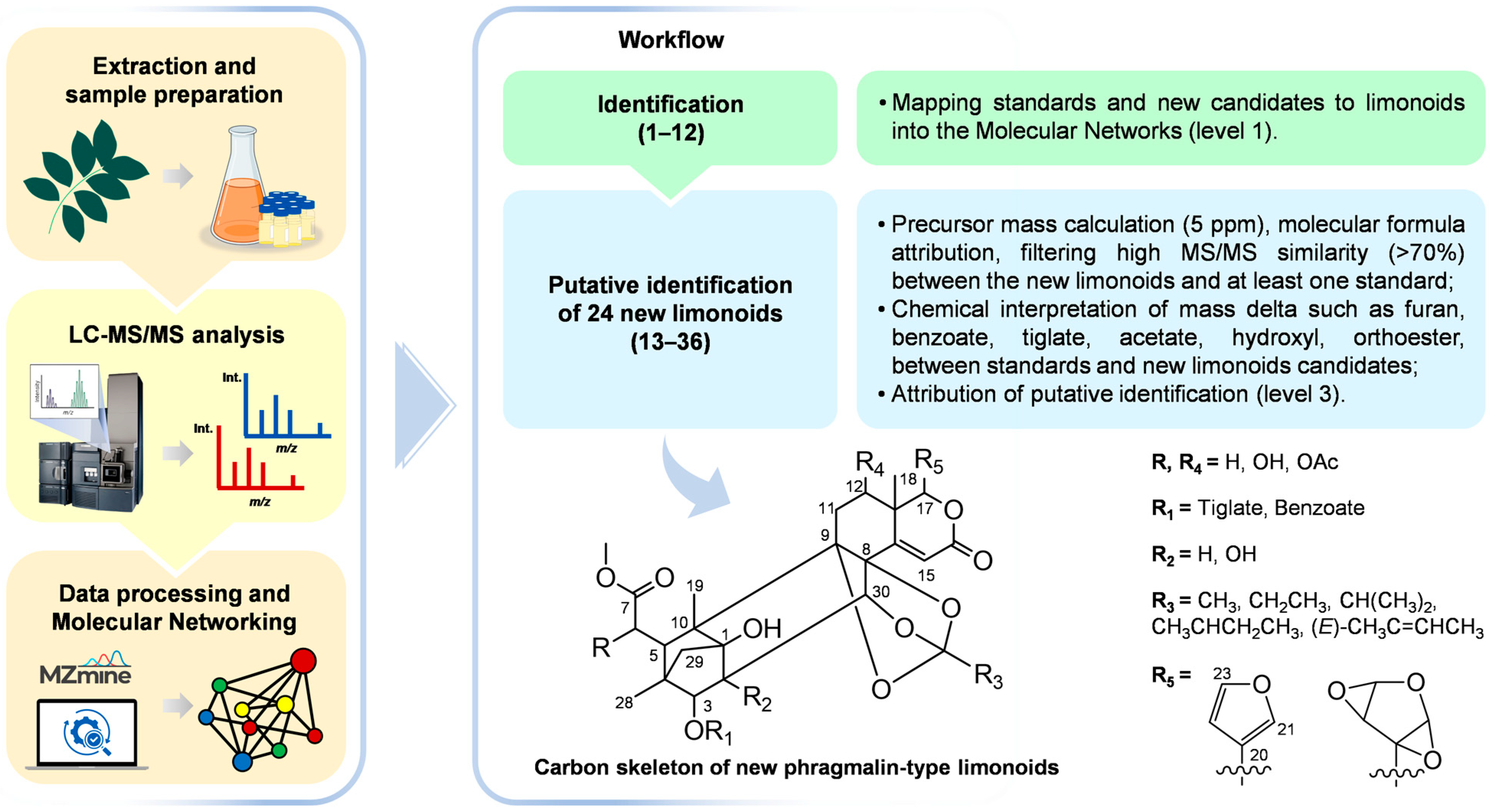
Abstract
:1. Introduction
2. Results
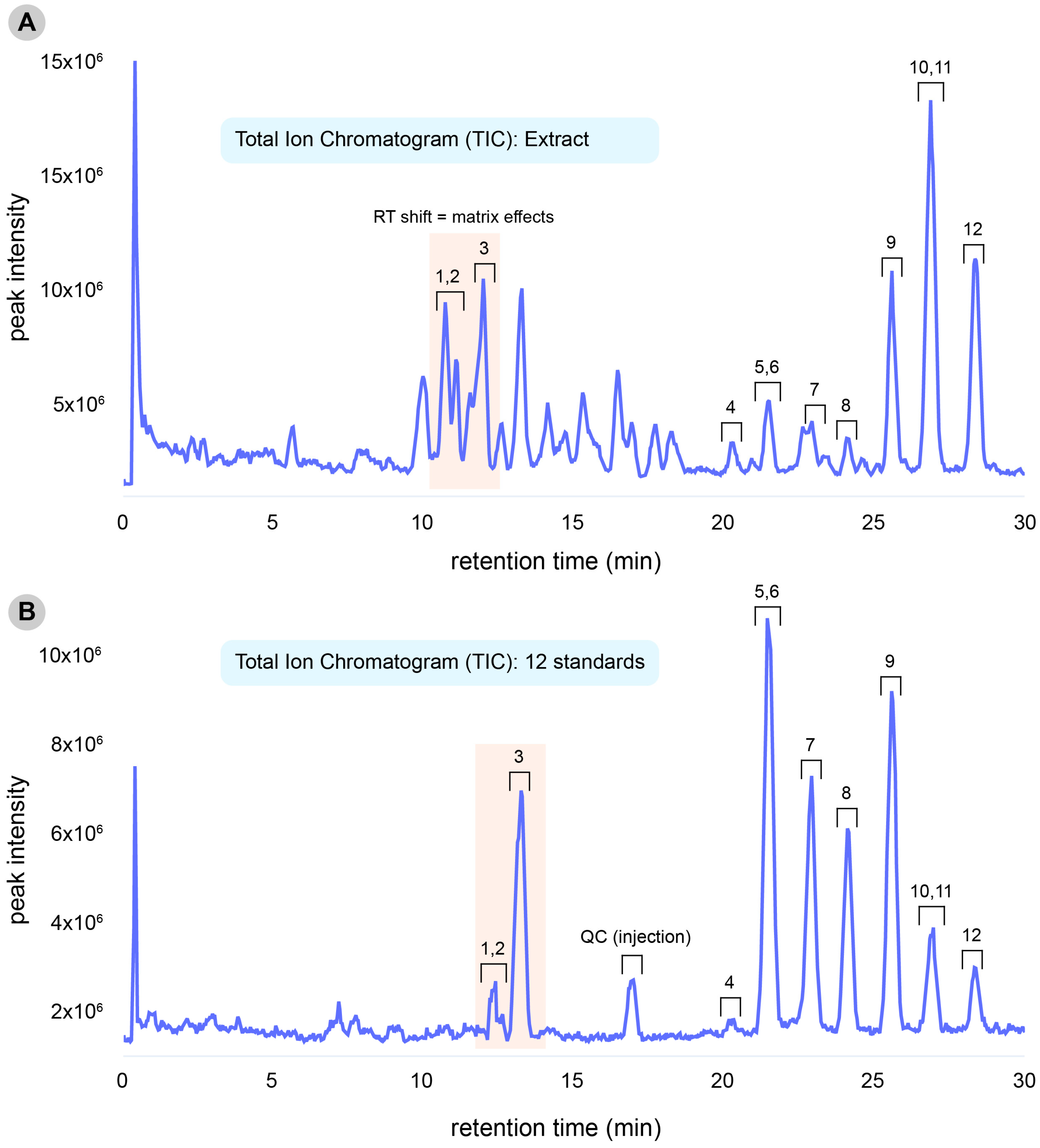
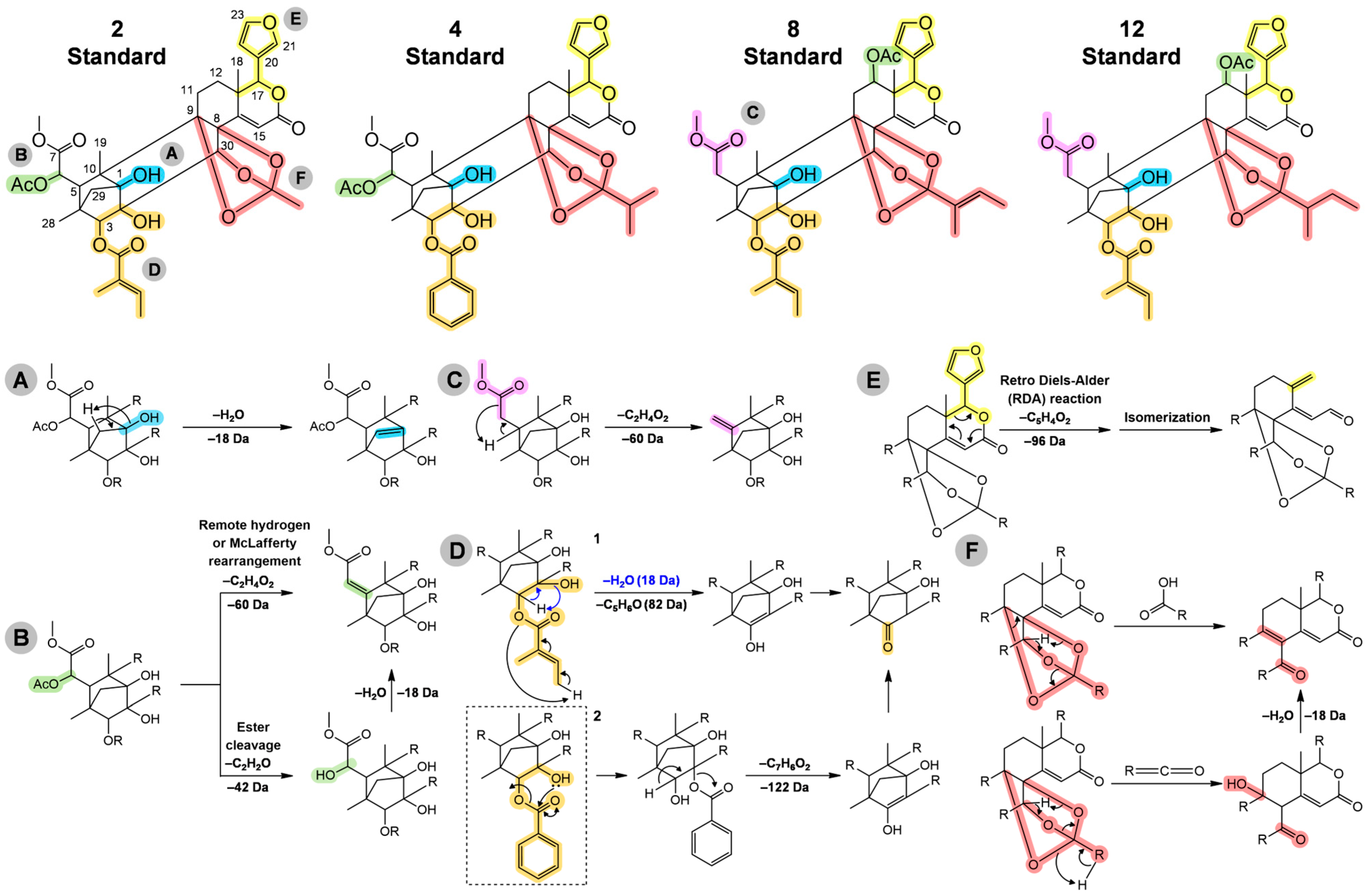
2.1. Standards and Their Structural Characteristics
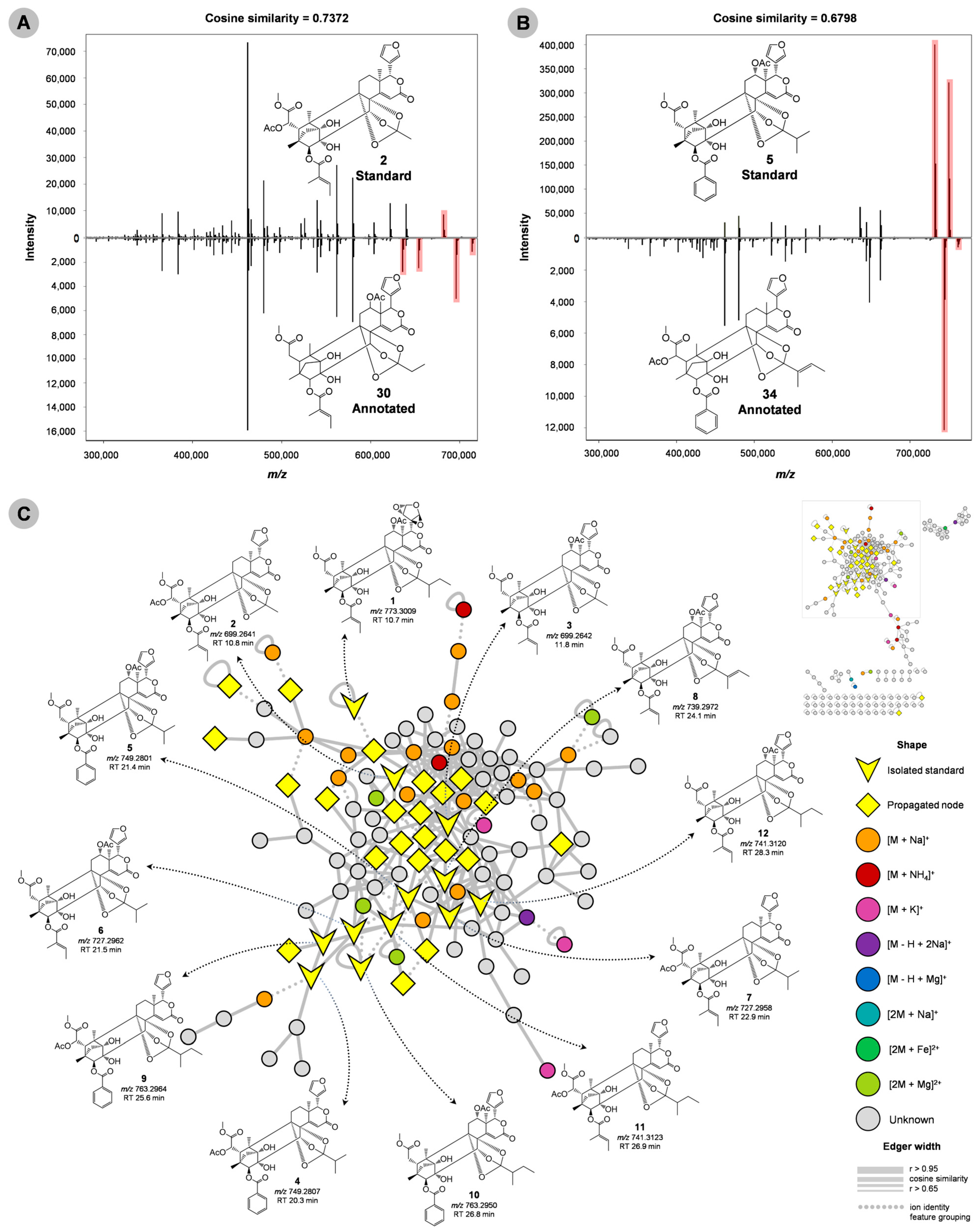
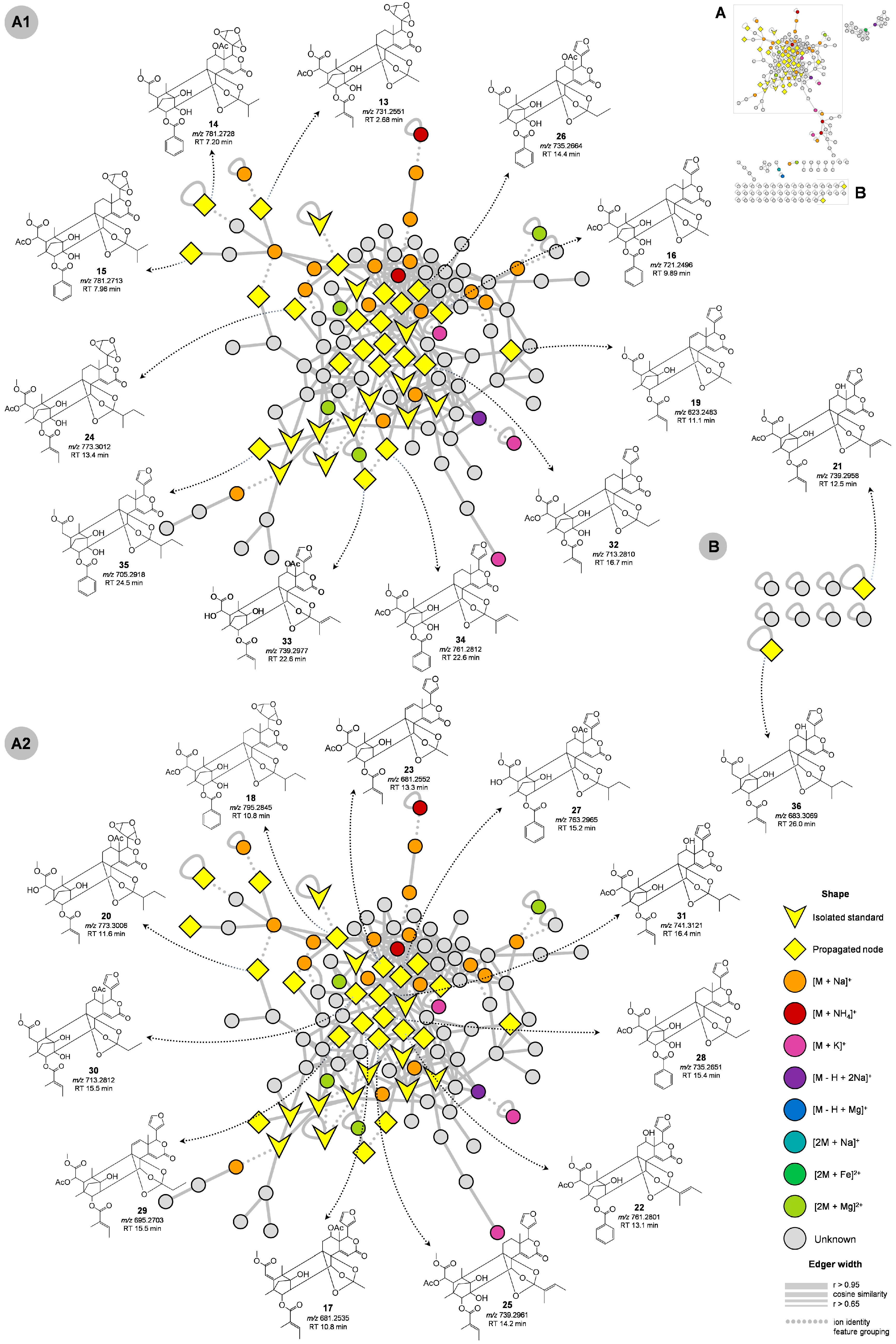
2.2. Molecular Networking and Putative Annotation
3. Discussion
4. Materials and Methods
4.1. Chemicals
4.2. Plant Collection
4.3. Extraction
4.4. LC-MS/MS Analysis
4.5. Data Processing (MZmine 3)
4.6. Ion Identity Molecular Networking (IIMN)
5. Conclusions
Supplementary Materials
Author Contributions
Funding
Institutional Review Board Statement
Informed Consent Statement
Data Availability Statement
Conflicts of Interest
References
- Flores, T.B. Meliaceae in Flora Do Brasil 2020 Em Construção. Available online: http://reflora.jbrj.gov.br/reflora/floradobrasil/FB23803 (accessed on 30 December 2020).
- Grogan, J.; Barreto, P.; Veríssimo, A. Mogno Na Amazônia Brasileira: Ecologia E Perspectivas de Manejo; Imazon: Belém, Brazil, 2002; ISBN 9788586212048. [Google Scholar]
- Eid, A.M.M.; Elmarzugi, N.A.; El-Enshasy, H.A. A Review on the Phytopharmacological Effect of Swietenia Macrophylla. Int. J. Pharm. Pharm. Sci. 2013, 5, 47–53. [Google Scholar]
- Tan, Q.-G.; Luo, X.-D. Meliaceous Limonoids: Chemistry and Biological Activities. Chem. Rev. 2011, 111, 7437–7522. [Google Scholar] [CrossRef] [PubMed]
- Champagne, D.E.; Koul, O.; Isman, M.B.; Scudder, G.G.E.; Neil Towers, G.H. Biological Activity of Limonoids from the Rutales. Phytochemistry 1992, 31, 377–394. [Google Scholar] [CrossRef]
- Dewanjee, S.; Maiti, A.; Das, A.K.; Mandal, S.C.; Dey, S.P. Swietenine: A Potential Oral Hypoglycemic from Swietenia Macrophylla Seed. Fitoterapia 2009, 80, 249–251. [Google Scholar] [CrossRef]
- Goh, B.H.; Kadir, H.A. In Vitro Cytotoxic Potential of Swietenia Macrophylla King Seeds against Human Carcinoma Cell Lines. J. Med. Plant Res. 2011, 5, 1395–1404. [Google Scholar]
- Cheng, Y.B.; Chien, Y.T.; Lee, J.C.; Wu, Y.C.; Chang, F.R. Anti-Dengue Virus Limonoids from the Actual Seeds of Swietenia Macrophylla. Planta Med. 2014, 80, P1L118. [Google Scholar] [CrossRef]
- Cheng, Y.B.; Chien, Y.T.; Lee, J.C.; Tseng, C.K.; Wang, H.C.; Lo, I.W.; Wu, Y.H.; Wang, S.Y.; Wu, Y.C.; Chang, F.R. Limonoids from the Seeds of Swietenia Macrophylla with Inhibitory Activity against Dengue Virus 2. J. Nat. Prod. 2014, 77, 2367–2374. [Google Scholar] [CrossRef]
- Chen, L.-C.; Liao, H.-R.; Chen, P.-Y.; Kuo, W.-L.; Chang, T.-H.; Sung, P.-J.; Wen, Z.-H.; Chen, J.-J. Limonoids from the Seeds of Swietenia Macrophylla and Their Anti-Inflammatory Activities. Molecules 2015, 20, 18551–18564. [Google Scholar] [CrossRef]
- Chen, J.-J.; Huang, S.-S.; Liao, C.-H.; Wei, D.-C.; Sung, P.-J.; Wang, T.-C.; Cheng, M.-J. A New Phragmalin-Type Limonoid and Anti-Inflammatory Constituents from the Fruits of Swietenia Macrophylla. Food Chem. 2010, 120, 379–384. [Google Scholar] [CrossRef]
- Pamplona, S.G.S.R.; Arruda, M.S.P.; Castro, K.C.F.; Silva, C.Y.Y.; Ferreira, A.G.; Silva, M.F.G.F.; Ohashi, O.S.; Silva, M.N. Phragmalin Limonoids from Swietenia Macrophylla and Their Antifeedant Assay against Mahogany Predator. J. Braz. Chem. Soc. 2018, 29, 1621–1629. [Google Scholar] [CrossRef]
- Abdelgaleil, S.A.M.; Doe, M.; Nakatani, M. Rings B,D-Seco Limonoid Antifeedants from Swietenia Mahogani. Phytochemistry 2013, 96, 312–317. [Google Scholar] [CrossRef] [PubMed]
- Shi, Z.; An, L.; Yang, X.; Xi, Y.; Zhang, C.; Shuo, Y.; Zhang, J.; Jin, D.-Q.; Ohizumi, Y.; Lee, D.; et al. Nitric Oxide Inhibitory Limonoids as Potential Anti-Neuroinflammatory Agents from Swietenia mahagoni. Bioorg. Chem. 2019, 84, 177–185. [Google Scholar] [CrossRef] [PubMed]
- Mootoo, B.S.; Ali, A.; Motilal, R.; Pingal, R.; Ramlal, A.; Khan, A.; Reynolds, W.F.; McLean, S. Limonoids from Swietenia Macrophylla and S. Aubrevilleana. J. Nat. Prod. 1999, 62, 1514–1517. [Google Scholar] [CrossRef] [PubMed]
- Kojima, K.; Isaka, K.; Ogihara, Y. Tetranortriterpenoids from Swietenia Macrophylla. Chem. Pharm. Bull. 1998, 46, 523–525. [Google Scholar] [CrossRef]
- Solomon, A.; Malathi, R.; Rajan, S.; Narasimhan, S.; Nethaji, M. Swietenine. Acta Crystallogr. Sect. E Struct. Rep. Online 2003, 59, o1519–o1521. [Google Scholar] [CrossRef]
- Chan, K.C.; Tang, T.S.; Toh, H.T. Isolation of Swietenolide Diacetate from Swietenia Macrophylla. Phytochemistry 1976, 15, 429–430. [Google Scholar] [CrossRef]
- Taylor, A.R.H.; Taylor, D.A.H. Limonoid Extractives from Swietenia Macrophylla. Phytochemistry 1983, 22, 2870–2871. [Google Scholar] [CrossRef]
- Fowles, R.G.; Mootoo, B.S.; Ramsewak, R.; Reynolds, W.; Lough, A.J. 3,6-Di-O-Acetylswietenolide 0.25-Hydrate. Acta Cryst. E 2007, 63, 660–661. [Google Scholar] [CrossRef]
- da Silva, M.N.; Arruda, M.S.P.; Castro, K.C.F.; da Silva, M.F.D.G.; Fernandes, J.B.; Vieira, P.C. Limonoids of the Phragmalin Type from Swietenia Macrophylla and Their Chemotaxonomic Significance. J. Nat. Prod. 2008, 71, 1983–1987. [Google Scholar] [CrossRef]
- Lau, W.K.; Goh, B.H.; Kadir, H.A.; Shu-Chien, A.C.; Muhammad, T.S.T. Potent PPARγ Ligands from Swietenia Macrophylla Are Capable of Stimulating Glucose Uptake in Muscle Cells. Molecules 2015, 20, 22301–22314. [Google Scholar] [CrossRef]
- Liu, J.-Q.; Wang, C.-F.; Chen, J.-C.; Qiu, M.-H. Limonoids from the Leaves of Swietenia Macrophylla. Nat. Prod. Res. 2012, 26, 1887–1891. [Google Scholar] [CrossRef] [PubMed]
- Liu, J.S.; Zhu, L.L.; Wang, G.K.; Wang, G. Studies on Chemical Constituents from the Fruit of Swietenia Macrophylla. Zhong Yao Cai 2016, 39, 1530–1535. [Google Scholar] [PubMed]
- Lin, B.-D.; Zhang, C.-R.; Yang, S.-P.; Zhang, S.; Wu, Y.; Yue, J.-M. D-Ring-Opened Phragmalin-Type Limonoid Orthoesters from the Twigs of Swietenia Macrophylla. J. Nat. Prod. 2009, 72, 1305–1313. [Google Scholar] [CrossRef] [PubMed]
- Tan, S.-K.; Osman, H.; Wong, K.-C.; Boey, P.-L. New Phragmalin-Type Limonoids from Swietenia Macrophylla King. Food Chem. 2009, 115, 1279–1285. [Google Scholar] [CrossRef]
- Goh, B.H.; Kadir, H.A.; Malek, S.N.A.; Ng, S.W. (αR,4R,4aR,6aS,7R,8S,10R,11S)-Methyl α-Acetoxy-4-(3-Furanyl)-10-Hydroxy-4a,7,9,9-Tetramethyl-2,13-Dioxo-1,4,4a,5,6,6a,7,8,9,10,11,12-Dodecahydro-7,11-Methano-2H-cycloocta[f][2]benzopyran-8-Acetate (6-O-Acetylswietenolide) from the Seeds ofSwietenia Macrophylla. Acta Cryst. E 2010, 66, 2802–2803. [Google Scholar]
- Goh, B.H.; Abdul Kadir, H.; Abdul Malek, S.N.; Ng, S.W. Swietenolide Diacetate from the Seeds of Swietenia Macrophylla. Acta Cryst. E 2010, 66, 1396. [Google Scholar] [CrossRef]
- Sun, Y.-P.; Zhu, L.-L.; Liu, J.-S.; Yu, Y.; Zhou, Z.-Y.; Wang, G.; Wang, G.-K. Limonoids and Triterpenoid from Fruit of Swietenia Macrophylla. Fitoterapia 2018, 125, 141–146. [Google Scholar] [CrossRef]
- Guevara, A.P.; Apilado, A.; Sakurai, H.; Kozuka, M.; Takuda, H. Anti-Inflammatory, Antimutagenicity, and Antitumor-Promoting Activities of Mahogany Seeds, Swietenia Macrophylla (Meliaceae). Philipp J. Sci. 1996, 125, 271–277. [Google Scholar]
- Ma, Y.-Q.; Liu, M.-H.; Jiang, K.; Guo, L.; Qu, S.-J.; Wan, Y.-Q.; Tan, C.-H. Limonoids from the Fruits of Swietenia Macrophylla with Inhibitory Activity against H2O2-Induced Apoptosis in HUVECs. Fitoterapia 2018, 129, 179–184. [Google Scholar] [CrossRef]
- Ma, Y.-Q.; Jiang, K.; Deng, Y.; Guo, L.; Wan, Y.-Q.; Tan, C.-H. Mexicanolide-Type Limonoids from the Seeds of Swietenia Macrophylla. J. Asian Nat. Prod. Res. 2018, 20, 299–305. [Google Scholar] [CrossRef]
- Mi, C.-N.; Li, W.; Chen, H.-Q.; Wang, J.; Cai, C.-H.; Li, S.-P.; Mei, W.-L.; Dai, H.-F. Two New Compounds from the Roots of Swietenia Macrophylla. J. Asian Nat. Prod. Res. 2019, 21, 1005–1012. [Google Scholar] [CrossRef] [PubMed]
- Mi, C.N.; Mei, W.L.; Li, W.; Wang, J.; Cai, C.H.; Li, S.P.; Dai, H.F. Chemical Constituents from the Roots of Swietenia Macrophylla King. J. Trop. Subtrop. Bot. 2017, 25, 610–616. [Google Scholar]
- Zhang, R.; Cao, M.; Yu, H.; Hongping, Z.; Di, Y.; Hao, X. Limonoids from the Twigs and Leaves of Swietenia Macrophylla. Nat. Prod. Res. Dev. 2013, 25, 969–971. [Google Scholar] [CrossRef]
- Lopes, N.P.; Silva, R.R. Mass Spectrometry in Chemical Biology: Evolving Applications; Royal Society of Chemistry: London, UK, 2017; ISBN 9781788013468. [Google Scholar]
- Clendinen, C.S.; Monge, M.E.; Fernández, F.M. Ambient Mass Spectrometry in Metabolomics. Analyst 2017, 142, 3101–3117. [Google Scholar] [CrossRef] [PubMed]
- Schmid, R.; Petras, D.; Nothias, L.-F.; Wang, M.; Aron, A.T.; Jagels, A.; Tsugawa, H.; Rainer, J.; Garcia-Aloy, M.; Dührkop, K.; et al. Ion Identity Molecular Networking for Mass Spectrometry-Based Metabolomics in the GNPS Environment. Nat. Commun. 2021, 12, 3832. [Google Scholar] [CrossRef] [PubMed]
- Yang, J.Y.; Sanchez, L.M.; Rath, C.M.; Liu, X.; Boudreau, P.D.; Bruns, N.; Glukhov, E.; Wodtke, A.; de Felicio, R.; Fenner, A.; et al. Molecular Networking as a Dereplication Strategy. J. Nat. Prod. 2013, 76, 1686–1699. [Google Scholar] [CrossRef] [PubMed]
- Ramos, A.E.F.; Alcover, C.; Evanno, L.; Maciuk, A.; Litaudon, M.; Duplais, C.; Bernadat, G.; Gallard, J.-F.; Jullian, J.-C.; Mouray, E.; et al. Revisiting Previously Investigated Plants: A Molecular Networking-Based Study of Geissospermum Laeve. J. Nat. Prod. 2017, 80, 1007–1014. [Google Scholar] [CrossRef]
- Sumner, L.W.; Amberg, A.; Barrett, D.; Beale, M.H.; Beger, R.; Daykin, C.A.; Fan, T.W.-M.; Fiehn, O.; Goodacre, R.; Griffin, J.L.; et al. Proposed Minimum Reporting Standards for Chemical Analysis Chemical Analysis Working Group (CAWG) Metabolomics Standards Initiative (MSI). Metabolomics 2007, 3, 211–221. [Google Scholar] [CrossRef]
- Rutz, A.; Sorokina, M.; Galgonek, J.; Mietchen, D.; Willighagen, E.; Gaudry, A.; Graham, J.G.; Stephan, R.; Page, R.; Vondrášek, J.; et al. The LOTUS Initiative for Open Natural Products Research: Knowledge Management through Wikidata. bioRxiv 2021. [Google Scholar] [CrossRef]
- Abdelgaleil, S.A.M.; Doe, M.; Morimoto, Y.; Nakatani, M. Rings B,D-Seco Limonoids from the Leaves of Swietenia Mahogani. Phytochemistry 2006, 67, 452–458. [Google Scholar] [CrossRef]
- Zhang, J.; Li, W.; Dai, Y.; Shen, L.; Wu, J. Twenty-Nine New Limonoids with Skeletal Diversity from the Mangrove Plant, Xylocarpus Moluccensis. Mar. Drugs 2018, 16, 38. [Google Scholar] [CrossRef] [PubMed]
- Yang, W.; Fang, D.-M.; He, H.-P.; Hao, X.-J.; Wu, Z.-J.; Zhang, G.-L. Analysis of Mexicanolide- and Phragmalin-Type Limonoids from Heynea Trijuga Using High-Performance Liquid Chromatography/electrospray Tandem Mass Spectrometry. Rapid Commun. Mass Spectrom. 2013, 27, 1203–1212. [Google Scholar] [CrossRef] [PubMed]
- Kumar, K.; Siva, B.; Rama Rao, N.; Suresh Babu, K. Rapid Identification of Limonoids from Cipadessa Baccifera and Xylocarpus Granatum Using ESI-Q-ToF-MS/MS and Their Structure-Fragmentation Study. J. Pharm. Biomed. Anal. 2018, 152, 224–233. [Google Scholar] [CrossRef] [PubMed]
- Demarque, D.P.; Crotti, A.E.M.; Vessecchi, R.; Lopes, J.L.C.; Lopes, N.P. Fragmentation Reactions Using Electrospray Ionization Mass Spectrometry: An Important Tool for the Structural Elucidation and Characterization of Synthetic and Natural Products. Nat. Prod. Rep. 2016, 33, 432–455. [Google Scholar] [CrossRef]
- Mak, K.-K.; Shiming, Z.; Balijepalli, M.K.; Dinkova-Kostova, A.T.; Epemolu, O.; Mohd, Z.; Pichika, M.R. Studies on the Mechanism of Anti-Inflammatory Action of Swietenine, a Tetranortriterpenoid Isolated from Swietenia Macrophylla Seeds. Phytomedicine Plus 2021, 1, 100018. [Google Scholar] [CrossRef]
- Sukardiman; Ervina, M. The Recent Use of Swietenia mahagoni (L.) Jacq. as Antidiabetes Type 2 Phytomedicine: A Systematic Review. Heliyon 2020, 6, e03536. [Google Scholar] [CrossRef]
- Ovalle-Magallanes, B.; Navarrete, A.; Haddad, P.S.; Tovar, A.R.; Noriega, L.G.; Tovar-Palacio, C.; Mata, R. Multi-Target Antidiabetic Mechanisms of Mexicanolides from Swietenia Humilis. Phytomedicine 2019, 58, 152891. [Google Scholar] [CrossRef]
- Pinto, L.C.; Mesquita, F.P.; Barreto, L.H.; Souza, P.F.N.; Ramos, I.N.F.; Pinto, A.V.U.; Soares, B.M.; da Silva, M.N.; Burbano, R.M.R.; Montenegro, R.C. Anticancer Potential of Limonoids from Swietenia Macrophylla: Genotoxic, Antiproliferative and Proapoptotic Effects towards Human Colorectal Cancer. Life Sci. 2021, 285, 119949. [Google Scholar] [CrossRef]
- Sung, H.; Ferlay, J.; Siegel, R.L.; Laversanne, M.; Soerjomataram, I.; Jemal, A.; Bray, F. Global Cancer Statistics 2020: GLOBOCAN Estimates of Incidence and Mortality Worldwide for 36 Cancers in 185 Countries. CA Cancer J. Clin. 2021, 71, 209–249. [Google Scholar] [CrossRef]
- Shannon, P.; Markiel, A.; Ozier, O.; Baliga, N.S.; Wang, J.T.; Ramage, D.; Amin, N.; Schwikowski, B.; Ideker, T. Cytoscape: A Software Environment for Integrated Models of Biomolecular Interaction Networks. Genome Res. 2003, 13, 2498–2504. [Google Scholar] [CrossRef]
- Holman, J.D.; Tabb, D.L.; Mallick, P. Employing ProteoWizard to Convert Raw Mass Spectrometry Data. Curr. Protoc. Bioinform. 2014, 46, 1–13. [Google Scholar] [CrossRef] [PubMed]
- Pluskal, T.; Castillo, S.; Villar-Briones, A.; Oresic, M. MZmine 2: Modular Framework for Processing, Visualizing, and Analyzing Mass Spectrometry-Based Molecular Profile Data. BMC Bioinform. 2010, 11, 395. [Google Scholar] [CrossRef] [PubMed]
- Allard, P.-M.; Péresse, T.; Bisson, J.; Gindro, K.; Marcourt, L.; Pham, V.C.; Roussi, F.; Litaudon, M.; Wolfender, J.-L. Integration of Molecular Networking and In-Silico MS/MS Fragmentation for Natural Products Dereplication. Anal. Chem. 2016, 88, 3317–3323. [Google Scholar] [CrossRef]
- Tian, Q.; Schwartz, S.J. Mass Spectrometry and Tandem Mass Spectrometry of Citrus Limonoids. Anal. Chem. 2003, 75, 5451–5460. [Google Scholar] [CrossRef] [PubMed]
- Tian, Q.; Li, D.; Barbacci, D.; Schwartz, S.J.; Patil, B.S. Electron Ionization Mass Spectrometry of Citrus Limonoids. Rapid Commun. Mass Spectrom. 2003, 17, 2517–2522. [Google Scholar] [CrossRef] [PubMed]
- Tian, Q.; Kent, K.D.; Bomser, J.A.; Schwartz, S.J. Characterization of Limonin Glucoside Metabolites from Human Prostate Cell Culture Medium Using High-Performance Liquid Chromatography/electrospray Ionization Mass Spectrometry and Tandem Mass Spectrometry. Rapid Commun. Mass Spectrom. 2004, 18, 3099–3104. [Google Scholar] [CrossRef] [PubMed]
- Wu, J.; Xiao, Q.; Huang, J.; Xiao, Z.; Qi, S.; Li, Q.; Zhang, S. Xyloccensins O and P, Unique 8,9,30-Phragmalin Ortho Esters from Xylocarpus Granatum. Org. Lett. 2004, 6, 1841–1844. [Google Scholar] [CrossRef]
- Wu, J.; Xiao, Q.; Zhang, S.; Li, X.; Xiao, Z.; Ding, H.; Li, Q. Xyloccensins Q–V, Six New 8,9,30-Phragmalin Ortho Ester Antifeedants from the Chinese Mangrove Xylocarpus Granatum. Tetrahedron 2005, 61, 8382–8389. [Google Scholar] [CrossRef]
- Wu, J.; Xiao, Q.; Li, Q. Limonoids from the Mangrove Xylocarpus Granatum. Biochem. Syst. Ecol. 2006, 34, 838–841. [Google Scholar] [CrossRef]
- Fan, C.-Q.; Wang, X.-N.; Yin, S.; Zhang, C.-R.; Wang, F.-D.; Yue, J.-M. Tabularisins A–D, Phragmalin Ortho Esters with New Skeleton Isolated from the Seeds of Chukrasia Tabularis. Tetrahedron 2007, 63, 6741–6747. [Google Scholar] [CrossRef]
- Schmid, R.; Heuckeroth, S.; Korf, A.; Smirnov, A.; Myers, O.; Dyrlund, T.S.; Bushuiev, R.; Murray, K.J.; Hoffmann, N.; Lu, M.; et al. Integrative Analysis of Multimodal Mass Spectrometry Data in MZmine 3. Nat. Biotechnol. 2023, 41, 447–449. [Google Scholar] [CrossRef] [PubMed]


Disclaimer/Publisher’s Note: The statements, opinions and data contained in all publications are solely those of the individual author(s) and contributor(s) and not of MDPI and/or the editor(s). MDPI and/or the editor(s) disclaim responsibility for any injury to people or property resulting from any ideas, methods, instructions or products referred to in the content. |
© 2023 by the authors. Licensee MDPI, Basel, Switzerland. This article is an open access article distributed under the terms and conditions of the Creative Commons Attribution (CC BY) license (https://creativecommons.org/licenses/by/4.0/).
Share and Cite
Reis, J.D.E.; Gomes, P.W.P.; Sá, P.R.d.C.; Pamplona, S.d.G.S.R.; Silva, C.Y.Y.e.; da Silva, M.F.d.G.F.; Bishayee, A.; da Silva, M.N. Putative Identification of New Phragmaline-Type Limonoids from the Leaves of Swietenia macrophylla King: A Case Study Using Mass Spectrometry-Based Molecular Networking. Molecules 2023, 28, 7603. https://doi.org/10.3390/molecules28227603
Reis JDE, Gomes PWP, Sá PRdC, Pamplona SdGSR, Silva CYYe, da Silva MFdGF, Bishayee A, da Silva MN. Putative Identification of New Phragmaline-Type Limonoids from the Leaves of Swietenia macrophylla King: A Case Study Using Mass Spectrometry-Based Molecular Networking. Molecules. 2023; 28(22):7603. https://doi.org/10.3390/molecules28227603
Chicago/Turabian StyleReis, José Diogo E., Paulo Wender P. Gomes, Paulo R. da C. Sá, Sônia das G. S. R. Pamplona, Consuelo Yumiko Y. e Silva, Maria Fátima das G. F. da Silva, Anupam Bishayee, and Milton Nascimento da Silva. 2023. "Putative Identification of New Phragmaline-Type Limonoids from the Leaves of Swietenia macrophylla King: A Case Study Using Mass Spectrometry-Based Molecular Networking" Molecules 28, no. 22: 7603. https://doi.org/10.3390/molecules28227603