Article

The Synthesis, Characterization, and Fluxional Behavior of a Hydridorhodatetraborane

by
Fatou Diaw-Ndiaye
,
Pablo J. Sanz Miguel
,
Ricardo Rodríguez
and
Ramón Macías
*
Departamento de Química Inorgánica, Instituto de Síntesis Química y Catálisis Homogénea (ISQCH), Universidad de Zaragoza-CSIC, 50009 Zaragoza, Spain
*
Author to whom correspondence should be addressed.
Molecules 2023, 28(18), 6462; https://doi.org/10.3390/molecules28186462
Submission received: 25 July 2023 / Revised: 30 August 2023 / Accepted: 31 August 2023 / Published: 6 September 2023

Abstract

:
The octahydridotriborate anion plays a crucial role in the field of polyhedral boron chemistry, facilitating the synthesis of higher boranes and the preparation of diverse transition metal complexes. Among the stable forms of this anion, CsB3H8 (or (n-C4H9)4N)[B3H8] have been identified. These salts serve as valuable precursors for the synthesis of metallaboranes, wherein the triborate anion acts as a ligand coordinating to the metal center. In this study, we have successfully synthesized a novel rhodatetraborane dihydride, [Rh(η2-B3H8)(H)2(PPh3)2] (1), which represents a Rh(III) complex featuring a bidentate chelate ligand fasormed by B3H8. Extensive characterization of this rhodatetraborane complex has been performed using NMR spectroscopy in solution and X-ray diffraction analysis in the solid state. Notably, the complex exhibits intriguing fluxional behavior, which has been investigated using NMR techniques. Moreover, we have explored the reactivity of complex 1 towards pyridine (py) and dimethylphenylphosphine (PMe2Ph). Our findings highlight the labile nature of this four-vertex rhodatetraborane as it undergoes disassembly upon attack from the corresponding Lewis base, resulting in the formation of borane adducts, LBH3, where L = py, PMe2Ph. Furthermore, in these reactions, we report the characterization of new cationic hydride complexes, such as [Rh(H)2(PPh3)2 (py)]+ (2) and [Rh(H)2(PMe2Ph)4]+. Notably, the latter complex has been characterized as the octahydridotriborate salt [Rh(H)2(PMe2Ph)4][B3H8] (3), which extends the scope of rhodatetraborane derivatives.

1. Introduction

The synthesis of [arachno-B3H8] has gathered significant attention in recent years due to its pivotal role in the preparation of higher boranes, such as closo-B12H122−, and a wide array of transition metal complexes [1,2,3,4].
Many salts of this anion have been prepared using B2H6, but concerns regarding the toxicity and flammability of diborane have prompted the exploration of alternative synthetic routes. Among the various methods, the reaction of Na[BH4] with I2 stands out [2]; however, the presence of iodide anions poses challenges in obtaining the desired product. Recent investigations have focused on replacing iodine with various metal halides as oxidants, offering improved efficiency and selectivity in the synthesis of [arachno-B3H8].
The crystal structure of [(H3N)2BH2][B3H8] has been determined by Peters and Nordman, shedding light on the structural composition of the octahydridotriborate anion. The anion consists of a triangular arrangement of boron atoms, with two bridging and six terminal hydrogens [3].
The inherent stability and accessibility of the arachno-B3H8 anion have paved the way for extensive investigations into its reactivity with various metal complexes [4]. The resulting arachno-2-metallaboranes can be synthesized through ligand substitution reactions, offering a general route for the incorporation of transition metals into the cluster framework:
LnMX + B3H8 → [Ln−1MB3H8] + L + X (L = neutral ligand; X = halide)
The reactivity of arachno-B3H8 has been explored with a wide range of transition metals, spanning across the periodic table, including Ti, Cr, Mo, W, Mn, Re, Fe, Ru, Os, Ir [5], Cu, Ag and Zn [1,3,4].
The reaction between a transition metal complex and the octahydridotriborate anion is, a priori, the most direct route to the synthesis of arachno-2-metallatetraboranes. However, some of the reported metallatetraboranes were prepared from reactions of either monocyclopentadienyl metal chlorides or hydride-ligated complexes of transition metals from groups 5–9 with monoboranes (LiBH4 or BH3THF) [6,7,8]. This synthetic procedure was developed mainly by Fehlener and co-workers at Notre Dame University (Notre Dam, IN, USA) [9]; and Gosh and co-workers have been using it for a good number of years, at the Indian Institute of Technology Madras (Chennai, India), in the pursuit of new metallaboranes [10].
Alternatively, reactions between metal complexes and larger boranes, such as pentaborane, were also a route to tetraboranes, via cluster dismantling processes [11].
To expand the scope of transition element arachno-metallaboranes and explore novel structures and dynamic processes, our study focused on investigating the reactivity between Wilkinson’s catalyst, [RhCl(PPh3)3], and the octahydridotriborate anion, [B3H8]. This investigation resulted in the successful synthesis of dihydridorhodatetraborane, [Rh(η2-B3H8)(H)2(PPh3)2] (1). The compound was comprehensively characterized using NMR spectroscopy and X-ray diffraction analysis. Notably, the newly synthesized metallatetraborane exhibited a chemical non-rigidity, which was studied using NMR spectroscopy at variable temperatures. In addition, we have carried out an exploratory study of the reactivity of 1 with Lewis bases, resulting in the characterization of hydride metal complexes.

2. Results and Discussion

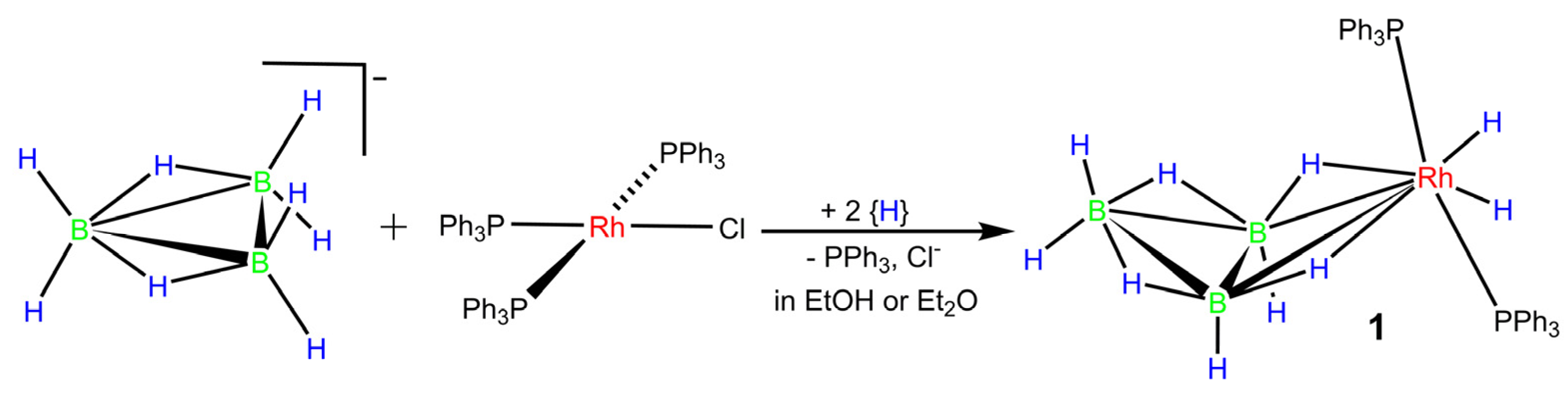
2.1. Synthesis of [Rh(η2-B3H8)(H)2(PPh3)2] (1)

The reaction of the Wilkinson’s catalyst with the cesium salt CsB3H8 in ethanol leads to the formation of dihydridorhodatetraborane (1) (Scheme 1).
Due to the limited solubility of the cesium salt in ethanol and the insolubility of the rhodium complex (Wilkinson’s catalyst) in the same solvent, the reaction proceeds in a heterogeneous solid–liquid phase. The reaction mixture initially forms a brick-red suspension, which transforms into a red-orange product, corresponding to the formation of dihydridorhodatetraborane (1). The product is then collected by filtration using a sintered disc filter funnel.
As an alternative approach, we conducted the reaction using the tris(dioxane) solvate NaB3H8.3(C4H8O2) as the starting material in diethyl ether, which also exhibits limited solubility for Wilkinson’s catalyst. Similar to the ethanol system, this synthesis is characterized by a heterogeneous reaction. The resulting yellow product, identified as hydridorhodathiaborane, is easily filtered under ambient conditions, yielding 1.

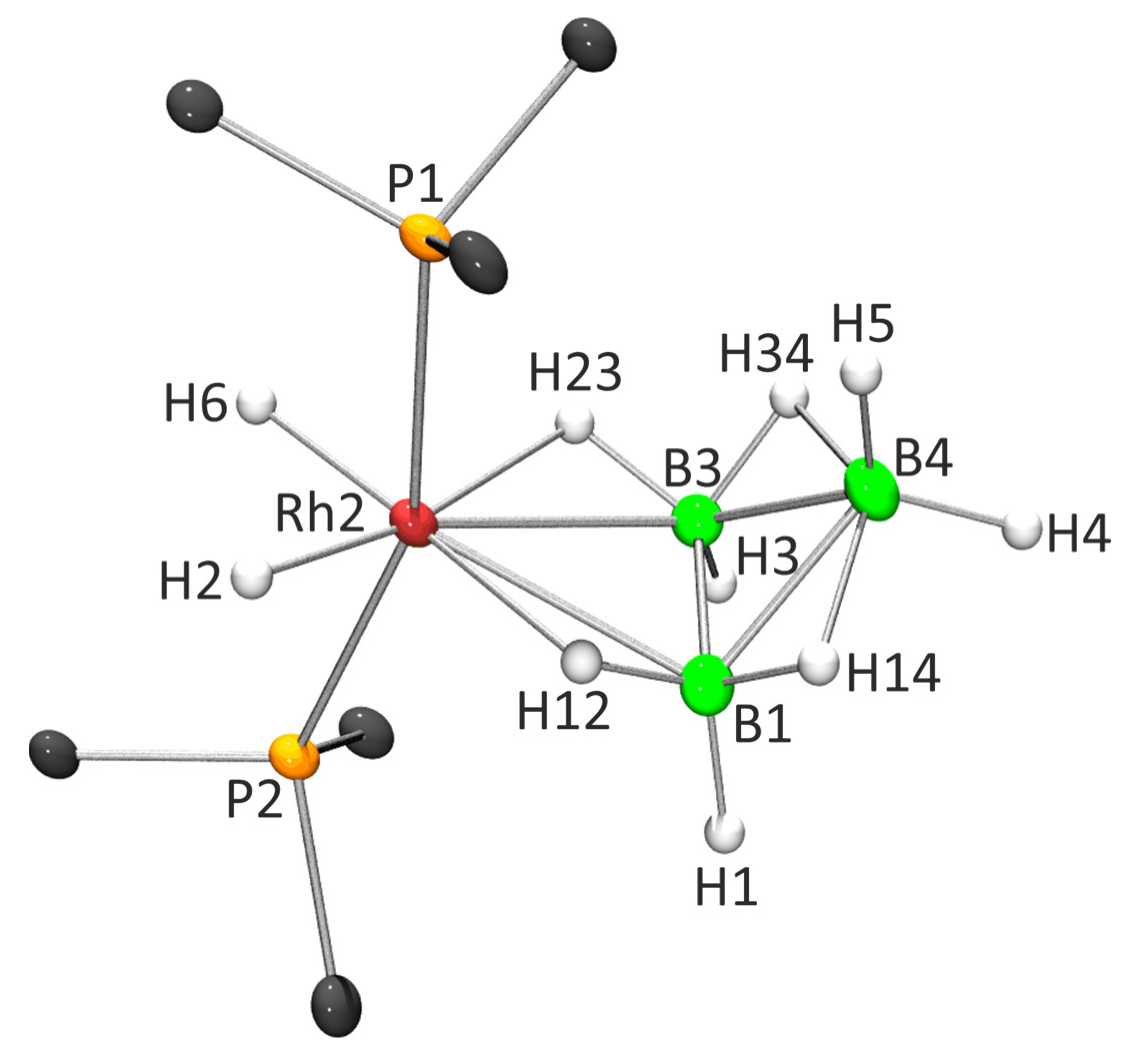
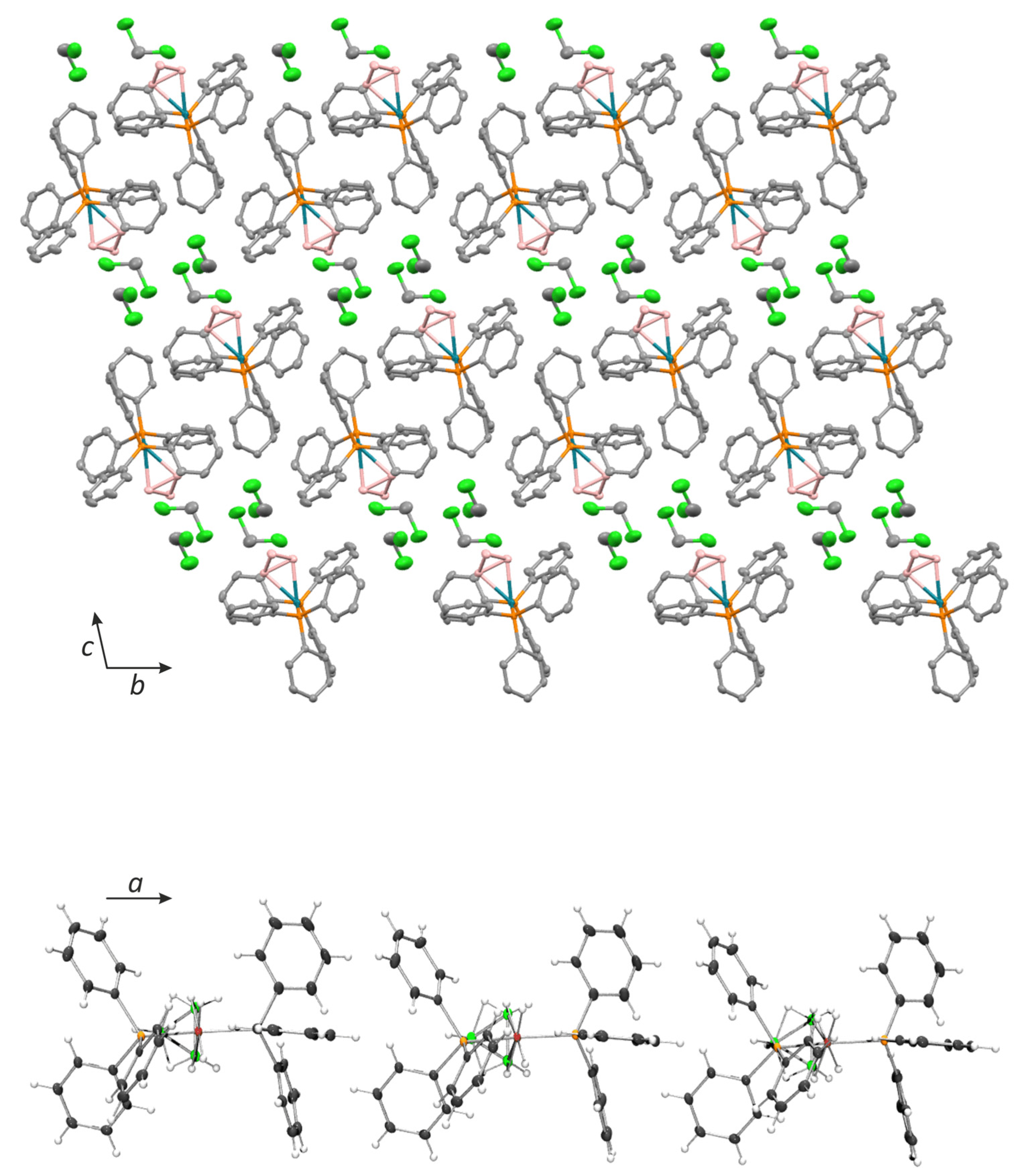
2.2. X-ray Diffraction Analysis

The Cambridge Crystallographic Data Centre (CCDC) provides X-ray diffraction analyses for fifteen arachno-metallaboranes, incorporating {MLn}-fragments of Nb [7], Cr [12], Mo [10], W [6,10], Mn [13], Re [8,13], Ru [11,14,15], Os [16] and Cu [17]. However, the availability of comparative structural data across the periodic table, for this particular class of four-vertex arachno-metallaboranes, is limited. It is important to emphasize that the crystal structure of compound 1 represents the first example of a Group 9 arachno-2-metallaborane characterized by X-ray diffraction analysis.
Single crystals of the compound were obtained by diffusing hexane into a solution of 1 in CH2Cl2. Figure 1 depicts an ORTEP-type drawing, illustrating selected interatomic distances and angles. The rhodium center in the compound exhibits an octahedral coordination sphere, where the B3H8 moiety acts as a bidentate η2-ligand through two B–H–Rh bridge bonds. These bridge bonds are located trans to the exo-polyhedral hydride ligands. Completing the coordination number 6 around the metal, two Ph3P ligands are mutually trans. Consequently, the molecule can be classified as an eighteen-electron, six-coordinate, octahedral d6 rhodium(III) complex.
Alternatively, compound 1 can be described as a four-vertex arachno-cluster, which can be related to the parent arachno-B4H10 by replacing a BH2 ‘wing-tip’ with the d6-{Rh(H)2(PPh3)2} fragment. This description follows the architectural patterns proposed by Williams [18], where the four-vertex butterfly type arachno-cluster is derived from an octahedron by removing two adjacent vertices. According to the polyhedral skeletal electron pair theory (PSEPT) [19,20], these clusters are expected to have seven skeletal electron pairs (n + 3; where n is the number of vertices of the polyhedral cluster). Applying the PSEPT electron-counting rules, the {Rh(H)2(PPh3)2} group in compound 1 can be considered as a vertex contributing three electrons to the cluster framework bonding [9 e(Rh) + 4 e(2PPh3) + 2 e(2H) − 12 e = 3 e], resembling the BH2 ‘wing-tip’ in arachno-B4H10.
The distances between Rh2 and P1, as well as Rh2 and P2, are determined to be 2.3103(11) Å and 2.2911(11) Å, respectively. These bond lengths are significantly shorter compared to the Ru–P lengths observed in the arachno-2-ruthenatetraborane, [Ru(η2-B3H8)(H)2(PPh3)2], which also features two mutually trans PPh3 ligands. In the ruthenatetraborane, the Ru–P bond lengths are measured to be 2.373(1) Å and 2.364(1) Å. However, when the hydrotris(pyrazol-1-yl)borate-ligated ruthenatetraborane, [Ru(η2-B3H8)(PPh3){K3-HB(pz)3}], is considered, the Ru–P bond distance is slightly shorter at 2.317(1) Å. Analysis from the Cambridge Crystallographic Data Centre (CCDC) reveals that the mean bond length for Rh–PPh3 is 2.318 Å, while the mean bond distance for Ru–PPh3 is slightly longer at 2.350 Å. Based on these findings, it can be concluded that the M–P bond distances fall within the crystallographic data, indicating that the Ru–PPh3 bonds are on average longer than the corresponding Rh–PPh3 lengths.
It is noteworthy to highlight that the {Ru(CO)(H)(PPh3)2} and {Ru(PPh3){K3-HB(pz)3} fragments present in the ruthenaboranes are isolobal and isoelectronic with the {Rh(H)2(PPh3)2} group observed in compound 1. These fragments contribute three electrons to the cluster framework, thus fulfilling the expected seven skeletal electron pairs (seps) characteristic of a four-vertex arachno-cluster.
In the crystal structure of compound 1, the rhodatetraborane clusters exhibit a packing arrangement along the crystallographic axis a. These clusters form ribbons that associate in pairs through sextuple phenyl embrace (SPE) interactions. The separation between the P atoms (P···P) and the collinearity of Rh–P···P–Rh are measured at 7.9 Å and 165°, respectively, falling within the reported range for this attractive edge-to-face interaction (Figure 2). The SPE interaction arises from intermolecular edge-to-face C–H···π attractive forces facilitated by the presence of phenyl rings [21,22].
All metal octahydridotriboranes stored in the CCDC exhibit a notable similarity, with mean distance values of 1.745 Å, 1.797 Å and 1.794 Å for the B1–B3, B1–B4 and B3–B4 linkages, respectively. Among these linkages, the B1–B3 edge involved in the M–B–B interaction (M = Nb, Cr, Mo, W, Mn, Re, Ru, Os, Cu) shows the shortest mean value. However, the length of the B1–B3 edge varies within the range of 1.707 Å to 1.833 Å, which is larger than the range of 1.772 Å to 1.817 Å observed for the other two B–B connections involving B–B–B bonds. This structural feature is expected due to the variation in the metal center across the metallatetraborane series. The M–B–B interaction, facilitated by two bridging hydrogen atoms, is expected to influence the B1–B3 distance, leading to significant differences among the compounds.
In the parent arachno-B4H10 cluster, the ‘hinge’ B1–B3 edge exhibits a mean value of 1.722 Å, which is the shortest among the five B–B bond distances present in this four-vertex arachno-cluster. The other B–B distances in arachno-B4H10 range between 1.844 Å and 1.847 Å, based on the average values derived from the five structures available in the CCDC. The butterfly dihedral angle, characterizing the molecular structure of B4H10, is measured to be 118.4 ± 0.4°. In comparison, the dihedral angle for the metal compounds falls within the range of 124.5 ± 5.2°. The smallest dihedral angle observed among the metallatetraboranes is 119.3°, which corresponds to the octahydridotriborato-bis(triphenylphosphine)copper(I) compound [17]. This compound features a {(PPh3)2Cu} vertex with both endo- and exo-triphenyl ligands, closely resembling the {BH2} vertex in arachno-tetraborane(10). The coordination of the {(PPh3)2Cu} and {BH2} vertices, bound to the {{η2-B3H8} fragment, exhibits a tetrahedral geometry. Consequently, significant structural similarities between these two molecules are expected, as evidenced by the similarity in their dihedral angles.
Among the tetrametallaboranes determined through crystallography and deposited in the CCDC, different metal fragments act as vertices, formally replacing the {BH2} vertex in arachno-B4H10. As a result, notable differences in the dihedral angles are observed, depending on the nature of the endo- and exo-ligands. For instance, the [Ru(η2-B3H8)(Cl)(η6-(CH3)6C6))] cluster features a pseudo-octahedral ruthenium(II) center, bonded to exo-hexamethylbenzene and endo-chloro ligands. This metal fragment leads to a relatively flat structure with a dihedral angle of 129.7°. Another tetrametallaborane, [Nb(η2-B3H8)(η5-(C5H5)2], exhibits a dihedral angle of 125.7°. In this case, the coordination number around the niobium(III) center is eight, with six positions occupied by the cyclopentadienyl ligands, each acting as a tridentate ligand, and the remaining two positions occupied by the bidentate octahydridotriborate anion.

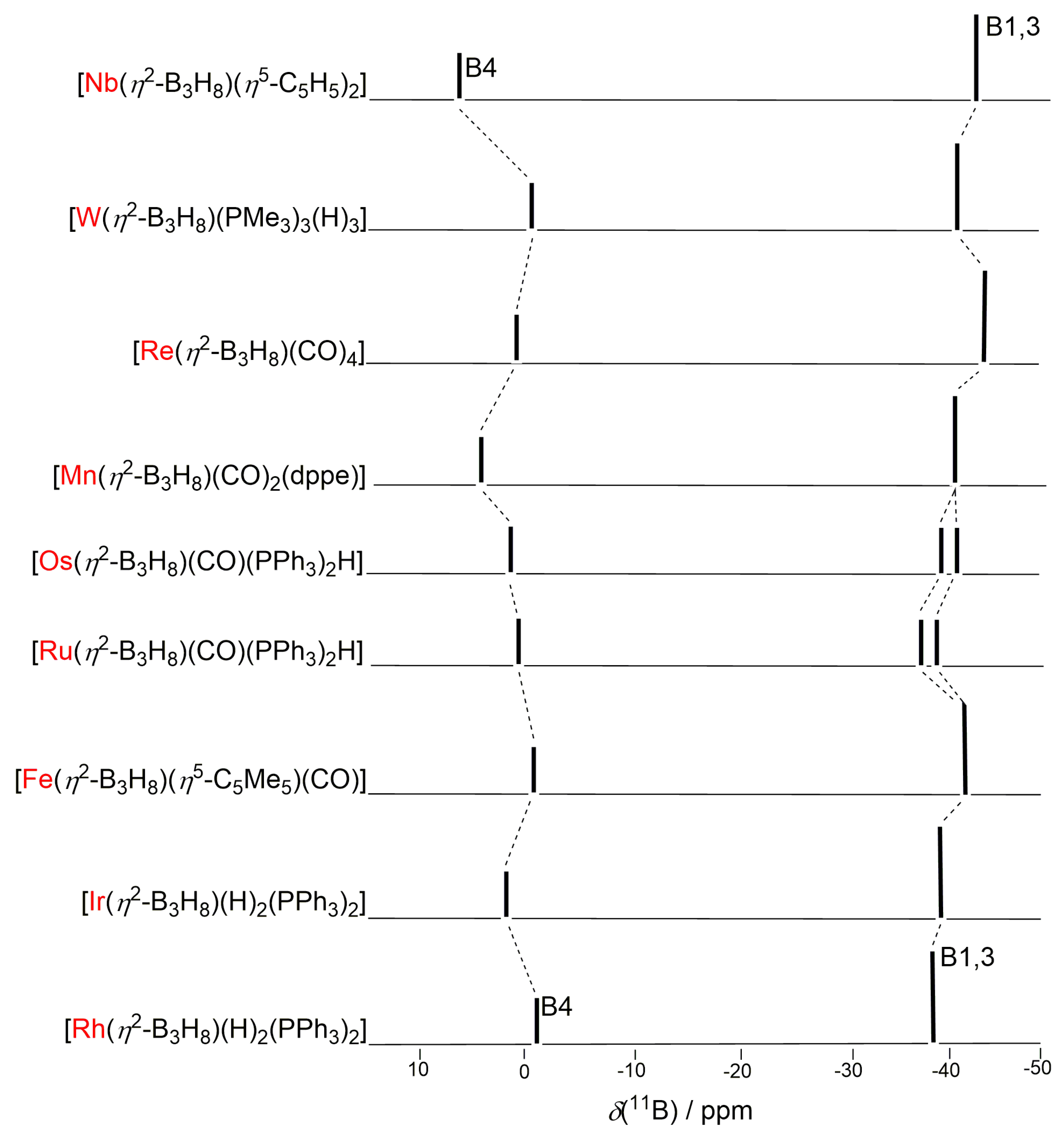
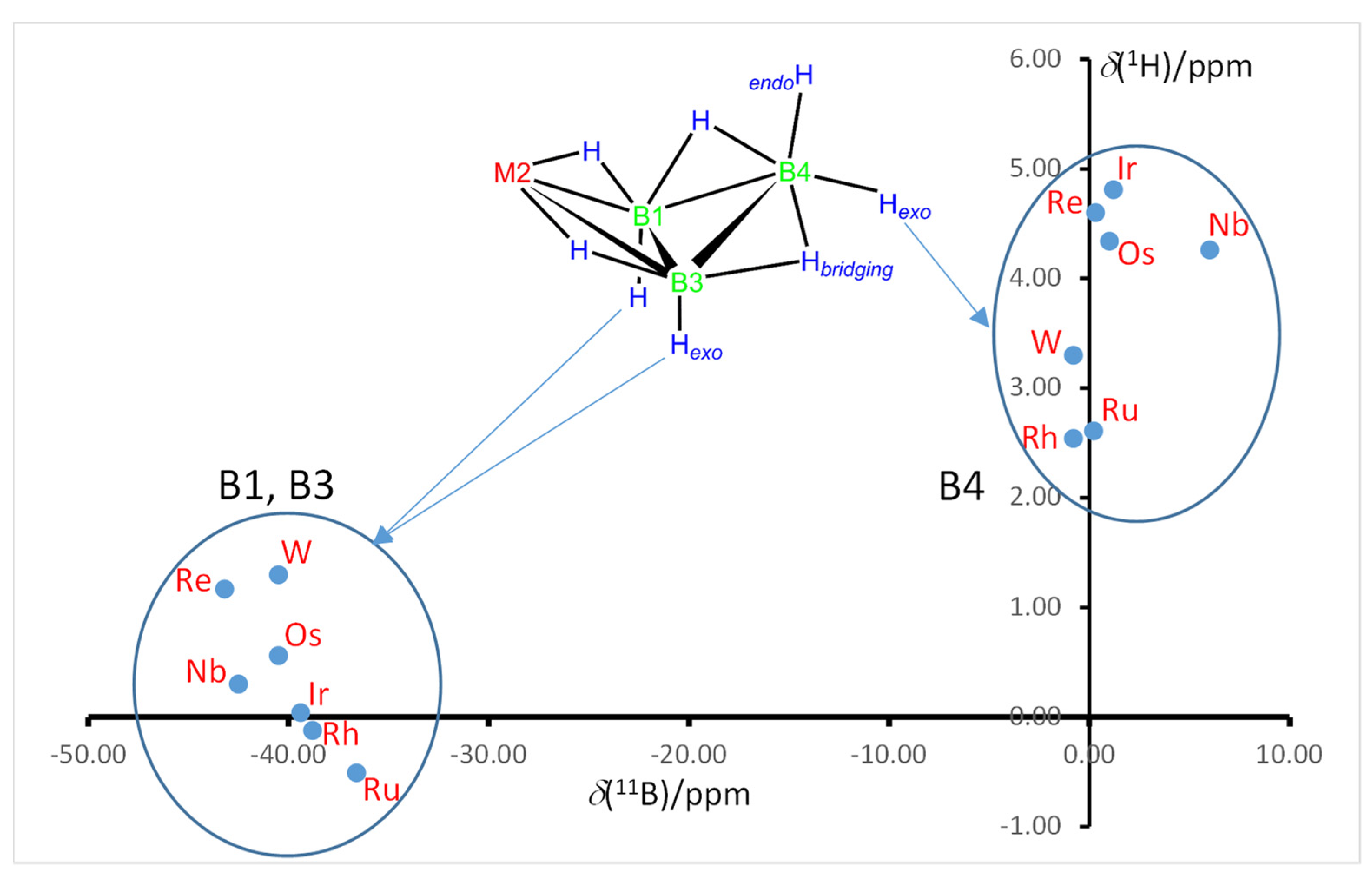
2.3. NMR Characterization and Comparison

The assignments provided in Table 1 for the resonances are reasonably determined based on their relative intensities and by comparing them with the resonances observed in the previously reported hydridoiridatetraborane, [Ir(η2-B3H8)(H)2(PPh3)2] [5]. To further support these assignments, DFT calculations were performed on a model compound, [Rh(η2-B3H8)(H)2(PH3)2]. The calculated data confirmed the resonance assignments and provided additional insight into the molecular structure and behavior of the compound.
The NMR data obtained for compound 1 are fully in accord with the solid-state structure determined by X-ray diffraction analysis. The room temperature 11B NMR spectrum shows two distinct resonances at δ(11B) −1.0 and −38.9 ppm, with a relative intensity ratio of 1:2. These resonances can be attributed to the B4 and B1,3 vertices, respectively. Interestingly, the uncoupled 11B spectrum does not display the expected 1J(11B-1H) coupling constants (refer to Figure S1). This observation can be rationalized considering the chemical non-rigidity of the endo- and bridging-hydrogen atoms present in the {(η2-B3H8)} ligand (vide infra).
Figure 3 illustrates a stick representation of the chemical shifts and relative intensities in the 11B spectra for a series of isostructural and isoelectronic arachno-metallatetraboranes similar to compound 1. These four-vertex clusters exhibit highly similar overall 11B shielding patterns. The resonances corresponding to the metal-bound B1–B3 positions are grouped within a narrow region of δ(11B) from −36.0 to −41.0 ppm. On the other hand, the signals associated with the “wing-tip” B4 position are observed between δ(11B) −1.0 and +6.0 ppm.
The observed marked similarity in the 11B NMR resonances is somewhat surprising, considering that the metal fragments change from early to late-transition elements, each bearing different ligands such as CO, PPh3, dppe, C5H5, C5Me5 and hydrides, and the fact that the spectra were recorded in different solvents such as toluene-d8, CD2Cl2 and CDCl3.
This finding suggests that the fundamental nature of the metal-to-{η2-B3H8} fragment interaction is maintained throughout this series of compounds. According to the electron-counting rules [19,20] the {Nb(η5-C5H5)2}- [7], {W(PMe3)3(H)3}- [6], {Re(CO)4}- [5], {Mn(CO)2(dppe)}- [13], {Os(CO)(PPh3)2(H)}- [5], {Ru(CO)(PPh3)2(H)}-, {Fe(η5-C5Me5)(CO)}-, {Ir(H)2(PPh3)2} [5] and {Rh(H)2(PPh3)2}-vertices contribute three electrons to the cluster framework. This electron count yields seven seps, as expected for four-vertex arachno-2-metallatetraboranes. The consistency in electron counting and the resultant 11B resonances further support the notion that the interaction between the metal fragment and the {η2-B3H8} ligand is maintained across this series of compounds.
At 298 K, the 1H-{11B} NMR spectrum of compound 1 exhibits three signals at δ(1H) +2.66 ppm, −6.96 ppm and −11.95 ppm, with a relative intensity ratio of 1:2:2. These spectroscopic data do not match the expected pattern based on the molecular structure of the dihydrorhodatetraborane. According to the Cs point group symmetry, we would anticipate five proton resonances with a relative intensity ratio of 1:1:2:2:2, along with aromatic Ph signals (30H). However, when the 1H-{11B} spectrum is measured at 223 K, the expected pattern is observed. Peaks appear at δ(1H) +2.54 (1H), +1.82 (1H), −0.11 (2H), −1.11 (2H), −7.07 (2H) and −11.79 ppm.
The lowest frequency signal at −11.79 ppm, corresponding to the Rh–H hydride ligands, does not broaden in the proton-coupled spectrum. This hydride resonance exhibits the characteristic pattern of a broad quintet, which appears as an apparent broad triplet in the 1H-{31P} spectrum (refer to Figure S2). However, the chemically equivalent hydride ligands, H2 and H6 in Figure 1, couple unequally to the H1,2 and H2,3 nuclei, resulting in magnetic non-equivalence (Figure S3). Consequently, the 1H-{31P} spectrum for the Rh–H ligands (H2, H6 in Figure 1) displays second-order behavior (Figure S3).
In the dihydridoiridatetraborane analogue, [Ir(η2-B3H8)(H)2(PPh3)2], the hydride signal appears at δ(1H) −13.30 p.p.m, appearing as a triplet of doublets due to cisoid coupling to two 31P nuclei with very similar coupling constants. Additionally, a small transoid coupling, 2J(1Hbridge-1Ht) = 7.0 Hz, is observed. In compound 1, there are also two 31P nuclei with similar coupling constants. However, the proton pattern of the hydride nuclei shows second-order effects, as discussed above. Interestingly, the calculated transoid 2J(1H1,2-1H6) coupling constant in compound 1 is significantly larger compared to that observed in the dihydridoiridatetraborane analogue [5].
The two 31P nuclei in compound 1 form an AB-spin system with strong coupling, which is evident from the presence of a “roof effect” in the 31P-{1H} spectrum at 202 MHz (refer to Figure S4). In a strong coupling regime, the separation of the two central states is determined by the formula C = [((δυ)2 + J2]½, where δυ represents the difference in resonance frequencies of the two spins and J is the scalar coupling constant [23]. The large 2J(31P1–103Rh–31P2) of 367 Hz indicates a mutually trans-disposition of the two phosphorus atoms, confirming the molecular structure determined by X-ray diffraction analysis (Figure 1).
The proton signals at δ(1H) +2.54 (1H), +1.82 (1H), −0.11 (2H), −1.11 (2H) and −7.07 (2H) exhibit significant broadening in the 1H spectrum compared to the 1H-{11B} spectrum. This indicates that these resonances correspond to 1H nuclei directly bound to boron atoms. The molecular structure of compound 1, along with the 1H-{11B} selective experiments and the observed broadening patterns, has facilitated the complete assignment of the proton resonances to their respective positions within the structure of 1.
In Figure 4, it is observed that the 1H resonances assigned to the B1,3 exo-hydrogen atoms are grouped together between −0.51 and +1.30 ppm, forming a “low-frequency” cluster. On the other hand, the B4–Hexo signals are grouped between +1.83 and +4.81 ppm, forming a “high-frequency” cluster. Within this high-frequency group, the B4 exo-hydrogen resonance experiences significant deshielding when the metal atom is Nb, Re, Os, or Ir. This results in a large chemical shift difference for this particular resonance between [Rh(η2-B3H8)(H)2(PPh3)2] (1) and [Ir(η2-B3H8)(H)2(PPh3)2], as well as between [Ru(η2-B3H8)(CO)(PPh3)2H] and [Os(η2-B3H8)(CO)(PPh3)2H]. If we consider that the metal center is located antipodal to the B4 vertex through an axis connecting M2 and B4, this effect can be attributed to the change from a second-row transition metal center to a third-row transition metal center.
An interesting observation was made regarding the anomalous low proton shielding of exo-terminal protons that are positioned antipodal to third-row metal centers in twelve-vertex closo-metallaheteroborane systems. This phenomenon has been recognized as a diagnostic characteristic of this structural feature [24,25,26,27,28,29,30]. Similarly, we can utilize the strong deshielding of B4-Hexo protons as a diagnostic indicator for the presence of third-row transition metal centers in four-vertex arachno-2-metallatetraboranes. This provides valuable insights into the structural composition of these compounds.

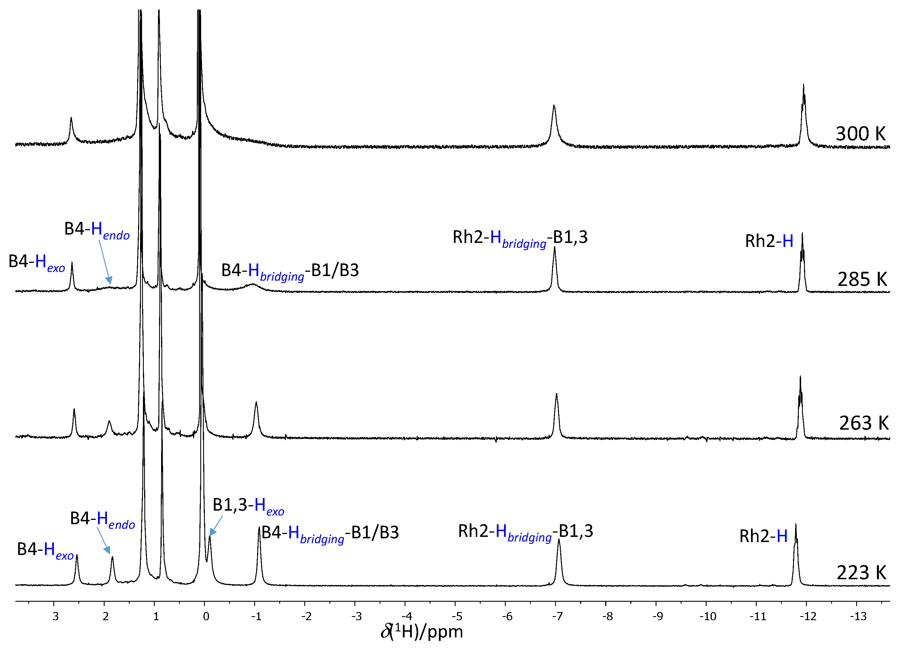
2.4. Fluxional Behavior

In order to investigate the chemical non-rigidity and fluxional behavior of compound 1, a variable temperature (VT) NMR study was conducted in CD2Cl2. Figure 5 illustrates the changes observed in the 1H-{11B} NMR spectrum as the temperature was varied. The proton signals corresponding to B4-Hendo, B1,3-Hexo, and B4-Hbridging-B1/B3 hydrogen atoms gradually broaden and eventually disappear, indicating an intramolecular proton exchange process in compound 1. Notably, this process does not involve the B4-Hexo and Rh-H-B1/B3 bridging hydrogen atoms nor the Rh-H hydride ligands.
The coalescence temperature, determined as the point at which the 1H signals merge, was estimated to be 300 K. Using this information, the activation energy (ΔG ) for the asymmetric population system was calculated to be 10 kcal/mol (see Supporting Information for the analysis) [31].
To investigate the possible exchange of B4–Hexo, the Rh–Hbriding–B1,3 and the Rh–H hydrogen atoms at higher temperatures, NMR spectra of compound 1 were measured at +67 °C in deuterated 1,1,2,2-tetrachloroethane. The 1H–{11B} spectrum revealed the formation of a new hydridorhodatetraborane, exhibiting proton resonances at δH −6.78 and −11.67 ppm, which were assigned to Rh-H-B and Rh-H hydrogen atoms, respectively (Figure S5).
A further increase in the temperature to +97 °C resulted in the decomposition of both compound 1 and the new hydridorhodaborane. The products of this decomposition included borane triphenylphosphine (Ph3P–BH3) and hydride-ligated complexes, as evidenced by the presence of several doublets in the 31P-{1H} spectrum (Figure S6). Additionally, the 11B NMR spectrum showed peaks between δB +2.5 and +10.0 ppm, which did not exhibit 1J(11B–1H) coupling, suggesting the formation of species containing O-B bonds (Figure S7).
The intramolecular hydrogen atom exchange observed in compound 1 shares similarities with the reported behavior of octahydridotriborate complexes such as [Mn(η2-B3H7Br)(CO)4] and [Ru(η2-B3H8)(CO)(H)(PPh3)2] [32], where the M2–H–B1/B3 bridging atoms also remain static. In these cases, the fluxional process occurs with an activation energy, ΔG, of approximately 12.2 kcal/mol at +23 °C for the manganesaborane. Interestingly, analogous compounds of third-row transition elements, such as [Os(η2-B3H8)(CO)(H)(PPh3)2] and [Ir(η2-B3H8) (H)2(PPh3)2], which are CO-ligated ruthenaborane and compound 1 analogues, respectively, do not exhibit fluxional behavior.
Several mechanisms have been proposed to explain hydrogen exchange in four-vertex arachno-2-metallatetraboranes. In the case of covalent metal-octahydridotriborate Be(B3H8)2, for instance, a rearrangement involving a Be-to-B3H8 bond change from η2 to η1, facilitated by Be–H–B bonds, followed by hydrogen atom exchange around the two BH3 units of the Be–{η1-B3H8} fragment, has been suggested. This mechanism ultimately leads to complete proton and boron exchange at high temperatures [33]. However, this mechanism cannot be applied to explain the observed exchange in compound 1, as the Rh–H–B hydrogen atoms do not participate in the dynamic process.
Similar fluxional behavior has been observed in LnCuB3H8 species, where low-energy exchange of hydrogen and boron atoms occurs [34]. This behavior is reminiscent of the “free” B3H8 anion, for which the energy barrier for complete scrambling of hydrogen and boron atoms has been calculated as 5.2 kcal/mol [35,36]. The fluxional process in copper-octahydridotriborate complexes involves a pseudo-rotatory motion of the {LnCu} fragment around the {B3H8} ligand, supported by Cu–H–B bonds of different hapticity. Additionally, (CH3)2GaB3H8 and (CH3)2AlB3H8 have been found to exhibit fluxional behavior in solution, and the mechanism explaining the intramolecular exchange of hydrogen and boron atoms also involves metal-to-octahydrotriborane hapticity [37].
The fluxional process observed in the complex [Mn(η2-B3H7Br)(CO)4], where the Mn–H–B hydrogen atoms are not involved in the exchange, was proposed to involve a rotation around the B4–Brexo bond coupled with rotation about either B–H bond in the metal-boron bridge. Similarly, in the case of compound 1, we can propose a concerted rotation of the B4–Hendo, B1,3–Hexo and B4–H–B1,3 bridging hydrogen atoms around the B4–Hexo bond as a mechanism to explain the observed fluxional exchange (Scheme 2). This rotational motion would allow for the dynamic rearrangement of hydrogen atoms without involving the Rh–H–B or Rh–H–B1,3 bonds.

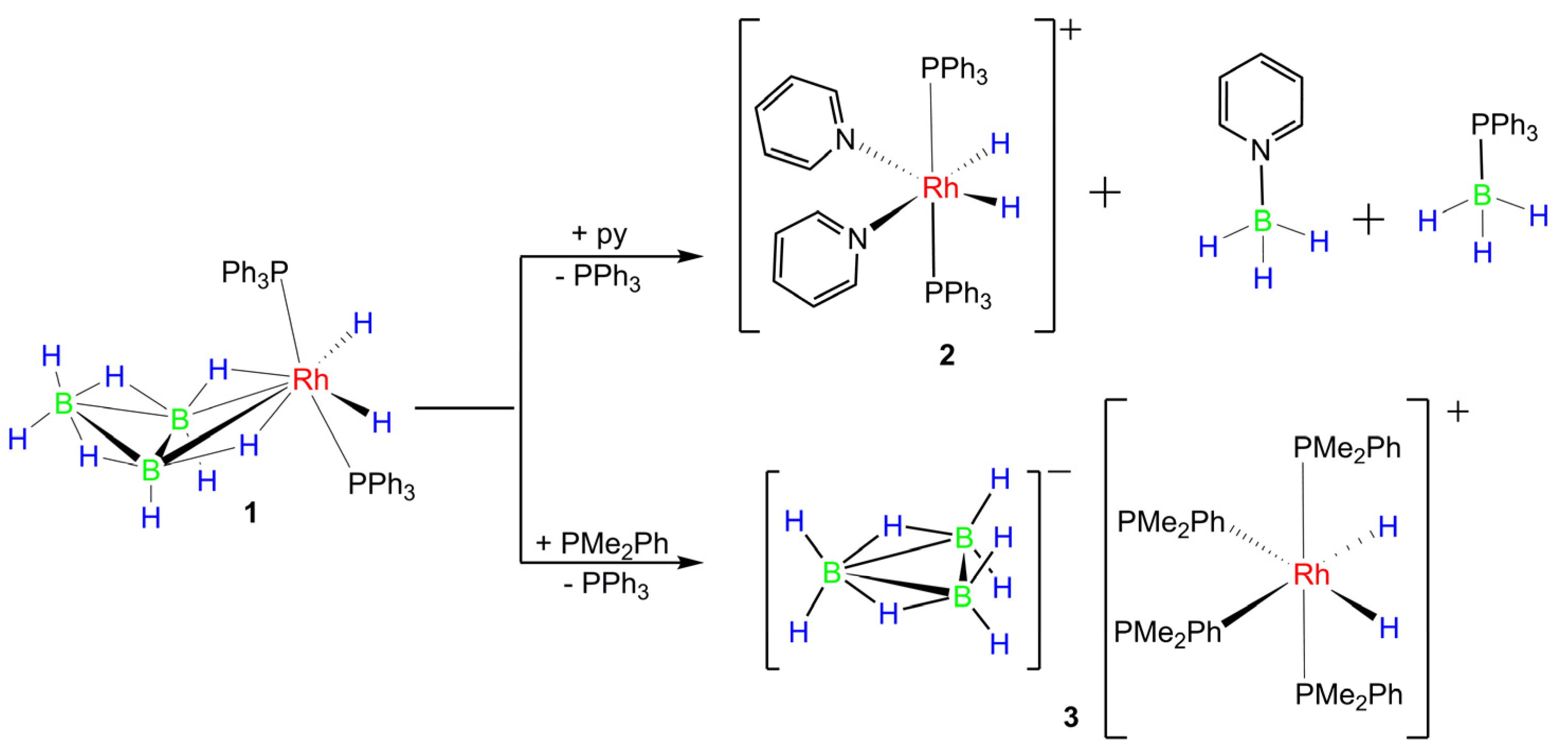
2.5. Reactions of 1 with Lewis Bases

We conducted preliminary and exploratory studies on the reactivity of arachno-2-rhodatetraborane (1) with pyridine (py) and dimethylphenylphosphine (PMe2Ph). The reactions were performed on a small scale in NMR tubes, and the results presented and discussed in this section should be considered as initial findings.
The 31P-{1H} NMR spectrum of the reaction mixture, obtained by adding pyridine to a CD2Cl2 solution of 1 in a 5 mm NMR tube at 233 K, reveals a doublet at δ(31P) +47.2 ppm, along with the resonances of the starting rhodatetraborane (Figure S8). In the 1H-{31P} NMR spectrum at 223 K, two new signals appear at δ(1H) −16.89 and −17.96, with a 1:1 relative intensity ratio, exhibiting the patterns of a pseudo-triplet and a doublet of doublets (dd), respectively (Figure S9). In the 1H-{11B} NMR spectrum, the apparent triplet transforms into an apparent quintet, and the dd becomes a triplet of doublets (Figure S10). The two-dimensional 1H–31P-HMBC spectrum reveals clear cross peaks between the 31P doublet and the two hydride signals, and the 1H-1H correlation further confirms the coupling between both hydrides (Figures S11 and S12).
In the 11B NMR spectrum, a broad peak of low intensity is observed at δ(11B) +19.3 ppm, accompanied by smaller intensity peaks between +2 and –5 ppm. The highest intensity signals correspond to a quartet at δ(11B) −12.1 p.p.m. and a multiplet at −37.8 ppm. The latter signal transforms into a doublet under 1H decoupling (Figure S13). The main 11B resonances can be confidently assigned to the pyridine and phosphine adducts, py-BH3 and PPh3-BH3. However, the assignment of the lower intensity triplet at −9.4 ppm (close to the quartet of the pyridine borane) remains uncertain.
Based on the observed NMR data, it is proposed that the reaction of compound 1 with pyridine results in the formation of borane adducts and a new cationic rhodium(III) complex, [Rh(H)2(PPh3)2(py)2]+ (compound 2), which exhibits an octahedral structure (Scheme 3). However, the formation of anionic species, in particular the borate anions, is not clearly observed in the NMR spectra. The low-intensity signals observed in the 11B NMR spectrum, some of which do not show 1J(1H-11B) coupling and others that appear as triplets, could potentially correspond to borate anions. Further characterization is required to determine the exact nature of the anionic species formed in the reaction with pyridine.
Upon addition of PMe2Ph to a CD2Cl2 solution of compound 1, the 31P-{1H} NMR spectrum shows the appearance of two new doublets of triplets at δ(31P) +0.3 and −10.6 ppm. The spectrum at 233 K also reveals signals corresponding to free PMe2Ph and PPh3 at δ(31P) −45.6 and −7.3 ppm, respectively (Figure S15). In the 1H-{11B} spectrum, a new hydride resonance is observed at δ(1H) −10.15 ppm, exhibiting the pattern of a doublet of pseudo-quartets. This hydride resonance appears as a simple doublet in the 1H–{31P} spectrum (Figures S16 and S17). These observations strongly suggest the formation of the octahedral rhodium(III) cationic complex [Rh(H)2(PMe2Ph)4]+, in which the hydride ligands occupy cis positions to each other (Scheme 3).
The 11B NMR spectrum exhibits a septet at δ(11B) −30.5 ppm, which can be assigned to the free octadecahydridoborate anion, B3H8. There is also a multiplet at −37.7 ppm, which becomes a doublet upon 1H decoupling, corresponding to PhMe2P–BH3. Additionally, the spectrum shows signals of low intensity at −40.6, −44.7 and −45.4 ppm, as well as a broad peak of higher intensity at 16.3 ppm (Figure S18). These signals may correspond to uncharacterized metallaborane species present at low concentrations.
Overall, the data described for the reaction between 1 and PMe2Ph strongly suggest the formation of the salt [Rh(H)2(PMe2Ph)4][B3H8] (3).

3. Conclusions

The reaction in ethanol of Cs[B3H8] with the Wilkinson’s catalyst provides a convenient method for the preparation of the arachno-2-rhodatetraborane, 1. This reaction involves the oxidative addition of two hydrogen atoms to the rhodium(I) center to form a {Rh(III)(H)2(PPh3)2}+ cationic fragment that binds the [B3H8] anionic ligand. The origin of the two additional hydrogen atoms is unclear and we can envision that some of the octahydridotriborate anion could donate them. Alternatively, the presence of ethanol could potentially act as a hydrogen transfer agent, facilitating the addition of hydrogen atoms to the rhodium center.
In the crystal structure, the sextuple phenyl embrace is an important driving force leading to the formation of ribbons in the lattice.
The fluxional behavior observed in compound 1 is similar to the non-rigid behavior found in other arachno-2-metallatetraboranes; based on the literature, we have proposed a probable mechanism of H atom exchange that involves the endo-H5 hydrogen atom, the exo-H1 and exo-H3 as well as the B1-H1,4-B4 and B3-H3,4-B4 bridging hydrogen atoms. It has been found that 1 is thermally unstable, decomposing at temperatures between +67 and +97 °C; this behavior suggests that the rhodatetraborane may exhibit a rich reaction chemistry versus different reagents.
We have explored this hypothesis in reactions of 1 with the Lewis bases, dimethylphenylphosphine, PMe2Ph and pyridine. In these reactions, we have found that the {η2-B3H8} anionic ligand is labile, and it is cleaved by PMe2Ph to form the salt [Rh(H)2(PMe2Ph)4][B3H8] (3). Alternatively, the reaction with pyridine demonstrates that dismantling of the η2-B3H8 ligand can also lead to the formation of pyridine and triphenylphosphine adducts, L–BH3, and to cationic complexes such as [Rh(H)2(PPh3)2(py)2]+ (2).
The observed fluxional behavior and thermal instability highlight the versatility and potential reactivity of the rhodatetraborane compound 1, making it an interesting candidate for further exploration in various chemical reactions and applications.

4. Materials and Methods

4.1. General

Reactions were carried out under an argon atmosphere using standard Schlenk line techniques. Solvents were obtained from a Solvent Purification System from Innovative Technology Inc. NaB₃H₈·3(C4H8O2) was purchased from Katchem spol. s r. o., and used as received. The deuterated solvent CD2Cl2 was deaerated, following freeze–pump–thaw methods, and dried over 3 Å molecular sieves.
Infrared spectra were recorded on a Perkin-Elmer 100 spectrometer, using a Universal ATR Sampling Accessory. Solution NMR spectra were recorded on Bruker Avance AV 300-MHz, AV 400-MHz and AV 500-MHz spectrometers, using 11B, 11B-{1H}, 1H, 1H-{11B}, 1H-{11B(selective)}, 1H-31P-HMBC and 1H-1H-COSY techniques. The 1H NMR chemical shifts were measured relative to the partially deuterated solvent peaks but are reported in ppm relative to tetramethylsilane. 11B chemical shifts are quoted relative to [BF3·OEt2].

4.2. Crystal Structure Determination

X-ray diffraction data were collected on an APEX DUO Bruker diffractometer, using graphite-monochromated Mo Kα radiation (λ = 0.71073 Å). Diffracted intensities were integrated [38] and corrected for absorption effects using the multi-scan method [39,40]. Both programs are included in the APEX4 package. All the structures were solved by direct methods with SHELXS [41] and refined by full-matrix least squares on F2 with SHELXL [42]. Hydrogen atoms were located from difference Fourier maps and refined isotropically.
Single crystals of 1 suitable for X-ray analysis were grown in a 5 mm NMR tube in a fridge at 4 °C by slow diffusion of hexane into a CH2Cl2 solution of the salt.
Structural data for [Rh(η2-B3H8)(H)2(PPh3)2]·2CH2Cl2 (1·2CH2Cl2, 100 K): Mr = 839.81, colorless prism, triclinic P−1, a = 12.3834(12) Å, b = 12.9413(12) Å, c = 13.9452(13) Å, α = 76.211(2)°, β = 85.014(2)°, γ = 66.7050(10)°, V = 1993.4(3) Å3, Z = 2, T = 100(2) K, Dcalcd = 1.399 g cm−3, µ = 0.803 mm−1, absorption correction factors min. 0.824 max. 0.924. 32,874 reflections, 8938 unique (Rint = 0.0659), 6581 observed, R1 = 0.0558 [I > 2σ(I)], wR2(F2) = 0.1562 (all data), GOF = 1.060. CCDC 2281251.

4.3. Mass Spectrometry

The mass spectrum for compound 1 was measured on a Thermo-Finnigan LCQ-Fleet Ion Trap instrument using electrospray ionization (ESI) with samples dissolved in acetonitrile (approximately 100 ng mL−1) and introduced to the ion source by infusion at a rate of 6 μL min−1: source voltage 3.2 kV, tube lens voltage −90.7 V, capillary voltage −32.0 V, capillary temperature 360 °C, drying gas flow 7 L min−1.

4.4. Computational Details

The calculations were performed using the Gaussian 09 package [43]. The structure of the model molecule, [Rh(B3H8)(H)2(PH3)2], was initially optimized using standard methods with the B3LYP/6-31+G(d) methodology and basis sets. The final optimization, including frequency analyses to confirm the true minima, together with GIAO nuclear-shielding calculations, was performed using B3LYP methodology with the 6-31++G(d) basis-set. GIAO nuclear shielding calculations were performed on the final optimized geometry, and computed 11B shielding values were related to chemical shifts by comparison with the computed value for B2H6, which was taken to be δ(11B) +16.6 ppm relative to the BF3(OEt2) = 0.0 ppm standard.

4.5. Preparation of [Rh(η2-B3H8)(H)2(PPh3)2] (1)

Method A: White powdery CsB₃H₈ [44] (0.0808 g, 0.470 mmol) was added to 10 mL of ethanol in a Schlenk tube (the ethanol was previously degassed with argon for five minutes). The tube was slightly heated to facilitate the formation of a solution, upon which [RhCl(PPh₃)₃] (0.4311 g; 0.470 mmol) was added to form a red-orange suspension. The reaction mixture was stirred at room temperature for three hours to give a yellow solid in suspension. The product was filtered through a frit, in air, to yield a yellow-mustard solid and an orange filtrate. The solid was collected in a Schlenk tube, dissolved in dichloromethane and filtered, under argon, through a silica gel layer. The resulting yellow solid was crystallized from CH2Cl2/Hexane (1:2). This final product was studied using NMR spectroscopy, demonstrating that its composition corresponded to the hydridrorhodatetraborane 1. The total yield after drying under vacuum for several hours was 0.26 mg (0.387 mmol, 82.34%).
1H-{11B} (500 MHz, CD2Cl2, 223 K): δ + 7.66 to +7.32 ppm (m, aromatic signals, C6H5, 30H). IR (ATR): νmax/cm−1 3054–2962 (w, C-H), 2508, 2452, 2378 (s, BH), 2451 (s, BH), 2418 (s, BH), 1585, 1568, 1480 (C=C aromatics). HR-MS(ESI): m/z calcd exact mass for C36H40B3P2Rh, [M]+, 670.1939; this anticipated parent ion is clearly absent. Instead, the spectrum exhibits high intensity peaks at 627.0867, 628.0884 and 629.0901 u, with an isotopic pattern that matches well that calculated for the ion [C36H30P2Rh]+, [M − (B3H8 + H2)]+. This ion corresponds to the {Rh(PPh3)2} fragment, demonstrating that the rhodatetraborane 1 undergoes facile cleavage upon ionization (Figure S19).
Method B: White powdery NaB₃H₈·3(C4H8O2) (0.1261 g, 0.385 mmol) was dissolved in 10 mL of dry ether, in a Schlenk tube, which was immersed in an isopropanol bath at −30 °C. Subsequently, the Wilkinson’s catalyst was added (0.3559 g, 0.385 mmol), under a flow of argon, to the sodium octahydridotriborate dioxane solution. The resulting brick-red suspension was stirred at room temperature under an atmosphere of argon. After one hour of stirring the temperature was increased to +5 °C and the color of the suspension became orange-red. The reaction was maintained for another three hours to give a brown-yellow suspension, immersed in the isopropanol bath at +10 °C. We allowed the solid to settle down and decanted the supernatant with a pipette, under a flow of argon. The decanted liquid was dried under vacuum to give an orange-yellow solid, whereas the sediment formed a beige solid, after drying. The NMR spectra of the former fraction (the decanted liquid) showed the presence of O=PPh3 and Ph3P–BH3, as major and minor components, respectively. The ether-insoluble product corresponded to the four-vertex rhodatetraborane. This method afforded 20 mg of 1 (8%).

4.6. Reactions of [Rh(η2-B3H8)(H)2(PPh3)2] (1) with Lewis Bases

  • Reaction of [Rh(η2-B3H8)(H)2(PPh3) 2] (1) with py. 10.2 mg of 1 (1.50 × 10² mmol) treated with 1.20 μg of pyridine (1.50 × 10² mmol), in a Schlenk tube immersed in a bath of isopropanol at −30 °C. The resulting yellow solution was stirred, under an atmosphere of argon for 5 h; during this time, the temperature was raised to +10 °C. The reaction was stirred for another 20 min at room temperature. The solvent was evaporated under vacuum to give an orange solid, which was dissolved in deuterated dichloromethane and studied using NMR spectroscopy. 31P-{1H} (162 MHz, 233 K): δ +47.2 ppm [1J(31P-103Rh) = 118 Hz], together with the signal of O=PPh3. 1H-{31P} (400 MHz, 233 K): δ +8.59 (d, J = 5.0 Hz, ortho-NC5H5, 2H), +8.59 (d, J = 5.0 Hz, o-NC5H5, 2H), +8.34 (br. s, o-NC5H5, 2H), +7.95 (t., p-NC5H5, 2H), +7.95 (t., p-NC5H5, 2H), +6.54 (br. s, m-NC5H5, 2H), between +7.73 and +6.80 (m, C6H5-rings and NC5H5), −16.89 (t, 1J(103Rh-1H2,6) + 2J(1H2-1H2,3) = 2J(1H6-1H1,2) = 12.9 Hz, 2H) and -17.96 (dd, 1J(103Rh-1H2,6) = 23.4, 2J(1H2-1H2,3) = 2J(1H6-1H1,2) = 10.9 Hz, 2H). 1H-{11B} (400 MHz, 233 K): δ −16.89 (app. quintet, 1J(103Rh-1H2,6) + 2J(31P-31P) + 2J(1H2-1H2,3) = 12.9 Hz, 2H) and −17.96 (td, J = 25.5, 13.1 Hz, Rh-H2). δ 11B (400 MHz, 298 K): δ +18.8 (br.), +1.47 (s), −1.5 (t), −9.31 (t, 95 Hz), −12.0 (q, 1J(11B-1H) = 98 Hz, py-BH3), −37.8 (dq, 62 Hz, Ph3PBH3), −27.4 (t, 98 Hz).
  • In situ characterization of [Rh(H)2(PMe2Ph)4][B3H8] (3). 12.6 mg of 1 (1.88 × 10⁻² mmol), dissolved in CD2Cl2, in a NMR tube, which was immersed in an isopropanol bath at −30 °C, and 2.6 mg (2.7 μL) of PMe2Ph (1.88 × 10² mmol) was added into the NMR tube, under a flow of argon. The reaction was studied using NMR spectroscopy, starting at 233 K and then heating the sample to room temperature. 31P-{1H} (162 MHz, 233 K): δ +26.7 ppm [s, O=PPh3], +19.7 (very br., PhMe2P-BH3), +0.3 [dt, 1J(103Rh–31P) = 97 Hz, 2J(31P1–31P2) = 24 Hz], −7.2 (s, PPh3), −10.6 p.pm [dt, 1J(103Rh–31P) = 86 Hz, 2J(31P1–31P2) = 24 Hz], together with the signals of 1 (Table 1). 1H-{31P} (400 MHz, 233 K): δ +7.84 to 6.94 (m, aromatics, C6H5), +1.56 (s, CH3), +1.46 (s, CH3), −10.25 (d, 1J(103Rh-1H) = 13.8 Hz. 1H-{11B} (400 MHz, 233 K): +1.2 (d, 2J(31P–1H), PhMe2-BH3), +0.23(s, B3H8), −10.25 (d of pseudo-quintets, second order, 2J(11P-1Htrans) = 147.9 Hz, 1J(103Rh-1H) = 14.0 Hz, 2J(11P-1Hcis) = 17.2 Hz, 2H) and −17.96 (dd, 1J(103Rh-1H2,6) = 23.4, 2J(1H2-1H2,3) = 2J(1H6-1H1,2) = 10.9 Hz, 2H) ppm. 11B (400 MHz, 298 K): δ −16.4 (br. s), −30.5 (sept, B3H8), −37.7 (quartet of d, 1J(11B-31P) = 59 Hz, 1J(11B-1H) = 102 Hz, PhMe2P–BH3), −45.2 (br. s).

Supplementary Materials

The following supporting information can be downloaded at https://www.mdpi.com/article/10.3390/molecules28186462/s1, Figures S1–S19: 11B, 1H, 31P, 1H-31P-HMBC, 1H-1H-COSY NMR spectra for 1, and for the reaction mixtures with pyridine and dimethylphenylphosphine; Table S1: calculated Cartesian coordinates for PH3-ligated model compound [Rh(η2-B3H8(H)2(PH3)2].

Author Contributions

F.D.-N. synthesis, spectroscopic characterization, single crystal growth, preliminary analysis and preparation of the experimental data. P.J.S.M. and R.M. conceptualization, formal analysis and supervision. R.R. project administration and funding acquisition. Writing—original draft preparation, review and editing. R.M. Visualization, proofreading. Additional conceptualization and discussion of the work, P.J.S.M. and R.R. All authors have read and agreed to the published version of the manuscript.

Funding

PID2021-122406NB-I00 funded by MCIN/AEI/10.13039/501100011033 and by “ERDF A way of making Europe” and Grupo de Referencia: Catálisis Homogénea Enantioselectiva, ChiralCat E05-23R, funded by Gobierno de Aragón.

Informed Consent Statement

Not applicable.

Data Availability Statement

Not applicable.

Acknowledgments

We want to acknowledge the use of Servicio General de Apoyo a la Investigación-SAI, University of Zaragoza. To an outstanding and creative scientist, John D. Kennedy, Emeritus Professor at University of Leeds, on his 80th birthday. He has been a superb mentor to a good number of scientists. We worked for long hours in “the 400 MHz”, we waited for the new results and we discussed exciting experiments at “The Pack Horse”. Those days have “dyed our souls” and will never leave us. Thank you hombre roto de Leeds.

Conflicts of Interest

The authors declare no conflict of interest.

Sample Availability

Not applicable.

References

  1. Barton, L.; Strivastava, D.K. Comprehensive Organometallic Chemistry II; Abel, E.W., Stone, F.G.A., Wilkinson, G., Eds.; Pergamon: New York, NY, USA, 1995; Volume 1, pp. 275–372. [Google Scholar]
  2. Cotton, F.A.; Wilkinson, G.; Murillo, C.A.; Bochmann, M. Advanced Inorganic Chemistry, 6th ed.; Wiley: New York, NY, USA, 1999. [Google Scholar]
  3. Kennedy, J.D. The Polyhedral Metalloboranes. 1. Metalloborane Clusters with 7 Vertices And Fewer. In Progress in Inorganic Chemistry; John Wiley & Sons: New York, NY, USA, 1984; Volume 32, pp. 519–679. [Google Scholar]
  4. Weller, A.S. d- and f-Block Metallaboranes. In Comprehensive Organometallic Chemistry III; Crabtree, R.H., Mingos, D.M.P., Eds.; Elsevier: Oxford, UK, 2007; Volume 3, pp. 133–174. [Google Scholar]
  5. Bould, J.; Greenwood, N.N.; Kennedy, J.D. The first osmaboranes and a new iridatetraborane. J. Organomet. Chem. 1983, 249, 11–21. [Google Scholar] [CrossRef]
  6. Grebenik, P.D.; Leach, J.B.; Green, M.L.H.; Walker, N.M. Transition metal mediated homologation of BH3·THF: Synthesis and crystal structure of [WH3(PMe3)3B3H8]. J. Organomet. Chem. 1988, 345, C31–C34. [Google Scholar] [CrossRef]
  7. Grebenik, P.D.; Leach, J.B.; Pounds, J.M.; Green, M.L.H.; Mountford, P. Niobium metallaboranes: A novel metallaborane analogue of pentaborane(11). J. Organomet. Chem. 1990, 382, C1–C5. [Google Scholar] [CrossRef]
  8. Ghosh, S.; Beatty, A.M.; Fehlner, T.P. The Reaction of Cp*ReH6, Cp* = C5Me5, with Monoborane to Yield a Novel Rhenaborane. Synthesis and Characterization of arachno-Cp*ReH3B3H8. Collect. Czech. Chem. Commun. 2002, 67, 808–812. [Google Scholar] [CrossRef]
  9. Fehlner, T.P. Systematic Metallaborane Chemistry. Organometallics 2000, 19, 2643–2651. [Google Scholar] [CrossRef]
  10. Geetharani, K.; Kumar Bose, S.; Pramanik, G.; Kumar Saha, T.; Ramkumar, V.; Ghosh, S. An Efficient Route to Group 6 and 8 Metallaborane Compounds: Synthesis of arachno-[Cp*Fe(CO)B3H8] and closo-[(Cp*M)2B5H9] (M = Mo, W). Eur. J. Inorg. Chem. 2009, 2009, 1483–1487. [Google Scholar] [CrossRef]
  11. Green, M.L.H.; Leach, J.B.; Kelland, M.A. Synthesis and Interconversion of Some Small Ruthenaboranes: Reaction of a Ruthenium Borohydride with Pentaborane(9) to Form Larger Ruthenaboranes. Organometallics 2007, 26, 4031–4037. [Google Scholar] [CrossRef]
  12. Guggenberger, L.J. Crystal structure of the tetramethylammonium salt of the octahydrotriborotetracarbonylchromium anion, (CO)4CrB3H8. Inorg. Chem. 1970, 9, 367–373. [Google Scholar] [CrossRef]
  13. Beckett, M.A.; Brassington, D.S.; Coles, S.J.; Gelbrich, T.; Hursthouse, M.B. Synthesis and characterisation of a series of Group 7 metal 2,2,2,2-dicarbonylbis(triorganophosphine)-arachno-2-metallatetraboranes, [M(CO)2L2(B3H8)] (M=Re, Mn); crystal and molecular structures of [Re(CO)2(dppf)(B3H8)] and [Mn(CO)2(dppe)(B3H8)]. Polyhedron 2003, 22, 1627–1632. [Google Scholar] [CrossRef]
  14. Burns, I.D.; Hill, A.F.; Williams, D.J. Ruthenatetraboranes:  Molecular Structure of [Ru(B3H8)(PPh3){κ3-HB(pz)3}] (pz = Pyrazol-1-yl). Inorg. Chem. 1996, 35, 2685–2687. [Google Scholar] [CrossRef]
  15. Bown, M.; Ingham, S.L.; Norris, G.E.; Waters, J.M. exo-2-([eta]6-Hexamethylbenzene)-endo-2-chloro-2-ruthena-arachno-tetraborane(8). Acta Crystallogr. Sect. C 1995, 51, 1503–1505. [Google Scholar] [CrossRef]
  16. Bould, J.; Rath, N.P.; Barton, L. [(CO)H(PPh3)2-arachno-OsB3H8]. Acta Crystallogr. Sect. C 1996, 52, 1388–1390. [Google Scholar] [CrossRef]
  17. Lippard, S.J.; Melmed, K.M. Transition metal borohydride complexes. III. Structure of octahydrotriboratobis(triphenylphosphine)copper(I). Inorg. Chem. 1969, 8, 2755–2762. [Google Scholar] [CrossRef]
  18. Williams, R.E. Coordination number pattern recognition theory of carborane structures. Adv. Inorg. Chem. Radiochem. 1976, 18, 67–142. [Google Scholar]
  19. Olah, G.A.; Wade, K.; Williams, R.E. Electron Deficient Boron and Carbon Clusters; John Wiley & Sons: New York, NY, USA, 1991. [Google Scholar]
  20. Wade, K. Structural and bonding patterns in cluster chemistry. Adv. Inorg. Chem. Radiochem. 1976, 18, 1–66. [Google Scholar]
  21. Dance, I.; Scudder, M. The sextuple phenyl embrace, a ubiquitous concerted supramolecular motif. J. Chem. Soc. Chem. Commun. 1995, 10, 1039–1040. [Google Scholar] [CrossRef]
  22. Dance, I.; Scudder, M. Molecules embracing in crystals. CrystEngComm 2009, 11, 2233–2247. [Google Scholar] [CrossRef]
  23. Hore, P.J. Nuclear Magnetic Resonance; Oxford University Press: Oxford, UK, 2011. [Google Scholar]
  24. Dhubhghaill, O.N.; Spalding, T.R.; Ferguson, G.; Kaitner, B.; Fontaine, X.L.R.; Kennedy, J.D.; Reed, D. Metallaheteroborane Chemistry. 4. The Synthesis of closo-[2,2-(PPh3)2-2-H-1,2-XMB10H10] (X = Se Or Te, M = Rh Or Ir) Compounds, their Characterization by Nuclear Magnetic-Resonance Techniques, and the crystal and molecular structure of the X = Te, M = Rh complex. J. Chem. Soc. Dalton Trans. 1988, 11, 2739–2745. [Google Scholar]
  25. Ferguson, G.; Kennedy, J.D.; Fontaine, X.L.R.; Faridoon; Spalding,, T.R. Metallaheteroborane chemistry.3. Synthesis of [2,2-(PEt3)2-1,2-TePtB10H10], [2,2-(PBun3)2-1,2-TePTtB10H10], [2,2-(PMe2Ph)2-1,2-TePtB10H10], their characterization by nuclear magnetic-resonance spectroscopy, and the crystal and molecular-structure of [2,2-(PEt3)2-1,2-TePtB10H10]. J. Chem. Soc. Dalton Trans. 1988, 10, 2555–2564. [Google Scholar]
  26. O’Connell, D.; Patterson, J.C.; Spalding, T.R.; Ferguson, G.; Gallagher, J.F.; Li, Y.; Kennedy, J.D.; Macías, R.; Thornton-Pett, M.; Holub, J. Conformational polymorphism and fluxional behaviour of M(PR3)2 units in closo-twelve-atom metallaheteroboranes with MX2B9 (X = C or As) and MZB10 cages (Z = S, Se or Te). J. Chem. Soc. Dalton Trans. 1996, 15, 3323–3333. [Google Scholar] [CrossRef]
  27. McGrath, M.; Spalding, T.R.; Fontaine, X.L.R.; Kennedy, J.D.; Thornton-Pett, M.J. Metallaheteroborane chemistry. Part 9. Syntheses and spectroscopy of platinum and palladium phosphine complexes containing η5-{As2B9}-based cluster ligands. Crystal structures of [3,3-L2-closo-3,1,2-PtAs2B9H9] (L = PPh3 or PMe2Ph) and [3-Cl-3,8-(PPh3)2-closo-3,1,2-PdAs2B9H8]. J. Chem. Soc. Dalton Trans. 1991, 3223–3233. [Google Scholar]
  28. Fontaine, X.L.R.; Kennedy, J.D.; McGrath, M.; Spalding, T.R. Metallheteroborane Chemistry. 8. NMR study of some arsena- and stibaboranes and of the rhodadiarsenaboranes [3,3-(PPh3)2-3-(H)-closo-3,1,2-RhAs2B9H9] and [3-(η5-C5Me5)-closo-3,1,2-RhAs2B9H9]. Magn. Reson. Chem. 1991, 29, 711–720. [Google Scholar] [CrossRef]
  29. Fontaine, X.L.R.; Greenwood, N.N.; Kennedy, J.D.; Nestor, K.; Thorntonpett, M.; Hermanek, S.; Jelinek, T.; Stibr, B. Polyhedral Metallacarbaborane Chemistry—Preparation, Molecular-Structure, And Nuclear Magnetic-Resonance Investigation of [3-(η5-C5Me5)-closo-3,1,2-M2B9H11] (M = Rh Or Ir). J. Chem. Soc. Dalton Trans. 1990, 2, 681–689. [Google Scholar] [CrossRef]
  30. Ferguson, G.; Gallagher, J.F.; McGrath, M.; Sheehan, J.P.; Spalding, T.R.; Kennedy, J.D. Metallaheteroborane chemistry. Part 11. Selective syntheses of the palladium heteroborane complexes [2,2-(PR3)2-closo-2,1-PdEB10H10] (R3= Ph3, MePh2 or Me2Ph; E = Se or Te) and [2-X-2-(PPh3)-closo-2,1-PdTeB10H9(PPh3)] (X = Cl, Br, I, CN, SCN or O2CMe). J. Chem. Soc. Dalton Trans. 1993, 27–34. [Google Scholar] [CrossRef]
  31. Shanan-Atidi, H.; Bar-Eli, K. A convenient method for obtaining free energies of activation by the coalescence temperature of an unequal doublet. J. Phys. Chem. 1970, 74, 961–963. [Google Scholar] [CrossRef]
  32. Chen, M.W.; Calabrese, J.C.; Gaines, D.F.; Hillenbrand, D.F. Low-temperature crystal and molecular structure of tetracarbonyl[2-bromoheptahydrotriborato(1-)]manganese, (CO)4MnB3H7Br, and a proton NMR study of the kinetics of its intramolecular hydrogen exchange in solution. J. Am. Chem. Soc. 1980, 102, 4928–4933. [Google Scholar] [CrossRef]
  33. Gaines, D.F.; Walsh, J.L.; Morris, J.H.; Hillenbrand, D.F. The chemistry of beryllaboranes. Characterization and reactions of beryllium bis(tetrahydroborate), Be(BH4)2, and beryllium bis(octahydrotriborate), Be(B3H8)2. Inorg. Chem. 1978, 17, 1516–1522. [Google Scholar] [CrossRef]
  34. Bushweller, C.H.; Beall, H.; Dewkett, W.J. Stereodynamics of L2CuB3H8. Rate of triborohydride ion (B3H8) rearrangement as a function of L. Inorg. Chem. 1976, 15, 1739–1740. [Google Scholar] [CrossRef]
  35. Beall, H.; Bushweller, C.H. Dynamical processes in boranes, borane complexes, carboranes, and related compounds. Chem. Rev. 1973, 73, 465–486. [Google Scholar] [CrossRef]
  36. Serrar, C.; Es-sofi, A.; Boutalib, A.; Ouassas, A.; Jarid, A.; Nebot-Gil, I.; Tomás, F. Theoretical Study of the Structural and Fluxional Behavior of Copper(I)-Octahydrotriborate Complex. J. Phys. Chem. A 2001, 105, 9776–9780. [Google Scholar] [CrossRef]
  37. Borlin, J.; Gaines, D.F. Internal exchange in new Group III metalloborane derivatives, dimethylaluminum ((CH3)2AlB3H8), and dimethylgallium triborane(8) (CH3)2GaB3H8). J. Am. Chem. Soc. 1972, 94, 1367–1369. [Google Scholar] [CrossRef]
  38. Bruker-AXS SAINT. Area-Detector Integration Software, version 6.01; Bruker-AXS SAINT: Madison, WI, USA, 2001. [Google Scholar]
  39. Bruker-AXS SADABS. Area Detector Absorption Program; Bruker-AXS SADABS: Madison, WI, USA, 1996. [Google Scholar]
  40. Krause, L.; Herbst-Irmer, R.; Sheldrick, G.M.; Stalke, D. Comparison of silver and molybdenum microfocus X-ray sources for single-crystal structure determination. J. Appl. Crystallogr. 2015, 48, 3–10. [Google Scholar] [CrossRef] [PubMed]
  41. Sheldrick, G.M. A short history of SHELX. Acta Crystallogr. Sect. A Found. Crystallogr. 2008, 64, 112–122. [Google Scholar] [CrossRef] [PubMed]
  42. Sheldrick, G. Crystal structure refinement with SHELXL. Acta Crystallogr. Sect. C 2015, 71, 3–8. [Google Scholar] [CrossRef] [PubMed]
  43. Frisch, M.J.; Trucks, G.W.; Schlegel, H.B.; Scuseria, G.E.; Robb, M.A.; Cheeseman, J.R.; Scalmani, G.; Barone, V.; Mennucci, B.; Petersson, G.A.; et al. Gaussian 09, Revision A.02; Gaussian, Inc.: Wallingford, CT, USA, 2009. [Google Scholar]
  44. Chen, X.-M.; Ma, N.; Liu, X.-R.; Wei, C.; Cui, C.-C.; Cao, B.-L.; Guo, Y.; Wang, L.-S.; Gu, Q.C. Facile Synthesis of Unsolvated Alkali Metal Octahydrotriborate Salts MB3H8 (M = K, Rb, and Cs), Mechanisms of Formation, and the Crystal Structure of KB3H8. Angew. Chem. 2019, 131, 2746–2750. [Google Scholar] [CrossRef]
Scheme 1. Reaction between the Wilkinson’s catalyst and the octahydridotriborate anion to give [Rh(η2-B3H8)(H)2(PPh3)2] (1).
Molecules 28 06462 sch001
Figure 1. ORTEP-type of drawing for [Rh(η2-B3H8)(H)2(PPh3)2] (1), showing the cluster numbering system employed, with 50% thermal ellipsoids for non-hydrogen atoms. The phenyl rings (except for the ipso carbon atoms) are omitted to aid clarity. Selected interatomic distances (Å) and angles (°) with esds in parenthesis: Rh2–P1 2.3103(11), Rh2–P2 2.2911(11), Rh2–H 1.58(2) (average value of the two hydride ligands), Rh2–B1 2.428(5), Rh1–B3 2.420(5), B1–B3 1.764(8), B1–B4 1.790(8), B3–B4 1.805(8), P1–Rh2–P2 158.68(4), B1–Rh2–P1 103.27(13), B1–Rh2–P2 95.72(13), B3–Rh2–P1 106.40(13), B3–Rh2–P2 94.10(13), H–Rh2–P1 81.1(18) (average value of the two hydride ligands), B1–Rh2–B3 42.67(18), B3–B1–Rh2 68.4(2), B1–B3–Rh2 68.9(2), B3–B1–B4 61.0(3), B1–B3–B4 60.2(3), B1–B4–B3 58.8(3). The dihedral angle between planes {B1B3B4} and {B1B3Rh2} is 121.91(3).
Molecules 28 06462 g001
Figure 2. Packing of individual molecules of 1 that form ribbons along the a axis (above); detail of edge-to-face phenyl interactions between the molecules within the ribbons (below).
Molecules 28 06462 g002
Figure 3. Stick representation of the 11B-{1H} NMR spectra of a series of arachno-metallaboranes. The NMR data were measured in different solvents such as toluene-d8, CD2Cl2 and CDCl3.
Molecules 28 06462 g003
Figure 4. Plot of δ(11B) versus δ (1H) for directly bound [BH(terminal)] units in the following arachno-metallatetraboranes: [Nb(η2-B3H8)(η5-C5H5)2], [W(η2-B3H8)(PMe3)3(H)3], [Re(η2-B3H8)(CO)4], [Os(η2-B3H8)(CO)(PPh3)2H], [Ru(η2-B3H8)(CO)(PPh3)2H], [Ir(η2-B3H8)(H)2(PPh3)2] and [Rh(η2-B3H8)(H)2(PPh3)2] (1).
Molecules 28 06462 g004
Figure 5. 1H-{11B} NMR spectra, in CD2Cl2, at different temperatures, which demonstrate an intramolecular fluxional process for compound 1.
Molecules 28 06462 g005
Scheme 2. A proposed mechanism of intramolecular hydrogen exchange in 1.
Molecules 28 06462 sch002
Scheme 3. Reactions of 1 with the ligands py and PMe2Ph.
Molecules 28 06462 sch003
Table 1. 11B, 1H and 31P NMR data for compound [Rh(η2-B3H8)(H)2(PPh3)2] (compound 1), compared to the corresponding DFT/GIAO 11B-nuclear shielding data, calculated for the PH3 model, [Rh(η2-B3H8)(H)2(PH3)2] [in brackets].
(a) Cluster Data:
Assignment 1δ (11B) 2Assignment 1δ (1H) 3
B4−1.0 [−3.8]exo-H4+2.54 [+2.74]
B1,3−38.9 [−41.0]endo-H5+1.82 [+2.33]
exo-H1, exo-H3−0.11 [+1.02]
H1,4; H3,4 (B–H–B)−1.11 [−0.65]
H1,2; H3,2 (Rh–H–B)−7.07 [−5.10]
H2, H6 (Rh–H)−11.79 4 [−6.37]
(b) Phosphorous-31 Data:
Assignmentδ (31P) 31J(103Rh–31P)/Hz2J(31P1–31P2)/Hz
P139.9111367
P244.7111
1 Based on the symmetry of 1, 1H–{11B} selective experiments and DFT calculations. 2 CD2Cl2 solution at 298 K. 3 CD2Cl2 solution at 223 K. 4 1H–{11B(off)}: broad, apparent quintet, J = 15.3 Hz; 1H–{31P}: broad, apparent triplet, which corresponds to a second order spectrum, analyzed to give 1J(103Rh-1H1,2) = 1J(103Rh-1H2,3) = 18 Hz, 1J(103Rh-1H2) = 1J(103Rh-1H6) = 16 Hz, 2J(1H1,2-1H2) = 2 Hz, 2J(1H1,2-1H6) = 12 Hz.
Disclaimer/Publisher’s Note: The statements, opinions and data contained in all publications are solely those of the individual author(s) and contributor(s) and not of MDPI and/or the editor(s). MDPI and/or the editor(s) disclaim responsibility for any injury to people or property resulting from any ideas, methods, instructions or products referred to in the content.

