
Synthesis, Characterization, and Anticancer Activity of New N,N′-Diarylthiourea Derivative against Breast Cancer Cells
 , ,
, ,  and
and
Abstract
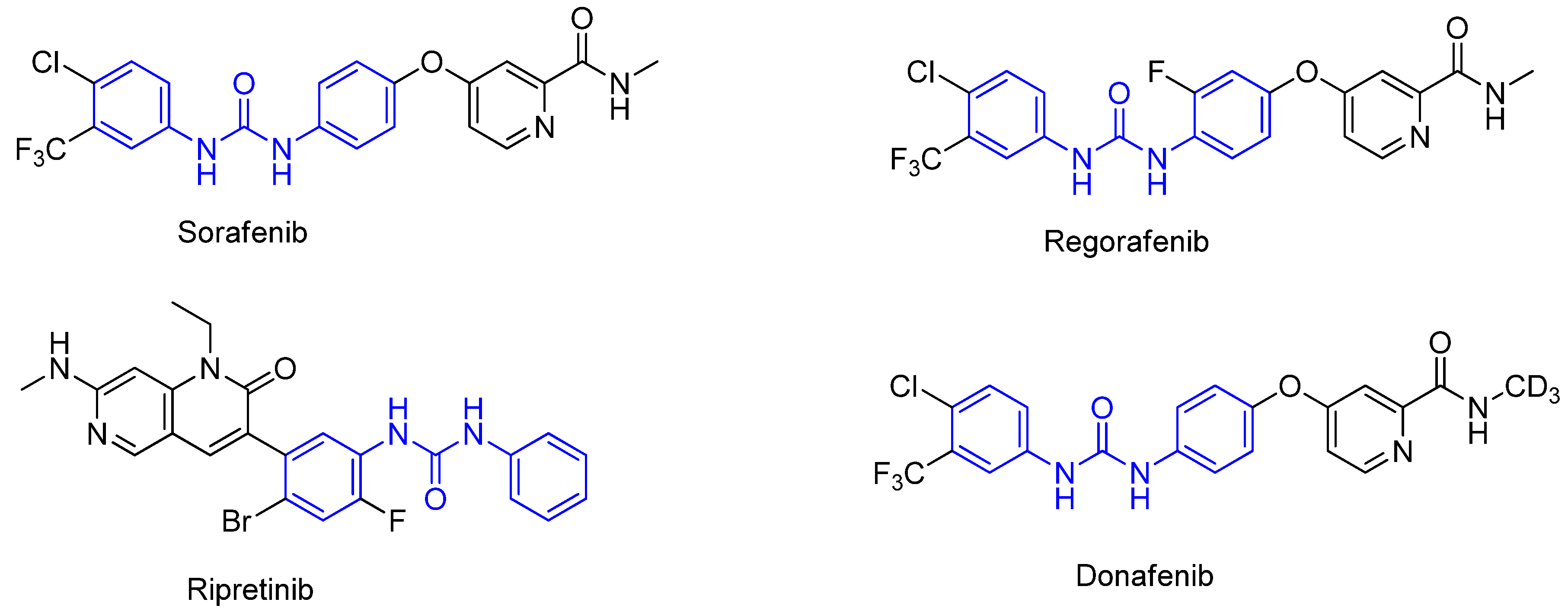
:1. Introduction
2. Results and Discussion
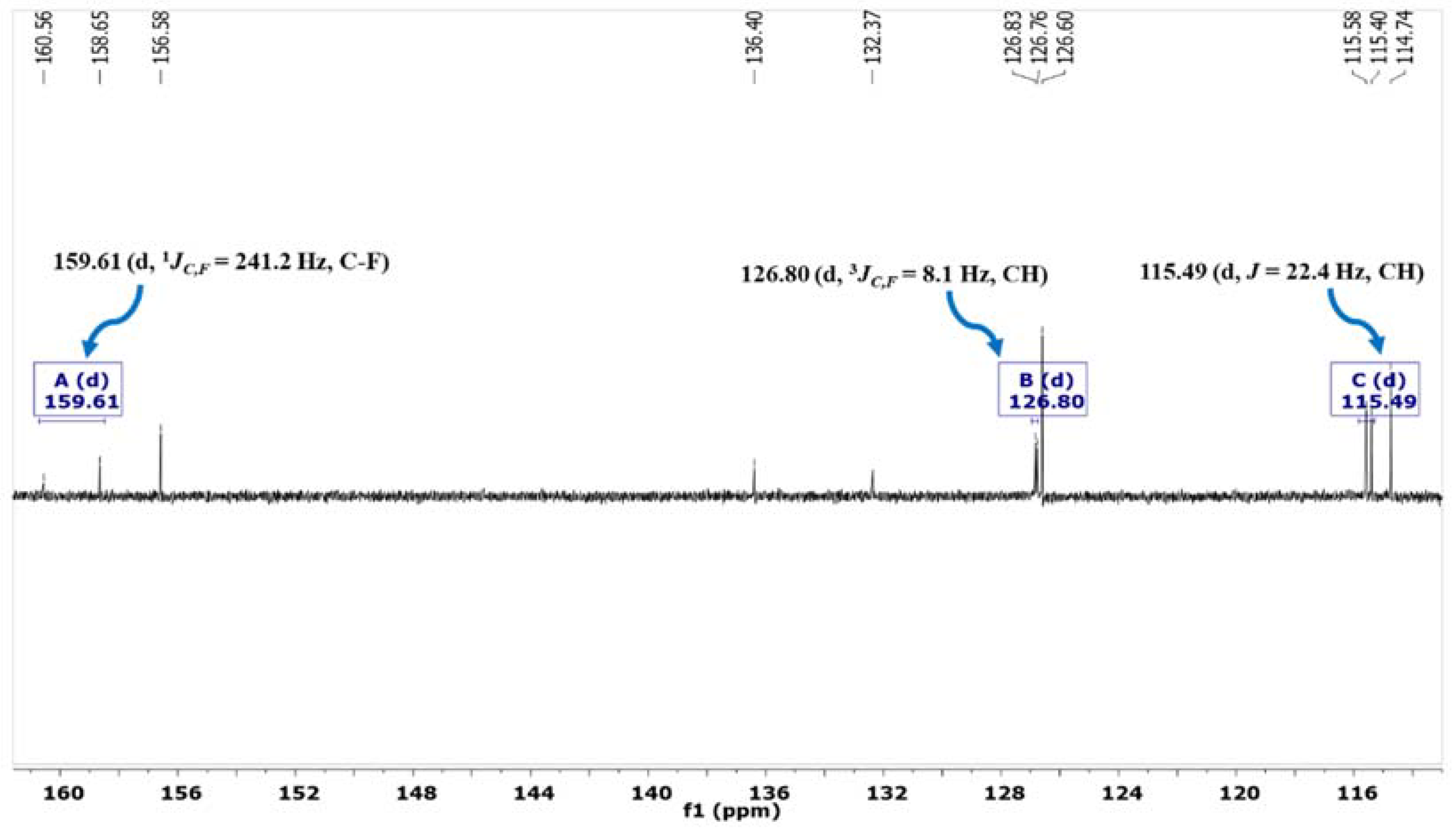
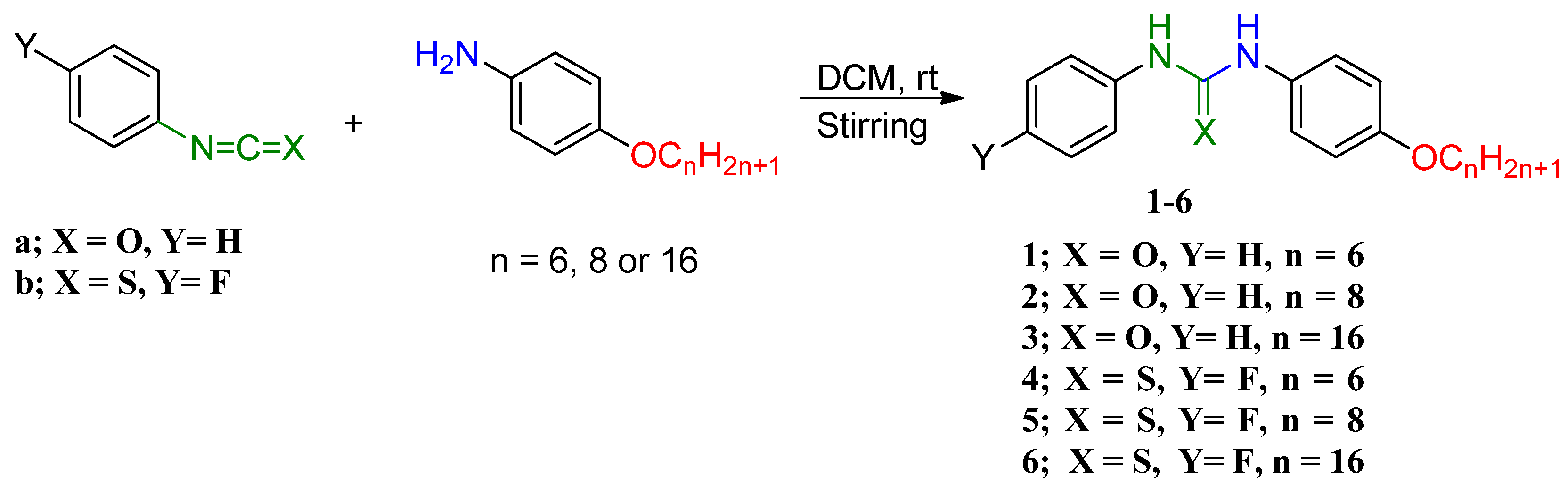
2.1. Chemistry
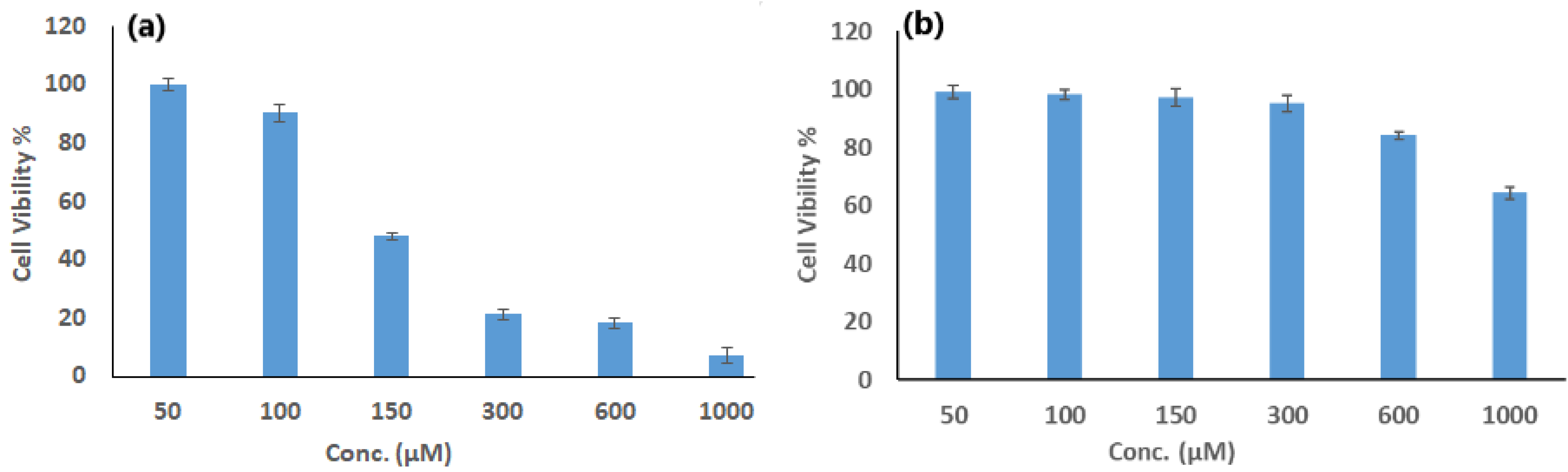
2.2. Cytotoxicity Analyzes
2.3. Estimation of Apoptosis and DNA Damage Generation in MCF-7 Cells
- -
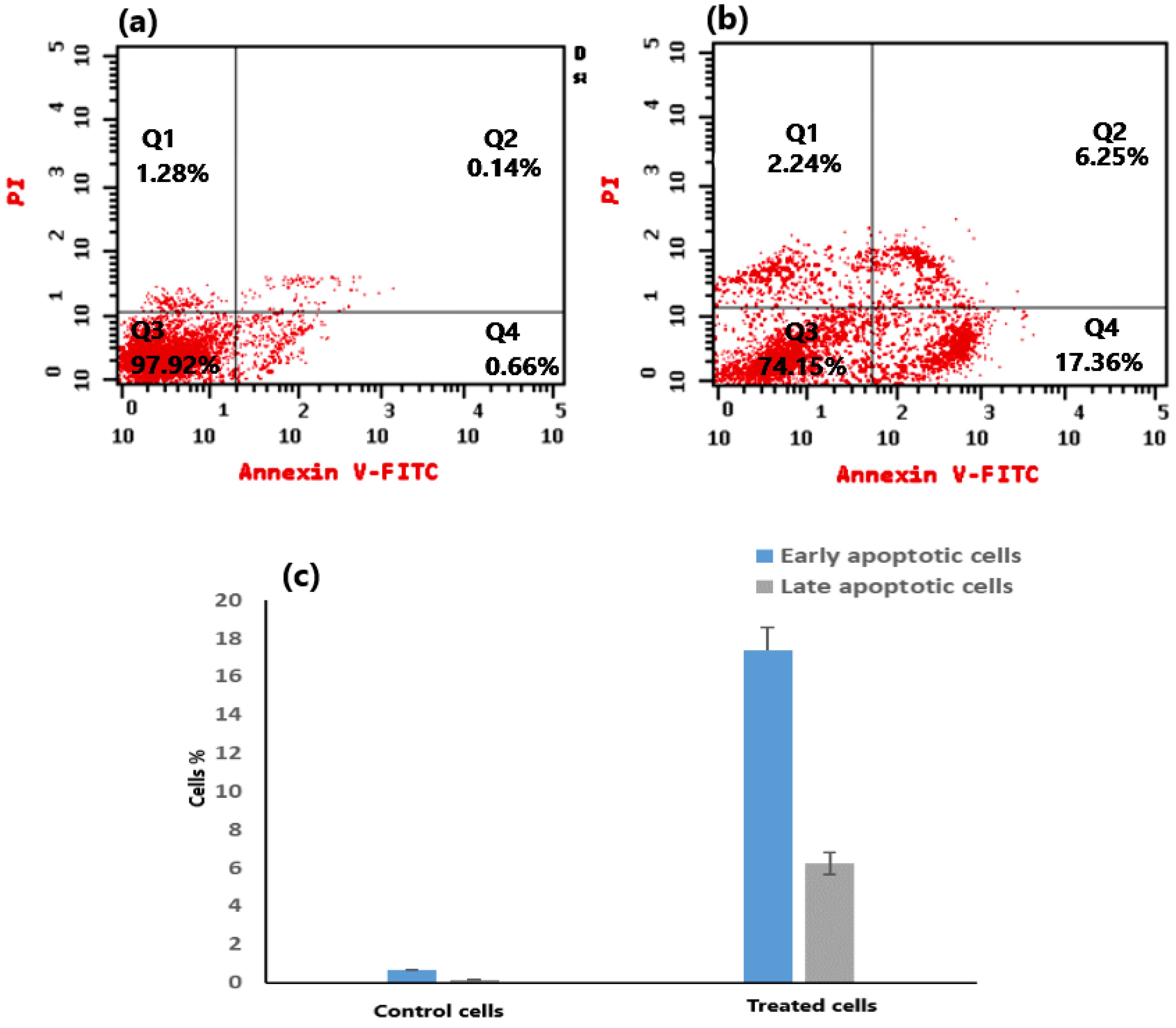
- The ability of compound 4 to cause apoptosis and damage DNA in MCF-7 cells was tested with Annexin V/PI and comet analyses. Figure 5a,b illustrate the outcomes of flow cytometry analysis using annexin V/PI staining for cells treated with compound 4, as compared to untreated cells. The proportion of apoptotic cells (including both early and late stages) was found to be significantly higher in the compound 4-treated MCF-7 cells in comparison to untreated ones. Moreover, the % of both early and late apoptotic cells in compound 4-treated cells in comparison to untreated ones is displayed in Figure 5c.
- -
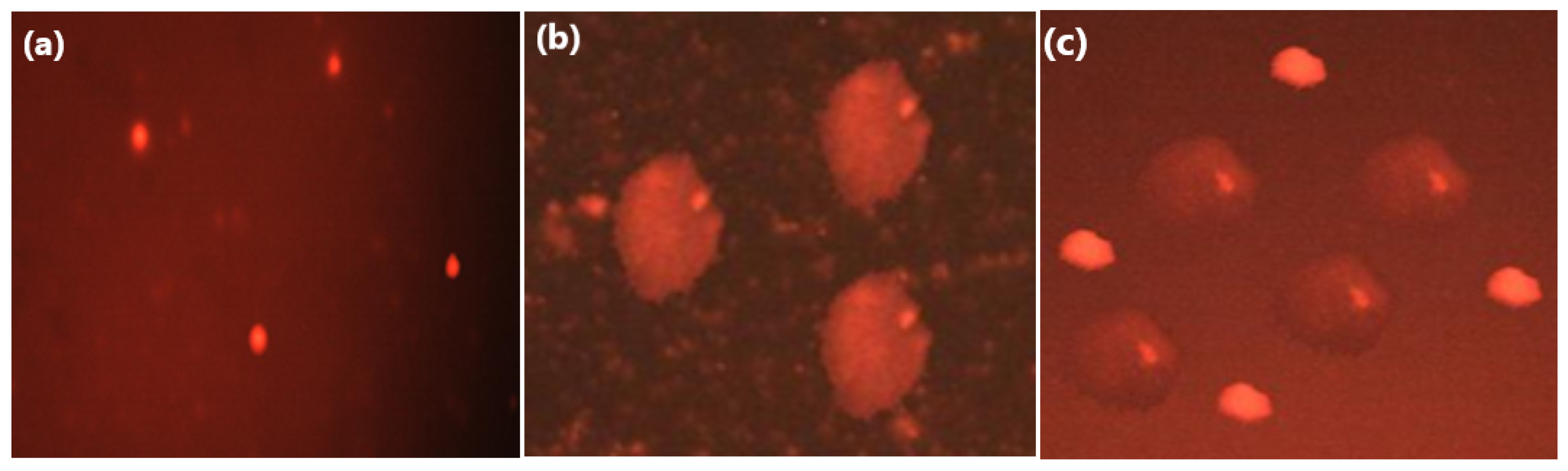
- The comet assay approach is also utilized for assessing the destruction of DNA generated in MCF-7 cells treated with compound 4 that was pretreated with a caspase-3 inhibitor (Z-DEVD-FMK). As shown in Table 1, there was a significant difference in the value of the Olive tail moment (OTM) between cells treated with compound 4, as well as cells treated with compound 4 that have been previously treated with caspase-3 inhibitors, and the untreated control ones. Moreover, the obtained images using fluorescence microscopy (Figure 6) exhibited the fact that intact nuclei were observed in the untreated control cells (Figure 6a), whereas a comet-resembling structure was observed in both the cells treated with compound 4 (Figure 6b) and the cells treated with compound 4 that were pretreated with the inhibitor (Figure 6c), indicating that compound 4 induced DNA damage in the treated cells independently of caspase-3 activity. Based on these findings, it is possible that compound 4 effectively promotes the death of breast cancer cells by generating DNA damage, which subsequently initiates an intrinsic apoptosis pathway and consequently activates caspase-3.
2.4. Possible Apoptotic Pathways of Compound 4 in MCF-7 Cells
- -
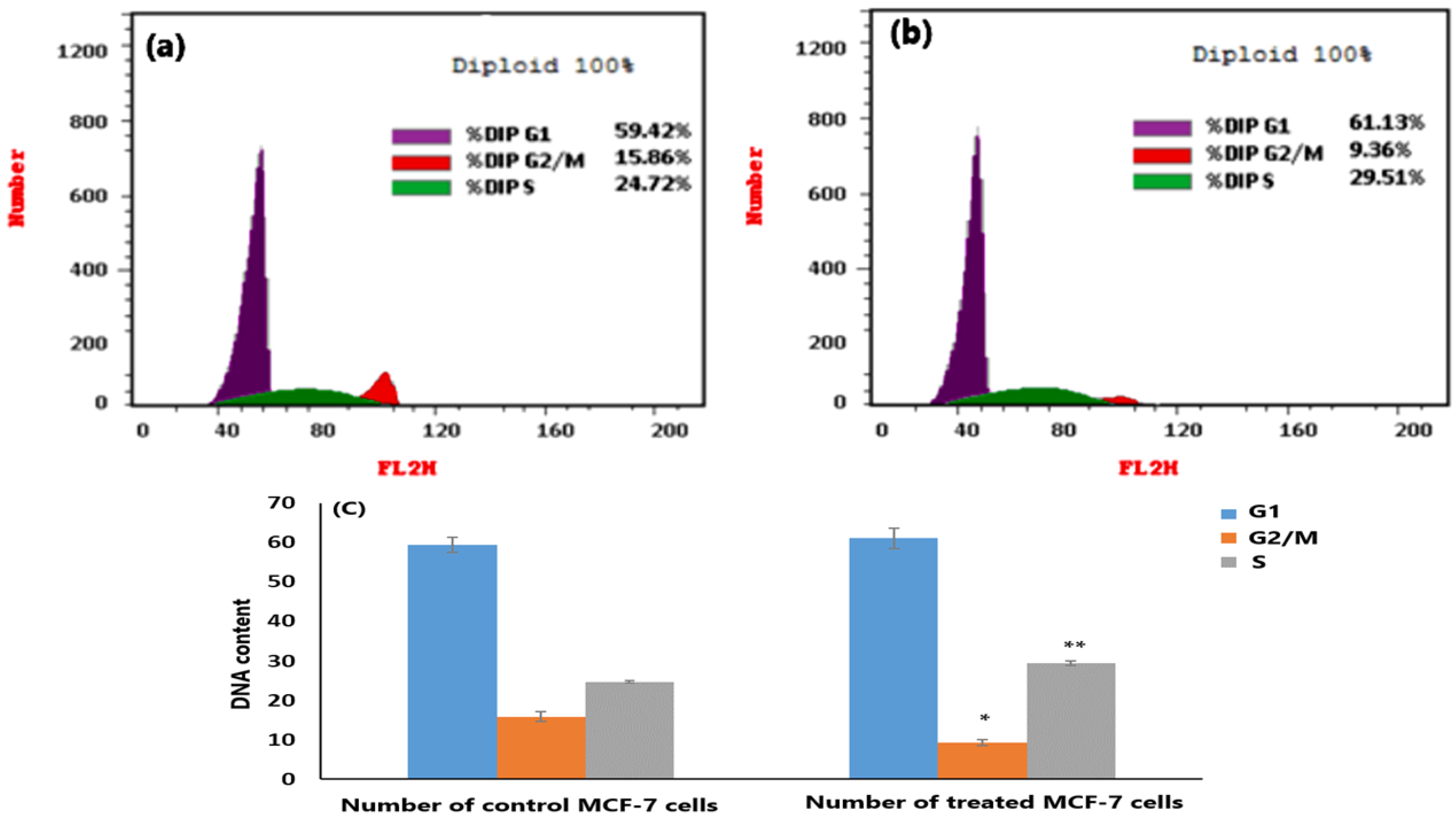
- Examining the Cell Cycle
- -
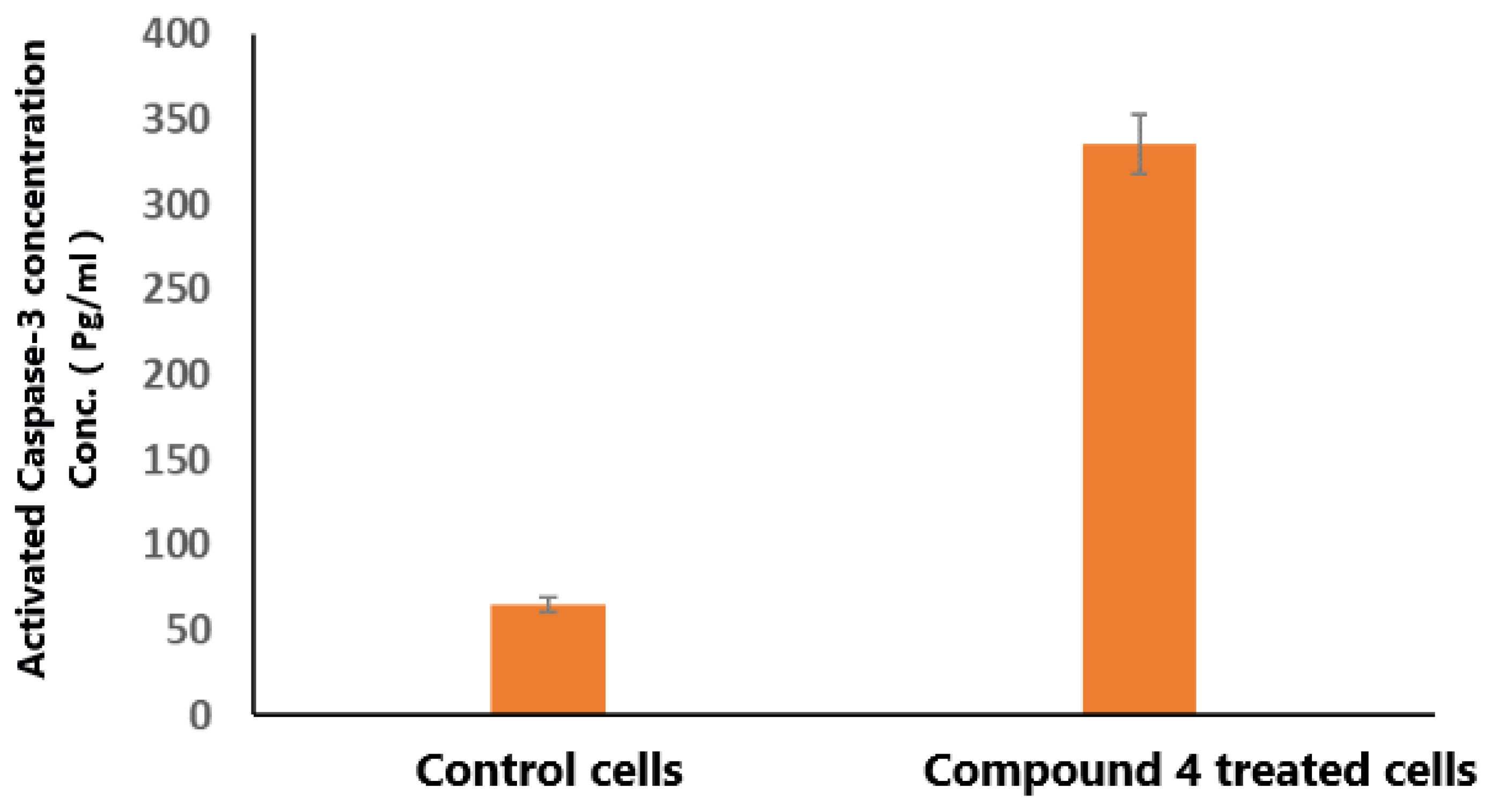
- Quantification analysis of the Caspase-3 ELISA assay
3. Experimental
3.1. Instruments and Apparatus
3.2. General Method for Synthesis of Diarylurea or Diarylthiourea Derivatives 1–6
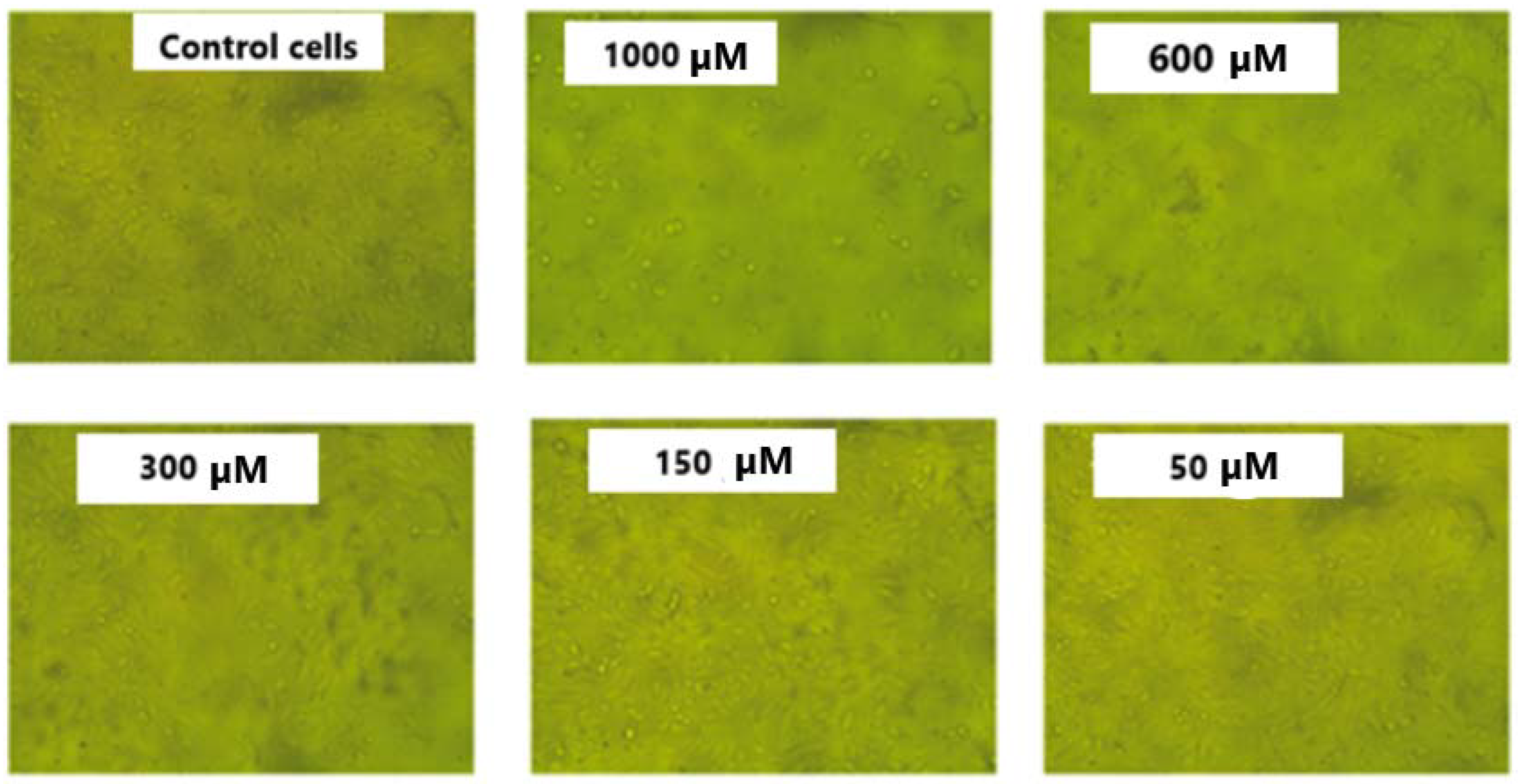
3.3. Cytotoxicity Assays
3.3.1. Cell Line Preparation
3.3.2. Cytotoxicity Test Using the MTT Approach
3.3.3. Measurement of Lactate Dehydrogenase (LDH) by Leakage Test
3.4. Apoptosis Assays
3.4.1. Apoptosis Analysis Using Annexin V and PI Staining
3.4.2. DNA Fragmentation Analysis Using a Comet Assay
3.5. Apoptosis: Underlying Mechanisms
3.5.1. Analysis of the Cell Cycle’s Distinct Stages
3.5.2. ELISA Examination for Quantifying the Caspase-3 Levels
4. Conclusions
Supplementary Materials
Author Contributions
Funding
Institutional Review Board Statement
Informed Consent Statement
Data Availability Statement
Conflicts of Interest
Sample Availability
References
- Sung, H.; Ferlay, J.; Siegel, R.L.; Laversanne, M.; Soerjomataram, I.; Jemal, A.; Bray, F. Global cancer statistics 2020: GLOBOCAN estimates of incidence and mortality worldwide for 36 cancers in 185 countries. CA Cancer J. Clin. 2021, 71, 209–249. [Google Scholar] [CrossRef] [PubMed]
- Malash, I.; Mansour, O.; Gaafar, R.; Shaarawy, S.; Abdellateif, M.S.; Ahmed, O.S.; Zekri, A.-R.N.; Bahnassy, A. Her2/EGFR-PDGFR pathway aberrations associated with tamoxifen response in metastatic breast cancer patients. J. Egypt. Natl. Cancer Inst. 2022, 34, 1–9. [Google Scholar] [CrossRef] [PubMed]
- Mohmmed, E.A.; Ramadan, S.S.; EL-Saiid, A.S.; Shousha, W.G. Frequency and Clinical Features of Over-Expressed HER2 in Egyptian Breast Cancer Women Patients. Egypt. J. Hosp. Med. 2021, 85, 3431–3435. [Google Scholar] [CrossRef]
- Early Breast Cancer Trialists’ Collaborative Group (EBCTCG). Effects of radiotherapy and of differences in the extent of surgery for early breast cancer on local recurrence and 15-year survival: An overview of the randomised trials. Lancet 2005, 366, 2087–2106. [Google Scholar] [CrossRef]
- Prasad, V.; Diener-West, M. Primary chemoprevention of breast cancer: Are the adverse effects too burdensome? CMAJ 2015, 187, E276–E278. [Google Scholar] [CrossRef] [PubMed]
- Sun, Y.; Shan, Y.; Li, C.; Si, R.; Pan, X.; Wang, B.; Zhang, J. Discovery of novel anti-angiogenesis agents. Part 8: Diaryl thiourea bearing 1H-indazole-3-amine as multi-target RTKs inhibitors. Eur. J. Med. Chem. 2017, 141, 373–385. [Google Scholar] [CrossRef] [PubMed]
- Adhikari, N.; Banerjee, S.; Amin, S.; Jha, T. Exploring Structural Requirements of Diarylurea Derivatives as VEGFR-2 Inhibitors through Comparative QSAR Modeling Study. In Proceedings of the International Conference on Drug Discovery (ICDD), Hyderabad, India, 29 February–2 March 2020. [Google Scholar]
- Zhang, L.; Shan, Y.; Li, C.; Sun, Y.; Su, P.; Wang, J.; Li, L.; Pan, X.; Zhang, J. Discovery of novel anti-angiogenesis agents. Part 6: Multi-targeted RTK inhibitors. Eur. J. Med. Chem. 2017, 127, 275–285. [Google Scholar] [CrossRef] [PubMed]
- Strzyga-Łach, P.; Chrzanowska, A.; Podsadni, K.; Bielenica, A. Investigation of the mechanisms of cytotoxic activity of 1,3-disubstituted thiourea derivatives. Pharmaceuticals 2021, 14, 1097. [Google Scholar] [CrossRef]
- Ghosh, A.K.; Brindisi, M. Urea derivatives in modern drug discovery and medicinal chemistry. J. Med. Chem. 2019, 63, 2751–2788. [Google Scholar] [CrossRef]
- Liu, S.; Louie, M.C.; Rajagopalan, V.; Zhou, G.; Ponce, E.; Nguyen, T.; Green, L. Synthesis and evaluation of the diarylthiourea analogs as novel anti-cancer agents. Bioorganic Med. Chem. Lett. 2015, 25, 1301–1305. [Google Scholar] [CrossRef]
- Yao, J.; Chen, J.; He, Z.; Sun, W.; Xu, W. Design, synthesis and biological activities of thiourea containing sorafenib analogs as antitumor agents. Bioorganic Med. Chem. 2012, 20, 2923–2929. [Google Scholar] [CrossRef]
- Listro, R.; Rossino, G.; Piaggi, F.; Sonekan, F.F.; Rossi, D.; Linciano, P.; Collina, S. Urea-based anticancer agents. Exploring 100-years of research with an eye to the future. Front. Chem. 2022, 10, 995351. [Google Scholar] [CrossRef] [PubMed]
- Chen, F.; Fang, Y.; Zhao, R.; Le, J.; Zhang, B.; Huang, R.; Chen, Z.; Shao, J. Evolution in medicinal chemistry of sorafenib derivatives for hepatocellular carcinoma. Eur. J. Med. Chem. 2019, 179, 916–935. [Google Scholar] [CrossRef] [PubMed]
- Liu, X.-J.; Zhao, H.-C.; Hou, S.-J.; Zhang, H.-J.; Cheng, L.; Yuan, S.; Zhang, L.-R.; Song, J.; Zhang, S.-Y.; Chen, S.-W. Recent development of multi-target VEGFR-2 inhibitors for the cancer therapy. Bioorganic Chem. 2023, 133, 106425. [Google Scholar] [CrossRef]
- Li, H.-Q.; Lv, P.-C.; Yan, T.; Zhu, H.-L. Urea derivatives as anticancer agents. Anti-Cancer Agents Med. Chem. (Former. Curr. Med. Chem. Anti-Cancer Agents) 2009, 9, 471–480. [Google Scholar] [CrossRef]
- Hassan, M.A.; Sayed, G.H.; El-Nagar, A.M.; Hussien, A.M. A convenient synthesis of some diarylurea and thiourea derivatives as antimicrobial compounds. Chem. Process Eng. Res. 2014, 25, 1–11. [Google Scholar]
- Khan, S.A.; Singh, N.; Saleem, K. Synthesis, characterization and in vitro antibacterial activity of thiourea and urea derivatives of steroids. Eur. J. Med. Chem. 2008, 43, 2272–2277. [Google Scholar] [CrossRef] [PubMed]
- Naramsetti, K.; Alluri, C.; Amperayani, K.R.; Sharma, G. Continuous-Flow Synthesis, Characterization, Antimicrobial Activity, and Docking Studies of Urea Derivatives of 4-(4-Aminophenyl)-3-morpholinone. Russ. J. Gen. Chem. 2023, 93, 1261–1273. [Google Scholar] [CrossRef]
- Lv, P.-C.; Li, H.-Q.; Sun, J.; Zhou, Y.; Zhu, H.-L. Synthesis and biological evaluation of pyrazole derivatives containing thiourea skeleton as anticancer agents. Bioorganic Med. Chem. 2010, 18, 4606–4614. [Google Scholar] [CrossRef]
- Saeed, S.; Rashid, N.; Jones, P.G.; Ali, M.; Hussain, R. Synthesis, characterization and biological evaluation of some thiourea derivatives bearing benzothiazole moiety as potential antimicrobial and anticancer agents. Eur. J. Med. Chem. 2010, 45, 1323–1331. [Google Scholar] [CrossRef]
- Manjula, S.; Noolvi, N.M.; Parihar, K.V.; Reddy, S.M.; Ramani, V.; Gadad, A.K.; Singh, G.; Kutty, N.G.; Rao, C.M. Synthesis and antitumor activity of optically active thiourea and their 2-aminobenzothiazole derivatives: A novel class of anticancer agents. Eur. J. Med. Chem. 2009, 44, 2923–2929. [Google Scholar] [CrossRef]
- Kumar, V.; Chimni, S.S. Recent developments on thiourea based anticancer chemotherapeutics. Anti-Cancer Agents Med. Chem. 2015, 15, 163–175. [Google Scholar] [CrossRef]
- Ghorab, M.M.; El-Gaby, M.S.A.; Alsaid, M.S.; Elshaier, Y.A.M.M.; Soliman, A.M.; El-Senduny, F.F.; Badria, F.A.; Sherif, A.Y.A. Novel thiourea derivatives bearing sulfonamide moiety as anticancer agents through COX-2 inhibition. Anti-Cancer Agents Med. Chem. (Former. Curr. Med. Chem. Anti-Cancer Agents) 2017, 17, 1411–1425. [Google Scholar] [CrossRef] [PubMed]
- Perez, S.A.; de Haro, C.; Vicente, C.; Donaire, A.; Zamora, A.; Zajac, J.; Kostrhunova, H.; Brabec, V.; Bautista, D.; Ruiz, J. New acridine thiourea gold (I) anticancer agents: Targeting the nucleus and inhibiting vasculogenic mimicry. ACS Chem. Biol. 2017, 12, 1524–1537. [Google Scholar] [CrossRef] [PubMed]
- Abbas, S.Y.; Al-Harbi, R.A.; El-Sharief, M.A.S. Synthesis and anticancer activity of thiourea derivatives bearing a benzodioxole moiety with EGFR inhibitory activity, apoptosis assay and molecular docking study. Eur. J. Med. Chem. 2020, 198, 112363. [Google Scholar] [CrossRef] [PubMed]
- Jin, J.; Hu, J.; Qin, Y.; Zhang, J.; Yue, L.; Hou, H. In vitro and in vivo anticancer activity of a thiourea tripyridyl dinuclear Cu (ii) complex. New J. Chem. 2019, 43, 19286–19297. [Google Scholar] [CrossRef]
- Kaur, B.; Singh, G.; Sharma, V.; Singh, I. Sulphur Containing Heterocyclic Compounds as Anticancer Agents. Anticancer Agents Med. Chem. 2023, 23, 869–881. [Google Scholar]
- Widiandani, T. The Potency of 4-Nitrobenzoyl-3-Allylthiourea as an Agent of Breast Cancer with Egfr/Her2: In Silico snd In Vitro Study. Int. J. Multidiscip. Innov. Res. Methodol. 2022, 1, 1–10. [Google Scholar] [CrossRef]
- Hanna, D.H.; Osailan, R.; Ahmed, H.A. Stevia rebaudiana Methanolic Leaf Extract in Egypt: Phytochemical Analysis, Antioxidant, Antilipid Peroxidation, Antihemolytic, Antimetastatic, and Anticancer Properties. J. Food Biochem. 2023, 2023, 7161091. [Google Scholar] [CrossRef]
- Hoshino, R.; Tanimura, S.; Watanabe, K.; Kataoka, T.; Kohno, M. Blockade of the extracellular signal-regulated kinase pathway induces marked G1 cell cycle arrest and apoptosis in tumor cells in which the pathway is constitutively activated: Up-regulation of p27Kip1. J. Biol. Chem. 2001, 276, 2686–2692. [Google Scholar] [CrossRef]
- Janicke, R.U.; Sprengart, M.L.; Wati, M.R.; Porter, A.G. Caspase-3 is required for DNA fragmentation and morphological changes associated with apoptosis. J. Biol. Chem. 1998, 273, 9357–9360. [Google Scholar] [CrossRef]
- Hanna, D.H.; Hamed, A.A.; Saad, G.R. Synthesis and characterization of poly (3-hydroxybutyrate)/chitosan-graft poly (acrylic acid) conjugate hyaluronate for targeted delivery of methotrexate drug to colon cancer cells. Int. J. Biol. Macromol. 2023, 240, 124396. [Google Scholar] [CrossRef] [PubMed]
- Al-Shammari, A.M.; Salman, M.I.; Saihood, Y.D.; Yaseen, N.Y.; Raed, K.; Shaker, H.K.; Ahmed, A.; Khalid, A.; Duiach, A. In vitro synergistic enhancement of Newcastle Disease Virus to 5-fluorouracil cytotoxicity against tumor cells. Biomedicines 2016, 4, 3. [Google Scholar] [CrossRef] [PubMed]
- Liu, B.; Han, M.; Sun, R.-H.; Wang, J.-J.; Zhang, Y.-P.; Zhang, D.-Q.; Wen, J.-K. ABL-N-induced apoptosis in human breast cancer cells is partially mediated by c-Jun NH 2-terminal kinase activation. Breast Cancer Res. 2010, 12, R9. [Google Scholar] [CrossRef]
- Chen, Y.C.; Chen, B.H. Preparation of curcuminoid microemulsions from Curcuma longa L. to enhance inhibition effects on growth of colon cancer cells HT-29. RSC Adv. 2018, 8, 2323–2337. [Google Scholar] [CrossRef]
- Worsley, C.M.; Veale, R.B.; Mayne, E.S. Inducing apoptosis using chemical treatment and acidic pH, and detecting it using the Annexin V flow cytometric assay. PLoS ONE 2022, 17, e0270599. [Google Scholar] [CrossRef]
- Hanna, D.H.; Saad, G.R. Induction of mitochondria mediated apoptosis in human ovarian cancer cells by folic acid coated tin oxide nanoparticles. PLoS ONE 2021, 16, e0258115. [Google Scholar] [CrossRef] [PubMed]
- Mantena, S.K.; Sharma, S.D.; Katiyar, S.K. Berberine, a natural product, induces G1-phase cell cycle arrest and caspase-3-dependent apoptosis in human prostate carcinoma cells. Mol. Cancer Ther. 2006, 5, 296–308. [Google Scholar] [CrossRef]
- Hanna, D.H.; Aziz, M.M.; Shafee, E.E. Effective-by-method for the preparation of folic acid-coated TiO2 nanoparticles with high targeting potential for apoptosis induction against bladder cancer cells (T24). Biotechnol. Appl. Biochem. 2023. Online ahead of print. [Google Scholar] [CrossRef]
- Raina, R.; Afroze, N.; Sundaram, M.K.; Haque, S.; Bajbouj, K.; Hamad, M.; Hussain, A. Chrysin inhibits propagation of HeLa cells by attenuating cell survival and inducing apoptotic pathways. Eur. Rev. Med. Pharmacol. Sci. 2021, 25, 2206–2220. [Google Scholar] [PubMed]


| The Tested Cells | Tail Length (PX) | % DNA in Tail | Tail Moment | Olive Tail Moment (OTM) |
|---|---|---|---|---|
| The untreated control MCF-7 cells | 7.52 ± 1.82 | 3.11 ± 1.73 | 0.22 ± 0.02 | 0.55 ± 0.02 |
| The treated MCF-7 cells with compond 4 without caspase-3 inhibitor pretreatment | 6.67 ± 1.94 | 17.26 ± 2.34 *** | 1.72 ± 0.05 *** | 2.38 ± 0.06 *** |
| The treated MCF-7 cells with compound 4 with caspase-3 inhibitor pretreatment | 7.13 ± 2.21 | 14.37 ± 1.33 *** | 1.04 ± 0.04 *** | 1.87 ± 0.34 ** |
Disclaimer/Publisher’s Note: The statements, opinions and data contained in all publications are solely those of the individual author(s) and contributor(s) and not of MDPI and/or the editor(s). MDPI and/or the editor(s) disclaim responsibility for any injury to people or property resulting from any ideas, methods, instructions or products referred to in the content. |
© 2023 by the authors. Licensee MDPI, Basel, Switzerland. This article is an open access article distributed under the terms and conditions of the Creative Commons Attribution (CC BY) license (https://creativecommons.org/licenses/by/4.0/).
Share and Cite
El-Atawy, M.A.; Alsubaie, M.S.; Alazmi, M.L.; Hamed, E.A.; Hanna, D.H.; Ahmed, H.A.; Omar, A.Z. Synthesis, Characterization, and Anticancer Activity of New N,N′-Diarylthiourea Derivative against Breast Cancer Cells. Molecules 2023, 28, 6420. https://doi.org/10.3390/molecules28176420
El-Atawy MA, Alsubaie MS, Alazmi ML, Hamed EA, Hanna DH, Ahmed HA, Omar AZ. Synthesis, Characterization, and Anticancer Activity of New N,N′-Diarylthiourea Derivative against Breast Cancer Cells. Molecules. 2023; 28(17):6420. https://doi.org/10.3390/molecules28176420
Chicago/Turabian StyleEl-Atawy, Mohamed A., Mai S. Alsubaie, Mohammed L. Alazmi, Ezzat A. Hamed, Demiana H. Hanna, Hoda A. Ahmed, and Alaa Z. Omar. 2023. "Synthesis, Characterization, and Anticancer Activity of New N,N′-Diarylthiourea Derivative against Breast Cancer Cells" Molecules 28, no. 17: 6420. https://doi.org/10.3390/molecules28176420