Article

Isolation and Identification of Phytocompounds from Maytenus dhofarensis and Their Biological Potentials

by
Fatma Al-Rubaiai
1,
Zakiya Zahran Al-Shariqi
1,
Khalsa S. Al-Shabibi
1,
John Husband
1,
Asmaa M. Al-Hattali
1,
Marcia Goettert
2,
Stefan Laufer
2,3,
Younis Baqi
1,
Syed Imran Hassan
1,* and
Majekodunmi O. Fatope
1,*
1
Department of Chemistry, College of Science, Sultan Qaboos University, Al Khod, P.O. Box 36, Muscat 123, Oman
2
Department of Pharmaceutical and Medicinal Chemistry, Institute of Pharmacy, Eberhard Karls Universität Tübingen, D-72076 Tübingen, Germany
3
Tübingen Center for Academic Drug Discovery (TüCAD2), D-72076 Tübingen, Germany
*
Authors to whom correspondence should be addressed.
Molecules 2023, 28(16), 6077; https://doi.org/10.3390/molecules28166077
Submission received: 17 July 2023 / Revised: 8 August 2023 / Accepted: 9 August 2023 / Published: 15 August 2023

Abstract

:
Maytenus dhofarensis Sebsebe (Celestraceae) is a naturally growing shrub in Oman. It is not a reputed medicinal plant in Oman, but it is regionally endemic and causes shivering attacks on goats that graze on it. The chemical investigation of the hexane and chloroform extracts of the fruits and stems of M. dhofarensis afforded dihydro-β-agarofuran-type sesquiterpene pyridine alkaloid (1), lupanyl myristoate (2) and lignanolactone (3). Compounds (13) are new isolates from M. dhofarensis. The structures of these compounds were assigned through comprehensive IR, NMR, and ESI-MS analyses, and the relative configurations of compounds 1 and 3 were deduced from density function theory (DFT) calculations and NMR experiments. Compound 1 was assayed against the kinase enzyme and showed no inhibition activity for p38 alpha and delta at a 10 µM test concentration. Compound 3 inhibited the 2,2′-diphenyl-1-picrylhydrazyl radical (DPPH) by 69.5%, compared to 70.9% and 78.0% for gallic acid and butylated hydroxyanisole, respectively, which were used as positive controls.

1. Introduction

Plants of the genus Maytenus (Celestraceae) are widely distributed in tropical and subtropical regions of the world and about 800–1300 species [1] are known. Several members of this genus are used in traditional medicine to treat cancer [2], gastric ulcers [3], and arthritis [4]. Maytenus species are known to contain a diverse group of triterpenoids [5,6,7], flavonoids [8,9], tannins [10], lignans [11,12], dihydro-β-agarofurans [4,13,14] and sesquiterpene pyridine alkaloids [15,16,17] that display remarkable structural diversities and cytotoxicity [18,19,20,21,22], as well as insecticidal [23], antitumor-promoting [13], MDR-reverting [24,25,26], antitubercular [27], neuroprotective [28], immunosuppressive [29], anti-HIV [30], anti-inflammatory [18], and medicinal properties [3,31,32,33,34,35,36,37].
Maytenus dhofarensis is a regionally endemic spiny shrub found growing naturally in the Dhofar region of Oman [38]. It is not a reputed medicinal plant in Oman but causes shivering attacks on goats that graze on it. To date, no reports on its secondary metabolites have previously been described. The lack of elaborate phytochemical and pharmacological work on this species stimulated our interest in examining the plant for structurally novel compounds and bioactivity. Herein, we report the isolation and structural characterization of a new dihydro-β-agarofuran-type sesquiterpene pyridine alkaloid (1), lupanyl myristoate (2), and lignanolactone (3) (Figure 1) from the fruits and stems of the plant, and the bioassay results of the more-abundant isolates for their antioxidant and kinase-inhibitory activities. Their structures were elucidated from the interpretation of spectral data and the relative configurations of compounds 1 and 3 were determined from observed and calculated chemical shift values for their diastereoisomers using DP4+ probability [39,40] analysis. Compound 3 showed 2,2′-diphenyl-1-picrylhydrazyl radical (DPPH)-scavenging activities.

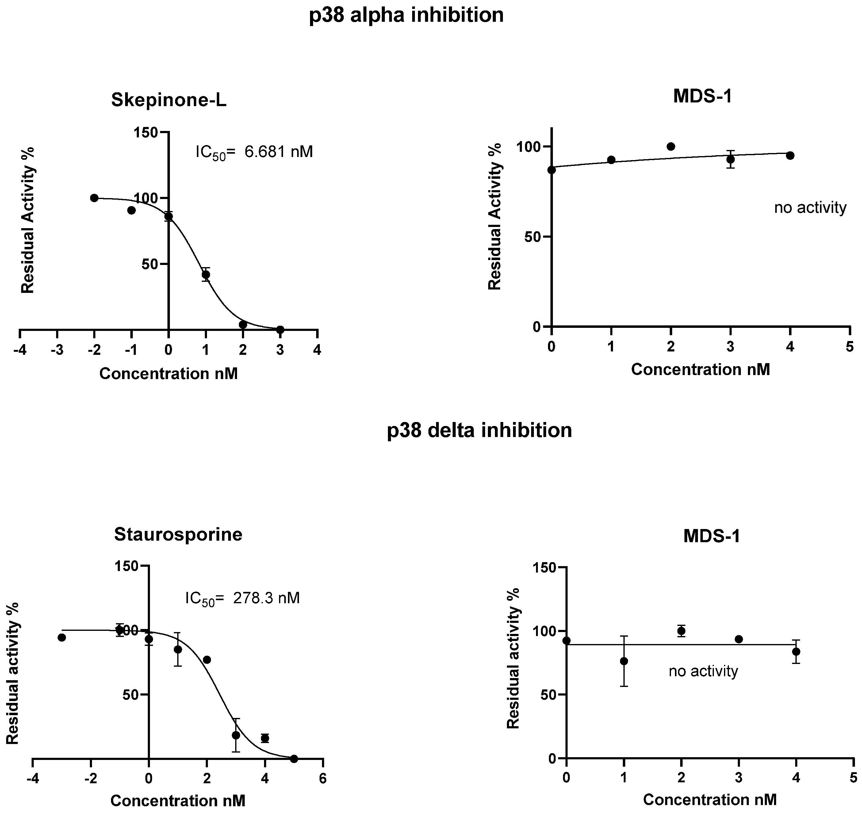
2. Results and Discussion

Compound 1 was isolated as a colorless gum, and it produced an alkaloid-positive test with Dragenddorrff’s reagent. Its molecular formula (C31H41NO11) was deduced from the ESIMS ion peak at m/z 645.1000 [M + CH3CN + H]+ (calc. m/z 645.1000 for C33H45N2O11), corresponding to an acetonitrile-adduct ion cluster with one proton. The UV spectrum exhibited characteristic absorption bands for aromatic moiety (λmax at 283 (1.6), 275 (1.9), and 246 (3.0) nm). The infrared (IR) spectrum displayed absorption bands at 3280 (broad), 1745, 1708, and 1596 cm−1 for free hydroxyl, multi-carbonyl esters, and an aromatic ring, respectively. Its 1H NMR spectrum data (Table 1) revealed three acetyl methyl groups at δH 1.78 (s), 2.08 (s), and 2.28 (s); four oxygenated methines at δH 5.53 (H-2, m), 6.22 (H-6,s), 5.28 (H-9, d, J = 6.8 Hz) and 5.44 (H-1, d, J = 3.2 Hz); a hydroxyl proton at δH 3.10; and one set of oxygenated methylene signals at δH 4.92 (H-15a, d, J = 12.8 Hz) and 4.39 (H-15b, d, J = 13.6 Hz), respectively. The signal at δH 2.35 was assigned to the aliphatic methine proton (H-7). There were also resonances for four sets of aliphatic methylene protons at δH 2.02 (H-3a, dd, J = 3.2, 15.1 Hz), 2.18 (H-3b, dd, 5.3, 16.0 Hz), 2.19 (H-8a, m) and 2.60 (H-8b, ddd, J = 3.8, 11.1 15.9 Hz) and five tertiary methyl protons at 1.52 (H-13, s), 1.57 (H-12, s), 1.49 (H-14, s), 1.81 (H-10′, s), and 1.85 (H-11′, s). The vinylic signal at δH 6.93 corresponded to the olefinic proton H-7′, which appeared as a doublet of a quartet (J = 7.0, 1.3 Hz). The 1H NMR spectrum data revealed the presence of three 2,3 disubstituted pyridine protons at δH 7.48 (H-5′, dd, J = 8.1, 7.6 Hz), 7.58 (H-4′, dd, J = 7.4, 2.5 Hz) and 8.18 (H-6′, dd, J = 8.3, 1.2 Hz). Additional signals for four ester carbonyls at δC 166.5, 169.8, 169.5, and 170.7; three oxygenated quaternary carbons at δC 69.9 (C-4), 84.7 (C-11), and 91.1 (C-5); one olefinic quaternary carbon at δC 129.8 (C-8′); and two aromatic quaternary carbons at δC 127 (C-3′) and 166.1 (C-2′) were observed in the 13C NMR spectrum (Table 1). Taken together, these data indicate that compound 1 is a dihydro-β-agarofuran-type sesquiterpene pyridine alkaloid [16,18]. A polyoxygenated dihydroagarofuran skeleton was determined by the 1H–1H COSY cross-signals for H-1/H-2, H-2/H-3, H-7/H-8, and H-8/H-9, coupling systems and the following HMBC interactions: H-1/ C-2, C-9, C-10, C-15, and C-9′; H-9/ C-5, C-7, C-8 C-10 and C-15; H-15/ C-1, C-5 and C-9; and H-6/ C-5, C-7, C-8, C-10 and C-11. The free hydroxyl groups were located at C-1 (δC 70.6) and C-4 (δC 69.9) by a comparison of the observed 13C NMR chemical shift with reported values [16,18] and the HMBC cross-signals of OH (δH 3.1) with C-4, C-5, and C-14, respectively.
The 2,3-disubstituted pyridine core unit was confirmed by 1H-1H COSY correlations and HMBC interactions. In the HMBC interactions (Table 1), H-5′ (δH 7.48) showed interaction with a methine carbon signal at δC 127.5 (C-3′). Signals at δH 8.18 (H-6′) and 6.93 (H-7′) showed interactions with δC 166.5 (C-2′). Also, the signal at δH 7.58 (H-4′) showed interaction with δC 130.19 (C-6′). The 1H−1H COSY spectrum of compound 1 revealed a separated spin−spin system (H-4′/H-5′/H-6′).
The relative configuration of 1 was established by the 2D NOESY spectrum and by comparing it with previous studies [16,18]. DFT-predicted chemical shifts for 9R and 9S diastereomers were compared with the observed values for the isolated compound, and the best match using the DP4+ probability [39] favored the 9R configuration for compound 1. The NOESY spectrum also did not show any correlation between H-1 and H-9, supporting the 9R configuration assignment. Predicted chemical shift values are listed in Table S1, while the results of the DP4+ analyses are summarized in Table S2. Based on the above assignments, the structure of compound 1 was identified as a dihydro-β-agarofuran-type sesquiterpene pyridine alkaloid, which was named Maytendhofarene (Figure 1).
Compound 1 was investigated for kinase-inhibition activity using the homogenous time-resolved fluorescence (HTRF) detection kit [41] but showed no inhibition activity for p38 alpha and delta at a 10 µM test concentration (Figure 2). p38 MAP kinases have been implicated in a wide range of complex biological processes, such as cell differentiation and proliferation, cell death, cell migration, and invasion [42]. The dysregulation of p38 MAPK is associated with diverse diseases such as chronic inflammation and cancer and can act as a tumor suppressor or tumor inducer [43].
Compound 2, whose molecular formula was identified as C44H76O3 from the HRESIMS sodium-adduct ion cluster at 675.6628 [M + Na]+ (calcd. for C44H76O3Na 675.5692), was obtained as a white solid (m.p. = 66-68 °C). The FTIR spectrum showed absorption bands at 3373 (broad), 2916, 2849, and 1702 cm−1 characteristic of hydroxyl, alkene, and ester groups, respectively. The 1H NMR spectrum (Table 2) displayed six tertiary methyl groups (each, 3 H, s) at δH 1.03, 0.94, 0.87, 0.85, 0.84, 0.78 and one isopropenyl methyl at δH 1.68 (3H, s). Two exocyclic vinyl protons resonated at δH 4.69 (1H, s) and 4.57 (1H, s). Two oxymethine signals at δH 4.53 (1H, dd, J = 14.0 and 7.0 Hz) and δ 3.99 (1H, m) and a typical lupene Hβ-19 resonance at δH 2.38 (1H, dd, J = 14.0 and 7.0 Hz) were also observed. These signals indicated a dihydroxy lupene triterpene substructure with one hydroxyl group masked as an ester for compound 1. The multiplicity and 1H-1H COSY connectivity of the Hβ-19 signal at δH 2.38 (dd, J = 14.0 and 7.0 Hz) to δH 3.99 (H-21) and δH 1.35 (H-18) showed that C-19 is flanked by oxymethine and methine groups. The resonances at δH at 1.42 and 1.32 were assigned to H-22a and H-22b based on their connectivity to δH 3.99 (H-21) in the 1H-1H COSY map. The exocyclic alkene group was confirmed by the resonances at δC 151.1 (C-20) and 109.5 (C-29) in the 13C NMR DEPT spectrum. The assignment of the resonances of the lupene substructure (Table 2) allowed the signals of the fatty ester to be easily recognized and assigned. The positions of the hydroxy and myristoxy groups were confirmed by H-3/H-2 correlation in the 1H-1H COSY, and H-3/C-2, H-3/C-4, H-3/C-23, H-3/C-24, H-21/C-1′, H-21/C-19, H-21/C-22 in the HMBC. Additional HMBC interactions between δH 2.38 (H-19) and δC 38.2 (C-13), 48.1 (C-18), 151.1 (C-20), 68.4 (C-21), 35.7 (C-22), 109.5 (C-29), 18.1 (C-30), and 173.0 (C-1′) corroborated the assigned structure. The orientations of C-3 hydroxyl and C-21 myristoxy groups were determined as β and α based on coupling constants or chemical shift values [44] of H-3 and H-21. Compound 2 is a 3β, 21α-dihydroxylup-20(29)-ene, with the C-21 hydroxyl group masked as an ester of myristic acid and named 3β-hydroxy-21α-myristoxylup-20(29)-ene (2).
Compound 3 was isolated from the CHCl3 extract of M. dhofarensis. It was a solid (m.p. = 75–80 °C) and exhibited IR absorption bands for an OH stretch (3982 and 3397 cm−1, broad), ester (1738 cm−1), and benzene ring (1599, 1508 cm−1). The molecular formula was determined to be C20H22O7 from the HRESIMS sodium-adduct ion cluster at m/z 397 [M + Na]+ and a protonated dimer molecule ion at m/z 749.2809 [2M + H]+ (calcd. for C40H45O14 749.2812). This formula is consistent with the presence of ten degrees of unsaturation. The 1H NMR spectrum (Table 3) showed signals for six aromatic protons and some phenolic hydroxy, two methoxy, one methane, and three methylene groups for compound 3. It also displayed isochronous signals at δH 4.04 and 3.99, δH 3.10, and 2.91, and δH 2.59 and 2.49 for oxymethylene, acetoxymethylene, and benzylmethylene protons (Table 3). All the methylene protons in compound 3 are thus diastereotopic atoms. A methine proton at δH 2.52 (H-8′) showed spin–spin couplings to the oxymethylene protons (H-9′a and H-9′b) and benzylmethylene protons (H-7′a and H-7′b). The diastereotopic acetoxy protons were only coupled to each other in the 1H-1H COSY map and must be flanked by quaternary carbons. The interpretation of the IR, 1H NMR chemical shifts, and coupling patterns suggested a β-hydroxy-β-phenyl-γ-benzyl δ-valerolactone substructure for compound 3. The two methoxy groups resonated as singlets at δH 3.64 and 3.85, and the six aromatic protons constituted two separate ABX coupling systems with characteristic splitting patterns of aromatic protons in 1,3,4 relative positions. Each of the four aromatic protons resonated as a doublet (J = 7.9 Hz) at δH 6.84, 6.82, 6.63, and 6.62, and the other aromatic protons as broad singlets at δH 6.69 and 6.60, respectively. The aromatic signals and spin–spin couplings are consistent with the presence of two 3, 4-di-substituted phenyl units in compound 3. The 13C NMR Broad Band and DEPT experiments resolved twenty carbon resonances for compound 3, which included one carbonyl at δC 179.1 (C-9) and two tri-substituted aromatic units (Table 3). The spectra also displayed the following carbon signals: one oxymethylene at δC 70.7 (C-9′), one acetoxy methylene at δC 42.4 (C-8), one benzylmethylene at δC 31.9 (C-7′), one methine at δC 44.2 (C-8′), one oxygenated quaternary carbon at δC 76.9 (C-7), and signals for two methoxy groups at δC 56.4 and 56.3, respectively. The assignment of methoxy groups to C-4 and C-4′ was complemented by connectivity between δH 3.85 and δC 144.7 (C-3) and δC 147.0 (C-4) or δH 3.64 and 147.0 (C-3′) and 145.4 (C-4′) in HMBC. All the methylene and methine protons showed connectivity to the quaternary carbon resonance at δC 76.9 (C-3) in the HMBC. Additional HMBC connectivities between the protons of benzylmethylene [δH 2.59 (H-7′a) and 2.49 (H-7′b)] and δC 126.5 (C-1′), 121.9 (C-6′), and 111.9 (C-2′); oxymethylene [δH 4.04 (H-9′b) and 3.99 (H-9′a)] and δC 179.1 (C-9); and acetoxymethylene [δH 3.10 (H-8a) and 2.91 (H-8b)] and δC 179.1 (C-9), 44.2 (C-8′), and 130.7 (C-1) resonances established a lignanolactone structure for compound 3. Positions C-7 and C-8′ are chirality centers. The peripheral diamagnetic anisotropy of the phenyl groups at C-7 and C-7′ should cause H-8′ and H-7′, in particular, to resonate at a lower field when the configuration of H-8′ is R (Figure S23), and the phenyl groups at C-7′ and C-7 are quasi syn-periplanar to H-8′, as revealed by the molecular model of compound 3. DFT-predicted chemical shifts for H-7R, H-8′R and H-7S, and H-8′R diastereomers were compared (Table S3) with the observed values for the isolated compound, and the best match (98.9%) using the DP4 probability [40] favored H-7S, H-8′R configuration for compound 3. Compound 3 demonstrated radical scavenging property, inhibiting [45] DPPH by 69.5%, compared to 70.9% and 78.0% for gallic acid and butylated hydroxyanisole, which were used as controls.

3. Experimental Section

3.1. General Experimental Procedures

IR spectra were obtained with a Nicolet FT-IR spectrometer. 1H and 13C NMR spectra were recorded in CDCl3 with Bruker Advance NMR spectrometer operating at 700 MH with TMS as the internal standard. ESIMS was recorded on a Quattro Ultima Platinum Tandem quadrupole mass spectrometer (Micromass, Wilmslow, UK). ESIMS data were acquired on an Agilent 6400 Triple Quad LC/MS and using HRESIMS. The column chromatography (CC) was performed using EM Science Silica gel 60 (70–230 mesh ASTM). Whatman precoated silica-gel (60A K6F) analytical plates (20 × 20 cm) were used for TLC, with compounds visualized by a UV lamp and spraying with 10% (v/v) H2SO4 or Molybdophosphoric acid-isopropanol followed by heating. All absorbance measurements were recorded using a Shimadzu UV spectrophotometer.

Solvents and Reagents

The solvents used in this investigation were of analytical grade. The hexane, diethyl ether, ethyl acetate, and deuterated chloroform (CDCl3) were purchased from Sigma Aldrich (Dorset, UK). Chloroform and ethanol were purchased from BDH (Hampshire, UK). Moreover, 2,2′-diphenyl-1-picrylhydrazyl radical (DPPH), butylhydroxy anisole, and gallic acid were purchased from Sigma Aldrich (Steinheim, Germany).

3.2. Plant Material

M. dhofarensis was collected from Gogeb in Salalah in the Dhofar region, Oman (GPS coordinates: 17°12′42″ N, 54°6′29″ E) in December 2018 at an altitude of 800 m. The plant was identified by Dr. S.A. Ghazanfar at the Royal Botanic Gardens, Kew Richmond, UK, and Dr. Amina A. Al Farsi at the Department of Biology, College of Science, Sultan Qaboos University, Muscat, Oman. A voucher specimen (SQUH00006216) is kept in the Herbarium of the Department of Biology, College of Science, Sultan Qaboos University, Muscat, Oman.

3.3. Extraction and Isolation

The fruits of M. dhofarensis were dried in a hot room (at 42 °C) for 3 weeks in the Department of Chemistry, Sultan Qaboos University, and the seeds were separated from the calyx and milled to give 576 g of powdered seed. The seeds (288 g) were extracted with chloroform (2 × 1800 mL) by maceration at room temperature for three days each and concentrated under vacuum at 25–30 °C to give a gummy residue (208.6 g). The column chromatography of a portion of the chloroform extract (44.75 g) on silica gel (895 g), using gradient mixtures of n-hexane—CHCl3, CHCl3, and CHCl3—EtOH as eluent, gave a variable number of fractions, which were combined based on their TLC profiles.
Preliminary cytotoxicity testing using the brine shrimp test [46] (BST) revealed that (Maytenus dhofarensis) Md-seed-CHCl3-F73 is the most active fraction. This fraction (Md- seed-CHCl3-F73) was chromatographed on a silica gel (91 g) column (2.5 cm × 35 cm), starting with n-hexane, and then the polarity was gradually increased using ethyl acetate. Compound 1 (23.4 mg; eluent hexane-diethyl ether 3:1) was obtained as a pure compound from subfraction F73-82 (384 mg) and purified by multi-column chromatography using hexane-diethyl ether mobile phase.
A portion of the dried and powdered calyx (275 g) of M. dhofarensis was extracted with hexane by Soxhlet (2 × 600 mL) for four hours. The solvent was removed in a vacuum to yield a hexane extract (11.5 g). The separation of the extract was undertaken using column chromatography with silica gel (120 g) and using hexane and gradient mixtures of hexane-ethyl acetate with a collection of 100 mL fractions. TLC analysis using hexane: EtOAc (1:4) as the eluting system allowed fractions (M.d-calyx-H-40 to M.d-calyx-H-50) to be combined with M.d-calyx-Hexane-50 (441 mg) and purified on silica gel (105 g). Then, they were eluted using gradients of diethyl ether (DEE) in hexane as a mobile phase to produce fractions 1–33. Fraction M.d-calyx-Hexane-50-9 (128.7 mg) eluted with hexane: DEE (1:1.5) was further purified on silica gel (54 g), eluting with gradients of DEE in hexane to afford M.d-calyx-Hexane-50-9-1 to M.d-calyx-Hexane-50-9-156. Fractions M.d-calyx-Hexane-50-9-91 to M.d-calyx-Hexane-50-9-93 gave (5.0 mg; eluent hexane-DEE 2:1) pure compound 1.
The dried and powdered twigs M. d (2.0 kg) were extracted with hexane (5 L) for one week to give a residue (12.7 g) after solvent removal. The defatted plant material was re-extracted with CHCl3 (5L× 2) for two weeks and then concentrated under vacuum to yield a brown residue (59.5 g).
The hexane extract (12.0 g) was chromatographed on a silica-gel (150 g) CC column (3.2 cm × 46.5 cm) eluted with hexane in a gradient mode with CHCl3 to give fractions MDp-1 to MDp-107. Fractions were combined in groups based on their TLC similarity. Fractions MDp-44 and MDp-45 were combined with MDp-45 (1.7 g) and loaded on a silica-gel (100 g) column (2.5 cm × 30 cm) (eluted with a Hex/ EtOAc gradient, yielding 58 fractions (MDp-45-1 to MDp-45-58). Fractions MDp-45-10 to MDp-45-12 (eluent hexane-EtOAc 10:2) were combined with MDp-45-12 (0.34 g) due to their TLC similarity. Fraction MDp-45-12 was further purified by CC with Hexane-EtOAc in increasing order of polarity on Si to produce a pure compound 2 (2.5 mg; eluent Hexane-EtOAc (10:1).
The CHCl3 extract of the powdered twigs (25.0 g) was fractionated with silica-gel (180 g) CC using hexane, CHCl3, EtOAc, and EtOH as eluent to give a primary fractioning of 36 fractions (M.d-1 to M.d-36). The purification of M.d-19 (2.3 g, eluent hexane 100%) with the gradient of EtOAc in hexane gave M.d-19-1 to M.d-19-79. M.d-19-36, eluted by hexane: EtOAc (7:3), showed a single spot on TLC and gave a pure compound 3 (2.3 mg).
Maytendhofarene (1): colorless gum. IR υmax cm−1 3280, 2921, 1745, 1708, 1594, 1371, 1224, 1067; UV (CHCl3) λmax 283 (1.6), 275 (1.9) and 246 (3.0); 1H NMR (CDCl3, 700 MHz); and 13C NMR (CDCl3, 176 MHz) data are given in Table 1; ESIMS: m/z 645.1000 [M + CH3CN + H]+ (calc. 645.3000 for C33H45N2O11).
3β-Hydroxy-21α-myristoxylup-20(29)-ene (2): white solid. mp 66-68 °C; FTIR υmax 3373 (br), 2916, 2849 and 1702 cm−1 ;1H (CDCl3, 700 MHz); and 13C NMR (CDCl3, 176 MHz) data are given in Table 2; HRESIMS m/z 675.6628 [M + Na]+ (calcd for C44H76O3Na 675.5692).
4,4′-Dimethoxy-3,3′,7-trihydroxy-7,8′-lignano-9,9′-lactone (3): whitish-yellow solid. mp 75-80 °C; FTIR υmax (cm−1) 3982, 3397 (hydroxyl), 1738 (ester) and 1599 and 1508 (aromatic ring); 1H NMR (CDCl3, 400 MHz); and 13C NMR (CDCl3, 100 MHz) data are given in Table 3; HRESIMS m/z 397 [M + Na]+, and m/z 749.2809 [2M + H]+ (calcd for C40H45O14 749.2812).

3.4. Computational Studies of Compounds 1 and 3

Density functional theory (DFT) was used to predict the NMR chemical shifts for the diastereomers of compound 1 (9R and 9S) and compound 3, (7R-8′R and 7S-8′R). For each, conformers up to an upper limit of 10 kcal/mol were identified using the MMFF force field as implemented by MarvinView software (version 17.2.6.0) [47]. The selected rotamers were optimized using Gaussian (G09W) software (Gaussian 09, revision E.01) [40] at the B3LYP/6-31G+(d,p) (gas-phase) level of theory, and subsequent frequency calculations confirmed the absence of any imaginary frequencies in the minimized structures. Isotropic shielding constants were calculated at the mPW1PW91/6-311+G(d,p)/PCM level using the GIAO method. The predicted values for each diastereomer were averaged using a Boltzmann weighting, and for each compound, the unscaled shielding constants were compared in the Bayesian-based DP4+ analyses utilizing the Excel file provided by the Sarotti group [39].

3.5. Biological Assays

3.5.1. Homogenous Time-Resolved Fluorescence (HTRF) Kinase Assay

Compound 1 was diluted with a kinase buffer (with the p38 alpha enzyme or p38 delta enzyme) arising out of a stock solution of 10 mM in MDSO, giving a final concentration of 10 µM. Subsequently, 10 µL of the compound dilution was added to the enzyme, producing a final volume of 20 µL. Instead of a compound, 10 µL of KB was distributed in the wells of negative (NSB) and positive controls (STIM). The 96-well non-binding plate was centrifuged before a preincubation for 10 min at 37 °C, gently shaking at 150 rounds. After the preincubation, an ATP/ATF 2 solution was added to the wells and incubated again for 30 min at 37 °C. The assays were performed using the homogenous time-resolved fluorescence (HTRF) detection kit (Cisbio, Bedford, MA) by adding 10 µL of the HTRF detection solution (2.5 µL of PAb Anti-phospho ATF 2-Eu cryptate (1:400); 5 µL of MAb Anti GST-d2 (1:200); and 992.5 µL of HTRF detection buffer). The plate was incubated for 30 min at room temperature in the dark. The HTRF signal was read from the Victor Nivo® and calculated as the ratio of signal from the 665 nm (acceptor) and 615 nm (donor) channels and multiplied by 10,000. The percent activity was calculated by normalizing the HTRF signal from each sample well to the mean HTRF signal from the DMSO-only control wells, using the following equation:
Inhibition (%) =100 − ((ODsample − NSB)/(ODstim − NSB)) × 100%

3.5.2. Antioxidant Assay Activity Using 2,2′-Diphenyl-1-picrylhydrazyl (DPPH) Radical-Scavenging Method

The free-radical scavenging activity of compound 3 was determined using the protocol reported by Morelli [45] with slight modifications: 8.65 mg of compound 3 was dissolved in CHCl3 (1 mL), and an aliquot of this was diluted with CHCl3 to give a solution of 0.4 mg/2.0 mL of compound 3. This solution was mixed with 2.0 mL of 100 µM DPPH solution prepared by dissolving 2 mg DPPH in 50 mL of 25% aqueous ethanol. Butyl hydroxyl anisole (BHA) and gallic acid solutions were used as positive controls, while 2.0 mL of 25% aqueous ethanol solution of 100 µM DPPH mixed with 2 mL of CHCl3 served as the blank solution. The absorbance at 517 nm of all prepared solutions was determined after 15 min of incubation in the dark against the blank solution.
The ability to scavenge DPPH was expressed as a percentage inhibition (% IP) of the DPPH radical.
Inhibition (%) = [(Absorbance of control – Absorbance of sample)/Absorbance of control] × 100%

4. Conclusions

Previously uninvestigated endemic M. dhofarensis yielded three new compounds (1-3), which are similar to the compounds found in the genus Mayteneus. However, compound 1 is structurally unique. It is a dihydro-β-agarofuran-type sesquiterpene pyridine alkaloid that differs structurally from the vast array of Celastraceous macrocyclic dihydro-β-agarofuran sesquiterpene pyridine alkaloids [48] due to the absence of a pyridine dicarboxylic acid macrocyclic bridge. This is structurally significant for an endemic plant of the genus Mayteneus. Compound 1 lacked kinase-inhibitory activity as hoped for in the original design of this work. It was obtained in the fruit and also detected in the alcohol extracts of the stem. The fruit is toxic to goats, and whenever a goat grazed the fruits, it fell ill with shivering attacks. Compound 3 is a new addition to the secondary metabolites from this genus. It was isolated from the stem and showed radical scavenging activity at a level comparable to gallic acid. This might help the plant to overcome oxidative stress.

Supplementary Materials

The following supporting information can be downloaded at: https://www.mdpi.com/article/10.3390/molecules28166077/s1.

Author Contributions

F.A-R., Z.Z.A.-S., K.S.A.-S. and A.M.A.-H. performed the experiments for the isolation, structural elucidation and antioxidant assay, F.A.-R., M.O.F. and S.I.H. wrote the original draft manuscript Y.B., M.G. and S.L. facilitated and conducted the kinase assay; J.H. performed DFT calculations M.O.F. conceived and designed the project, and M.O.F. and S.I.H. supervised the work. All authors have read and agreed to the published version of the manuscript.

Funding

This research work was funded in part with a graduate research grant from TRC (BFP/GRG/EBR/21/035) to F.A.-R, and His Majesty’s Strategic Research Trust Fund (Grant SR/SCI/CHEM/01/01) to M.O.F.

Institutional Review Board Statement

Not applicable.

Informed Consent Statement

Not applicable.

Data Availability Statement

Not applicable.

Acknowledgments

We would like to thank Samuel Premkumar of CAARU, Sultan Qaboos University for NMR and MS measurements.

Conflicts of Interest

The authors declare no conflict of interest.

Sample Availability

Samples of the compound Maytendhofarene is available from the corresponding authors.

References

  1. Simmons, M.P.; Savolainen, V.; Clevinger, C.C.; Archer, R.H.; Davis, J.I. Phylogeny of the Celastraceae inferred from 26S nuclear ribosomal DNA, phytochrome B, rbcL, atpB, and morphology. Mol. Phylogenetics Evol. 2001, 19, 353–366. [Google Scholar] [CrossRef] [PubMed]
  2. Gonzalez, J.G.; Delle Monache, G.; Delle Monache, F.; Marini-Bettolo, G.B. Chuchuhuasha—A drug used in folk medicine in the Amazonian and Andean areas. A chemical study of Maytenus laevis. J. Ethnopharmacol. 1982, 5, 73–77. [Google Scholar] [CrossRef]
  3. Veloso, C.; Oliveira, M.; Rodrigues, V.; Oliveira, C.; Duarte, L.; Teixeira, M.; Ferreira, A.; Perez, A. Evaluation of the effects of extracts of Maytenus imbricata (Celastraceae) on the treatment of inflammatory and metabolic dysfunction induced by high-refined carbohydrate diet. Inflammopharmacology 2019, 27, 539–548. [Google Scholar] [PubMed]
  4. Malaník, M.; Treml, J.; Rjašková, V.; Tížková, K.; Kaucká, P.; Kokoška, L.; Kubatka, P.; Šmejkal, K. Maytenus macrocarpa (Ruiz & Pav.) Briq.: Phytochemistry and Pharmacological Activity. Molecules 2019, 24, 2288. [Google Scholar] [PubMed] [Green Version]
  5. de Figueiredo, P.T.; Silva, E.W.; Cordeiro, L.V.; Barros, R.P.; Lima, E.; Scotti, M.T.; da Silva, M.S.; Tavares, J.F.; Costa, V.C.d.O. Lupanes and friedelanes, the first chemical constituents of the aerial parts of Maytenus erythroxylon Reissek. Phytochem. Lett. 2021, 45, 19–24. [Google Scholar] [CrossRef]
  6. Taddeo, V.A.; Castillo, U.G.; Martínez, M.L.; Menjivar, J.; Jiménez, I.A.; Núñez, M.J.; Bazzocchi, I.L. Development and validation of an HPLC-PDA method for biologically active quinonemethide triterpenoids isolated from Maytenus chiapensis. Medicines 2019, 6, 36. [Google Scholar] [CrossRef] [Green Version]
  7. Zhang, L.; Ji, M.-Y.; Qiu, B.; Li, Q.-Y.; Zhang, K.-Y.; Liu, J.-C.; Dang, L.-S.; Li, M.-H. Phytochemicals and biological activities of species from the genus Maytenus. Med. Chem. Res. 2020, 29, 575–606. [Google Scholar] [CrossRef]
  8. De Sousa, G.F.; de Aguilar, M.G.; Takahashi, J.A.; Alves, T.M.; Kohlhoff, M.; Vieira Filho, S.A.; Silva, G.D.; Duarte, L.P. Flavonol triglycosides of leaves from Maytenus robusta with acetylcholinesterase inhibition. Phytochem. Lett. 2017, 19, 34–38. [Google Scholar]
  9. Pino, L.L.; García, T.H.; Delgado-Roche, L.; Rodeiro, I.; Hernández, I.; Vilegas, W.; Spengler, I. Polyphenolic profile by FIA/ESI/IT/MSn and antioxidant capacity of the ethanolic extract from the barks of Maytenus cajalbanica (Borhidi & O. Muñiz) Borhidi & O. Muñiz. Nat. Prod. Res. 2020, 34, 1481–1485. [Google Scholar]
  10. Olivaro, C.; Escobal, M.; de Souza, G.; Mederos, A. Chemical characterisation and in vitr o anthelmintic activity of phenolic-rich extracts from the leaves and branches of Maytenus ilicifolia, a native plant from South America. Nat. Prod. Res. 2022, 36, 3168–3172. [Google Scholar] [CrossRef]
  11. Vazdekis, N.E.; Chavez, H.; Estevez-Braun, A.; Ravelo, A.G. Triterpenoids and a lignan from the aerial parts of Maytenus apurimacensis. J. Nat. Prod. 2009, 72, 1045–1048. [Google Scholar] [CrossRef]
  12. Okoye, F.B.C.; Agbo, M.O.; Nworu, C.S.; Nwodo, N.J.; Esimone, C.O.; Osadebe, P.O.; Proksch, P. New neolignan glycoside and an unusual benzoyl malic acid derivative from Maytenus senegalensis leaves. Nat. Prod. Res. 2015, 29, 109–115. [Google Scholar] [CrossRef] [PubMed]
  13. Perestelo, N.R.; Jiménez, I.A.; Tokuda, H.; Vázquez, J.T.; Ichiishi, E.; Bazzocchi, I.L. Absolute configuration of dihydro-β-agarofuran sesquiterpenes from Maytenus jelskii and their potential antitumor-promoting effects. J. Nat. Prod. 2016, 79, 2324–2331. [Google Scholar] [CrossRef] [PubMed]
  14. Alarcón-Enos, J.; Muñoz-Núñez, E.; Gutiérrez, M.; Quiroz-Carreño, S.; Pastene-Navarrete, E.; Céspedes Acuña, C. Dyhidro-β-agarofurans natural and synthetic as acetylcholinesterase and COX inhibitors: Interaction with the peripheral anionic site (AChE-PAS), and anti-inflammatory potentials. J. Enzyme Inhib. Med. Chem. 2022, 37, 1845–1856. [Google Scholar] [CrossRef] [PubMed]
  15. Núñez, M.J.; Guadaño, A.; Jiménez, I.A.; Ravelo, A.G.; González-Coloma, A.; Bazzocchi, I.L. Insecticidal Sesquiterpene Pyridine Alkaloids from Maytenus c hiapensis. J. Nat. Prod. 2004, 67, 14–18. [Google Scholar] [CrossRef]
  16. Santos, V.A.F.F.M.; Regasini, L.O.; Nogueira, C.u.R.; Passerini, G.D.; Martinez, I.; Bolzani, V.S.; Graminha, M.r.A.; Cicarelli, R.M.; Furlan, M. Antiprotozoal sesquiterpene pyridine alkaloids from Maytenus ilicifolia. J. Nat. Prod. 2012, 75, 991–995. [Google Scholar] [CrossRef]
  17. Din, A.U.; Siddiqui, B.S. Royleanine A, an Antitumor Dihydro-β-agarofuran Sesquiterpene Pyridine Alkaloid from Maytenus royleanus. J. Braz. Chem. Soc. 2022, 33, 1386–1391. [Google Scholar]
  18. Wang, C.; Li, C.-J.; Yang, J.-Z.; Ma, J.; Chen, X.-G.; Hou, Q.; Zhang, D.-M. Anti-inflammatory sesquiterpene derivatives from the leaves of Tripterygium wilfordii. J. Nat. Prod. 2013, 76, 85–90. [Google Scholar] [CrossRef]
  19. González, A.G.; Jiménez, I.A.; Ravelo, A.G.; Sazatornil, J.G.; Bazzocchi, I.L. New sesquiterpenes with antifeedant activity from Maytenus canariensis (Celastraceae). Tetrahedron 1993, 49, 697–702. [Google Scholar] [CrossRef]
  20. Wibowo, M.; Wang, Q.; Holst, J.; White, J.M.; Hofmann, A.; Davis, R.A. Dihydro-β-agarofurans from the roots of the Australian endemic rainforest tree Maytenus bilocularis act as leucine transport inhibitors. Phytochem 2018, 148, 71–77. [Google Scholar] [CrossRef] [Green Version]
  21. Touré, S.; Nirma, C.; Falkowski, M.; Dusfour, I.; Boulogne, I.; Jahn-Oyac, A.; Coke, M.; Azam, D.; Girod, R.; Moriou, C. Aedes aegypti larvicidal sesquiterpene alkaloids from Maytenus oblongata. J. Nat. Prod. 2017, 80, 384–390. [Google Scholar] [PubMed]
  22. Zhu, Y.D.; Miao, Z.H.; Ding, J.; Zhao, W.M. Cytotoxic Dihydroagarofuranoid Sesquiterpenes from the Seeds of Celastrus orbiculatus. J. Nat. Prod. 2008, 71, 1005–1010. [Google Scholar] [CrossRef] [PubMed]
  23. Wu, W.J.; Wang, M.G.; Zhu, J.B.; Zhou, W.M.; Hu, Z.N.; Ji, Z.Q. Five New Insecticidal Sesquiterpenoids from Celastrus angulatus. J. Nat. Prod. 2001, 64, 364–367. [Google Scholar] [CrossRef] [PubMed]
  24. Bazzocchi, I.L.; Nunez, M.J.; Pardo, L.; Castanys, S.; Campillo, M.; Jimenez, I.A. Biological Evaluation, Structure–Activity Relationships, and Three-Dimensional Quantitative Structure–Activity Relationship Studies of Dihydro-β-agarofuran Sesquiterpenes as Modulators of P-Glycoprotein-Dependent Multidrug Resistance. J. Med. Chem. 2007, 50, 4808–4817. [Google Scholar]
  25. Callies, O.; Sanchez-Canete, M.P.; Gamarro, F.; Jimenez, I.A.; Castanys, S.; Bazzocchi, I.L. Restoration of Chemosensitivity in P-Glycoprotein-Dependent Multidrug-Resistant Cells by Dihydro-β-agarofuran Sesquiterpenes from Celastrus vulcanicola. J. Nat. Prod. 2015, 78, 736–745. [Google Scholar] [CrossRef] [PubMed]
  26. Callies, O.; Sanchez-Canete, M.P.; Gamarro, F.; Jimenez, I.A.; Castanys, S.; Bazzocchi, I.L. Optimization by Molecular Fine Tuning of Dihydro-β-agarofuran Sesquiterpenoids as Reversers of P-Glycoprotein-Mediated Multidrug Resistance. J. Med. Chem. 2016, 59, 1880–1890. [Google Scholar]
  27. Chen, J.J.; Chou, T.H.; Peng, C.F.; Chen, I.S.; Yang, S.Z. Antitubercular Dihydroagarofuranoid Sesquiterpenes from the Roots of Microtropis fokienensis. J. Nat. Prod. 2007, 70, 202–205. [Google Scholar] [CrossRef] [PubMed]
  28. Fu, Y.; Wang, W.; Gong, Q.; Zhang, H.; Zhao, W.M. Neuroprotective Dihydro-β-agarofuran-Type Sesquiterpenes from the Seeds of Euonymus maackii. J. Nat. Prod. 2019, 82, 3096–3103. [Google Scholar] [CrossRef]
  29. Luo, Y.; Pu, X.; Luo, G.; Zhou, M.; Ye, Q.; Liu, Y.; Gu, J.; Qi, H.; Li, G.; Zhang, G. Nitrogen-Containing Dihydro-β-agarofuran Derivatives from Tripterygium wilfordii. J. Nat. Prod. 2014, 77, 1650–1657. [Google Scholar]
  30. Gutierrez-Nicolas, F.; Oberti, J.C.; Ravelo, A.G.; EstevezBraun, A. β-Agarofurans and Sesquiterpene Pyridine Alkaloids from Maytenus spinosa. J. Nat. Prod. 2014, 77, 1853–1863. [Google Scholar]
  31. Corsino, J.; da Silva Bolzani, V.; Pereira, A.M.S.; França, S.C.; Furlan, M. Bioactive sesquiterpene pyridine alkaloids from Maytenus aquifolium. Phytochemistry 1998, 48, 137–140. [Google Scholar]
  32. Malebo, H.M.; Wiketye, V.; Katani, S.J.; Kitufe, N.A.; Nyigo, V.A.; Imeda, C.P.; Ogondiek, J.W.; Sunguruma, R.; Mhame, P.P.; Massaga, J.J. In vivo antiplasmodial and toxicological effect of Maytenus senegalensis traditionally used in the treatment of malaria in Tanzania. Malar. J. 2015, 14, 1–7. [Google Scholar] [CrossRef] [PubMed] [Green Version]
  33. Da Silva, G.; Serrano, R.; Silva, O. Maytenus heterophylla and Maytenus senegalensis, two traditional herbal medicines. J. Nat. Sci. Biol. Med. 2011, 2, 59. [Google Scholar]
  34. Abreu-Naranjo, R.; Arteaga-Crespo, Y.; Bravo-Sanchez, L.R.; Pérez-Quintana, M.L.; García-Quintana, Y. Response surface methodology for optimisation of total polyphenol content and antioxidant activity of extracts from Maytenus macrocarpa bark by means of ultrasound-assisted extraction. Wood Sci. Technol. 2018, 52, 1359–1376. [Google Scholar] [CrossRef]
  35. Niero, R.; Faloni de Andrade, S.; Cechinel Filho, V. A review of the ethnopharmacology, phytochemistry and pharmacology of plants of the Maytenus genus. Curr. Pharm. Des. 2011, 17, 1851–1871. [Google Scholar] [CrossRef] [PubMed]
  36. Veloso, C.C.; Soares, G.L.; Perez, A.C.; Rodrigues, V.G.; Silva, F.C. Pharmacological potential of Maytenus species and isolated constituents, especially tingenone, for treatment of painful inflammatory diseases. Rev. Bras. Farmacogn. 2017, 27, 533–540. [Google Scholar] [CrossRef]
  37. Moo-Puc, J.A.; Martín-Quintal, Z.; Mirón-López, G.; Moo-Puc, R.E.; Quijano, L.; Mena-Rejón, G.J. Isolation and antitrichomonal activity of the chemical constituents of the leaves of Maytenus phyllanthoides Benth. (Celastraceae). Quim. Nova 2014, 37, 85–88. [Google Scholar] [CrossRef] [Green Version]
  38. Anthony, G.; Miller, M.M. Plants of Dhofar the Southern Region of Oman Traditional, Economic and Medicinal Uses; The Office of the Adviser for Conservation of the Environment, Diwan of Royal Court: Muscat, Oman, 1988; Volume 1. [Google Scholar]
  39. Grimblat, N.; Zanardi, M.M.; Sarotti, A.M. Beyond DP4: An improved probability for the stereochemical assignment of isomeric compounds using quantum chemical calculations of NMR shifts. J. Org. Chem. 2015, 80, 12526–12534. [Google Scholar] [CrossRef]
  40. Frisch, M.J.; Trucks, G.; Schlegel, H.B.; Scuseria, G.E.; Robb, M.A.; Cheeseman, J.; Scalmani, G.; Barone, V.; Mennucci, B.; Petersson, G.A.; et al. Gaussian 09 Revision E.01.; Gaussian Inc.: Wallingford, CT, USA, 2009. [Google Scholar]
  41. Degorce, F.; Card, A.; Soh, S.; Trinquet, E.; Knapik, G.P.; Xie, B. HTRF: A technology tailored for drug discovery–a review of theoretical aspects and recent applications. Curr. Chem. Genom. 2009, 3, 22. [Google Scholar] [CrossRef]
  42. Koul, H.K.; Pal, M.; Koul, S. Role of p38 MAP Kinase Signal Transduction in Solid Tumors. Genes Cancer 2013, 4, 342–359. [Google Scholar] [CrossRef]
  43. Bulavin, D.V.; Fornace, A.J., Jr. p38 MAP kinase’s emerging role as a tumor suppressor. Adv. Cancer Res. 2004, 92, 95–118. [Google Scholar] [PubMed]
  44. Clayden, J.; Greeves, N.; Warren, S. Organic Chemistry; Oxford University Press Inc.: New York, NY, USA, 2012; p. 796. [Google Scholar]
  45. Braca, A.; De Tommasi, N.; Di Bari, L.; Pizza, C.; Politi, M.; Morelli, I. Antioxidant principles from bauhinia t arapotensis. J. Nat. Prod. 2001, 64, 892–895. [Google Scholar] [CrossRef] [PubMed]
  46. Ferrigni, N.; McLaughlin, J.; Powell, R.; Smith, C., Jr. Use of potato disc and brine shrimp bioassays to detect activity and isolate piceatannol as the antileukemic principle from the seeds of Euphorbia lagascae. J. Nat. Prod. 1984, 47, 347–352. [Google Scholar] [CrossRef] [PubMed]
  47. MarvinView (Version 17.2.6.0) Developed by ChemAxon. 2017. Available online: http://www.chemaxon.com (accessed on 10 June 2023).
  48. Yuan-Yuan, H.; Lu, C.; Guo-Xu, M.; Xu-Dong, X.; Xue-Gong, J.; Fu-Sheng, D.; Xue-Jian, L.; Jing-Quan, Y. A Review on Phytochemicals of the Genus Maytenus and Their Bioactive Studies. Molecules 2021, 26, 4563. [Google Scholar]
Figure 1. Structures of isolated compounds 1–3.
Molecules 28 06077 g001
Figure 2. Kinase Inhibition of Compound 1 against p38 alpha and delta.
Molecules 28 06077 g002
Table 1. 1H NMR (700 MHz) and 13C NMR (176 MHz) Data for Compound 1 in CDCl3.
PositionδC (ppm)δH (ppm) (J in Hz)HMBC (1H → 13C)
170.65.44, d (3.2)2, 9′, 10, 15
268.35.53, m-
342.3Ha, 2.02, dd (3.2, 15.1)
Hb, 2.18, dd (5.3, 16.0)
-
469.9--
591.1--
678.76.22, s5, 7, 8, 10, 11, 16
749.22.35, m-
834.6Ha, 2.19, m
Hb, 2.60, ddd (3.8, 11.1, 15.9)
-
968.25.28, d (7.2)5, 7, 8, 10, 15
1054.9--
1184.7--
1229.51.57, s7, 11, 13
1325.71.52, s7, 11
1424.91.49, s3, 5
1565.5Ha, 4.92, d (12.8)
Hb, 4.39, d (13.6)
5, 9
5, 10
2`166.1--
3`127.5--
4`133.47.58, dd (7.4, 2.5)1′
5`128.77.48, dd (8.1, 7.6)3′
6`130.28.18, dd (8.3, 1.2)2′, 4′, 6′
7`140.26.93, dq (7.0, 1.3)2′, 10′, 11′
8`129.8--
9`170.7--
10`11.91.81, s-
11`14.71.85, s9`
OAc-6CH3: 20.61.78, s-
C=O:169.5-
OAc-9CH3: 21.42.28, s-
C=O:166.5--
OAc-15CH3: 21.22.08, s-
C=O:169.8--
OH-3.105, 7, 14
Table 2. 1H NMR (700 MHz) and 13C NMR (176 MHz) Data for Compound 2 in CDCl3.
PositionδC (ppm)δH (ppm) (J in Hz)HMBC (1H → 13C)
1a38.5, CH20.98 a
1b 1.61 a
223.9, CH21.62 a, m
381.6, CH4.53, dd (14.0, 7.0)2, 4, 23, 24
437.9, C
555.6, CH0.85 a
621.1, CH21.36 a
741.8, CH22.41, 2.39, dd (14.0, 7.0)
843.1, C
950.5, CH1.29 a, m
1037.2, C
1125.6, CH21.32
1227.6, CH20.88, t (7)
1338.2, CH1.61 a, m
1440.9, C
1534.3, CH21.35 a, m
1640.1, CH21.15–1.30 a, m
1742.9, C
1848.1, CH1.35 a, m
1948.41, CH2.38 dd (14.0, 7.0)1′, 13, 18, 20, 21, 22, 29, 30
20151.1, C
2168.4, CH3.99, m
22a35.7, CH21.42 a, m
22b 1.32 a, m
2328.2, CH31.03, s
2418.3, CH30.78, s
2516.1, CH30.84, s
2616.3, CH30.85, s
2714.3, CH30.94, s
2816.9, CH30.87, s
29a109.5, CH24.69, s
29b 4.57, s
3018.1, CH31.68, s
1′173.0, C
2′36.7, CH22.45, t (7)
3′25.2, CH21.62, m
4′29.5, CH21.18–1.28 a, m
5′29.6, CH21.18–1.28 a, m
6′29.7, CH21.18–1.28 a, m
7′29.9, CH21.18–1.28 a, m
8′29.8, CH21.18–1.28 a m
9′29.8, CH21.18–1.28 a, m
10′29.8, CH21.18–1.28 a, m
11′29.7, CH21.18–1.28 a, m
12′32.1, CH21.32 a, m
13′22.8, CH20.79
14′14.7, CH30.88, t (7.0)
OH2.94, br
a Partially overlapped signal.
Table 3. 1H NMR (400 MHz) and 13C NMR (100 MHz) Data for Compound 3 in CDCl3.
PositionδC (ppm)δH (ppm) (J in Hz)HMBC (1H → 13C)
1130.7, C
2114.7, CH6.69, (br s)
3144.7, C
4147.0, C
5113.1, CH6.63, d (7.9)
6123.6, CH6.84, d (7.9)
776.9, C
8a42.4, CH23.10, d (13.7)1, 2, 6, 7, 8′, 9′
8b 2.91, d (13.7)1, 2, 6, 7, 8′, 9′
9179.1, C
1′126.5, C
2′111.9, CH6.60, (br s)
3′147.0, C
4′145.4, C
5′114.9, CH6.82, d (7.9)
6′121.9, CH6.62, d (7.9)
7′ a31.9, CH22.59, dd (9.4, 4.6)1′, 2′, 6′, 8′, 9′, 7, 8
7′ b 2.49, dd (9.1, 4.5)1′, 2′, 6′, 8′, 9′, 7, 8
8′44.2, CH2.52, m1, 7, 8, 9, 2′, 6′, 7′, 9′
9′ a70.7, CH24.04, dd (8.9, 6.7)7, 9, 7′, 8′
9′ b 3.99, dd (8.9, 7.8)7, 9, 7′, 8′
4-OCH356.4, CH33.85, s3, 4
4′-OCH356.3, CH33.64, s3′, 4′
OH5.5–5.8, (broad)
Disclaimer/Publisher’s Note: The statements, opinions and data contained in all publications are solely those of the individual author(s) and contributor(s) and not of MDPI and/or the editor(s). MDPI and/or the editor(s) disclaim responsibility for any injury to people or property resulting from any ideas, methods, instructions or products referred to in the content.

