
Enzyme-Instructed Aggregation/Dispersion of Fluorophores for Near-Infrared Fluorescence Imaging In Vivo
Abstract
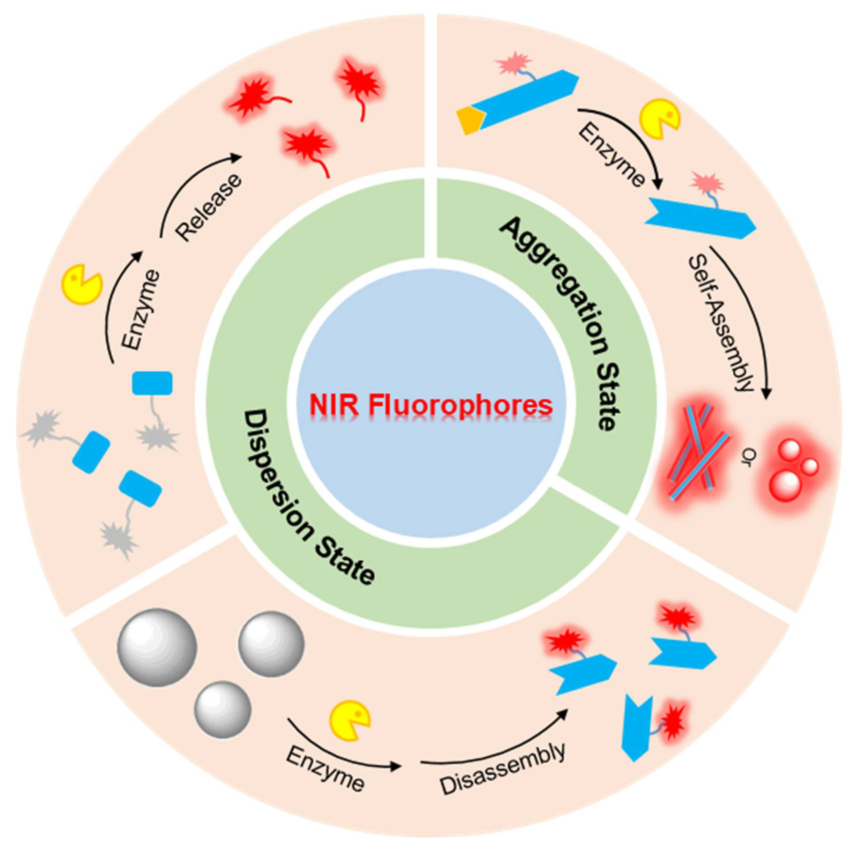
:1. Introduction
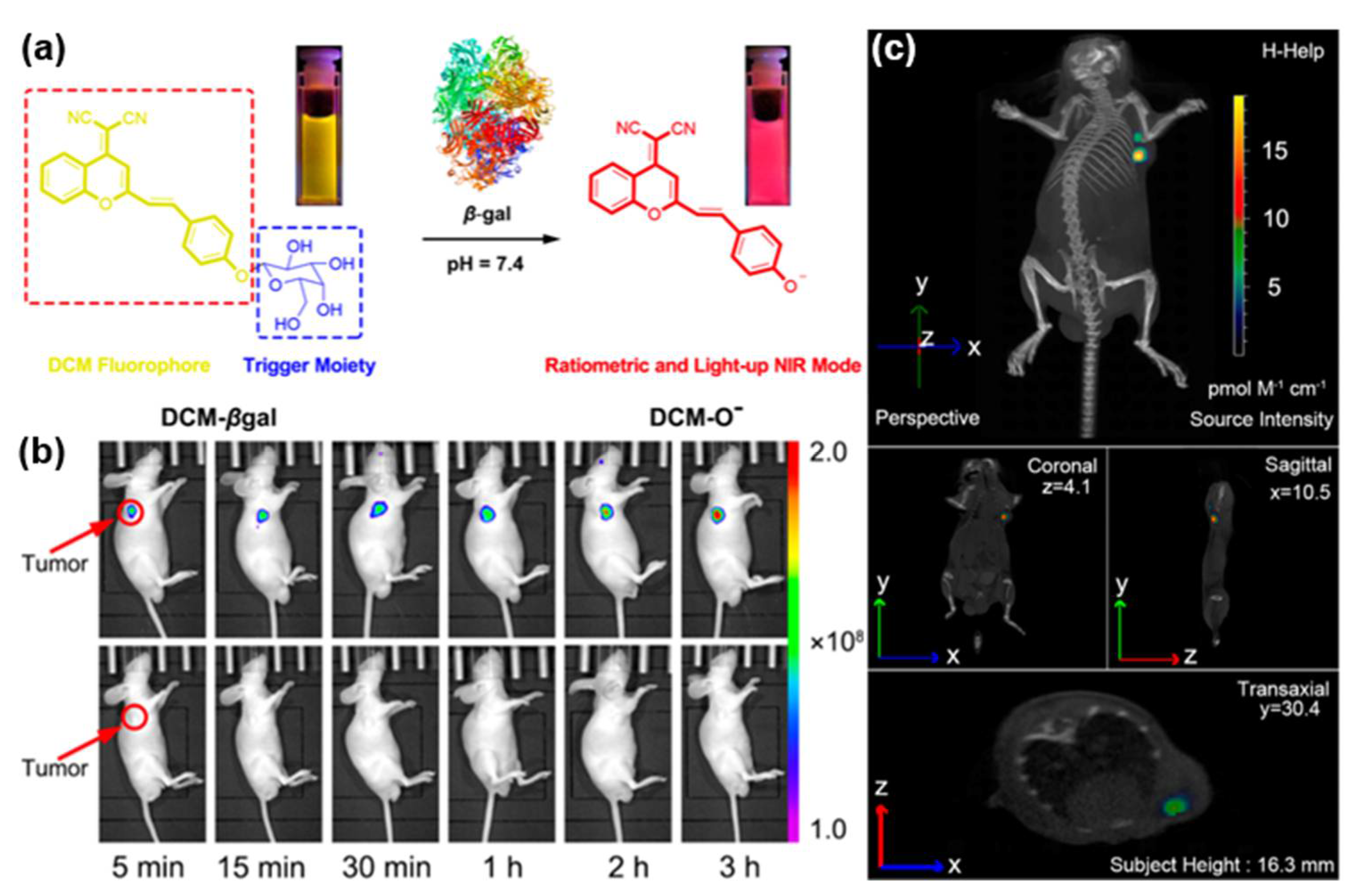
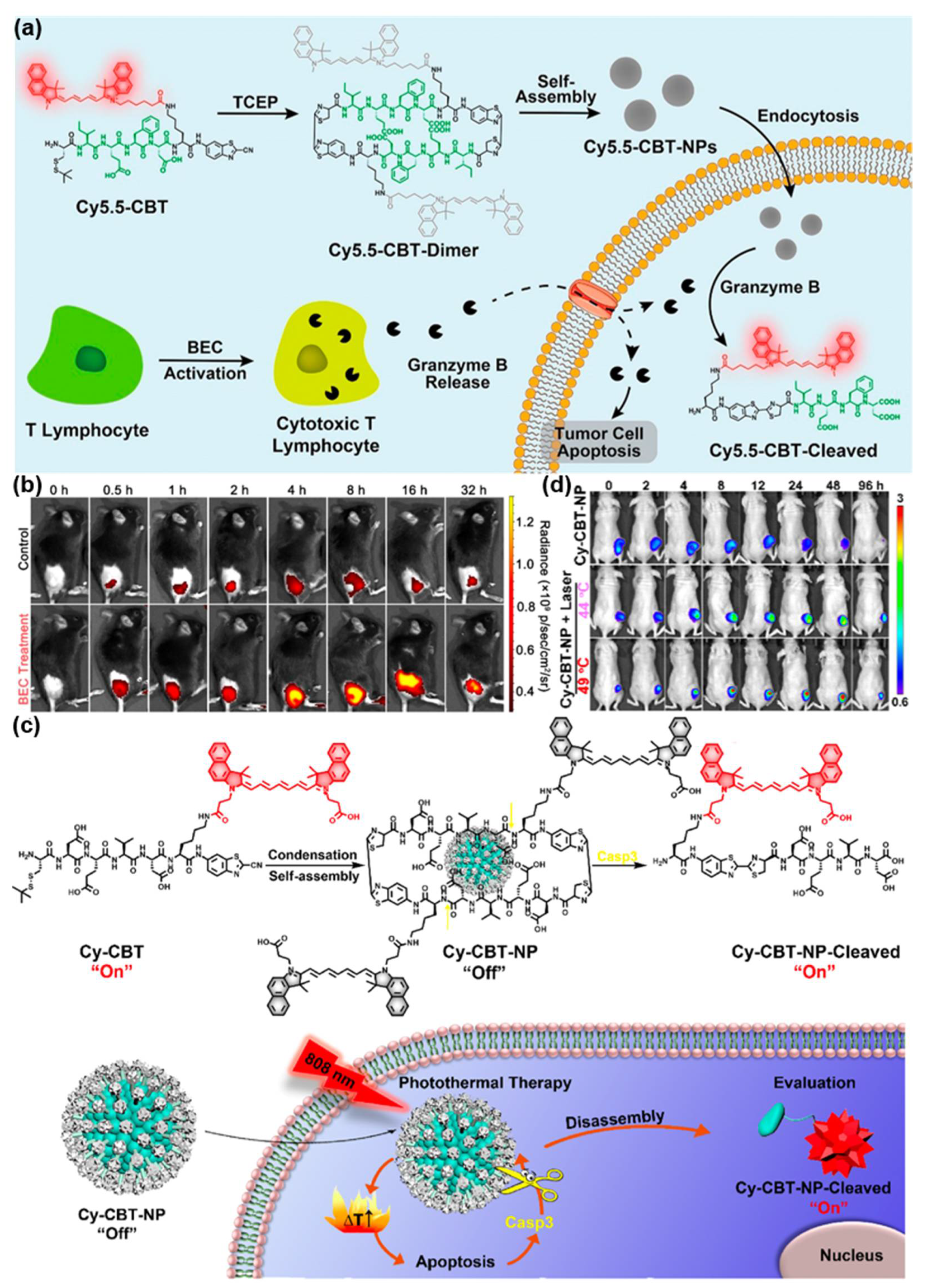
2. Enzyme-Instructed Release of Free NIR Fluorophores In Vivo
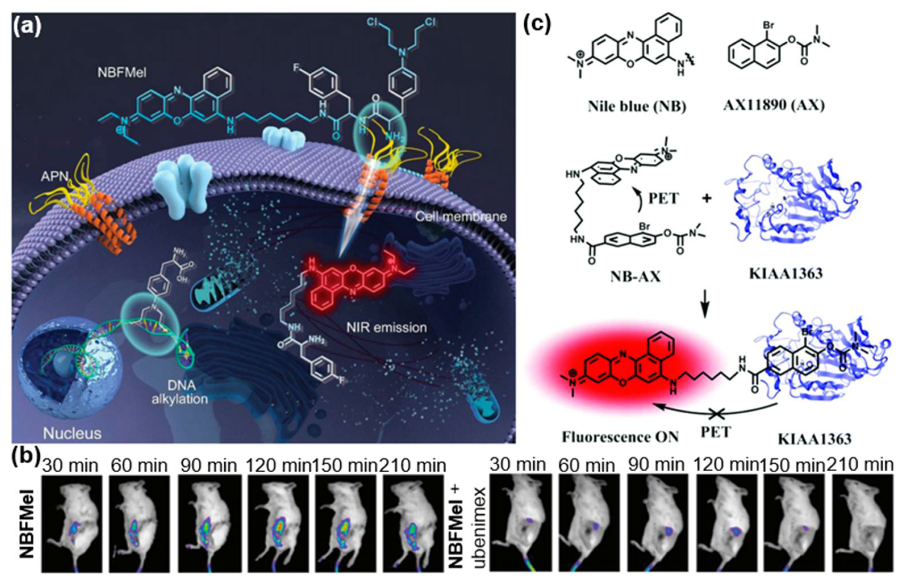
3. Enzyme-Instructed Self-Assembly of NIR Fluorophores In Vivo
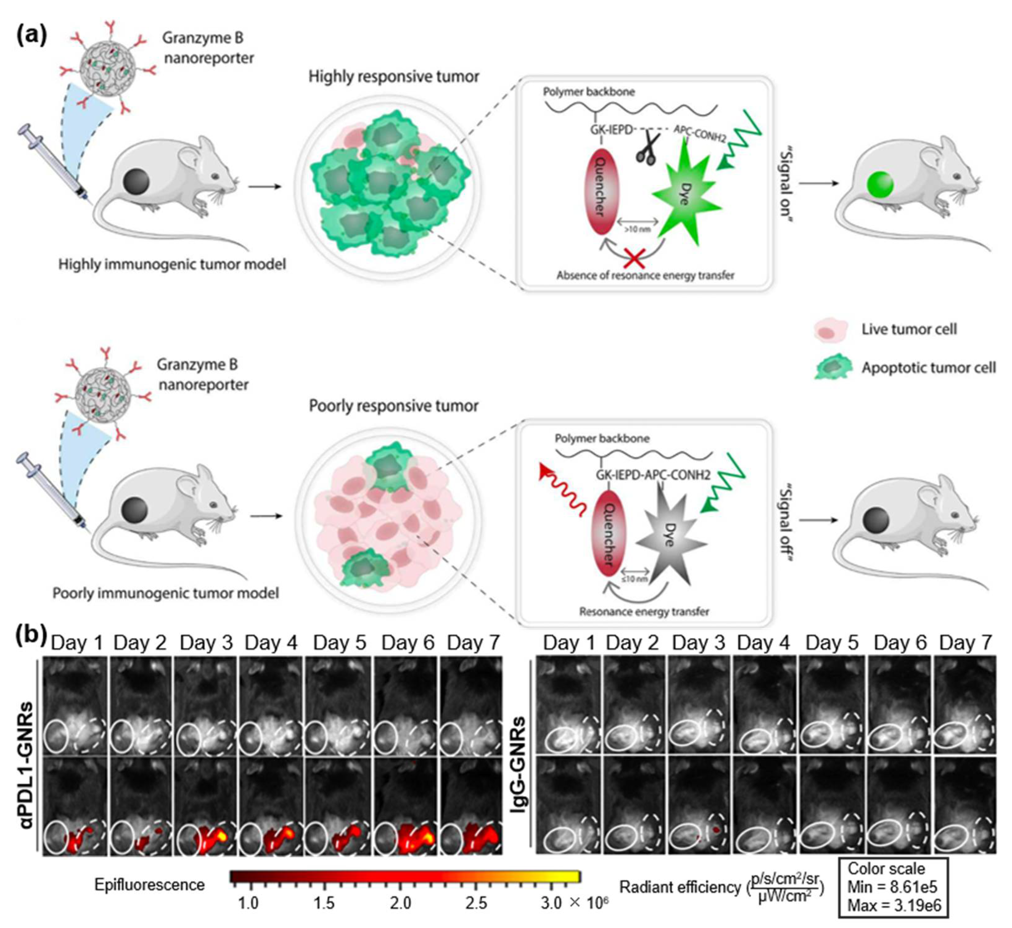
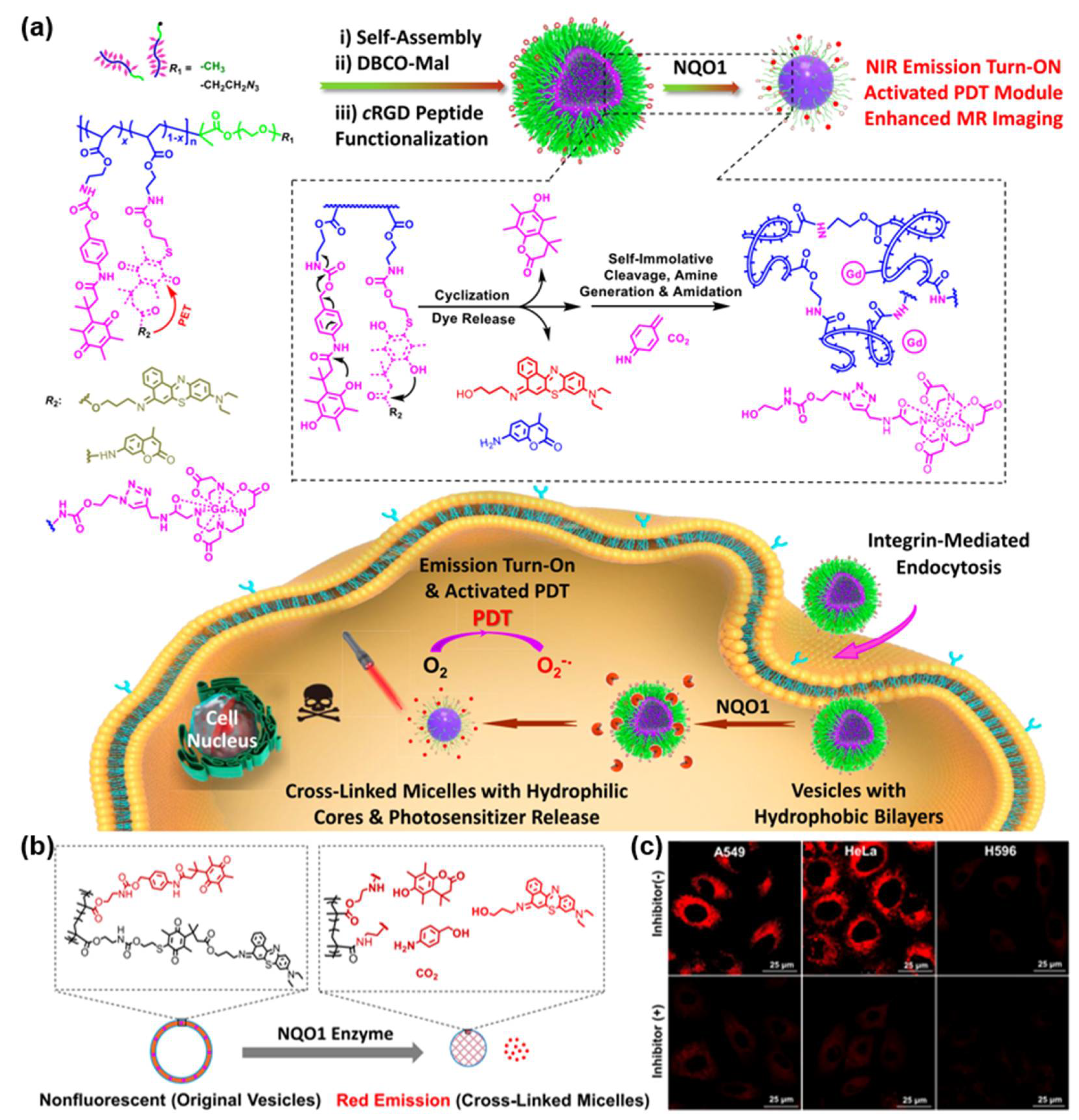
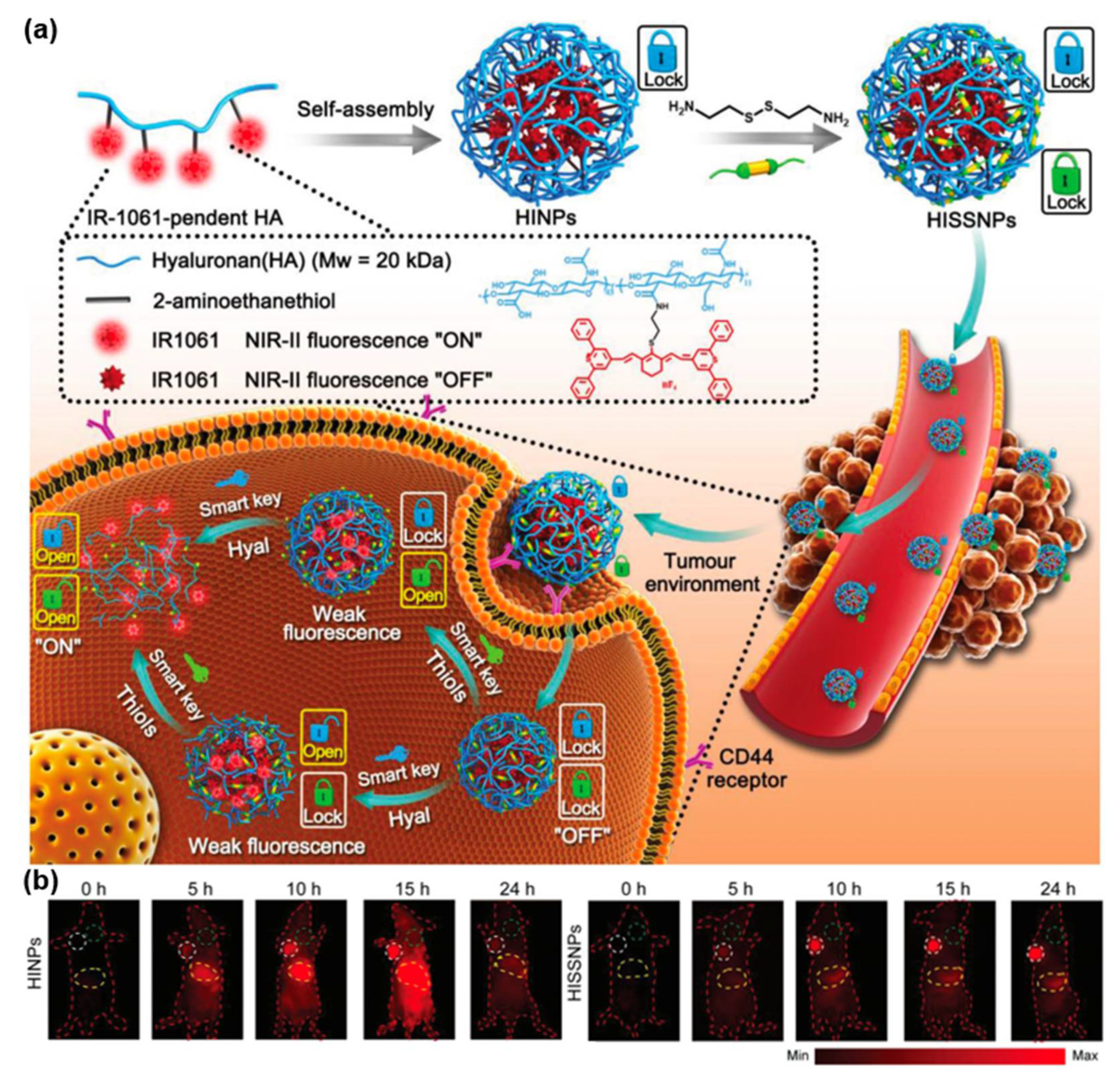
4. Enzyme-Instructed Disassembly of NIR Fluorophores In Vivo
5. Conclusions
Author Contributions
Funding
Institutional Review Board Statement
Informed Consent Statement
Data Availability Statement
Conflicts of Interest
References
- Li, C.; Pang, Y.; Xu, Y.; Lu, M.; Tu, L.; Li, Q.; Sharma, A.; Guo, Z.; Li, X.; Sun, Y. Near-infrared metal agents assisting precision medicine: From strategic design to bioimaging and therapeutic applications. Chem. Soc. Rev. 2023, 52, 4392–4442. [Google Scholar] [CrossRef] [PubMed]
- Xu, Y.; Tuo, W.; Yang, L.; Sun, Y.; Li, C.; Chen, X.; Yang, W.; Yang, G.; Stang, P.J.; Sun, Y. Design of a Metallacycle-Based Supramolecular Photosensitizer for In Vivo Image-Guided Photodynamic Inactivation of Bacteria. Angew. Chem. Int. Ed. 2022, 61, e202110048. [Google Scholar]
- Li, H.; Kim, Y.; Jung, H.; Hyun, J.Y.; Shin, I. Near-infrared (NIR) fluorescence-emitting small organic molecules for cancer imaging and therapy. Chem. Soc. Rev. 2022, 51, 8957–9008. [Google Scholar] [CrossRef]
- Li, K.; Xu, S.; Xiong, M.; Huang, S.; Yuan, L.; Zhang, B. Molecular engineering of organic-based agents for in situ bioimaigng and phototherapeutics. Chem. Soc. Rev. 2021, 50, 11766–11784. [Google Scholar] [CrossRef]
- Xu, Y.; Li, C.; An, J.; Ma, X.; Yang, J.; Luo, L.; Deng, Y.; Kim, J.S.; Sun, Y. Construction of a 980 nm laser-activated Pt(II) metallacycle nanosystem for efficient and safe photo-induced bacteria sterilization. Sci. China Chem. 2023, 66, 155–163. [Google Scholar] [CrossRef]
- Guo, Z.; Park, S.; Yoon, J.; Shin, I. Recent progress in the development of near-infrared fluorescent probes for bioimaging applications. Chem. Soc. Rev. 2014, 43, 16–29. [Google Scholar] [CrossRef]
- Weng, J.; Wang, Y.; Zhang, Y.; Ye, D. An Activatable Near-Infrared Fluorescence Probe for in Vivo Imaging of Acute Kidney Injury by Targeting Phosphatidylserine and Caspase-3. J. Am. Chem. Soc. 2021, 143, 18294–18304. [Google Scholar] [CrossRef] [PubMed]
- Xu, Y.; Li, C.; Ma, X.; Tuo, W.; Tu, L.; Li, X.; Sun, Y.; Stang, P.J.; Sun, Y. Long wavelength-emissive Ru(II) metallacycle-based photosensitizer assisting in vivo bacterial diagnosis and antibacterial treatment. Proc. Natl. Acad. Sci. USA 2022, 119, e2209904119. [Google Scholar] [CrossRef]
- He, L.; He, L.; Xu, S.; Ren, T.; Zhang, X.; Qin, Z.; Zhang, X.; Yuan, L. Engineering of Reversible NIR-II Redox-Responsive Fluorescent Probes for Imaging of Inflammation In Vivo. Angew. Chem. Int. Ed. 2022, 61, e202211409. [Google Scholar] [CrossRef]
- Li, C.; Xu, L.; Tu, L.; Choi, M.; Fan, Y.; Chen, X.; Sessler, J.L.; Kim, J.S.; Sun, Y. Rationally designed Ru(II)-metallacycle chemo-phototheranostic that emits beyond 1000 nm. Chem. Sci. 2022, 13, 6541–6549. [Google Scholar] [CrossRef]
- Du, J.; Hu, M.; Fan, J.; Peng, X. Fluorescent chemodosimeters using “mild” chemical events for the detection of small anions and cations in biological and environmental media. Chem. Soc. Rev. 2012, 41, 4511–4535. [Google Scholar] [CrossRef]
- Urano, Y.; Asanuma, D.; Hama, Y.; Koyama, Y.; Barrett, T.; Kamiya, M.; Nagano, T.; Watanabe, T.; Hasegawa, A.; Choyke, P.L.; et al. Selective molecular imaging of viable cancer cells with pH-activatable fluorescence probes. Nat. Med. 2009, 15, 104–109. [Google Scholar] [CrossRef] [Green Version]
- Zhang, Z.; Guan, R.; Li, J.; Sun, Y. Engineering Rational SERS Nanotags for Parallel Detection of Multiple Cancer Circulating Biomarkers. Chemosensors 2023, 11, 110. [Google Scholar] [CrossRef]
- Luo, R.; Ou, C.; Li, X.; Wang, Y.; Du, W.; Liang, G.; Gong, C. An acidity-initiated self-assembly/disassembly nanoprobe to switch on fluorescence for tumor-targeted near-infrared imaging. Nano Lett. 2022, 22, 151–156. [Google Scholar] [CrossRef]
- Zhang, Y.; Bi, J.; Xia, S.; Mazi, W.; Wan, S.; Mikesell, L.; Luck, R.L.; Liu, H. A near-infrared fluorescent probe based on a FRET rhodamine donor linked to a cyanine acceptor for sensitive detection of intracellular pH alternations. Molecules 2018, 23, 2679. [Google Scholar] [CrossRef] [Green Version]
- Xu, L.; Zhan, W.; Deng, Y.; Liu, X.; Gao, G.; Sun, X.; Liang, G. ROS turn nanoparticle fluorescence on for imaging Staphylococcus aureus infection in vivo. Adv. Healthc. Mater. 2022, 11, 2200453. [Google Scholar] [CrossRef]
- Pang, Y.; Li, C.; Deng, H.; Sun, Y. Recent advances in luminescent metallacycles/metallacages for biomedical imaging and cancer therapy. Dalton Trans. 2022, 51, 16428–16438. [Google Scholar] [CrossRef] [PubMed]
- Liu, K.; Shang, H.; Kong, X.; Ren, M.; Wang, J.-Y.; Liu, Y.; Lin, W. A novel near-infrared fluorescent probe for H2O2 in alkaline environment and the application for H2O2 imaging in vitro and in vivo. Biomaterials 2016, 100, 162–171. [Google Scholar] [CrossRef] [PubMed]
- Huang, Y.; Zhang, Y.; Huo, F.; Chao, J.; Yin, C. A near-infrared ratiometric fluorescent probe with large stokes based on isophorone for rapid detection of ClO− and its bioimaging in cell and mice. Sens. Actuator B-Chem. 2019, 287, 453–458. [Google Scholar] [CrossRef]
- Ding, W.; Yao, S.; Chen, Y.; Wu, Y.; Li, Y.; He, W.; Guo, Z. A near-infrared fluorescent and photoacoustic probe for visualizing biothiols dynamics in tumor and liver. Molecules 2023, 28, 2229. [Google Scholar] [CrossRef] [PubMed]
- Zhang, X.; Li, C.; Zhang, Y.; Guan, X.; Mei, L.; Feng, H.; Li, J.; Tu, L.; Feng, G.; Deng, G.; et al. Construction of Long-Wavelength Emissive Organic Nanosonosensitizer Targeting Mitochondria for Precise and Efficient In Vivo Sonotherapy. Adv. Funct. Mater. 2022, 32, 2207259. [Google Scholar] [CrossRef]
- Wu, X.; Sun, X.; Guo, Z.; Tang, J.; Shen, Y.; James, T.D.; Tian, H.; Zhu, W. In vivo and in situ tracking cancer chemotherapy by highly photostable NIR fluorescent theranostic prodrug. J. Am. Chem. Soc. 2014, 136, 3579–3588. [Google Scholar] [CrossRef]
- Yin, J.; Kwon, Y.; Kim, D.; Lee, D.; Kim, G.; Hu, Y.; Ryu, J.H.; Yoon, J. Cyanine-based fluorescent probe for highly selective detection of glutathione in cell cultures and live mouse tissues. J. Am. Chem. Soc. 2014, 136, 5351–5358. [Google Scholar] [CrossRef]
- Lee, H.; Kim, J.; Kim, H.; Kim, Y.; Choi, Y. A folate receptor-specific activatable probe for near-infrared fluorescence imaging of ovarian cancer. Chem. Commun. 2014, 50, 7507–7510. [Google Scholar] [CrossRef]
- Li, Z.; He, X.; Wang, Z.; Yang, R.; Shi, W.; Ma, H. In vivo imaging and detection of nitroreductase in zebrafish by a new near-infrared fluorescence off–on probe. Biosens. Bioelectron. 2015, 63, 112–116. [Google Scholar] [CrossRef] [PubMed]
- Zeng, W.; Ye, D. Seeing cancer via sonoafterglow. Nat. Biomed. Eng. 2023, 7, 197–198. [Google Scholar] [CrossRef]
- Zeng, Z.; Mizukami, S.; Fujita, K.; Kikuchi, K. An enzyme-responsive metal-enhanced near-infrared fluorescence sensor based on functionalized gold nanoparticles. Chem. Sci. 2015, 6, 4934–4939. [Google Scholar] [CrossRef] [Green Version]
- Zhen, X.; Zhang, J.; Huang, J.; Xie, C.; Miao, Q.; Pu, K. Macrotheranostic probe with disease-activated near-infrared fluorescence, photoacoustic, and photothermal signals for imaging-guided therapy. Angew. Chem. Int. Ed. 2018, 130, 7930–7934. [Google Scholar] [CrossRef]
- Li, Y.; Song, H.; Xue, C.; Fang, Z.; Xiong, L.; Xie, H. A self-immobilizing near-infrared fluorogenic probe for sensitive imaging of extracellular enzyme activity in vivo. Chem. Sci. 2020, 11, 5889–5894. [Google Scholar] [CrossRef] [PubMed]
- Liu, H.W.; Chen, L.; Xu, C.; Li, Z.; Zhang, H.; Zhang, B.; Tan, W. Recent progresses in small-molecule enzymatic fluorescent probes for cancer imaging. Chem. Soc. Rev. 2018, 47, 7140–7180. [Google Scholar] [CrossRef]
- Hanash, S. Disease proteomics. Nature 2003, 422, 226–232. [Google Scholar] [CrossRef] [Green Version]
- Zhao, J.; Ma, T.; Chang, B.; Fang, J. Recent progress on NIR fluorescent probes for enzymes. Molecules 2022, 27, 5922. [Google Scholar] [CrossRef]
- Dong, Z.; Wang, Y.; Wan, W.; Wu, J.; Wang, B.; Zhu, H.; Xie, M.; Liu, L. Resveratrol ameliorates oxaliplatin-induced neuropathic pain via anti-inflammatory effects in rats. Exp. Ther. Med. 2022, 24, 1–10. [Google Scholar] [CrossRef] [PubMed]
- Zhang, J.; Cheng, P.; Pu, K. Recent advances of molecular optical probes in imaging of β-galactosidase. Bioconjugate Chem. 2019, 30, 2089–2101. [Google Scholar] [CrossRef] [PubMed]
- Yao, Y.; Zhang, Y.; Yan, C.; Zhu, W.-H.; Guo, Z. Enzyme-activatable fluorescent probes for β-galactosidase: From design to biological applications. Chem. Sci. 2021, 12, 9885–9894. [Google Scholar] [CrossRef] [PubMed]
- Ichim, G.; Tait, S.W.G. A fate worse than death: Apoptosis as an oncogenic process. Nat. Rev. Cancer 2016, 16, 539–548. [Google Scholar] [CrossRef] [Green Version]
- Wang, P.; Yang, H.; Liu, C.; Qiu, M.; Ma, X.; Mao, Z.; Sun, Y.; Liu, Z. Recent advances in the development of activatable multifuntional probes for in vivo imaging of caspase-3. Chin. Chem. Lett. 2021, 32, 168–178. [Google Scholar] [CrossRef]
- Wang, Y.; Gao, W.; Shi, X.; Ding, J.; Liu, W.; He, H.; Wang, K.; Shao, F. Chemotherapy drugs induce pyroptosis through caspase-3 cleavage of a gasdermin. Nature 2017, 547, 99–103. [Google Scholar] [CrossRef]
- Li, C.; Tu, L.; Yang, J.; Liu, C.; Xu, Y.; Li, J.; Tuo, W.; Olenyuk, B.; Sun, Y.; Stang, P.J.; et al. Acceptor engineering of metallacycles with high phototoxicity indices for safe and effective photodynamic therapy. Chem. Sci. 2023, 14, 2901–2909. [Google Scholar] [CrossRef]
- Fu, Q.; Feng, H.; Su, L.; Zhang, X.; Liu, L.; Fu, F.; Yang, H.; Song, J. An activatable hybrid organic–inorganic nanocomposite as early evaluation system of therapy effect. Angew. Chem.Int. Ed. 2022, 61, e202112237. [Google Scholar] [CrossRef]
- Gao, T.; Yi, L.; Wang, Y.; Wang, W.; Zhao, Q.; Song, Y.; Ding, M.; Deng, C.; Chen, Y.; Xie, Y.; et al. Granzyme B-responsive fluorescent probe for non-invasive early diagnosis of transplant rejection. Biosens. Bioelectron. 2023, 232, 115303. [Google Scholar] [CrossRef]
- Chen, Y.; Pei, P.; Yang, Y.; Zhang, H.; Zhang, F. Noninvasive early diagnosis of allograft rejection by a granzyme B protease responsive NIR-II bioimaging nanosensor. Angew. Chem.Int. Ed. 2023, 62, e202301696. [Google Scholar] [CrossRef]
- Nguyen, A.; Ramesh, A.; Kumar, S.; Nandi, D.; Brouillard, A.; Wells, A.; Pobezinsky, L.; Osborne, B.; Kulkarni, A.A. Granzyme B nanoreporter for early monitoring of tumor response to immunotherapy. Sci. Adv. 2020, 6, eabc2777. [Google Scholar] [CrossRef] [PubMed]
- Xu, L.; Liu, N.; Zhan, W.; Deng, Y.; Chen, Z.; Liu, X.; Gao, G.; Chen, Q.; Liu, Z.; Liang, G. Granzyme B turns nanoparticle fluorescence “on” for imaging cytotoxic T lymphocyte activity in vivo. ACS Nano 2022, 16, 19328–19334. [Google Scholar] [CrossRef] [PubMed]
- Mills, B.; Norberg, D.; Dhaliwal, K.; Akram, A.R.; Bradley, M.; Megia-Fernandez, A. A matrix metalloproteinase activation probe for painting human tumours. Chem. Commun. 2020, 56, 9962–9965. [Google Scholar] [CrossRef]
- Myochin, T.; Hanaoka, K.; Komatsu, T.; Terai, T.; Nagano, T. Design strategy for a near-infrared fluorescence probe for matrix metalloproteinase utilizing highly cell permeable boron dipyrromethene. J. Am. Chem. Soc. 2012, 134, 13730–13737. [Google Scholar] [CrossRef] [PubMed]
- Zhao, X.; Yang, C.; Chen, L.; Yan, X. Dual-stimuli responsive and reversibly activatable theranostic nanoprobe for precision tumor-targeting and fluorescence-guided photothermal therapy. Nat. Commun. 2017, 8, 14998. [Google Scholar] [CrossRef] [PubMed] [Green Version]
- Chen, J.; Chen, L.; Zeng, F.; Wu, S. Aminopeptidase N activatable nanoprobe for tracking lymphatic metastasis and guiding tumor resection surgery via optoacoustic/NIR-II fluorescence dual-mode imaging. Anal. Chem. 2022, 94, 8449–8457. [Google Scholar] [CrossRef]
- Li, H.; Yao, Q.; Sun, W.; Shao, K.; Lu, Y.; Chung, J.; Kim, D.; Fan, J.; Long, S.; Du, J.; et al. Aminopeptidase N activatable fluorescent probe for tracking metastatic cancer and image-guided surgery via in situ spraying. J. Am. Chem. Soc. 2020, 142, 6381–6389. [Google Scholar] [CrossRef]
- Algar, W.R.; Hildebrandt, N.; Vogel, S.S.; Medintz, I.L. FRET as a biomolecular research tool—Understanding its potential while avoiding pitfalls. Nat. Methods 2019, 16, 815–829. [Google Scholar] [CrossRef]
- Myochin, T.; Hanaoka, K.; Iwaki, S.; Ueno, T.; Komatsu, T.; Terai, T.; Nagano, T.; Urano, Y. Development of a series of near-infrared dark quenchers based on Si-rhodamines and their application to fluorescent probes. J. Am. Chem. Soc. 2015, 137, 4759–4765. [Google Scholar] [CrossRef]
- Tuo, W.; Xu, Y.; Fan, Y.; Li, J.; Qiu, M.; Xiong, X.; Li, X.; Sun, Y. Biomedical applications of Pt(II) metallacycle/metallacage-based agents: From mono-chemotherapy to versatile imaging contrasts and theranostic platforms. Coord. Chem. Rev. 2021, 443, 214017. [Google Scholar] [CrossRef]
- Sun, W.; Li, M.; Fan, J.; Peng, X. Activity-based sensing and theranostic probes based on photoinduced electron transfer. Acc. Chem. Res. 2019, 52, 2818–2831. [Google Scholar] [CrossRef] [PubMed]
- Xiao, M.; Sun, W.; Fan, J.; Cao, J.; Li, Y.; Shao, K.; Li, M.; Li, X.; Kang, Y.; Zhang, W.; et al. Aminopeptidase-N-activated theranostic prodrug for NIR tracking of local tumor chemotherapy. Adv. Funct. Mater. 2018, 28, 1805128. [Google Scholar] [CrossRef]
- Fan, J.; Guo, S.; Wang, S.; Kang, Y.; Yao, Q.; Wang, J.; Gao, X.; Wang, H.; Du, J.; Peng, X. Lighting-up breast cancer cells by a near-infrared fluorescent probe based on KIAA1363 enzyme-targeting. Chem. Commun. 2017, 53, 4857–4860. [Google Scholar] [CrossRef]
- Pei, X.; Pan, Y.; Zhang, L.; Lv, Y. Recent advances in ratiometric luminescence sensors. Appl. Spectrosc. Rev. 2021, 56, 324–345. [Google Scholar] [CrossRef]
- Gu, K.; Xu, Y.; Li, H.; Guo, Z.; Zhu, S.; Zhu, S.; Shi, P.; James, T.D.; Tian, H.; Zhu, W.H. Real-time tracking and in vivo visualization of β-galactosidase activity in colorectal tumor with a ratiometric near-infrared fluorescent probe. J. Am. Chem. Soc. 2016, 138, 5334–5340. [Google Scholar] [CrossRef] [Green Version]
- He, L.; Dong, B.; Liu, Y.; Lin, W. Fluorescent chemosensors manipulated by dual/triple interplaying sensing mechanisms. Chem. Soc. Rev. 2016, 45, 6449–6461. [Google Scholar] [CrossRef] [PubMed]
- Yao, C.; Li, Y.; Wang, Z.; Song, C.; Hu, X.; Liu, S. Cytosolic NQO1 enzyme-activated near-infrared fluorescence imaging and photodynamic therapy with polymeric vesicles. ACS Nano 2020, 14, 1919–1935. [Google Scholar] [CrossRef]
- Sletten, E.M.; Bertozzi, C.R. Bioorthogonal chemistry: Fishing for selectivity in a sea of functionality. Angew. Chem. Int. Ed. 2009, 48, 6974–6998. [Google Scholar] [CrossRef] [Green Version]
- Liang, G.; Ren, H.; Rao, J. A biocompatible condensation reaction for controlled assembly of nanostructures in living cells. Nat. Chem. 2010, 2, 54–60. [Google Scholar] [CrossRef]
- Zhang, M.; Liang, G. Applications of CBT-Cys click reaction: Past, present, and future. Sci. China. Chem. 2018, 61, 1088–1098. [Google Scholar] [CrossRef]
- Ye, D.; Shuhendler, A.J.; Cui, L.; Tong, L.; Tee, S.S.; Tikhomirov, G.; Felsher, D.W.; Rao, J. Bioorthogonal cyclization-mediated in situ self-assembly of small-molecule probes for imaging caspase activity in vivo. Nat. Chem. 2014, 6, 519–526. [Google Scholar] [CrossRef] [Green Version]
- Chen, Z.; Chen, M.; Cheng, Y.; Kowada, T.; Xie, J.; Zheng, X.; Rao, J. Exploring the condensation reaction between aromatic nitriles and amino thiols to optimize in situ nanoparticle formation for the imaging of proteases and glycosidases in cells. Angew. Chem. Int. Ed. 2020, 59, 3272–3279. [Google Scholar] [CrossRef]
- Chen, Z.; Chen, M.; Zhou, K.; Rao, J. Pre-targeted imaging of protease activity through in situ assembly of nanoparticles. Angew. Chem. Int. Ed. 2020, 59, 7864–7870. [Google Scholar] [CrossRef]
- Jiang, Q.; Liu, X.; Liang, G.; Sun, X. Self-assembly of peptide nanofibers for imaging applications. Nanoscale 2021, 13, 15142–15150. [Google Scholar] [CrossRef] [PubMed]
- Deng, Y.; Zhan, W.; Liang, G. Intracellular self-assembly of peptide conjugates for tumor imaging and therapy. Adv. Healthc. Mater. 2021, 10, 2001211. [Google Scholar] [CrossRef] [PubMed]
- An, H.-W.; Hou, D.; Zheng, R.; Wang, M.-D.; Zeng, X.-Z.; Xiao, W.-Y.; Yan, T.-D.; Wang, J.-Q.; Zhao, C.-H.; Cheng, L.-M.; et al. A near-infrared peptide probe with tumor-specific excretion-retarded effect for image-guided surgery of renal cell carcinoma. ACS Nano 2020, 14, 927–936. [Google Scholar] [CrossRef] [PubMed]
- Ren, H.; Zeng, X.-Z.; Zhao, X.-X.; Hou, D.-Y.; Yao, H.; Yaseen, M.; Zhao, L.; Xu, W.-H.; Wang, H.; Li, L.-L. A bioactivated in vivo assembly nanotechnology fabricated NIR probe for small pancreatic tumor intraoperative imaging. Nat. Commun. 2022, 13, 418. [Google Scholar] [CrossRef] [PubMed]
- Zhao, X.-X.; Li, L.-L.; Zhao, Y.; An, H.-W.; Cai, Q.; Lang, J.-Y.; Han, X.-X.; Peng, B.; Fei, Y.; Liu, H.; et al. In situ self-assembled nanofibers precisely target cancer-associated fibroblasts for improved tumor imaging. Angew. Chem. Int. Ed. 2019, 58, 15287–15294. [Google Scholar] [CrossRef] [PubMed]
- Zheng, R.; Yang, J.; Mamuti, M.; Hou, D.-Y.; An, H.-W.; Zhao, Y.; Wang, H. Controllable self-assembly of peptide-cyanine conjugates in vivo as fine-tunable theranostics. Angew. Chem. Int. Ed. 2021, 133, 7888–7898. [Google Scholar] [CrossRef]
- Li, K.; Lyu, Y.; Huang, Y.; Xu, S.; Liu, H.-W.; Chen, L.; Ren, T.-B.; Xiong, M.; Huan, S.; Yuan, L.; et al. A de novo strategy to develop NIR precipitating fluorochrome for long-term in situ cell membrane bioimaging. Proc. Natl. Acad. Sci. USA 2021, 118, e2018033118. [Google Scholar] [CrossRef]
- Gao, Z.; Gao, H.; Zheng, D.; Xu, T.; Chen, Y.; Liang, C.; Wang, L.; Ding, D.; Yang, Z. β-galactosidase responsive AIE fluorogene for identification and removal of senescent cancer cells. Sci. China Chem. 2020, 63, 398–403. [Google Scholar] [CrossRef]
- Yuan, Y.; Zhang, J.; Cao, Q.; An, L.; Liang, G. Intracellular disassembly of self-quenched nanoparticles turns NIR fluorescence on for sensing furin activity in cells and in tumors. Anal. Chem. 2015, 87, 6180–6185. [Google Scholar] [CrossRef] [PubMed]
- Jiang, J.; Zhao, Z.; Hai, Z.; Wang, H.; Liang, G. Intracellular proteolytic disassembly of self-quenched near-infrared nanoparticles turning fluorescence on for tumor-targeted imaging. Anal. Chem. 2017, 89, 9625–9628. [Google Scholar] [CrossRef] [PubMed]
- Zhao, Y.; Hai, Z.; Wang, H.; Su, L.; Liang, G. Legumain-specific near-infrared fluorescence “turn on” for tumor-targeted imaging. Anal. Chem. 2018, 90, 8732–8735. [Google Scholar] [CrossRef] [Green Version]
- Wang, Y.; Du, W.; Zhang, T.; Zhu, Y.; Ni, Y.; Wang, C.; Sierra Raya, F.M.; Zou, L.; Wang, L.; Liang, G. A self-evaluating photothermal therapeutic nanoparticle. ACS Nano 2020, 14, 9585–9593. [Google Scholar] [CrossRef] [PubMed]
- Shimizu, Y.; Temma, T.; Hara, I.; Makino, A.; Kondo, N.; Ozeki, E.-I.; Ono, M.; Saji, H. In vivo imaging of membrane type-1 matrix metalloproteinase with a novel activatable near-infrared fluorescence probe. Cancer Sci. 2014, 105, 1056–1062. [Google Scholar] [CrossRef] [Green Version]
- Zhang, T.; Zhang, W.; Zheng, M.; Xie, Z. Near-infrared BODIPY-paclitaxel conjugates assembling organic nanoparticles for chemotherapy and bioimaging. J. Colloid Interface Sci. 2018, 514, 584–591. [Google Scholar] [CrossRef]
- Tang, Y.; Li, Y.; Hu, X.; Zhao, H.; Ji, Y.; Chen, L.; Hu, W.; Zhang, W.; Li, X.; Lu, X.; et al. “Dual lock-and-key”-controlled nanoprobes for ultrahigh specific fluorescence imaging in the second near-infrared window. Adv. Mater. 2018, 30, 1801140. [Google Scholar] [CrossRef]
- Mu, J.; Xiao, M.; Shi, Y.; Geng, X.; Li, H.; Yin, Y.; Chen, X. The chemistry of organic contrast agents in the NIR-II window. Angew. Chem. Int. Ed. 2022, 61, e202114722. [Google Scholar] [CrossRef] [PubMed]
- Fan, Y.; Li, C.; Bai, S.; Ma, X.; Yang, J.; Guan, X.; Sun, Y. NIR-II Emissive Ru(II) Metallacycle Assisting Fluorescence Imaging and Cancer Therapy. Small 2022, 18, 2201625. [Google Scholar] [CrossRef] [PubMed]
- Tu, L.; Li, C.; Xiong, X.; Kim, J.H.; Li, Q.; Mei, L.; Li, J.; Liu, S.; Kim, J.S.; Sun, Y. Engineered Metallacycle-Based Supramolecular Photosensitizers for Effective Photodynamic Therapy. Angew. Chem. Int. Ed. 2023, 62, e202301560. [Google Scholar] [CrossRef]
- Singh, H.; Tiwari, K.; Tiwari, R.; Pramanik, S.K.; Das, A. Small molecule as fluorescent probes for monitoring intracellular enzymatic transformations. Chem. Rev. 2019, 119, 11718–11760. [Google Scholar] [CrossRef] [PubMed]
- Zhao, M.; Li, B.; Fan, Y.; Zhang, F. In vivo assembly and disassembly of probes to improve near-infrared optical bioimaging. Adv. Healthc. Mater. 2019, 8, 1801650. [Google Scholar] [CrossRef] [PubMed]


Disclaimer/Publisher’s Note: The statements, opinions and data contained in all publications are solely those of the individual author(s) and contributor(s) and not of MDPI and/or the editor(s). MDPI and/or the editor(s) disclaim responsibility for any injury to people or property resulting from any ideas, methods, instructions or products referred to in the content. |
© 2023 by the authors. Licensee MDPI, Basel, Switzerland. This article is an open access article distributed under the terms and conditions of the Creative Commons Attribution (CC BY) license (https://creativecommons.org/licenses/by/4.0/).
Share and Cite
Zhang, Z.; Chen, P.; Sun, Y. Enzyme-Instructed Aggregation/Dispersion of Fluorophores for Near-Infrared Fluorescence Imaging In Vivo. Molecules 2023, 28, 5360. https://doi.org/10.3390/molecules28145360
Zhang Z, Chen P, Sun Y. Enzyme-Instructed Aggregation/Dispersion of Fluorophores for Near-Infrared Fluorescence Imaging In Vivo. Molecules. 2023; 28(14):5360. https://doi.org/10.3390/molecules28145360
Chicago/Turabian StyleZhang, Zhipeng, Peiyao Chen, and Yao Sun. 2023. "Enzyme-Instructed Aggregation/Dispersion of Fluorophores for Near-Infrared Fluorescence Imaging In Vivo" Molecules 28, no. 14: 5360. https://doi.org/10.3390/molecules28145360