Article

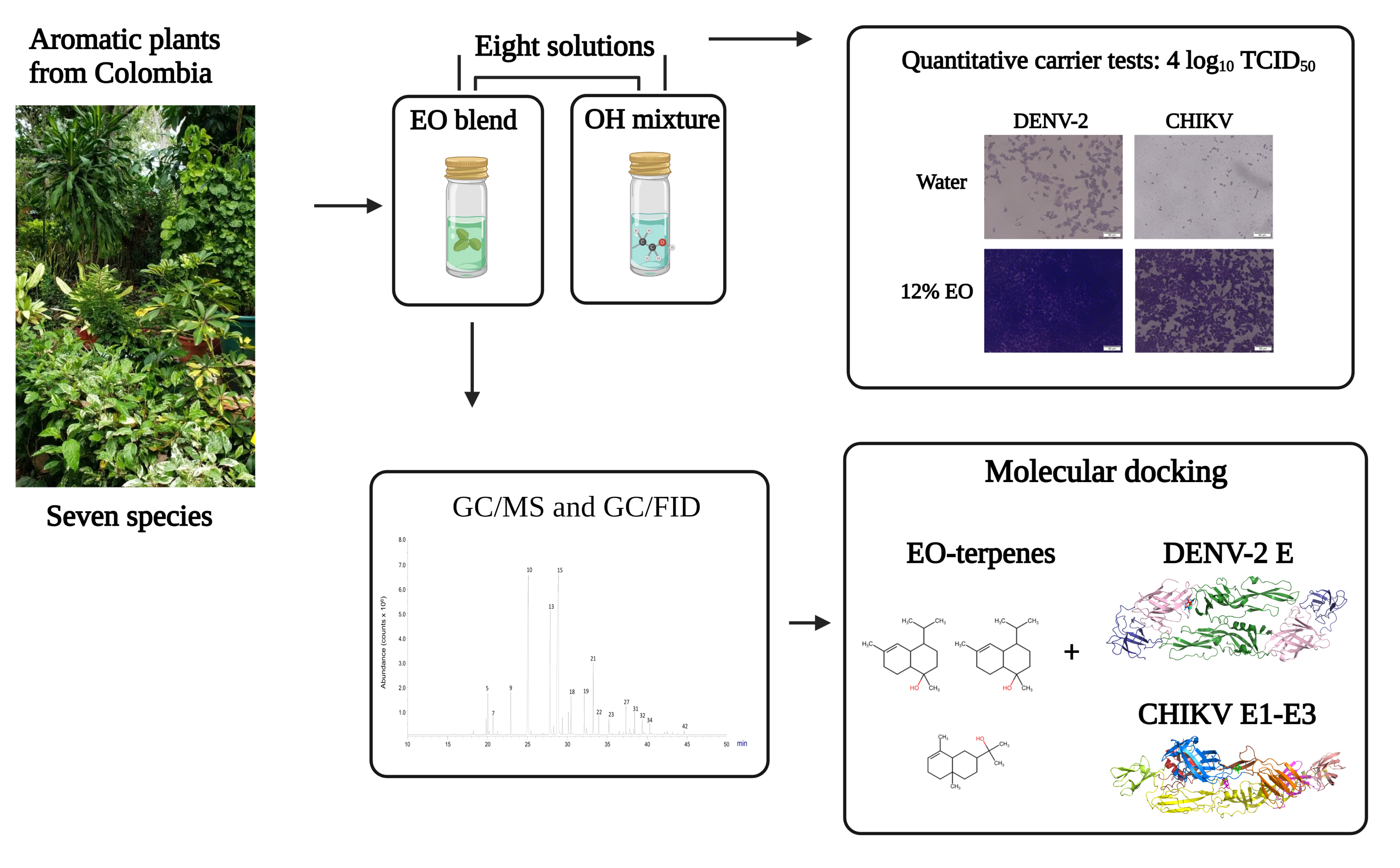
Comparative Virucidal Activities of Essential Oils and Alcohol-Based Solutions against Enveloped Virus Surrogates: In Vitro and In Silico Analyses

by
Valentina Parra-Acevedo
1,
Raquel E. Ocazionez
1,*,
Elena E. Stashenko
1,
Lina Silva-Trujillo
1 and
Paola Rondón-Villarreal
2
1
Centro de Cromatografía y Espectrometría de Masas—CROM-MASS, Universidad Industrial de Santander, Bucaramanga 680002, Colombia
2
Facultad de Ciencias Médicas y de la Salud, Instituto de Investigación Masira, Universidad de Santander, Bucaramanga 680003, Colombia
*
Author to whom correspondence should be addressed.
Molecules 2023, 28(10), 4156; https://doi.org/10.3390/molecules28104156
Submission received: 14 April 2023 / Revised: 10 May 2023 / Accepted: 15 May 2023 / Published: 18 May 2023
(This article belongs to the Topic Biological Activity of Plant Extracts)

Abstract

:
The large-scale use of alcohol (OH)-based disinfectants to control pathogenic viruses is of great concern because of their side effects on humans and harmful impact on the environment. There is an urgent need to develop safe and environmentally friendly disinfectants. Essential oils (EOs) are generally recognized as safe (GRAS) by the FDA, and many exhibit strong antiviral efficacy against pathogenic human enveloped viruses. The present study investigated the virucidal disinfectant activity of solutions containing EO and OH against DENV-2 and CHIKV, which were used as surrogate viruses for human pathogenic enveloped viruses. The quantitative suspension test was used. A solution containing 12% EO + 10% OH reduced > 4.0 log10 TCID50 (100% reduction) of both viruses within 1 min of exposure. In addition, solutions containing 12% EO and 3% EO without OH reduced > 4.0 log10 TCID50 of both viruses after 10 min and 30 min of exposure, respectively. The binding affinities of 42 EO compounds and viral envelope proteins were investigated through docking analyses. Sesquiterpene showed the highest binding affinities (from −6.7 to −8.0 kcal/mol) with DENV-2 E and CHIKV E1-E2-E3 proteins. The data provide a first step toward defining the potential of EOs as disinfectants.

Graphical Abstract

1. Introduction

Enveloped RNA viruses such as coronavirus, influenza A (HIN1) virus, and Ebola virus are responsible for pandemics and epidemics, which are transmitted primarily through close person-to-person contact as well as through aerosolized respiratory droplets [1,2]. A susceptible person can also be infected by indirect transmission by self-inoculation through the mucous membranes of the nose and mouth by touching contaminated surfaces. Viruses can persist for hours or even days on inanimate surfaces [1]. Therefore, the use of disinfecting agents for surface cleaning and personal care is one of the first-line strategies to limit virus transmission during an epidemic [3].
The World Health Organization recommends alcohol (OH)-based hand sanitizers to control the transmission of human pathogenic enveloped viruses [4]. Generally, OH-based virucidal disinfectants contain high concentrations of ethanol (80% v/v) or isopropanol (70% v/v) or a combination of these [5,6]. Because of their lipophilicity, OHs damage the phospholipid membrane of viruses by the delipidation and denaturation of proteins. Although OH-based disinfectants exhibit strong virucidal activity, they have limitations and their excessive use can be a threat to living beings [7,8,9]. OHs are flammable liquids, and prolonged exposure to ethanol causes skin and eye irritation; alcohol evaporates rapidly when exposed to air, thereby reducing the efficacy of the disinfectant; and fomites with prolonged exposure to OH may compromise their integrity. Mitigation strategies are required to reduce these effects.
Essential oils (EOs) distilled from aromatic plants are complex mixtures of monoterpene and sesquiterpene hydrocarbons and oxygenated compounds such as phenols, alcohols, aldehydes, ethers, and ketones [10]. EOs were proposed as starting points for drug discovery to prevent and treat viral infections. This is because numerous EOs exhibit in vitro antiviral activity against pathogenic human enveloped viruses such as herpesvirus, flavivirus, coronavirus, influenza A virus, and human immunodeficiency virus [11,12,13].
EOs have applications in industries other than pharmaceuticals, including the cleaning products industry [10,14,15,16]. EO-based disinfectants have been proposed as sanitizing agents for disinfection [5,14,16]. They can be used as an ingredient in OH-based disinfectants against viruses, reducing the adverse effects of the OH, but maintaining the virucidal action [17,18]. EO and ethanol mixtures were effective in reducing the concentration of viral particles when applied to ceramic, stainless steel, and laminate surfaces [17]. The EO of tea tree (Melaleuca alternifolia) combined with ethanol was effective in inactivating feline coronavirus (FCoVII) and the human coronavirus HCoV-OC4 [18].
In the current study, we investigated the virucidal disinfectant activity of an EO blend combined or not with OH against dengue virus type 2 (DENV-2) and chikungunya virus (CHIKV), which were used as pathogenic enveloped RNA virus surrogates. In addition, using an in silico approach, the possible activity of 42 EO compounds against viral envelope proteins was also investigated.

2. Results

2.1. Test Solutions

Table 1 shows the solutions tested for virucidal disinfectant activity. A single EO blend was used, which contained pure EOs from seven Colombian aromatic plants. In addition, an OH preparation was used, which contained ethanol (70%) and a mixture (25%) of isopropanol and glycerol. Eight solutions containing EO (3%, 6%, and 12%) combined or not with the OH preparation (1%, 5% and 10%) were analyzed. The cytotoxicity assay revealed that none of the test solutions were cytotoxic to Vero cells (Table 2). The cell viability ranged from 80% up to 100%, relative to untreated cells, after incubation of the cells with the lowest dilution (1:10) of each solution.

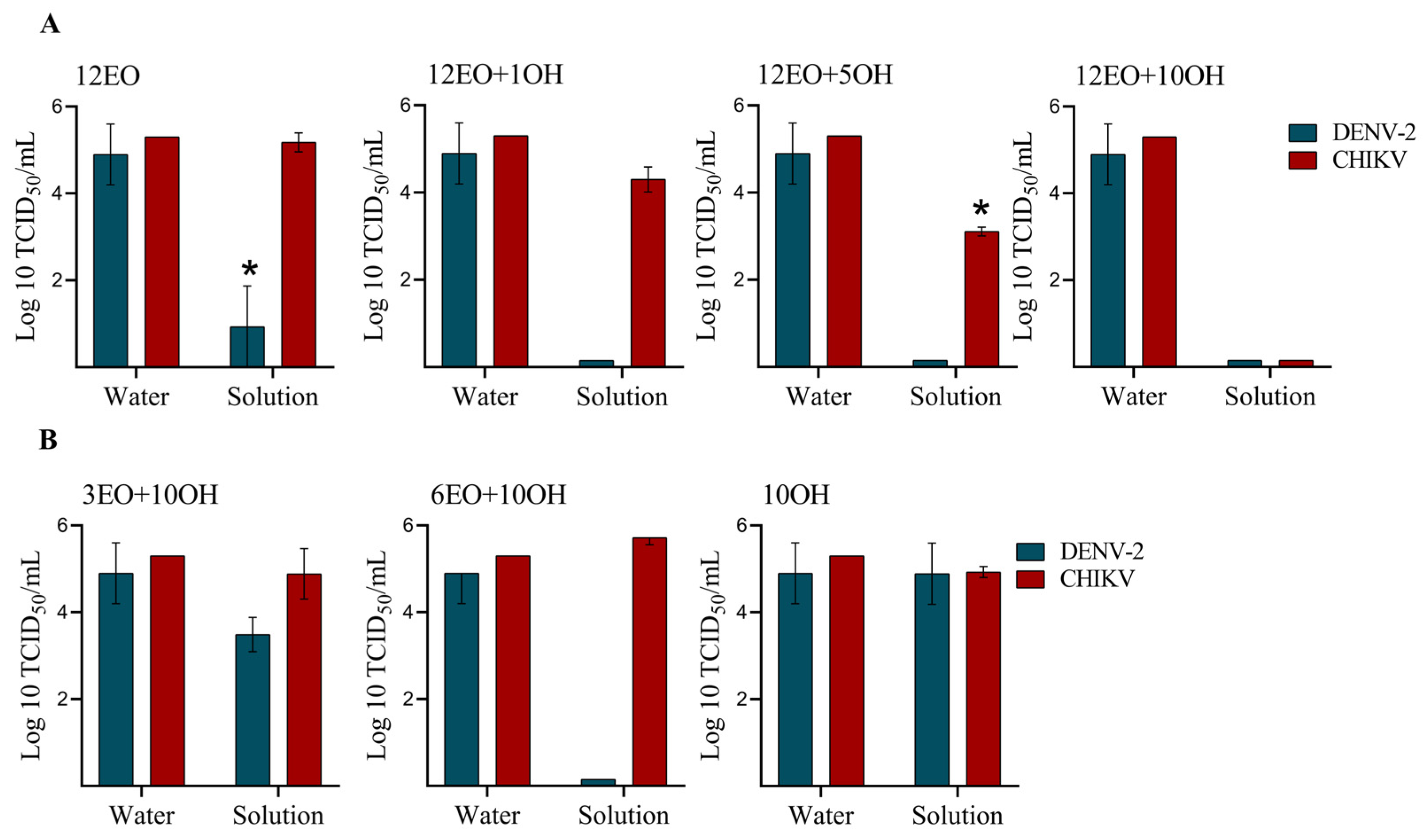
2.2. Virucidal Disinfectant Activity of the Test Solutions

The quantitative suspension test was used to evaluate the antiviral disinfectant activity following the German DVV/RKI guideline [19], and limits were 3.8–5 ± 1.2 and 5.3 ± 0.55 TCID50 (log10) per mL of DENV-2 and CHIKV, respectively (Table 3). A reduction factor of 4-log10 was the cutoff value for disinfectant activity [19]. First, we evaluated the activity of solutions containing 12% EO combined or not with OH within 1 min of exposure (Figure 1A). The solution containing 12% EO without OH (12EO) was sufficient to achieve a 3.9-log10 (81.6%) reduction in the DENV-2 titer, but was insufficient to reduce the CHIKV titer. The addition of 1% (12EO + 1OH solution) and 5% (12EO + 5OH solution) OH increased the reduction of DENV-2 to >4-log10 (100%), whereas the addition of 10% OH (12EO + 10OH solution) was required to achieve a 4-log10 (100%) reduction of CHIKV. Next, we evaluated solutions containing EO at concentrations lower than 12% combined with 10% OH after 1 min of exposure (Figure 1B). A reduction of 4-log10 of DENV-2, but not CHIKV, was achieved with the 6% EO + 10% OH (6EO + 10OH) solution, whereas the reduction of both viruses was not observed with the 3% EO + 10% OH (3EO − 10OH) solution. Solutions containing 3% EO without OH (3EO) and 10% OH without EO (10OH) did not show a virucidal effect against either virus after 1 min of exposure.
As the solutions containing 12% EO and 3% EO without OH did not show disinfectant effects against DENV-2 and CHIKV after 1 min of exposure, we assessed the activity of these solutions by increasing the exposure time in four intervals (Table 4). For the 12% EO solution (12EO), a reduction of >4-log10 (100%) of DENV-2 was achieved after 5 min of exposure, whereas a 100% reduction of CHIKV was achieved after 10 min of exposure. For the 3% EO solution, an exposure time of 30 min was required to achieve a 100% reduction in DENV-2 and CHIKV.

2.3. Chemical Composition of the EO Blend

Table 5 presents the linear retention indices and relative amounts of compounds in order of their elution on the DB-5MS column. A chromatogram of the EO blend is presented (Figure S1, Supplementary Materials). Forty-two compounds were identified. Monoterpene alcohols (52%) and aldehydes (23.8%) were the most abundant terpenes, especially geraniol (35.4%), citronellal (22.6%), and citronellol (14.1%), followed by monoterpene acetates (7.2%) and hydrocarbons (4.2%). Sesquiterpenoids were identified in low concentration (8.4%) and sesquiterpene hydrocarbons (5.7%), mostly germacrene D and δ-cadinene, were in higher concentration than oxygenated sesquiterpenes.

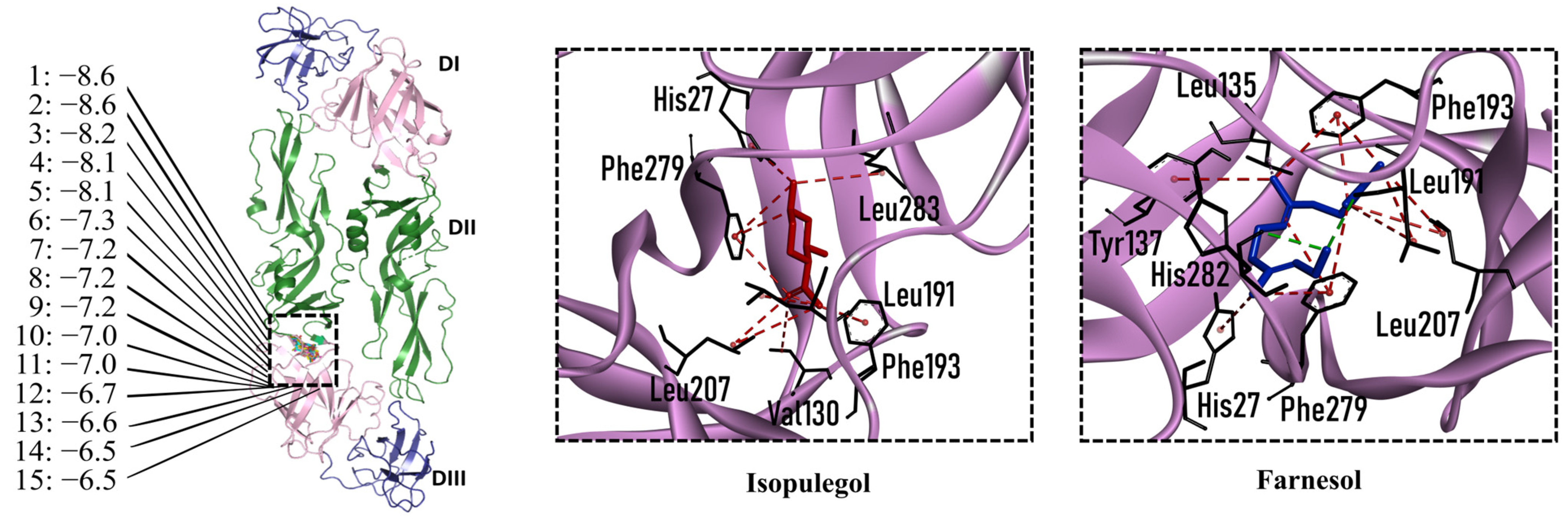
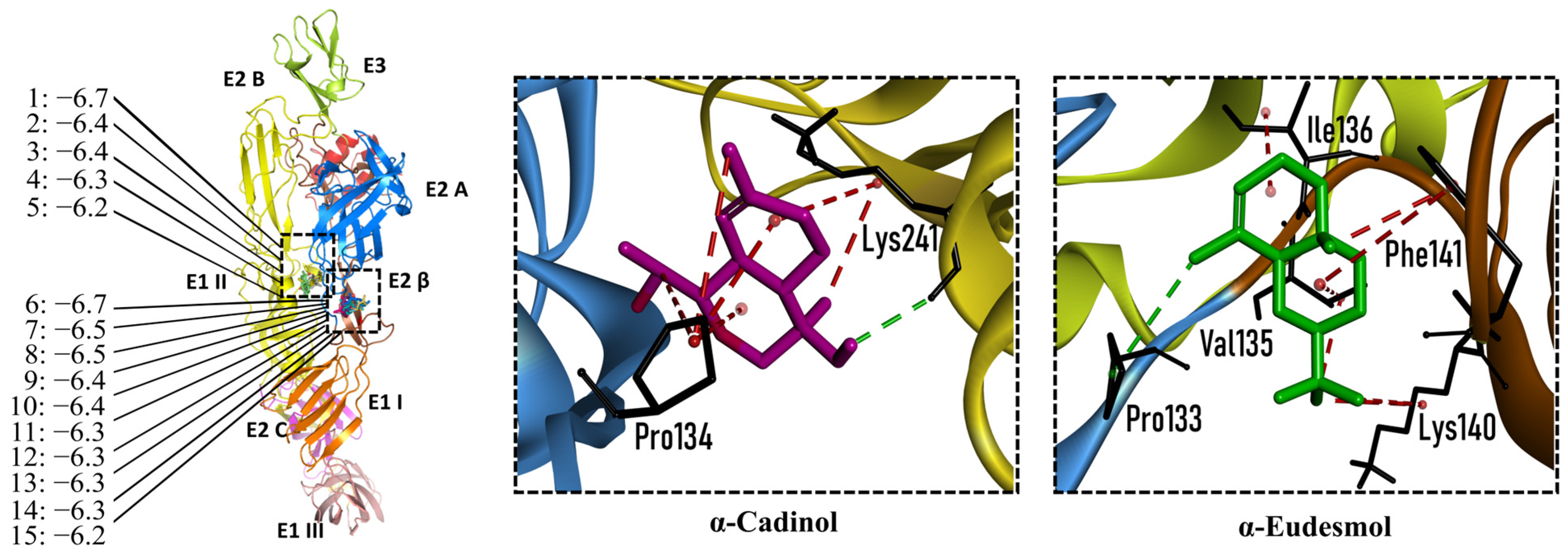
2.4. Molecular Interactions of EO Compounds and Viral Proteins

The DENV particle has a capsid surrounded by a lipid envelope, which contains the envelope (E) and membrane (prM/M) proteins [24]. The forty-two compounds identified in the EO blend were subjected to molecular docking simulation against E and prM/M of DENV-2. Table S1 presents the AutoDock Vina binding energies. Twenty-five (60%) compounds bound to the E protein, of which twelve bound with a strong binding energy (−7.03 to −8.61 kcal/mol) and thirteen with a weak binding energy (−6.0 to −6.7 kcal/mol). The compounds were accommodated in a consensus site corresponding to the detergent beta-octylglucoside (βOG) pocket in the hinge region of the E protein, which formed hydrophobic bonds with amino acid residues (Figure 2). Sesquiterpene hydrocarbons such as cadinene (δ and γ), α-guaiene and α-bulnesene, and the oxygenated sesquiterpene epi-α-muurolol showed the strongest binding affinities (−8.10 to −8.61 kcal/mol) with E, followed by monoterpene hydrocarbons (limonene, p-cymene, and γ-terpinene: −7.16 to −7.22 kcal/mol) and phenolic compounds (carvacrol and thymol: −7.03 to −7.32 kcal/mol). Other oxygenated sesquiterpenes (farnesol, α-cadinol, and α-eudesmol) and oxygenated monoterpenes (isopulegol, citronellyl acetate, neral, geranial, and citronellol) showed weak (−6.50 to −6.70 kcal/mol) binding affinities with the E protein. Farnesol and α-cadinol formed hydrogen bonds, the first with Gyl190, Leu191, and His282, and the second with Thr48 and Tyr137. Twenty compounds that bind to E were also identified in a previous study on the anti-DENV activity of EOs from other Colombian plant species [25]. Table 6 presents the EO compounds that docked DENV-2 E identified in this study. Docking analyses did not predict the binding of EO compounds to the prM/M protein (docking scores ranged from −4.08 to −5.47 kcal/mol).
The CHIKV particle has a capsid surrounded by a lipid envelope, which contains the E1-E2-E3 glycoprotein complex [26]. The forty-two compounds identified in the EO blend were subjected to molecular docking against E1-E2-E3. Table S1 presents the AutoDock Vina binding energies. Eight of the ten oxygenated sesquiterpenes identified in the EO blend bound to E1-E2-E3, and α-cadinol, α-eudesmol, and caryophyllene oxide exhibited the lowest binding energies (−6.50 to −6.70 kcal/mol) followed by patchoulol, germacrene D-4-ol, and epi-α-muurolol (−6.37 to −6.45 kcal/mol). In addition, nine of the ten sesquiterpenes hydrocarbons bound to the E1-E2-E3 complex, and α-guaiene, α-humulene, and trans-β-caryophyllene exhibited the lowest binding energies (−6.32 to −6.38 kcal/mol) followed by α-bulnesene, germacrene D, and δ-cadinene (−6.25 to −6.26 kcal/mol). The sesquiterpenes were accommodated in two consensus sites corresponding to a pocket in the domain II of the E1 protein (six sesquiterpenes) and a pocket in the β-ribbon connector of E2 protein (eleven sesquiterpenes). All EO compounds formed hydrophobic bonds with amino acid residues and five of the top compounds formed hydrogen bonds with amino acids lining the pocket (Figure 3). Table 7 presents the EO compounds with the lowest binding energy with the CHIKV E1-E2-E3 complex.

3. Discussion

Cleaning virus-contaminated hands and surfaces is essential for infection control and viral disease prevention [4]. OH-based solutions are utilized as disinfectants to control the transmission of human pathogenic viruses. However, frequent and prolonged use of OH-based disinfectants may be harmful to health and the environment [7,8,9]. EOs in the form of natural products are generally recognized as safe (GRAS) by the FDA (Food and Drug Administration, Silver Spring, MD, USA), and their use is permitted [27]. Many studies have explored using EOs as potential antibacterial and antifungal alternatives to commercial disinfectants [14,28]. In contrast, scientific evidence supporting the potential of EOs as disinfectants against enveloped viruses is very limited. Our study focused on enveloped viruses; studies show that enveloped viruses tend to infect more host species and are more likely to be pandemic than non-enveloped viruses [1,29].
The present study evaluated the virucidal disinfectant activity of solutions containing EO and OH against two surrogate viruses for pathogenic enveloped viruses. The results show that a solution of 12% EO combined with 10% OH reduced up to >4.0 log10 TCID50 (100% reduction) of both viruses within 1 min of exposure. In addition, the solutions containing EO without OH also exhibited virucidal action (100% reduction) against both viruses after 10 min (12% EO) and 30 min (3% EO) of exposure. We did not observe a 100% reduction in either virus with the 10% OH solution, but when combined with 12% EO, a strong virucidal activity was observed. It appears that low concentrations of EO and OH are insufficient to inactivate human pathogenic enveloped viruses. Romeo et al. [18] did not observe virucidal activity of a formulation containing 3.3% EO (Melaleuca alternifolia) combined with 5.3% ethanol against the coronavirus HCov-OC43 after 30 min of exposure.
To evaluate the virucidal disinfectant activity, we used two enveloped viruses, which differ in the lipid content [30,31] and protein structure [24,26] that comprise the viral envelope. The results indicated that DENV-2 was more sensitive to the action of test solutions than CHIKV. We hypothesized that differences in the viral envelope structure and its hydrophobic/hydrophilic nature might explain the variation in sensitivity. The DENV-2 particle assembles and buds into the endoplasmic reticulum of the infected cells where the envelope is formed. The envelope has 90 head-to-tail dimers of the E protein organized in a herringbone, with the M protein bound at the dimer interface [32]. On the other hand, CHIKV assembles and budding occurs at the cytoplasmic membrane, and the viral envelope comprises the E1 and E2 glycoproteins and a peptide (E3) arranged in trimers to make 80 E1/E2 spikes [27]. A recent study [18] showed differences in the sensitivity of enveloped viruses (human and feline coronaviruses) to treatment with a mixture of tea tree oil and ethanol.
Enveloped viruses enter host cells primarily via endocytosis following attachment to a cellular receptor [2,29,33]. Upon attachment, viruses are engulfed into endosomes where the low pH triggers conformational changes of the envelope proteins to drive fusion of the viral envelope and endosomal membrane. The viral envelope plays an important role in the membrane fusion process [33], and envelope proteins are potential extracellular drug targets with multiple strategies to inhibit entry of the virus into host cells [34]. Studies suggest that EOs could cause the morphological alteration of the viral particle by destroying the viral envelope through interactions between their terpene constituents and viral proteins [11,13]. In silico and in vitro evidence suggests that sesquiterpene hydrocarbons and oxygenated monoterpenes in specific ratios may account for the antiviral action of the EOs [11,12,13]. Recently, we documented a variation in the anti-DENV effect related to variation in oxygenated monoterpene content [25]. We also documented [35] a better in vitro anti-DENV effect of trans-β-caryophyllene and geranyl acetate compared to p-cymene, limonene, and neral, all of which were identified in the test EO blend.
We performed a primary docking analysis to describe the interactions between the 42 compounds of the EO blend and the envelope proteins of DENV-2 (E) and CHIKV (E1-E2-E3). As in a previous study [25], in the present study, we again found sesquiterpene hydrocarbons and oxygenated monoterpenes showing good binding affinities (−6.7 to −8.6 kcal/mol) with the DENV-2 E protein. These terpenes were accommodated in the βOG pocket and molecules that dock this pocket can block the conformational change of the E protein required for the fusion process [36]. As for CHIKV, seventeen EO compounds docked the E1-E2-E3 glycoprotein complex. Some bound to the E2 protein in a pocket of the β-ribbon connector peptides, which play a role during virus entry, helping to trigger E1 conformational changes during the fusion process [37]. Other EO compounds bound to the E1 protein of CHIKV near the hydrophobic fusion loop, which mediates membrane fusion [37]. According to the docking score values, EO compounds exhibited better binding affinities against DENV-2 than against CHIKV, which could partly explain the differences in sensitivity to the test solutions revealed in the virucidal disinfectant assays.
Little is known about the specific mechanism of action of EOs against enveloped viruses. Mechanisms other than alterations of the envelope protein structure have been proposed [11,12,13]. Being lipophilic, EOs can penetrate the viral envelope and cause membrane disintegration; they can cause viral expansion, which interferes with the attachment process by which viruses gain entry into host cells; moreover, EO components can inhibit host lipid metabolism pathways, which are crucial to ensure the availability of lipids to complete the assembly of new enveloped virions. On the other hand, OH causes protein denaturation and the disruption of the viral envelope [5]. Ethanol (95%, v/v) has broader and stronger virucidal activity than propanols (75% v/v); isopropanol, due to its lipophilic nature, interacts favorably with viral envelopes, and glycerol (80% v/v) and glycerol derivatives have been described as virucidal agents against enveloped viruses [38,39]. We hypothesize that the EO and OH mechanisms mentioned here could be involved in the strong virucidal disinfectant activity of the 12% EO + 10% OH solution.
The results of this study demonstrate that EO alone not only has disinfectant activity, but also shows synergistic activity with OH against two enveloped viruses. This synergistic activity may involve all of the aforementioned mechanisms of action of EOs. Further analysis is needed to investigate the contribution of each EO compound and their additive, synergistic, or antagonistic effects on the disinfectant action of a pure EO.

4. Materials and Methods

4.1. Plant Material and EO Blend

Pure EOs from seven aromatic plants grown in Colombia were used to prepare an EO blend. Then, a stock solution (6 × 10⁶ µg/mL) of the EO blend was prepared in DMSO and it was used to prepare the test solutions for analyses of the disinfectant activity (Table 1). EOs were distilled from Cymbopogon martinii (Roxb.) Will Watson (Poaceae family), Cymbopogon winterianus (Java citronella) Jowitt ex Bor (Poaceae family), Pogostemon cablin (Blanco) Benth, Lippia origanoides Kunth (Verbenaceae family), Elettaria cardamomum (L.) Maton (Zingiberaceae family) Swinglea glutinosa (Blanco) Merr (Rutaceae family), and Citrus × aurantium L. (Rutaceae family). The plants were grown in the experimental plots at the Agroindustrial Pilot Complex of the National Center for Agroindustrialization of Aromatic and Medicinal Tropical Vegetal Species (CENIVAM) in the Industrial University of Santander (Bucaramanga, Colombia). The taxonomic identification of these plants was performed at the Colombian National Herbarium (Bogotá, Colombia), where their vouchers were placed. EOs were obtained through the hydrodistillation (2 h) of plant leaves and stems on a Clevenger apparatus as described elsewhere [40,41].

4.2. Chemical Composition of the EO Blend

The analysis of the EO blend was performed by gas chromatography using mass spectrometric (GC/MS) and flame ionization detection (GC/FID) systems. Previous studies described the conditions of the process and data analysis [25,42,43]. Before the analysis, the EO blend was dissolved in dichloromethane (1 mL). n-Tetradecane (0.5 µL) was added as an internal standard. The injection volume was 2 µL in split mode (30:1). A 6890 Plus Gas Chromatograph (Agilent Technologies, AT, Palo Alto, CA, USA) equipped with a mass selective detector MSD 5975 (Electron ionization, EI, 70 eV), (AT, Palo Alto, CA, USA), a 7863 automatic injector, and an MSChemStation G1701DA data system (AT, Palo Alto, CA, USA) were used. The identification of EO compounds was accomplished by the comparison of their linear retention indices (LRIs) with those of standard compounds, and by the comparison of their mass spectral fragmentation patterns with those described in the literature and databases [20,21,22,23,24].

4.3. Preparation of the Test Solutions

Pure EOs from seven aromatic plant species were mixed in various proportions to obtain an EO blend using dimethyl sulfoxide (DMSO) as the solvent. The EO blend was mixed with the desired amount of an OH mixture (ethanol ca. 70%; isopropanol + glycerol ca. 2.5%) in a glass vial to give five different percentage ratios of EO/OH. Each solution was stirred using a vortex mixer until complete mixing took place. In addition, the EO blend was diluted to give solutions of 12% and 3%, and the OH mixture was diluted in water to prepare a 10% solution.

4.4. Cells and Viruses

Vero cells (African green monkey kidney cells; CCL-81™. ATCC, Manassas, VA, USA) were cultured in minimum essential medium (MEM) supplemented with 10% fetal bovine serum (FBS) and 1% antibiotic at 37 °C in a humidified atmosphere of 5% CO2. DENV-2 NGC (CDC, San Juan, Puerto Rico) was propagated in C6/36 Aedes albopictus cells (Pedro Kourí Institute for Tropical Medicine, La Habana, Cuba). CHIKV, a local strain isolated from a patient in our laboratory [44], was propagated in Vero cells. Both viruses were titrated using a protocol of the median tissue culture infectious dose (TCID50)—Spearman Karber assay [45].

4.5. Cytotoxicity Controls

As EO and OH can cause cytotoxic effects, the test solutions were first evaluated in Vero cells without the addition of virus. Briefly, the test solution was serially diluted, and an aliquot was added to cells seeded in 96-well plates. Following 1 h of incubation at 37 °C, the solution was discarded by washing and the cells were overlaid with fresh culture medium and incubated for 72 h at 37 °C and 5% CO2. Next, the cell viability was determined by staining with crystal violet, as in a previous study [25]. Briefly, 100 µL of 0.05% crystal violet solution was added to cells for 20 min at room temperature. After washing, the plates were aspirated and allowed to air dry at room temperature, and 200 µL of methanol was added to each well for 20 min. The optical density at 570 nm in each well was measured on a microplate reader (570 nm) to quantify crystal violet staining.

4.6. Evaluation of Virucidal Disinfectant Activity

The quantitative suspension test was used following the German DVV/RKI guideline [13,18]. The test was performed in five intervals (1, 5, 10, 20, and 30 min) of exposure of the virus with the test solution with fixed amounts of DENV-2 (8.4 log10 TCID50/mL) and CHIKV (7.8 log10 TCID50/mL). Briefly, 10 µL of a virus preparation was mixed with 80 µL of solution and 10 µL of water, and a virus control with 90 µL of water without test solution was included. At the end of the exposure times, 900 µL of ice-cold culture medium was added to each mixture and immediately diluted 10-fold to determine viral infectivity using end-point dilution titration. Vero cells were seeded in 96-well plates for 24 h at 37 °C under 5% CO2 and infected with serial dilutions of treated DENV-2 and treated CHIKV in triplicate on a logarithmic scale at base 10. Noninfected cells were included as controls. The plates were incubated at 37 °C and 5% CO2 for five days. After washing, the plates were aspirated and allowed to air dry at room temperature, and the crystal violet dye uptake was determined as described above. The quantity of virus was calculated as TCID50 (log10) per milliliter by the Spearman–Karber method [45].

4.7. Docking Analysis

Three-dimensional structures of DENV-2 E protein (PDB ID: 10AN) and the CHIKV E1-E2-E3 complex (PDB ID: 3N42) were downloaded from the Protein Data Bank. Structures of chemical constituents of the EO blend were retrieved from the PubChem (https://pubchem.ncbi.nlm.nih.gov/ (accessed on 13 April 2023)) database. The preparation of the target and ligands and molecular docking analyses were carried out using AutoDock Vina (Version 1.5.6, La Jolla, CA, USA), as described in a previous study [25]. The optimized protein structure was saved in the PDBQT file format for docking analysis. Default parameters were used, and the search exhaustiveness parameter was set to 100. For each ligand, 27 docked conformations were generated using global docking simulations. Three simulations were performed for each ligand–protein pair using seeds 6, 12, and 18. The average docking scores for each protein approximated the binding free energy. Discovery Studio Visualizer v21.1.0.20298 was used to view the ligand–protein interactions.

4.8. Statistical Analyses

A one-way ANOVA and a Tukey–Kramer post hoc test of viral titer values were used to compare the virucidal effect of each test solution, adopting a significance level of 0.05. The data were analyzed using GraphPad Prism software (version 8.0, San Diego, CA, USA).

5. Conclusions

The inadequate and inappropriate use of OH-based and other disinfectants has been associated with harmful effects on humans and the environment. There is an urgent need to develop safe and environmentally friendly disinfectants to minimize adverse effects. The data from this study provide a first step in defining the potential utility of EOs as disinfectants to control the transmission of human pathogenic enveloped viruses. We conclude that a solution containing 12% of a mixture of seven EOs and 10% of a mixture of OHs (ethanol, isopropanol, and glycerol) is highly effective for the inactivation of DENV-2 and CHIKV, which could be extended to enveloped viruses of similar structure that are transmitted person-to-person. In addition to reducing virus titers to 100%, the solution acts within one minute, making it practical for use in environments where rapid disinfection is needed. The hydrocarbon sesquiterpenes, oxygenated sesquiterpenes, and oxygenated monoterpenes present in the EO blend showed binding affinities for DENV-2 and CHIKV envelope proteins, suggesting that these types of terpenes could act as inhibitors of virus adsorption and entry into host cells. Further analysis is needed to better define the potential of EOs as virucidal disinfectant alternatives to commercial OH-based disinfectants.

Supplementary Materials

The following supporting information can be downloaded at: https://www.mdpi.com/article/10.3390/molecules28104156/s1: Table S1: AutoDock Vina binding energies values of essential oil compounds for target proteins. Figure S1: Chromatogram of the blend of seven essential oils used for virucidal activity study. DB-5 capillary column (60 m). Split 1:30.

Author Contributions

R.E.O., E.E.S. and V.P.-A. designed the study; V.P.-A. performed the cell-based experiments with support from R.E.O.; V.P.-A. and L.S.-T. performed the docking analyses with support from P.R.-V.; E.E.S. performed the GC/MS analysis and R.E.O. component identification; R.E.O. and V.P.-A. wrote the manuscript with support from L.S.-T. All authors contributed to the final version of the manuscript. All authors have read and agreed to the published version of the manuscript.

Funding

The authors are grateful for funding from the Ministry of Science, Technology and Innovation, the Ministry of Education, the Ministry of Industry, Commerce and Tourism, and ICETEX, Programme Ecosistema Científico-Colombia Científica, from the Francisco José de Caldas Fund, Grant RC-FP44842-212-2018.

Institutional Review Board Statement

The Ministry of Environment and Sustainable Development of Colombia supported the Universidad Industrial de Santander by permitting access to genetic resources and derivatives for bioprospecting (Contract No. 270). Project RC-FP44842-212-2018 was approved by the Scientific Research Ethical Committee (Record No. 15-2017, File No. 4110) of the Universidad Industrial de Santander. The experiments and chemical management were performed according to national law (Resolution No. 008430-1993) from the Ministry of Health of Colombia and the Institutional Manual of Integrated Management and Processes (PGOR-PGGA.05).

Informed Consent Statement

Not applicable.

Data Availability Statement

All data, tables, and figures are originals.

Conflicts of Interest

The authors declare no conflict of interest.

Sample Availability

Samples of the compounds are not available from the authors.

References

  1. Bhadoria, P.; Gupta, G.; Agarwal, A. Viral pandemics in the past two decades: An overview. J. Fam. Med. Prim. Care 2021, 10, 2745–2750. [Google Scholar] [CrossRef] [PubMed]
  2. Leung, N.H.L. Transmissibility and transmission of respiratory viruses. Nat. Rev. Microbiol. 2021, 19, 528–545. [Google Scholar] [CrossRef] [PubMed]
  3. Querido, M.M.; Aguiar, L.; Neves, P.; Pereira, C.C.; Teixeira, J.P. Self-disinfecting surfaces and infection control. Colloids Surf. B 2019, 178, 8–21. [Google Scholar] [CrossRef] [PubMed]
  4. Suchomel, M.; Eggers, M.; Maier, S.; Kramer, A.; Dancer, S.J.; Pittet, D. Evaluation of world health organization-recommended hand hygiene formulations. Emerg. Infect. Dis. 2020, 26, 2064–2068. [Google Scholar] [CrossRef]
  5. Lin, Q.; Lim, J.Y.C.; Xue, K.; Yew, P.Y.M.; Owh, C.; Chee, P.L.; Loh, X.J. Sanitizing agents for virus inactivation and disinfection. View 2020, 1, e16. [Google Scholar] [CrossRef]
  6. Kampf, G. Efficacy of ethanol against viruses in hand disinfection. J. Hosp. Infect. 2018, 98, 331–338. [Google Scholar] [CrossRef]
  7. Dhama, K.; Patel, S.K.; Kumar, R.; Masand, R.; Rana, J.; Yatoo, M.I.; Tiwari, R.; Sharun, K.; Mohapatra, R.K.; Natesan, S.; et al. The role of disinfectants and sanitizers during COVID-19 pandemic: Advantages and deleterious effects on humans and the environment. Environ. Sci. Pollut. Res. 2021, 28, 34211–34228. [Google Scholar] [CrossRef]
  8. Mahmood, A.; Eqan, M.; Pervez, S.; Alghamdi, H.A.; Tabinda, A.B.; Yasar, A.; Brindhadevi, K.; Pugazhendhi, A. COVID-19 and frequent use of hand sanitizers; human health and environmental hazards by exposure pathways. Sci. Total Environ. 2020, 742, 140561. [Google Scholar] [CrossRef]
  9. Jing, J.L.J.; Pei Yi, T.; Bose, R.J.; McCarthy, J.R.; Tharmalingam, N.; Madheswaran, T. Hand Sanitizers: A review on formulation aspects, adverse effects, and regulations. Int. J. Environ. Res. Public Health 2020, 17, 3326. [Google Scholar] [CrossRef]
  10. Manion, C.R.; Widder, R.M. Essentials of essential oils. Am. J. Health Syst. Pharm. 2017, 74, 153–162. [Google Scholar] [CrossRef]
  11. Wani, A.R.; Yadav, K.; Khursheed, A.; Rather, M.A. An updated and comprehensive review of the antiviral potential of essential oils and their chemical constituents with special focus on their mechanism of action against various influenza and coronaviruses. Microb. Pathog. 2021, 152, 104620. [Google Scholar] [CrossRef]
  12. Reichling, J. Antiviral and virucidal properties of essential oils and isolated compounds—A scientific approach. Planta Med. 2022, 88, 587–603. [Google Scholar] [CrossRef]
  13. Ma, L.; Yao, L. Antiviral effects of plant-derived essential oils and their components: An updated review. Molecules 2020, 25, 2627. [Google Scholar] [CrossRef] [PubMed]
  14. Bolouri, P.; Salami, R.; Kouhi, S.; Kordi, M.; Asgari-Lajayer, B.; Hadian, J.; Astatkie, T. Applications of essential oils and plant extracts in different industries. Molecules 2022, 27, 8999. [Google Scholar] [CrossRef] [PubMed]
  15. Pizzo, J.S.; Visentainer, J.V.; da Silva, A.L.B.R.; Rodrigues, C. Application of essential oils as sanitizer alternatives on the postharvest washing of fresh produce. Food Chem. 2023, 407, 135101. [Google Scholar] [CrossRef] [PubMed]
  16. Maurya, A.; Prasad, J.; Das, S.; Dwivedy, A.K. Essential oils and their application in food safety. Front. Sustain. Food Syst. 2021, 5, 653420. [Google Scholar] [CrossRef]
  17. Bailey, E.S.; Curcic, M.; Biros, J.; Erdogmuş, H.; Bac, N.; Sacco, A. Essential oil disinfectant efficacy against SARS-CoV-2 microbial surrogates. Front. Public Health 2021, 9, 2054. [Google Scholar] [CrossRef] [PubMed]
  18. Romeo, A.; Iacovelli, F.; Scagnolari, C.; Scordio, M.; Frasca, F.; Condò, R.; Ammendola, S.; Gaziano, R.; Anselmi, M.; Divizia, M.; et al. Potential use of tea tree oil as a disinfectant agent against coronaviruses: A combined experimental and simulation study. Molecules 2022, 27, 3786. [Google Scholar] [CrossRef] [PubMed]
  19. Rabenau, H.F.; Schwebke, I.; Blümel, J.; Eggers, M.; Glebe, D.; Rapp, I.; Sauerbrei, A.; Steinmann, E.; Steinmann, J.; Willkommen, H.; et al. Guideline for testing chemical disinfectants regarding their virucidal activity within the field of human medicine: As of December 1st, 2014, prepared by the German Association for the Control of Virus Diseases (DVV) and the Robert Koch Institute (RKI). Bundesgesundheitsblatt Gesundh. 2020, 63, 645–655. [Google Scholar] [CrossRef] [PubMed]
  20. Adams, R. Identification of Essential Oil Components by Gas Chromatography/Mass Spectrometry, 4th ed.; Allured Publishing Corporation: Carol Stream, IL, USA, 2007; ISBN 1932633219. [Google Scholar]
  21. Babushok, V.I.; Linstrom, P.J.; Zenkevich, I.G. Retention indices for frequently reported compounds of plant essential oils. J. Phys. Chem. 2011, 40, 043101. [Google Scholar] [CrossRef]
  22. Wallace, W. NIST Standard Reference Database 1A; Version 2.3; National Institute of Standards and Technology: Gaithersburg, MD, USA, 2017. [Google Scholar]
  23. Davies, N. Gas chromatographic retention indices of monoterpenes and sesquiterpenes on methyl silicone and Carbowax 20M phases. J. Chromatogr. A 1990, 503, 1–24. [Google Scholar] [CrossRef]
  24. Nasar, S.; Rashid, N.; Iftikhar, S. Dengue proteins with their role in pathogenesis, and strategies for developing an effective anti-dengue treatment: A review. J. Med. Virol. 2020, 92, 941–955. [Google Scholar] [CrossRef]
  25. Silva-Trujillo, L.; Quintero-Rueda, E.; Stashenko, E.E.; Conde-Ocazionez, S.; Rondón-Villarreal, P.; Ocazionez, R.E. Essential oils from Colombian plants: Antiviral potential against dengue virus based on chemical composition, in vitro and in silico analyses. Molecules 2022, 27, 6844. [Google Scholar] [CrossRef]
  26. Vaney, M.C.; Duquerroy, S.; Rey, F.A. Alphavirus structure: Activation for entry at the target cell surface. Curr. Opin. Virol. 2013, 3, 151–158. [Google Scholar] [CrossRef] [PubMed]
  27. Singh, S.; Chaurasia, P.K.; Bharati, S.L.; Golla, U.R. A mini-review on the safety profile of essential oils. MOJ Biol. Med. 2022, 7, 33–36. [Google Scholar]
  28. Cho, T.J.; Park, S.M.; Yu, H.; Seo, G.H.; Kim, H.W.; Kim, S.A.; Rhee, M.S. Recent advances in the application of antibacterial complexes using essential oils. Molecules 2020, 25, 1752. [Google Scholar] [CrossRef]
  29. Valero-Rello, A.; Sanjuán, R. Enveloped viruses show increased propensity to cross-species transmission and zoonosis. Proc. Natl. Acad. Sci. USA 2022, 119, e2215600119. [Google Scholar] [CrossRef]
  30. Hitakarun, A.; Williamson, M.K.; Yimpring, N.; Sornjai, W.; Wikan, N.; Arthur, C.J.; Pompon, J.; Davidson, A.D.; Smith, D.R. Cell type variability in the incorporation of lipids in the Dengue virus virion. Viruses 2022, 14, 2566. [Google Scholar] [CrossRef]
  31. Sousa, I.P., Jr.; Carvalho, C.A.M.; Gomes, A.M.O. Current understanding of the role of cholesterol in the life cycle of alphaviruses. Viruses 2020, 13, 35. [Google Scholar] [CrossRef] [PubMed]
  32. Zhang, X.; Ge, P.; Yu, X.; Brannan, J.M.; Bi, G.; Zhang, Q.; Schein, S.; Zhou, Z.H. CryoEM structure of the mature dengue virus at 3.5-å resolution. Nat. Struct. Mol. Biol. 2013, 20, 105–110. [Google Scholar] [CrossRef]
  33. Rey, F.A.; Lok, S.M. Common features of enveloped viruses and implications for immunogen design for next-generation vaccines. Cell 2018, 172, 1319–1334. [Google Scholar] [CrossRef] [PubMed]
  34. Verma, J.; Subbarao, N.; Rajala, M.S. Envelope proteins as antiviral drug target. J. Drug Target. 2020, 10, 1046–1052. [Google Scholar] [CrossRef] [PubMed]
  35. Flechas, M.C.; Ocazionez, R.E.; Stashenko, E.E. Evaluation of in vitro antiviral activity of essential oil compounds against Dengue Virus. Pharmacogn. J. 2018, 10, 55–59. [Google Scholar] [CrossRef]
  36. Naresh, P.; Selvaraj, A.; Shyam-Sundar, P.; Murugesan, S.; Sathianarayanan, S.; Namboori, P.K.K.; Jubie, S. Targeting a conserved pocket (n-octyl-β-d–glucoside) on the dengue virus envelope protein by small bioactive molecule inhibitors. J. Biomol. Struct. Dyn. 2022, 40, 4866–4878. [Google Scholar] [CrossRef]
  37. Mangala, P.V.; Blijleven, J.S.; Smit, J.M.; Lee, K.K. Visualization of conformational changes and membrane remodeling leading to genome delivery by viral class-II fusion machinery. Nat. Commun. 2022, 13, 4772. [Google Scholar] [CrossRef]
  38. Golin, A.P.; Choi, D.; Ghahary, A. Hand sanitizers: A review of ingredients, mechanisms of action, modes of delivery, and efficacy against coronaviruses. Am. J. Infect. Control 2020, 48, 1062–1067. [Google Scholar] [CrossRef]
  39. Welch, J.L.; Xiang, J.; Okeoma, C.M.; Schlievert, P.M.; Stapleton, J.T. Glycerol monolaurate, an analog to a factor secreted by lactobacillus, is virucidal against enveloped viruses, including HIV-1. mBio 2020, 11, e00686-20. [Google Scholar] [CrossRef]
  40. Hernández, R.; Pájaro, N.; Caballero, K.; Stashenko, E.E.; Olivero, J. Essential Oils from plants of the genus Cymbopogon as natural insecticides to control stored product pest. J. Stored Prod. Res. 2015, 62, 81–83. [Google Scholar] [CrossRef]
  41. Pinzón, J.A.; Contreras, N.J.; Durán, D.C.; Martínez, J.R.; Stashenko, E.E. Green biomass production and quality of essential oils of palmarrosa (Cymbopogon martinii Roxb.) with application of synthetic fertilizers and organic fertilizers. Acta Agron. 2014, 63, 335–342. [Google Scholar] [CrossRef]
  42. Stashenko, E.E.; Martínez, J.R.; Cala, M.; Durán, D.C.; Caballero, D. Chromatographic and mass spectrometric characterization of essential oils and extracts from Lippia (Verbenaceae) aromatic plants. J. Sep. Sci. 2013, 36, 192–202. [Google Scholar] [CrossRef]
  43. Stashenko, E.E.; Martínez, J.R.; Medina, J.D.; Durán, D.C. Analysis of essential oils isolated by steam distillation from Swinglea glutinosa fruits and leaves. J. Essent. Oil Res. 2015, 27, 276–282. [Google Scholar] [CrossRef]
  44. Carreño, M.F.; Jiménez-Silva, C.L.; Rey-Caro, L.A.; Conde-Ocazionez, S.A.; Flechas-Alarcón, M.C.; Velandia, S.A.; Ocazionez, R.E. Dengue in Santander State, Colombia: Fluctuations in the prevalence of virus serotypes are linked to dengue incidence and genetic diversity of the circulating viruses. Trop. Med. Int. Health 2019, 12, 1400–1410. [Google Scholar] [CrossRef] [PubMed]
  45. Ramakrishnan, M.A. Determination of 50% endpoint titer using a simple formula. WJV World J. Virol. 2016, 5, 85–86. [Google Scholar] [CrossRef] [PubMed]
Figure 1. Comparison of virucidal disinfectant activities of the test solutions against DENV-2 and CHIKV within 1 min of exposure. Table 1 presents the content of essential oil (EO) and alcohol (OH) in each solution. (A) Solutions based on 12% EO with increasing alcohol concentration (1%, 5% and 10%). (B) Solutions based on 10% alcohol with increasing EO concentration (3% and 6%). The residual infectivity was determined by virus titer using the TCID50 assay. Data are averages ± SDs from three independent assays in triplicate. * One-way ANOVA: DENV-2: F9,17 = 21.64; and CHIKV: F7,31 = 14.57, p < 0.001; Tukey’s post hoc test, p < 0.001.
Molecules 28 04156 g001
Figure 2. Docked poses of representative EO compounds in complex with DENV-2 E protein into the βOG pocket. Interacting amino acids are shown as black sticks; hydrogen bonding interactions are depicted as green dotted lines and hydrophobic interactions are depicted as red dotted lines. Compounds: 1, δ-cadinene; 2, α-guaiene; 3, γ-cadinene; 4, α-bulnesene; 5, carvacrol; 6, epi-α-muurolol; 7, γ-terpinene; 8, limonene; 9, p-cymene; 10, germacrene D; 11, thymol; 12, farnesol; 13, isopulegol; 14, α-cadinol; 15, α-eudesmol.
Molecules 28 04156 g002
Figure 3. Docked poses of representative EO compounds in complex with the E3-E2-E1 protein complex of CHIKV. Interacting amino acids are shown as black sticks; hydrogen bonding interactions are depicted as green dotted lines and hydrophobic interactions are depicted as red dotted lines. Compounds into a pocket of E1 domain II: 1, α-cadinol; 2, α-guaiene, 3: epi-α-muurolol; 4: epi-α-cadinol; 5, γ-cadinene. Compounds into a pocket of the β-ribbon connector of E2: 6, α-eudesmol; 7, caryophyllene oxide; 8, patchoulol; 9, germacrene D-4-ol; 10, α-humulene; 11, trans-β-caryophyllene; 12, α-bulnesene; 13, germacrene D; 14, δ-cadinene; 15, α-muurolene.
Molecules 28 04156 g003
Table 1. Solutions tested for virucidal disinfectant activity.
No.ContentDMSO, %Identifier
112% EO + 1% OH7.912EO + 1OH
212% EO + 5% OH7.612EO + 5OH
312% EO + 10% OH7.212EO + 10OH
46% EO + 10% OH7.26EO + 10OH
53% EO + 10% OH1.83EO + 10OH
612% EO8.012EO
73% EO2.03EO
810% OH-10OH
EO is a blend of pure EOs from seven aromatic plants. OH is a mixture of ethanol (ca. 70%) and other OHs (ca. 2.5%: isopropanol and glycerol). Dimethyl sulfoxide (DMSO) was used as an EO solvent and the final concentration in the solution is presented.
Table 2. Viability of Vero cells exposed to the test solutions for 72 h.
SolutionDilution/Percentage of Viability
1:101:1001:1000
12EO80 ± 2690 ± 13100 ± 10
12EO + 1OH80 ± 1192 ± 8.496 ± 6.0
12EO + 5OH80 ± 1693 ± 7.397 ± 2.7
12EO + 10OH80 ± 2590 ± 10100 ± 10
6EO + 10OH92 ± 9.296 ± 7.198 ± 3.3
3EO + 10OH92 ± 8.995 ± 9.195 ± 7.3
3EO100 ± 0.094 ± 8.990 ± 33
10OH94 ± 5.490 ± 8.890 ± 10
Acetic acid0.0 ± 3.280 ± 3293 ± 4.9
Acetic acid (5%) was used as a virucidal agent. Data are averages ± SDs from three independent assays in triplicate.
Table 3. Disinfectant activity of the test solutions after one minute of virus exposure.
SolutionDENV-2: TCID50/mLCHIKV: TCID50/mL
Log10RF% RLog10RF% R
Water3.8 ± 0.4 and 5 ± 1.25.3 ± 0.5--
Acetic acid0.04.9 ± 0.01000.05.3 ± 0.5100
12EO1 ± 1.63.9 ± 0.481.65.3 ± 0.50.00.0
12EO + 10OH0.04.9 ± 0.01004.3 ± 0.70.9 ± 0.718.8
12EO + 5OH0.04.9 ± 0.01003.1 ± 0.2 *2.2 ± 0.241.5
12EO + 10OH0.04.9 ± 0.01000.05.3 ± 0.0100
6EO + 10OH0.04.9 ± 0.01005.7 ± 0.20.00.0
3EO + 10OH3.5 ± 0.70.5 ± 0.728.55 ± 1.00.4 ± 1.09.5
3EO3.1 ± 0.30.7 ± 0.618.4NDND-
10OH4.2 ± 0.00.7 ± 0.00.05.3 ± 0.50.00.0
Virus control for the 3EO solution. RF: reduction factor, Log10 TCID50 virus control—Log 10 TCID50 treated virus; % R, percentage reduction in virus titer relative to virus control; 0.0, the virus was not detected. ND, not determined. Data are averages ± SDs from three independent assays in triplicate. * One-way ANOVA: DENV-2: F9,17 = 21.64; and CHIKV: F7,31 = 14.57, p < 0.001; Tukey’s post hoc test, p < 0.001.
Table 4. Virucidal disinfectant activity of essential oil solutions without alcohol according to time of exposure.
SolutionDENV-2: Log10 TCID50/mLCHIKV: Log10 TCID50/mL
ControlTreatedRFR, %ControlTreatedRFR, %
12EO
5 min5.4 ± 0.20.05.4 ± 0.21005.7 ± 0.73.0 ± 0.7 *2.7 ± 0.147.3
10 min5.0 ± 1.00.05.0 ± 1.51004.8 ± 0.40.04.8 ± 0.3100
20 min5.4 ± 0.50.05.4 ± 0.51004.9 ± 0.10.04.0 ± 0.1100
30 min4.5 ± 0.90.04.5 ± 0.91004.6 ± 0.50.04.6 ± 0.5100
3EO
5 min4.0 ± 0.13.0 ± 0.60.9 ± 0.5256.1 ± 0.65.4 ± 0.50.6 ± 0.611.4
10 min5.0 ± 1.53.3 ± 0.61.0 ± 0.726.65.1 ± 0.25.1 ± 0.40.00.0
20 min3.7 ± 0.12.1 ± 0.80.4 ± 1.043.26.1 ± 0.33.3 ± 0.7 *2.8 ± 0.845.9
30 min3.7 ± 0.40.0 ± 1.43.7 ± 0.11005.0 ± 1.50.0 ± 0.05.0 ± 1.5100
RF: reduction factor; R, %: percentage reduction in virus titer; 0.0, the virus was not detected. Data are averages ± SDs from three independent assays in triplicate. * One-way ANOVA: DENV-2: F7,16 = 20.27; and CHIKV: F7,14 = 46, p < 0.001; Tukey’s post hoc test, p < 0.001.
Table 5. Chemical composition of the EO blend.
No.CompoundTypeLinear Retention IndicesGC/FID Relative Peak Area, %
Exp.Lit.
1α-Pinene *MH934932 a0.1
26-Methyl-hept-5-en-2-oneOC984985 a0.1
3β-Myrcene *MH989990 a0.2
4p-Cymene *MH10261027 a0.8
5Limonene *MH10311029 a2.0
61,8-Cineole *OM10361034 a0.3
7trans-β-OcimeneMH10471050 a0.9
8γ-Terpinene *MH10601059 a0.2
9Linalool *OM11001096 a2.2
10Citronellal *OM11571153 a22.6
11IsopulegolOM11651155 a0.3
12n-DecanalOC12071201 a0.1
13CitronellolOM12201233 a14.1
14NeralOM12411242 b0.4
15Geraniol *OM12581255 b35.4
16Geranial *OM12711270 b0.8
17Thymol *PhC12921290 a1.1
18Carvacrol *PhC13011298 a2.1
19Citronellyl acetateOM13461350 a2.5
20EugenolPhC13541356 a0.4
21Geranyl acetateOM13771379 a4.7
22β-ElemeneSH13961389 a1.0
23trans-β-Caryophyllene *SH14321428 d0.9
24α-GuaienoSH14441440 b0.1
25α-Humulene *SH14681465 d0.2
26γ-MuuroleneSH14841478 a0.1
27Germacrene D *SH14921481 a1.5
28α-MuuroleneSH15061500 a0.4
29α-BulneseneSH15111509 a0.1
30γ-CadineneSH15231513 a0.3
31δ-CadineneSH15261522 a1.1
32ElemolOS15571548 a0.9
33trans-Nerolidol *OS15651561 a0.2
34Germacrene D-4-olOS15781574 a0.7
35Caryophyllene oxide *OS15861582 a0.1
36epi-α-CadinolOS16531650 c0.1
37epi-α-MuurololOS16551642 c0.2
38α-CadinolOS16671653 c0.2
39α-EudesmolOS16691659 c0.1
40PatchoulolOS16911660 b0.1
41FarnesolOS17191723 b0.1
42Neryl hexanoateOM17501732 c0.2
1. Monoterpenoids87.7
1.1 Monoterpene hydrocarbons (MH)4.2
1.2 Oxygenated monoterpenes (OM)83.5
 Alcohols52
 Acetates7.2
 Aldehydes23.8
 Others (ethers, esters, epoxides)0.5
2. Sesquiterpenoids8.4
2.1 Sesquiterpene hydrocarbons (SH)5.7
2.2 Oxygenated sesquiterpenes (OS)2.7
 Alcohols2.6
 Others (Oxides)0.1
3. Phenolic compounds (PhC)
 (Thymol, carvacrol, eugenol)
3.6
4. Other oxygenated compounds (OC)
 (n-Decanal, 6-methyl-5-hepten-2-one)
0.2
LRI, linear retention indices calculated using n-alkanes C8–C25 mixture on the DB-5MS (non-polar) column. Exp., experimental. Lit., literature: a [20]; b [21]; c [22]; d [23]. * Use of standard compounds.
Table 6. EO compounds with binding affinity to the E protein of DENV-2.
CompoundStructural FormulaAmino Acid Residues. H-Bond in Bold FontKcal/mol
IsopulegolMolecules 28 04156 i001Thr189, Leu191, Phe193,
Leu207, Phe279
−6.70 ± 0.6
FarnesolMolecules 28 04156 i002Thr48, Leu135, Tyr137, Gly190, Leu191, Phe193, Leu207, Phe279, His282, Leu283−6.57 ± 0.6
α-CadinolMolecules 28 04156 i003Thr48, Tyr137, Thr189, Leu191, Phe193, Leu207, Phe279−6.54 ± 0.5
EugenolMolecules 28 04156 i004Thr48, Leu135, Thr189, Leu191, Phe193, Phe279, Gly281, His282−6.54 ± 0.5
α-EudesmolMolecules 28 04156 i005Thr48, Val130, Phe193, Leu207, Phe279, Leu283−6.50 ± 0.6
The twelve compounds with a strong binding energy (−7.03 to −8.61 kcal/mol) and another eight compounds with a weak binding energy (−6.0 to −6.7 kcal/mol) were reported in a previous study [25].
Table 7. EO compounds with the lowest binding energy with the E1-E2-E3 glycoprotein complex of CHIKV.
CompoundStructural FormulaProteinAmino Acid Residues. H-Bond in Bold FontKcal/mol
α-CadinolMolecules 28 04156 i006E1, domain IIAsn39, Thr42, Pro133, Pro134, Lys241, Leu244−6.70 ± 0.2
α-EudesmolMolecules 28 04156 i007E2, β-ribbon connectorPro133, Val135, Ile136, Lys140, Phe141−6.70 ± 0.3
Caryophyllene oxideMolecules 28 04156 i008E2, β-ribbon connectorArg104, Pro133, Val135, Ile136, Lys40, Phe141−6.50 ± 0.31
PatchoulolMolecules 28 04156 i009E2, β-ribbon connectorPro133, Ile136, Lys140, Phe141, Asp43−6.45 ± 0.2
α-GuaieneMolecules 28 04156 i010E1, domain IIPro134, Lys241, Tyr242, Lys245−6.38 ± 0.2
Germacrene D-4-olMolecules 28 04156 i011E2, β-ribbon connectorThr42, Pro134, Lys241, Leu244, Lys245, Asn39−6.38 ± 0.2
epi-α-MuurololMolecules 28 04156 i012E1, domain IIPro134, Lys241, Leu244, Lys245−6.37 ± 0.2
α-HumuleneMolecules 28 04156 i013E2, β-ribbon connectorIle136, Phe141−6.37 ± 0.3
trans-β-CaryophylleneMolecules 28 04156 i014E2, β-ribbon connectorIle136, Phe141, Arg144−6.32 ± 0.1
Disclaimer/Publisher’s Note: The statements, opinions and data contained in all publications are solely those of the individual author(s) and contributor(s) and not of MDPI and/or the editor(s). MDPI and/or the editor(s) disclaim responsibility for any injury to people or property resulting from any ideas, methods, instructions or products referred to in the content.

