
Chemical Composition and Antimicrobial Properties of Honey Bee Venom


Abstract
:1. Introduction
2. Results and Discussion
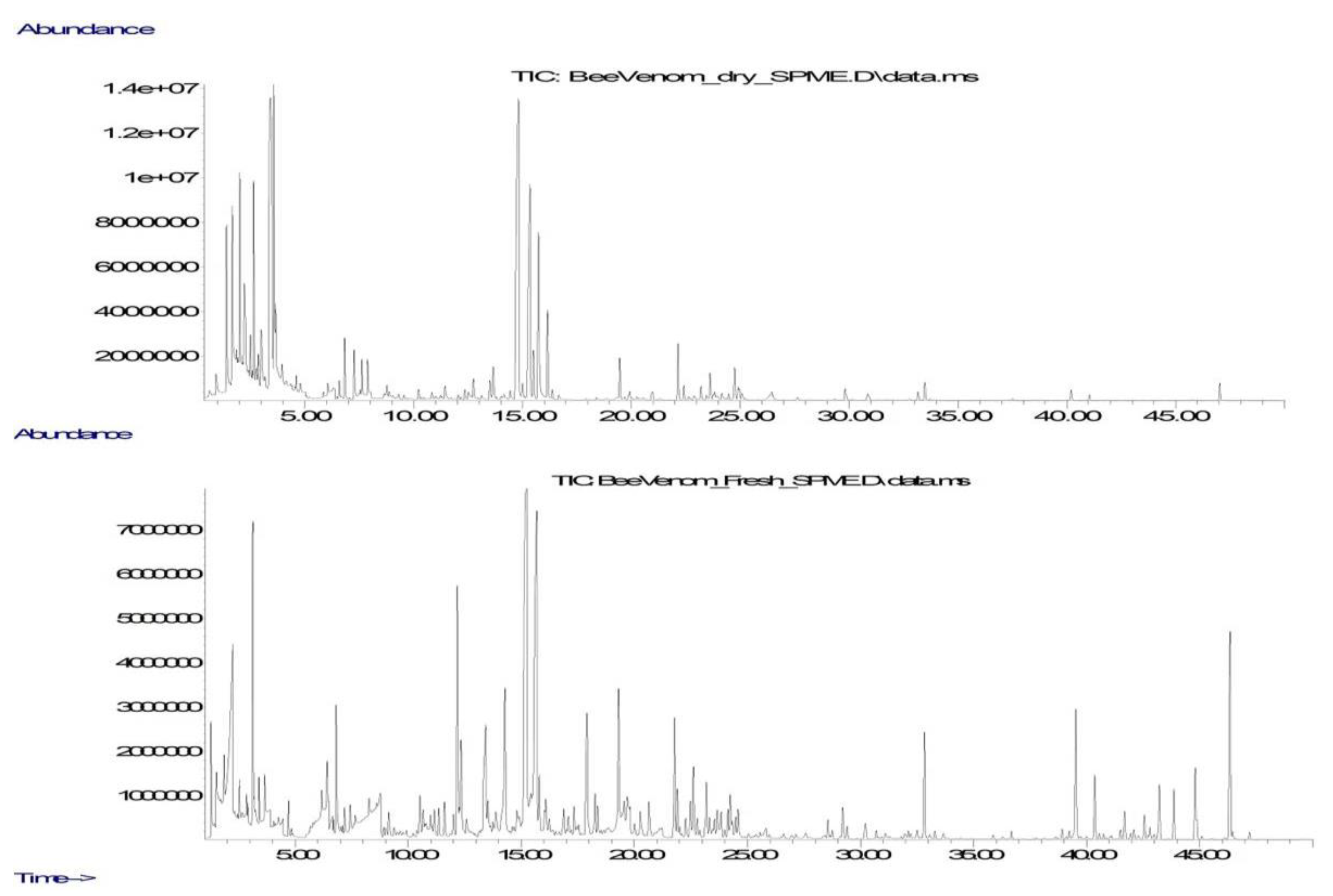
2.1. Chemical Composition of Volatile Compounds
2.2. Chemical Composition of Extractive Compounds
2.2.1. Chemical Composition of Extracts with Diethyl Ether
2.2.2. Chemical Composition of Methanol Extracts
2.3. Antimicrobial Activity of BV and Extracts
3. Materials and Methods
3.1. Chemicals and Materials for Microbiological Research
3.2. Bee Venom Preparations
3.3. Determination of Volatile Compounds
3.4. Determination of Extractive Compounds
3.5. Component Identification
3.6. Component Quantification
3.7. Determination of Antimicrobial Activity of Bee Venom and Extracts
4. Conclusions
Supplementary Materials
Author Contributions
Funding
Institutional Review Board Statement
Informed Consent Statement
Data Availability Statement
Acknowledgments
Conflicts of Interest
References
- Nelson, D.A. The Venom of the Honeybee Apis mellifera. Ph.D. Thesis, Montana State University, Bozeman, MO, Canada, 1966. [Google Scholar]
- Pucca, M.B.; Cerni, F.A.; Oliveira, I.S.; Jenkins, T.P.; Argemí, L.; Sørensen, C.V.; Ahmadi, S.; Barbosa, J.E.; Laustsen, A.H. Bee Updated: Current Knowledge on Bee Venom and Bee Envenoming Therapy. Front. Immunol. 2019, 10, 2090. [Google Scholar] [CrossRef] [PubMed]
- Mauchamp, B.; Grandperrin, D. Chromatographie en phase gazeuse des composes volatils des glandes a pheromones des abeilles: Methods d’analyse directe. Apidologie 1982, 13, 29–37. [Google Scholar] [CrossRef]
- Carpena, M.; Nunez-Estevez, B.; Soria-Lopez, A.; Simal-Gandara, J. Bee venom: An updating review of its bioactive molecules and its health applications. Nutrients 2020, 12, 3360. [Google Scholar] [CrossRef]
- Klupczynska, A.; Plewa, S.; Derezinski, P.; Garrett, T.J.; Rubio, V.Y.; Kokot, Z.J.; Matysiak, J. Identification and quantification of honeybee venom constituents by multiplatform metabolomics. Sci. Rep. 2020, 10, 21645. [Google Scholar] [CrossRef]
- Ghentt, R.L.; Gary, N.E. A chemical alarm releaser in honeybee sting (Apis mellifera L.). J. Entomol. 1962, 69, 039293. [Google Scholar] [CrossRef]
- Grandperrin, D. Sting alarm pheromone of the honeybee, the recruting effect of an artificial blend of volatile compounds of the worker sting (Apis mellifera L., Hymenoptera, Apidae). Experimentia 1983, 39, 219–221. [Google Scholar] [CrossRef]
- Bellik, Y. Bee and its potential use in alternative medicine. Anti-Infect. Agents 2015, 13, 3–16. [Google Scholar] [CrossRef]
- Sung, S.H.; Kim, J.W.; Han, J.E.; Shin, B.C.; Park, J.K.; Lee, G. Animal venom for medical usage in pharmacopuncture in Korean medicine: Current status and clinical implication. Toxins 2021, 13, 105. [Google Scholar] [CrossRef] [PubMed]
- Son, D.J.; Lee, J.W.; Lee, Y.H.; Song, H.S.; Lee, C.K.; Hong, J.T. Therapeutic application of anti-arthritis, pain-releasing, and anti-cancer effects of bee venom and its constituent compounds. Pharmacol. Therap. 2007, 115, 246–270. [Google Scholar] [CrossRef] [PubMed]
- He, S.; Tan, N.; Sun, C.; Liao, K.; Zhu, H.; Luo, X.; Zhang, J.; Li, D.; Huang, S. Treatment with melittin induces apoptosis and autophagy of fibroblast-like synoviocytes in patients with rheumatoid arthritis. Curr. Pharm. Biotechnol. 2020, 21, 734–740. [Google Scholar] [CrossRef]
- Jagua-Gualdron, A.; Pena-Latorre, J.A.; Fernadez-Bernal, R.E. Apitherapy for osteoarthritis: Perspectives from basic research. Complement. Med. Res. 2020, 27, 184–191. [Google Scholar] [CrossRef] [PubMed]
- Aufschnaiter, A.; Kohler, V.; Khalifa, S.; Abd El-Wahed, A.; Du, M.; El-Seedi, H.; Buttner, S. Apitoxin and its components against cancer, neurodegeneration and rheumatoid arthritis: Limitations and possibilities. Toxins 2020, 12, 66. [Google Scholar] [CrossRef] [PubMed]
- Ku, Y.H.; Kang, J.H.; Lee, H. Effect of bee venom on an experimental cellular model of Alzheimer’s disease. Am. J. Chin. Med. 2020, 48, 1803–1819. [Google Scholar] [CrossRef] [PubMed]
- El-Seedi, H.R.; Khalifa, S.A.M.; Abd El-Wahed, A.; Gao, R.; Guo, Z.; Tahir, H.E.; Zhao, C.; Du, M.; Farag, M.A.; Musharraf, S.G.; et al. Honeybee products: An updated review of neurological actions. Trends Food Sci. Technol. 2020, 101, 17–27. [Google Scholar] [CrossRef]
- El-Seedi, H.; Abd El-Wahed, A.; Yosri, N.; Musharraf, S.G.; Chen, L.; Moustafa, M.; Zou, X.; Al-Mousawi, S.; Guo, Z.; Khatib, A.; et al. Antimicrobial properties of Apis mellifera’s bee venom. Toxins 2020, 12, 451. [Google Scholar] [CrossRef] [PubMed]
- Memariani, H.; Memariani, M. Anti-fungal properties and mechanisms of melittin. Appl. Microbiol. Biotechnol. 2020, 104, 6513–6526. [Google Scholar] [CrossRef] [PubMed]
- Nainu, F.; Masyita, A.; Bahar, M.A.; Raihan, M.; Prova, S.R.; Mitra, S.; Bin Emran, T.; Simal-Gandara, J. Pharmaceutical prospects of bee products: Special focus on anticancer, antibacterial, antiviral, and antiparasitic properties. Antibiotics 2021, 10, 822. [Google Scholar] [CrossRef] [PubMed]
- Ratajczak, M.; Kaminska, D.; Matuszewska, E.; Holderna-Kedzia, E.; Rogacki, J.; Matysiak, J. Promising antimicrobial properties of bioactive compounds from different honeybee products. Molecules 2021, 26, 4007. [Google Scholar] [CrossRef] [PubMed]
- Tanugur-Samanc, A.E.; Kekecoglu, M. An evaluation of the chemical content and microbiological contamination of Anatolian bee venom. PLoS ONE 2021, 16, e0255161. [Google Scholar] [CrossRef] [PubMed]
- Haktanir, I.; Masoura, M.; Mantzouridou, F.T.; Gkatzionis, K. Mechanism of antimicrobial activity of honeybee (Apis mellifera) venom on Gram-negative bacteria: Escherichia coli and Pseudomonas spp. AMB Express 2021, 11, 54. [Google Scholar] [CrossRef] [PubMed]
- Elswaby, S.; Sadik, M.; Azouz, A.; Emam, N.; Ali, M. In vitro evaluation of antimicrobial and antioxidant activities of honeybee venom and propolis collected from various regions in Egypt. Egypt. Pharm. J. 2022, 21, 207–213. [Google Scholar] [CrossRef]
- Abdel-Monsef, M.M.; Darwish, D.A.; Zidan, H.A.; Hamed, A.A.; Ibrahim, M.A. Characterization antimicrobial and antitumor activity of superoxide dismutase extracted from Egyptian honeybee venom (Apis mellifera lamarckii). J. Genet. Eng. Biotechnol. 2023, 21, 21. [Google Scholar] [CrossRef]
- Kurek-Gorecka, A.; Komosinska-Vassev, K.; Rzepecka-Stojko, A.; Olczyk, P. Bee venom in wound healing. Molecules 2021, 26, 148. [Google Scholar] [CrossRef]
- Abdelsattar, A.S.; Makky, S.; Nofal, R.; Hebishy, M.; Agwa, M.M.; Aly, R.G.; EI-Naga, M.Y.A.; Heikal, Y.A.; Fayez, M.S.; Rezk, N.; et al. Enhancement of wound healing via topical application of natural products: In Vitro and in vivo evaluations. Arab. J. Chem. 2022, 15, 103869. [Google Scholar] [CrossRef]
- Kurek-Gorecka, A.; Gorecki, M.; Rzepecka-Stojko, A.; Balwierz, R.; Stojko, J. Bee products in dermatology and skin care. Molecules 2020, 25, 556. [Google Scholar] [CrossRef]
- Badawi, J.K. Bee venom components as therapeutic tools against prostate cancer. Toxins 2021, 13, 337. [Google Scholar] [CrossRef] [PubMed]
- Roy, A.; Bharadvaja, N. Venom-derived bioactive compounds as potential anticancer agents: A review. Int. J. Pept. Res. Ther. 2021, 27, 129–147. [Google Scholar] [CrossRef]
- Zhu, H.X.; Chen, D.T.; Xie, X.L.; Li, Y.M.; Fan, T.Y. Melittin inhibits lung metastasis of human osteosarcoma: Evidence of wnt/beta-catenin signalling pathway participation. Toxicon 2021, 198, 132–142. [Google Scholar] [CrossRef]
- Oršolić, N. Bee venom in cancer therapy. Cancer Metastasis Rev. 2012, 321, 173–194. [Google Scholar] [CrossRef]
- Sung, J.; Kim, Y.; Yu, P.F.; Kim, Y.; Han, I.H.; Bae, H. Subcutaneous toxicity of melittin-dKLA in ICR mice. Mol. Cell. Toxicol. 2021, 17, 417–428. [Google Scholar] [CrossRef]
- Duarte, D.; Falcao, S.I.; El Mehdi, I.; Vilas-Boas, M.; Vale, N. Honeybee venom synergistically enhances the cytotoxic effect of CNS drugs in HT-29 colon and MCF-7 breast cancer cell lines. Pharmaceutics 2022, 14, 511. [Google Scholar] [CrossRef]
- Erkoc, P.; von Reumont, B.M.; Luddecke, T.; Henke, M.; Ulshöfer, T.; Vilcinskas, A.; Fürst, R.; Schiffmann, S. The pharmacological potential of novel melittin variants from the honeybee and solitary bees against inflammation and cancer. Toxins 2023, 14, 818. [Google Scholar] [CrossRef] [PubMed]
- Viegas, S.; Ladeira, C.; Costa-Veiga, A.; Perelman, J.; Gajski, G. Forgotten public health impacts of cancer—An overview. Arh. Hig. Rada Toksikol. 2017, 68, 287–297. [Google Scholar] [CrossRef] [PubMed]
- Pawlak, M.; Klupczynska, A.; Kokot, Z.J.; Matysiak, J. Extending matabolimic studies of Apis mellifera venom: LC-MS-based target analysis of organic acids. Toxins 2020, 12, 14. [Google Scholar] [CrossRef] [PubMed]
- El-Wahed, A.A.A.; Mohamed, A.; Farag, M.A.; Walaa, A.; Eraqi, W.A.; Gaber, A.M.; Mersal, G.A.M.; Zhao, C.; Khalifa, S.A.M.; El-Seedi, H.R. Unravelling the beehive air volatiles profile as analysed via solid-phase microextraction (SPME) and chemometrics. J. King Saud Univ. Sci. 2021, 33, 101449. [Google Scholar] [CrossRef]
- Maschwitz, U. Gefahrenalarmstoffe und Alarmierung bei sozialen Hymenopteren. Z. Vgl. Physiol. 1964, 47, 596–655. [Google Scholar] [CrossRef]
- Boch, R.; Stone, B.C.; Shearer, D.A. Identification of isoamyl acetate as an active component in the sting pheromone of the honey bee. Nature 1962, 195, 1018–1020. [Google Scholar] [CrossRef]
- Free, J.B.; Simpson, J. The alerting pheromones of the honeybee. Z. Vgl. Physiol. 1968, 61, 361–365. [Google Scholar] [CrossRef]
- Collins, A.M.; Blum, M.S. Alarm responses caused by newly identified compounds derived from the honeybee sting. J. Chem. Ecol. 1983, 9, 57–65. [Google Scholar] [CrossRef]
- Camargos, A.F.; Cossolin, J.F.S.; Martinez, L.C.; Goncalves, W.G.; dos Santos, M.H.; Serrao, J.E. Morphology and chemical composition of the Koschewnikow gland of the honey bee Apis mellifera (Hymenoptera: Apidae) workers engaged in different tasks. J. Apic. Res. 2020, 59, 1037–1048. [Google Scholar] [CrossRef]
- Chen, Q.C.; Liu, L.; Yu, T.Y.; Lang, L.; Yin, M.L.; Zhu, W.H.; Jiang, X.Y.; Wang, H.Y. High-level expression and purification of melittin in Escherichia coli using SUMO fusion partner. Intern. J. Pept. Res. Ther. 2021, 27, 9–15. [Google Scholar] [CrossRef]
- Askari, P.; Namaei, M.H.; Ghazvini, K.; Hosseini, M. In Vitro and in vivo toxicity and antibacterial efficacy of melittin against clinical extensively drug-resistant bacteria. BMC Pharmacol. Toxicol. 2021, 22, 42. [Google Scholar] [CrossRef] [PubMed]
- Pereira, A.F.M.; Albano, M.; Alves, F.C.B.; Andrade, B.F.M.T.; Furlanetto, A.; Rall, V.L.M.; Dos Santos, L.D.; de Oliverira Orsi, R.; Júnior, A.F. Influence of apitoxin and melittin from Apis mellifera bee on Staphylococcus aureus strains. Microb. Pathog. 2020, 141, 104011. [Google Scholar] [CrossRef] [PubMed]
- Miyazaki, Y.; Shinoda, W. Cooperative antimicrobial action of melittin on lipid membranes: A coarse-grained molecular dynamics study. Biochim. Biophys. Acta Biomembr. 2022, 1864, 183955. [Google Scholar] [CrossRef]
- Isidorov, V.A.; Buczek, K.; Zambrowski, G.; Miastkowski, K.; Swiecicka, I. In Vitro study of the antimicrobial activity of European propolis against Paenibacillus larvae. Apidologie 2017, 48, 411–422. [Google Scholar] [CrossRef]
- Isidorov, V.A.; Maslowiecka, J.; Szoka, L.; Pellizzer, N.; Miranda, D.; Olchowik-Grabarek, E.; Zambrzycka, M.; Swiecicka, I. Chemical composition and biological activity of Argentinian propolis of four species of stingless bees. Molecules 2022, 27, 7686. [Google Scholar] [CrossRef]
- Isidorov, V.A.; Bakier, S.; Pirożnikow, E.; Zambrzycka, M.; Swiecicka, I. Selective behaviour of honeybees in acquiring European propolis plant precursors. J. Chem. Ecol. 2016, 42, 475–485. [Google Scholar] [CrossRef]
- De Graaf, D.C.; Braga, M.R.B.; Magalhães, R.M.; de Abreu, R.M.M.; Blank, S.; Bridts, C.H.; De Clerck, L. Standard methods for Apis mellifera venom research. J. Apic. Res. 2020, 60, 1–31. [Google Scholar] [CrossRef]
- Isidorov, V.; Bakier, S.; Grzech, I. Gas chromatographic –mass spectrometric investigation of volatile and extractable compounds of crude royal jelly. J. Chromatogr. B 2012, 885–886, 109–117. [Google Scholar] [CrossRef]
- Isidorov, V.A.; Stocki, M.; Vetchinnikova, L. Inheritance of specific secondary volatile metabolites in buds of white birch Betula pendula and Betula pubescens hybrids. Trees 2019, 55, 1329–1344. [Google Scholar] [CrossRef]
- Isidorov, V.A.; Nazaruk, J.; Stocki, M.; Bakier, S. Secondary metabolites of downy birch buds (Betula pubescens Erch.). Z. Naturforsch. C 2021, 77, 145–155. [Google Scholar] [CrossRef] [PubMed]
- Adams, R.A. Identification of Essential Oil Components by Gas Chromatography/Mass Spectrometry, 4th ed.; Allured Publishing Corporation: Carol Stream, IL, USA, 2007. [Google Scholar]
- Tkachev, A.V. Investigation of Plant’s Volatile Compounds; Ofset Publ.: Novosibirsk, Russia, 2008. [Google Scholar]
- Isidorov, V.A. GC-MS of Biologically and Environmentally Significant Organic Compounds/TMS Derivatives; Wiley & Sons Ltd.: Hoboken, NJ, USA, 2020; 706p. [Google Scholar]
- M100-S21; Performance Standards for Antimicrobial Susceptibility Testing. Twenty-First International Supplement; No 1. CLSI (Clinical and Laboratory Standard Institute): Wayne, PA, USA, 2011; Volume 31.
- Alanis, A.J. Resistance to antibiotics: Are we in the post-antibiotic era? Arch. Med. Res. 2005, 36, 697–705. [Google Scholar] [CrossRef] [PubMed]
- Sabtu, N.; Enoch, D.A.; Brown, N.M. Antibiotic resistance: What, why, where, when and how? Br. Med. Bull. 2015, 16, 105–113. [Google Scholar] [CrossRef] [PubMed]
- Garaj-Vrhovac, V.; Gajski, G. Evaluation of the cytogenetic status of human lymphocytes after exposure to a high concentration of bee venom in vitro. Arh. Hig. Rada Toksikol. 2009, 60, 27–34. [Google Scholar] [CrossRef] [PubMed]
- Gajski, G.; Garaj-Vrhovac, V. Bee venom induced cytogenetic damage and decreased cell viability in human white blood cells after treatment in vitro: A multi-biomarker approach. Environ. Toxicol. Pharm. 2011, 32, 201–211. [Google Scholar] [CrossRef]
- Sjakste, N.; Gajski, G. A review on genotoxic and genoprotective effects of biologically active compounds of animal origin. Toxins 2023, 15, 165. [Google Scholar] [CrossRef]


| Groups of Compounds | Dried Venom | Fresh Venom | ||
|---|---|---|---|---|
| Dv-1 | Dv-2 | Fv-1 | Fv-2 | |
| Aliphatic carbonyls, including: | 26.67 | 8.14 | 5.51 | 19.13 |
| - acetone | 0.59 | 0.50 | 0.15 | 2.30 |
| - 2-butanone | trace * | 0.18 | 1.18 | 1.56 |
| - 2-pentanone | 1.37 | trace | 0.40 | 0.73 |
| - 2-heptanone | 16.08 | 0.49 | 0.76 | 1.53 |
| - 2-nonanone | 1.99 | 1.57 | 1.19 | 10.94 |
| - 2-undecanone | - ** | - | 0.57 | 0.76 |
| - isobutanal | 1.88 | 0.02 | - | - |
| - isopentanal | 0.89 | 0.71 | - | - |
| - hexanal | 1.73 | 0.01 | - | - |
| - nonanal | 0.71 | trace | - | - |
| - (2E)-decenal | - | - | 0.30 | 0.23 |
| Aliphatic alcohols, including: | 1.85 | 50.37 | 15.66 | 20.11 |
| - ethanol | 1.46 | trace | 0.33 | 0.26 |
| - isopentanol | 0.40 | 28.90 | 3.56 | 5.10 |
| - (3Z)-hexen-1-ol | - | 0.73 | 0.34 | 0.23 |
| - 2-heptanol | trace | 0.21 | 0.15 | 0.40 |
| - 2-nonanol | trace | 0.57 | 5.79 | 8.46 |
| - 2-ethyl-1-hexanol | - | - | 1.84 | 1.38 |
| - 2-undecanol | - | - | 0.42 | 0.40 |
| - (2E)-decen-1-ol | - | - | 0.55 | 0.79 |
| Aliphatic acids, including: | 32.70 | 4.53 | 12.50 | 9.24 |
| - formic acid | 2.37 | trace | - | - |
| - acetic acid | 6.60 | 3.00 | 7.97 | 7.83 |
| - isobutyric acid | - | - | 0.60 | 0.91 |
| - butyric acid | trace | trace | 0.59 | 0.50 |
| - hexanoic acid | 1.43 | 0.30 | 0.76 | - |
| - octanoic acid | 20.72 | 0.05 | 0.93 | 0.50 |
| - 2-octenoic acid | 0.50 | trace | 0.50 | 0.15 |
| Aliphatic esters, including: | 12.49 | 14.43 | 9.13 | 9.80 |
| - ethyl acetate | 0.95 | trace | 0.54 | trace |
| - isoamyl acetate | trace | 0.13 | 1.45 | 4.92 |
| - isoamyl isobutanoate | trace | 0.76 | 0.25 | 0.38 |
| - ethyl 2-methylvalerate (manzanate) | - | - | 1.23 | 1.51 |
| - isoamyl pentanoate | - | - | 0.45 | 0.83 |
| - isoamyl 3-methy-2-butenoate | - | 2.67 | 1.15 | 0.14 |
| - isoamyl octanoate | trace | trace | 0.08 | 0.08 |
| - isoamyl benzoate | - | 0.23 | 0.16 | 0.29 |
| - isopropyl tetradecanoate | - | - | 0.60 | 1.09 |
| - ethyl octanoate | 10.59 | - | - | - |
| Aromatics, including: | 18.34 | 1.46 | 9.45 | 5.51 |
| - toluene | 3.40 | 1.15 | 1.93 | 1.42 |
| - p-cymene | trace | trace | 0.64 | 0.28 |
| - benzaldehyde | 2.62 | - | 0.15 | 0.11 |
| - acetophenone | 0.33 | trace | 0.43 | 0.58 |
| - 1-phenyl ethanol | - | - | 0.12 | 0.21 |
| - 2-phenyl ethanol | trace | trace | 1.39 | 0.82 |
| - cresol | - | - | 1.22 | 1.29 |
| - p-ethylguaiacol | - | - | 0.37 | 0.37 |
| - methyl benzoate | 1.88 | - | - | - |
| -methyl salicylate | 9.52 | - | - | - |
| Terpenoids, including: | 3.91 | 0.56 | 12.60 | 12.48 |
| - α-pinene | 1.78 | trace | 2.06 | 2.39 |
| - β-pinene | 0.54 | - | 0.47 | trace |
| - 3-carene | 0.36 | trace | 0.28 | 0.32 |
| - limonene | 0.56 | 0.35 | 2.90 | 3.12 |
| - dihydromyrcenol | - | - | 2.37 | 3.08 |
| - camphor | -- | - | 0.28 | 0.33 |
| - borneol | - | - | 0.32 | 0.37 |
| - bornyl acetate | - | - | 0.64 | 0.38 |
| - β-caryophyllene | trace | 0.21 | 0.06 | 0.17 |
| - γ-cadinene | - | - | 0.07 | 0.11 |
| Alkanes and alkenes, including: | 2.95 | 19.66 | 14.66 | 8.45 |
| - n-hexane | 2.62 | - | - | - |
| - n-decane | - | trace | - | 0.48 |
| - n-dodecane | - | trace | 2.00 | 0.26 |
| - n-tridecane | 0.33 | 0.19 | 0.16 | 0.28 |
| - n-tetradecane | - | - | 0.17 | 0.11 |
| - n-pentadecane | - | 0.49 | 1.44 | 1.36 |
| - n-heptadecane | - | 0.18 | 1.47 | 1.95 |
| - 1-pentadecene | - | 16.99 | 0.17 | 0.10 |
| - nonadecene | - | 0.45 | 5.51 | 2.87 |
| Lactones, including: | 0.30 | trace | 5.53 | 5.68 |
| - valerolactone | trace | - | 0.16 | - |
| - γ-caprolactone | trace | - | 1.71 | 2.56 |
| - γ-heptalactone | - | trace | 2.05 | 2.05 |
| - γ-octalactone | 0.30 | trace | 1.05 | 0.95 |
| Other, including: | 1.42 | 1.85 | - | 2.35 |
| - pyridine | 0.46 | 0.52 | - | 0.71 |
| - 2,3-butanediol | - | - | - | 0.24 |
| - 2-pentylfuran | 0.20 | - | - | - |
| - dimethyl sulfide | - | 0.81 | - | - |
| - acetoin | - | 0.52 | - | trace |
| NN | 0.55 | 1.42 | 6.38 | 7.25 |
| Groups of Compounds | Dried Venom | Fresh Venom | ||
|---|---|---|---|---|
| Dv-1 | Dv-2 | Fv-1 | Fv-2 | |
| Aliphatic Alcohols, Including: | 16.90 (12) * | 13.37 (7) | 18.04 (16) | 19.00 (11) |
| - isopentanol | - ** | - | 0.93 | 0.16 |
| - oleyl alcohol | 0.10 | 0.09 | 0.29 | 0.23 |
| - octadecanol | 0.08 | 0.06 | 0.27 | 0.12 |
| - (9Z)-eicosen-1-ol | 14.75 | 11.71 | 12.40 | 16.16 |
| - 1-docosanol | 0.06 | - | 0.33 | 0.57 |
| - 1-tetracosanol | 0.19 | 0.16 | 0.47 | 0.35 |
| - 1-hexacosanol | 0.23 | 0.17 | 0.37 | 0.33 |
| Aliphatic acids, including: | 3.23 (7) | 4.90 (9) | 27.31 (37) | 31.90 (19) |
| - lactic | 0.05 | 0.05 | 0.48 | trace *** |
| - citric | 1.88 | 3.79 | - | - |
| - palmitic | 0.34 | 0.29 | 2.40 | 4.93 |
| - α-linoleic | - | - | 2.12 | 1.94 |
| - oleic | 0.80 | 0.65 | 12.15 | 20.14 |
| - stearic | 0.06 | 0.06 | 3.18 | 2.76 |
| Aliphatic esters, including: | 17.31 (31) | 20.77 (6) | 8.07 (7) | 6.17 (7) |
| - octadecyl oleate | 2.59 | 0.22 | 0.62 | 0.22 |
| - eicosenyl oleate | 13.18 | 17.42 | 1.89 | 1.37 |
| - eicosyl oleate | 1.54 | 1.51 | 0.92 | 1.11 |
| - tetracosyl palmitate | - | 0.39 | 3.42 | 1.14 |
| Glycerol & glycerides, including: | 5.47 (4) | 8.22 (5) | 1.02 (2) | 0.58 (2) |
| - glycerol | 0.36 | 0.13 | 0.07 | trace |
| - 1-hexadecyl glycerol | 0.07 | 0.07 | - | - |
| - 1-eicosyl glycerol | 1.86 | 1.43 | 0.95 | 0.58 |
| - 2-eicosyl glycerol | 3.16 | 2.43 | - | - |
| Aromatic compounds, including: | 1.76 (2) | 1.05 (2) | 4.62 (10) | 0.58 (4) |
| - benzoic acid | - | - | 1.60 | 0.30 |
| - p-hydroxybenzoic acid | - | - | 0.28 | 0.22 |
| - gallic acid | 1.52 | 0.88 | - | - |
| - ellagic acid | 0.24 | 0.17 | - | - |
| Sterols, including: | 0.23 (1) | 0.10 (1) | 2.89 (3) | 6.51 (4) |
| - 24-methylenecholesterol? | - | - | 1.94 | 3.26 |
| - β-sitosterol | - | - | trace | 1.29 |
| - avenasterol | 0.23 | 0.10 | 0.94 | 1.65 |
| Alkanes and alkenes, including: | 45.30 (25) | 32.07 (30) | 25.82 (33) | 27.10 (34) |
| - n-pentacosane | 3.41 | 2.63 | 2.16 | 1.75 |
| - n-heptacosane | 5.51 | 4.27 | 3.55 | 3.39 |
| - n-nonacosane | 3.28 | 2.46 | 3.35 | 2.31 |
| - n-hentriacotane | 2.23 | 1.76 | 2.40 | 2.31 |
| - 7-hentriacontene | 4.42 | 3.46 | 0.58 | 2.45 |
| - 9-hentriacontene | 3.34 | 2.62 | 3.70 | 2.30 |
| Other compounds, including | 3.90 | 12.54 | 3.78 | 2.84 |
| - ethylamine | - | 8.44 | 0.30 | 0.47 |
| - uracil | - | - | 0.04 | - |
| - indole-3-acetic acid | - | - | 0.06 | - |
| NN | 5.90 (10) | 6.98 (15) | 8.45 (27) | 5.90 (10) |
| Groups of Compouns | Dry Venom | Fresh Venom | ||
|---|---|---|---|---|
| Dv-1 | Dv-2 | Fv-1 | Fv-2 | |
| Aliphatic Alcohols, Including: | 3.56 (2) * | 2.15 (3) | 5.51 (12) | 5.58 (2) |
| - glycerol | 0.42 | 0.15 | 2.25 | 2.24 |
| - (9Z)-eicosen-1-ol | 3.14 | 0.97 | 1.82 | 3.34 |
| - oleyl alcohol | - ** | 0.04 | - | - |
| Aliphatic acids, including: | 33.60 (18) | 28.62 (18) | 17.17 (26) | 12.61 (17) |
| - lactic | 0.49 | 0.22 | 4.00 | 0.38 |
| - glycolic | 0.11 | 0.05 | - | 0.10 |
| - glyceric | 0.62 | 0.04 | - | - |
| - succinic | 0.67 | 0.16 | 1.04 | 0.52 |
| - malic | 0.34 | 0.23 | 0.14 | 0.24 |
| - citric | 29.51 | 27.60 | 0.09 | - |
| Aminoacids, including: | 3.97 (10) | 1.13 (14) | 22.81 (21) | 19.10 (24) |
| - glycine | 0.14 | 0.08 | 1.12 | 0.76 |
| - alanine | 0.91 | 0.07 | 3.52 | 2.80 |
| - prolinę | 2.05 | 0.45 | 4.30 | 4.98 |
| - 5-oxoproline | 0.77 | 0.09 | 0.10 | 1.28 |
| - β-alanine | 0.10 | 0.15 | 1.59 | 0.73 |
| - glutamine | trace | 0.10 | 0.74 | 1.65 |
| - lysine | trace | 0.02 | 0.49 | 1.05 |
| - hydroxyproline | - | - | 0.09 | 0.92 |
| - γ-aminobutanoic (GABA) | - | 0.01 | Trace *** | 0.27 |
| Other N-containing compounds, including: | 12.10 (15) | 23.71 (21) | 7.63 (13) | 7.36 (16) |
| - 2-aminoethanol | 0.12 | 0.10 | 0.40 | trace |
| - putrescine | 0.72 | 0.63 | 1.54 | 1.49 |
| - cadaverine | 0.33 | 0.56 | trace | trace |
| - histaminę | 5.74 | 18.25 | 1.81 | 0.35 |
| - uridine | 0.99 | 0.32 | - | - |
| - uracil | - | - | 0.40 | 0.43 |
| - adenine | - | - | trace | 0.29 |
| - serotonin | 1.91 | 0.80 | - | 0.56 |
| - inosine | 1.91 | 0.80 | - | 0.56 |
| P-containing compounds, including: | 12.23 (2) | 7.17 (2) | 5.48 (5) | 9.29 (5) |
| - H3PO4 | 11.54 | 6.89 | 2.48 | 5.13 |
| - α-glycerophosphate | 0.69 | 0.28 | 2.33 | 3.31 |
| - myo-inositol phosphate | - | - | - | 0.72 |
| Carbohydrate & related compounds, including: | 27.20 (21) | 32.37 (30) | 30.34 (44) | 38.50 (35) |
| - α- and β-fructose | 1.96 | 14.26 | 4.58 | 7.23 |
| - α- and β-glucose | 0.14 | 4.11 | 12.13 | 16.51 |
| - gluconic acid | 0.51 | 4.05 | 2.81 | 3.18 |
| - mannitol | 0.04 | 0.59 | 0.77 | 1.34 |
| - glucitol | trace | 0.24 | 2.00 | 2.76 |
| - myo-inositol | 0.85 | 0.44 | 0.31 | 0.21 |
| - sucrose | 15.85 | 2.97 | - | - |
| - trehalose | 2.91 | 1.52 | 0.26 | 1.02 |
| - 1-kestose | 0.25 | 0.07 | - | - |
| - erlose | 1.36 | 0.02 | - | - |
| - melizitose | 0.77 | trace | - | - |
| Other compounds, including: | 3.13 (5) | 0.12 (4) | 1.14 (5) | 3.25 (7) |
| - 1-O-eicosyl glycerol | 0.62 | 0.04 | - | 0.62 |
| - β-sitosterol | - | - | 0.16 | 0.14 |
| - avenasterol | - | - | 0.37 | - |
| - unidentified P-containing compound | - | - | 0.30 | - |
| - 9-hentriacontene | 0.22 | 0.03 | 0.10 | - |
| - 7-hentriacontene | 0.14 | 0.03 | 0.21 | - |
| NN | 4.24 (12) | 4.75 (26) | 5.59 (27) | 4.31 (15) |
| Sample | Gram-Positive Bacteria | Gram-Negative Bacteria | Fungus | ||||
|---|---|---|---|---|---|---|---|
| P. larvae ATCC 9545 | S. aureus ATCC 6538 | B. cereus ATCC 10987 | B. subtilis ATCC 6633 | P. aeruginosa ATCC 19582 | E. coli ATCC 11229 | C. albicans ATCC 90029 | |
| MIC, ng mL−1 | |||||||
| Bee venom Bv-1 | 1.91 | 7.63 | 7.63 | 0.12 | >500 | 122.07 | 30.52 |
| - ether extract | 122.07 | 500 | - | 125 | >500 | >500 | 488.28 |
| - methanol extract | 31.25 | 122.07 | 122.07 | 7.63 | 500 | 500 | 125 |
| Bee venom Bv-2 | 0.48 | 1.91 | 0.48 | 0.12 | >500 | 7.63 | 7.63 |
| - ether extract | 31.25 | 125 | 125 | 125 | 500 | 500 | 7.63 |
| - methanol extract | 7.81 | 31.25 | 125 | 0.49 | >500 | 125 | 1.95 |
| MBC/MFC, ng mL−1 | |||||||
| Bee venom Bv-1 | 7.63 | 122.07 | 122.07 | 0.48 | >500 | 488.28 | 122.07 |
| - methanol extract | 488.28 | >500 | 488.28 | 30.52 | >500 | >500 | >500 |
| Bee venom Bv-2 | 1.91 | 1.91 | 1.91 | 0.48 | >500 | 30.52 | 30.52 |
| - methanol extract | 31.25 | 31.25 | 500 | 30.52 | >500 | - | 500 |
Disclaimer/Publisher’s Note: The statements, opinions and data contained in all publications are solely those of the individual author(s) and contributor(s) and not of MDPI and/or the editor(s). MDPI and/or the editor(s) disclaim responsibility for any injury to people or property resulting from any ideas, methods, instructions or products referred to in the content. |
© 2023 by the authors. Licensee MDPI, Basel, Switzerland. This article is an open access article distributed under the terms and conditions of the Creative Commons Attribution (CC BY) license (https://creativecommons.org/licenses/by/4.0/).
Share and Cite
Isidorov, V.; Zalewski, A.; Zambrowski, G.; Swiecicka, I. Chemical Composition and Antimicrobial Properties of Honey Bee Venom. Molecules 2023, 28, 4135. https://doi.org/10.3390/molecules28104135
Isidorov V, Zalewski A, Zambrowski G, Swiecicka I. Chemical Composition and Antimicrobial Properties of Honey Bee Venom. Molecules. 2023; 28(10):4135. https://doi.org/10.3390/molecules28104135
Chicago/Turabian StyleIsidorov, Valery, Adam Zalewski, Grzegorz Zambrowski, and Izabela Swiecicka. 2023. "Chemical Composition and Antimicrobial Properties of Honey Bee Venom" Molecules 28, no. 10: 4135. https://doi.org/10.3390/molecules28104135