
A Near-Infrared Fluorescent Probe for Recognition of Hypochlorite Anions Based on Dicyanoisophorone Skeleton
 ,
,  and
and 
Abstract
:1. Introduction
2. Results and Discussion
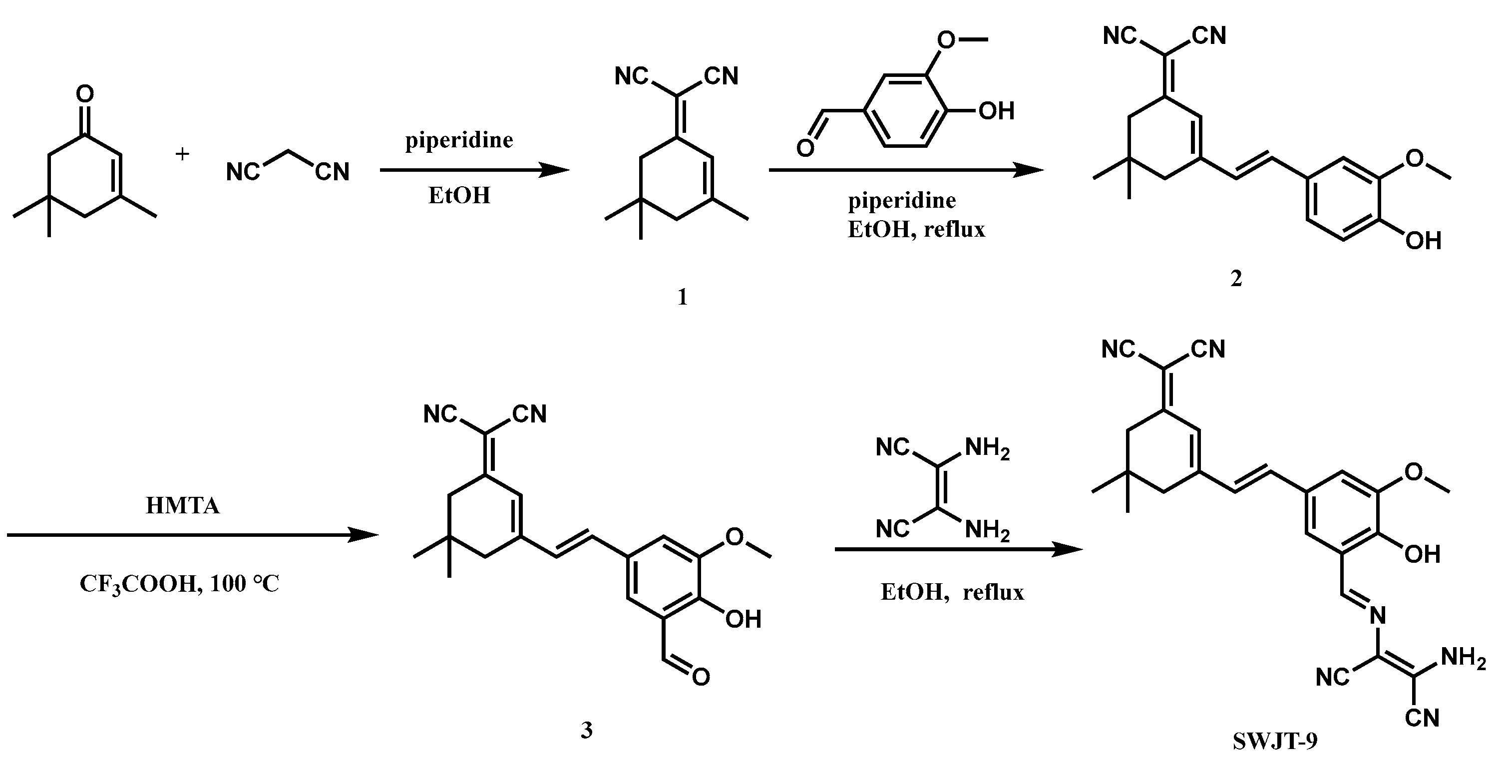
2.1. Design SWJT-9
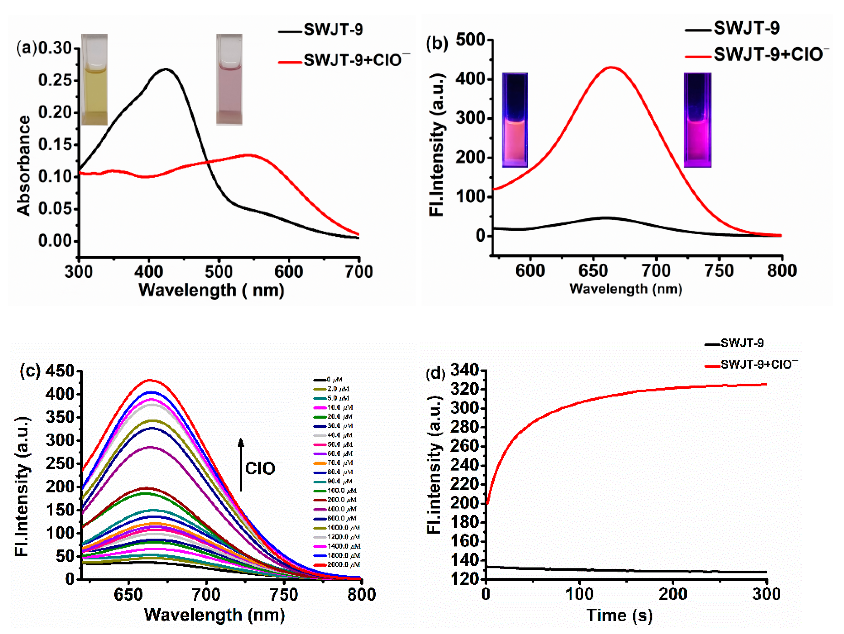
2.2. Photoproperties of Probe
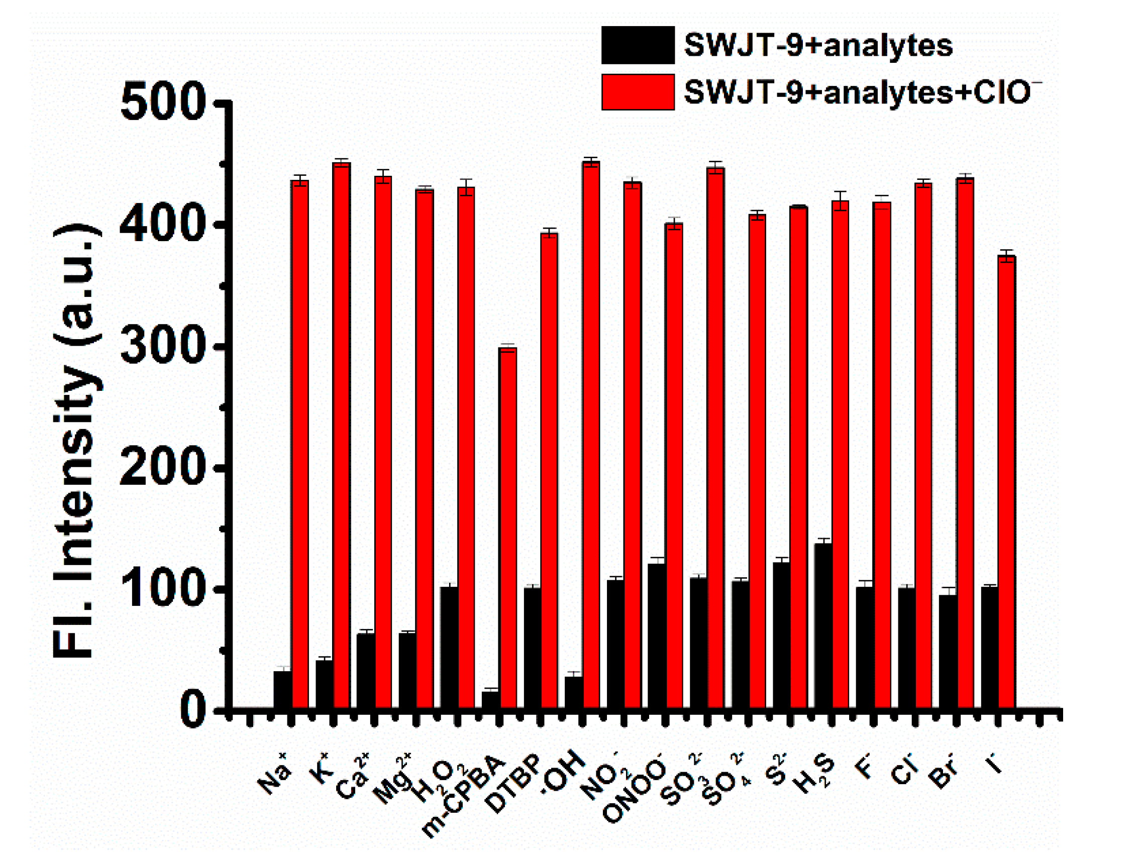
2.3. Competition Experiments
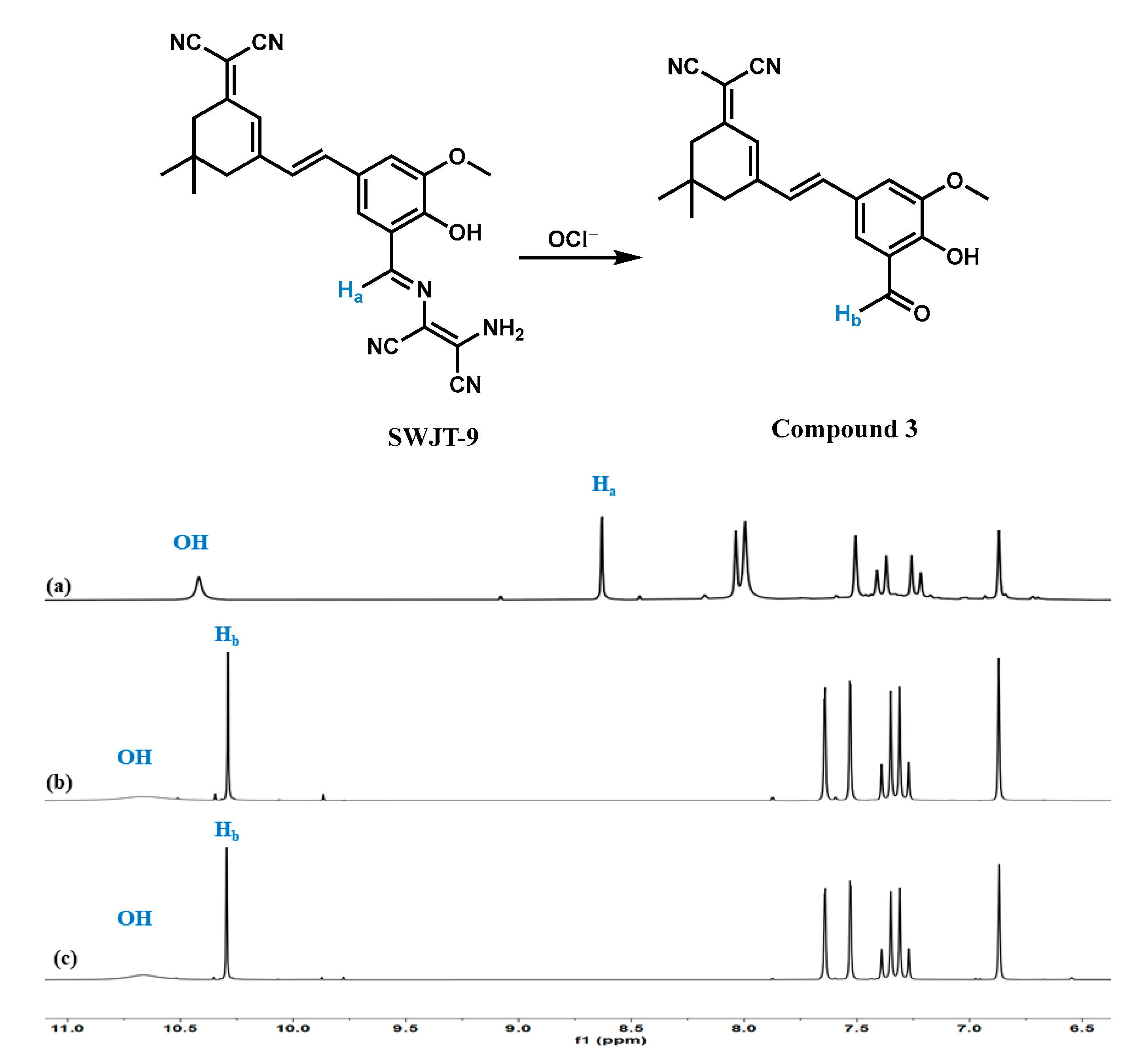
2.4. Response Mechanism
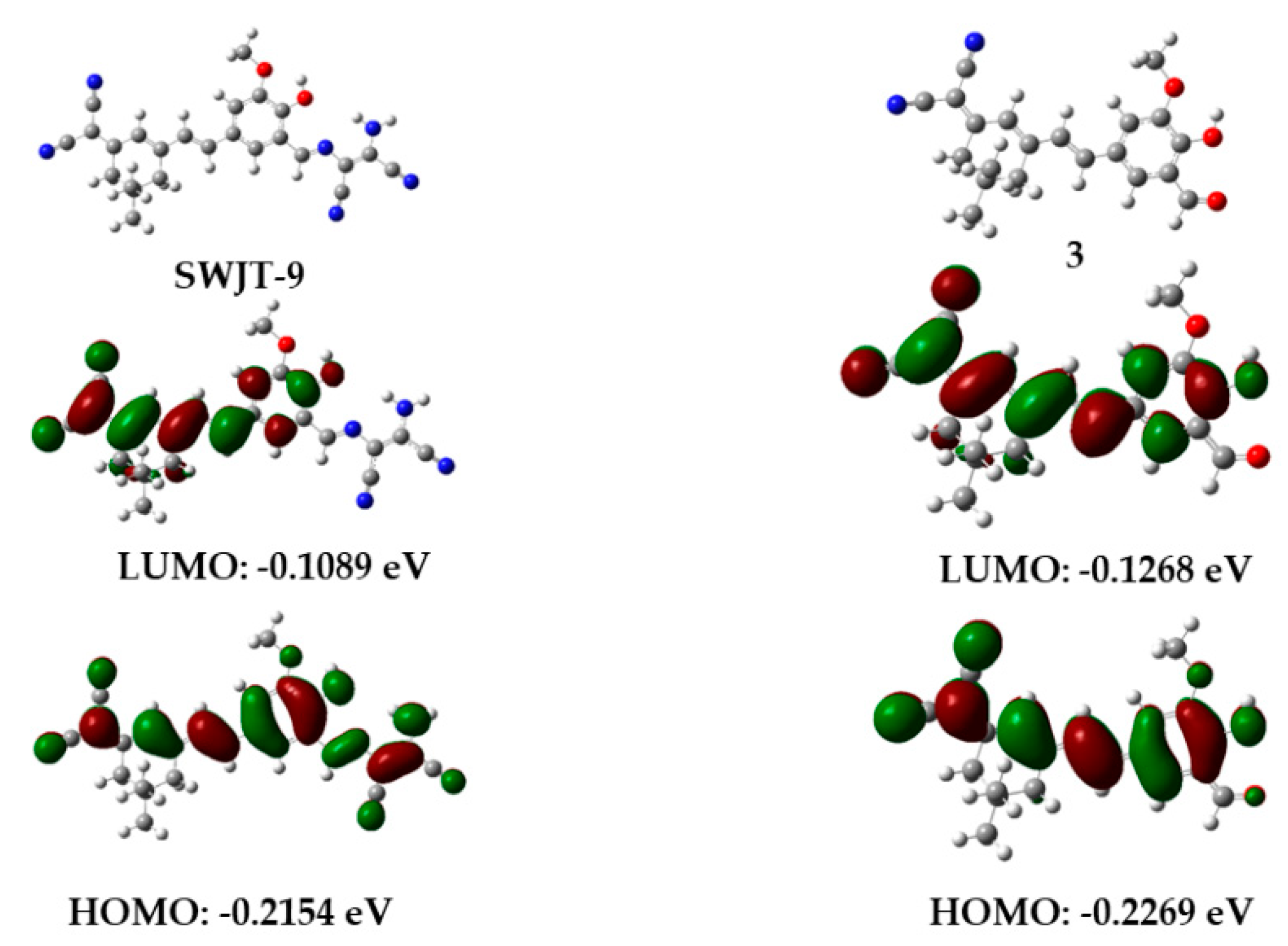
2.5. DFT Calculations
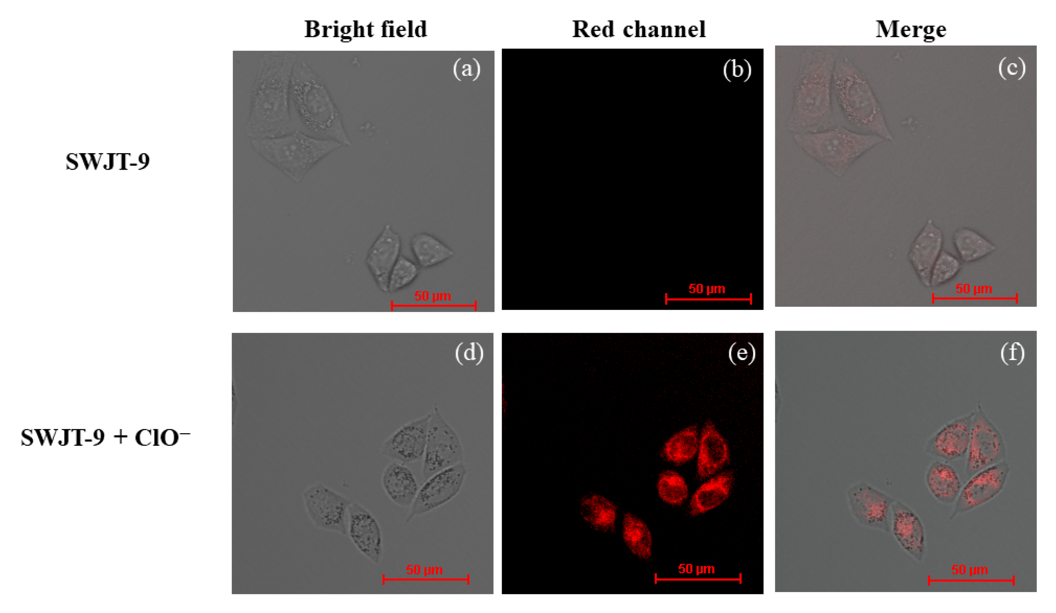
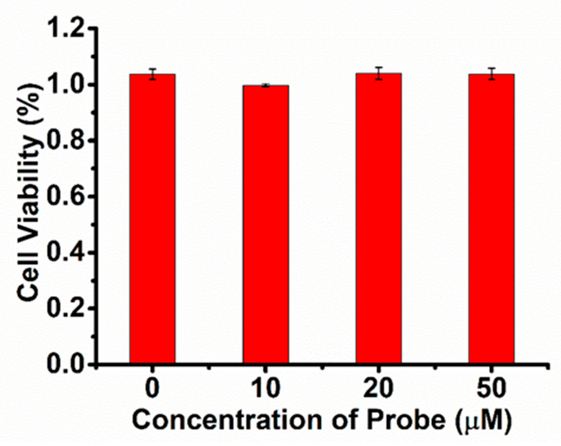
2.6. Cytotoxicity of SWJT-9 and Its Imaging in HeLa Cells
3. Materials and Methods
3.1. Materials and Reagents
3.2. Synthesis of Probe SWJT-9
4. Conclusions
Supplementary Materials
Author Contributions
Funding
Institutional Review Board Statement
Informed Consent Statement
Data Availability Statement
Acknowledgments
Conflicts of Interest
References
- Xu, R.; Huang, H.; Zhang, Z.; Wang, F.-S. The role of neutrophils in the development of liver diseases. Cell. Mol. Immunol. 2014, 11, 224–231. [Google Scholar] [CrossRef] [PubMed] [Green Version]
- Li, A.; Grönlund, E.; Brattsand, G. Automated white blood cell counts in cerebrospinal fluid using the body fluid mode on the platform Sysmex XE-5000. Scand. J. Clin. Lab. Investig. 2014, 74, 673–680. [Google Scholar] [CrossRef] [PubMed]
- Leliefeld, P.H.C.; Wessels, C.M.; Leenen, L.P.H.; Koenderman, L.; Pillay, J. The role of neutrophils in immune dysfunction during severe inflammation. Crit. Care 2016, 20, 73. [Google Scholar] [CrossRef] [PubMed] [Green Version]
- Lippolis, J.D. Immunological signaling networks: Integrating the body’s immune response. J. Anim. Sci. 2008, 86, 53–63. [Google Scholar] [CrossRef] [PubMed]
- Shao, B.H.; Belaaouaj, A.; Verlinde, C.M.L.J.; Fu, X.J.; Heinecke, J.W. Methionine sulfoxide and proteolytic cleavage contribute to the inactivation of cathepsin G by hypochlorous acid: An oxidative mechanism for regulation of serine proteinases by myeloperoxidase. J. Biol. Chem. 2005, 280, 29311–29321. [Google Scholar] [CrossRef] [PubMed] [Green Version]
- Winterbourn, C.C. Biological reactivity and biomarkers of the neutrophil oxidant, hypochlorous acid. Toxicology 2002, 181, 223–227. [Google Scholar] [CrossRef]
- Miyamoto, G.; Zahid, N.; Uetrecht, J.P. Oxidation of Diclofenac to Reactive Intermediates by Neutrophils, Myeloperoxidase, and Hypochlorous Acid. Chem. Res. Toxicol. 1997, 10, 414–419. [Google Scholar] [CrossRef]
- Seminko, V.; Maksimchuk, P.; Grygorova, G.; Avrunin, O.; Semenets, V.; Klochkov, V.; Malyukin, Y. Catalytic Decomposition of Hypochlorite Anions by Ceria Nanoparticles Visualized by Spectroscopic Techniques. J. Phys. Chem. C 2019, 123, 20675–20681. [Google Scholar] [CrossRef]
- Stanley, N.R.; Pattison, D.I.; Hawkins, C.L. Ability of Hypochlorous Acid and N-Chloramines to Chlorinate DNA and Its Constituents. Chem. Res. Toxicol. 2010, 23, 1293–1302. [Google Scholar] [CrossRef]
- Stéliana, G.; Carole, R.; Catherine, V.; Marianne, Z.; Cottin, Y.; Luc, R. Antioxidant properties of an endogenous thiol: Al-pha-lipoic acid, useful in the prevention of cardiovascular diseases. J. Cardiovasc. Pharm. 2009, 54, 391–398. [Google Scholar]
- Pattison, D.; Davies, M.J. Kinetic analysis of the role of histidine chloramines in hypochlorous acid mediated protein oxidation. Biochemistry 2005, 44, 7378–7387. [Google Scholar] [CrossRef] [PubMed]
- Aiken, M.L.; Painter, R.G.; Zhou, Y.; Wang, G. Chloride transport in functionally active phagosomes isolated from Human neutrophils. Free. Radic. Biol. Med. 2012, 53, 2308–2317. [Google Scholar] [CrossRef] [PubMed] [Green Version]
- Pattison, D.; Davies, M.J. Absolute rate constants for the reaction of hypochlorous acid with protein side chains and peptide bonds. Chem. Res. Toxicol. 2001, 14, 1453–1464. [Google Scholar] [CrossRef] [PubMed]
- Savenkova, M.; Mueller, D.; Heinecke, J. Tyrosyl radical generated by myeloperoxidase is a physiological catalyst for the initiation of lipid peroxidation in low density lipoprotein. J. Biol. Chem. 1994, 269, 20394–20400. [Google Scholar] [CrossRef] [PubMed]
- Wu, S.M.; Pizzo, S.V. α2-Macroglobulin from Rheumatoid Arthritis Synovial Fluid: Functional Analysis Defines a Role for Oxidation in Inflammation. Arch. Biochem. Biophys. 2001, 391, 119–126. [Google Scholar] [CrossRef]
- Claver, J.B.; Valencia Miron, M.C.; Capit´an-Vallvey, L.F. Determination of hypochlorite in water using a chemiluminescent test strip. Anal. Chim. Acta. 2004, 522, 267–273. [Google Scholar] [CrossRef]
- Chen, P.; Wei, W.-Z.; Yao, S.-Z. Different valency chlorine species analysis by non-suppressed ion-chromatography with double cell quartz crystal detector. Talanta 1999, 49, 571–576. [Google Scholar] [CrossRef]
- Hallaj, T.; Amjadi, M.; Manzoori, J.; Shokri, R. Chemiluminescence reaction of glucose-derived graphene quantumdots with hypochlorite, and its application to the determination of free chlorine. Microchim. Acta. 2015, 182, 789–796. [Google Scholar] [CrossRef]
- Dikalov, S.I.; Harrison, D.G. Methods for Detection of Mitochondrial and Cellular Reactive Oxygen Species. Antioxid. Redox Signal. 2014, 20, 372–382. [Google Scholar] [CrossRef] [Green Version]
- Qu, Z.; Ding, J.; Zhao, M.; Li, P. Development of a selenide-based fluorescent probe for imaging hypochlorous acid in lyso-somes. J. Photoch. Photobio. A 2015, 299, 1–8. [Google Scholar] [CrossRef]
- Wu, L.; Sedgwick, A.C.; Sun, X.; Bull, S.D.; He, X.-P.; James, T.D. Reaction-Based Fluorescent Probes for the Detection and Imaging of Reactive Oxygen, Nitrogen, and Sulfur Species. Accounts Chem. Res. 2019, 52, 2582–2597. [Google Scholar] [CrossRef] [PubMed]
- Ren, T.; Wang, Z.; Xiang, Z.; Lu, P.; Lai, H.; Yuan, L.; Zhang, X.; Tan, W. A General Strategy for Development of Activatable NIR-II Fluorescent Probes for In Vivo High-Contrast Bioimaging. Angew. Chem. Int. Ed. 2020, 60, 800–805. [Google Scholar] [CrossRef] [PubMed]
- Wei, Y.; Liu, Y.; He, Y.; Wang, Y. Mitochondria and lysosome-targetable fluorescent probes for hydrogen peroxide. J. Mater. Chem. B 2021, 9, 908–920. [Google Scholar] [CrossRef] [PubMed]
- Pang, Q.; Li, T.; Yin, C.; Ma, K.; Huo, F. Comparing the abundance of HClO in cancer/normal cells and visualizing in vivo using a mitochondria-targeted ultra-fast fluorescent probe. Analyst 2021, 146, 3361–3367. [Google Scholar] [CrossRef]
- Wu, P.; Zhu, Y.; Chen, L.; Tian, Y.; Xiong, H. A Fast-Responsive OFF–ON Near-Infrared-II Fluorescent Probe for In Vivo Detection of Hypochlorous Acid in Rheumatoid Arthritis. Anal. Chem. 2021, 93, 13014–13021. [Google Scholar] [CrossRef] [PubMed]
- Xu, H.; Wu, S.-L.; Song-Ling, W.; Lu, Y.; Xiao, J.; Wang, Y.-W.; Peng, Y. A NIR fluorescent probe for rapid turn-on detection and bioimaging of hypochlorite anion. Sens. Actuators B Chem. 2021, 346, 130484. [Google Scholar] [CrossRef]
- Duan, C.; Won, M.; Verwilst, P.; Xu, J.; Kim, H.S.; Zeng, L.; Kim, J.S. In Vivo Imaging of Endogenously Produced HClO in Zebrafish and Mice Using a Bright, Photostable Ratiometric Fluorescent Probe. Anal. Chem. 2019, 91, 4172–4178. [Google Scholar] [CrossRef]
- Gao, W.; Ma, Y.; Liu, Y.; Ma, S.; Lin, W. Observation of endogenous HClO in living mice with inflammation, tissue injury and bacterial infection by a near-infrared fluorescent probe. Sens. Actuators B Chem. 2021, 327, 128884. [Google Scholar] [CrossRef]
- Cheng, G.; Fan, J.; Sun, W.; Sui, K.; Jin, X.; Wang, J.; Peng, X. A highly specific BODIPY-based probe localized in mitochondria for HClO imaging. Analyst 2013, 138, 6091–6096. [Google Scholar] [CrossRef]
- Wang, L.; Ding, H.; Ran, X.; Tang, H.; Cao, D. Recent progress on reaction-based BODIPY probes for anion detection. Dye. Pigment. 2020, 172, 107857. [Google Scholar] [CrossRef]
- Wang, L.; Liu, J.; Zhang, H.; Guo, W. Discrimination between cancerous and normal cells/tissues enabled by a near-infrared fluorescent HClO probe. Sens. Actuators B Chem. 2021, 334, 129602. [Google Scholar] [CrossRef]
- Li, H.; Kim, H.; Han, J.; Nguyen, V.N.; Peng, X.; Yoon, J. Activity-based smart AIEgens for detection, bioimaging, and therapeutics: Recent progress and outlook. Aggregate 2021, 2, 51. [Google Scholar] [CrossRef]
- Hu, Q.; Qin, C.; Huang, L.; Wang, H.; Liu, Q.; Zeng, L. Selective visualization of hypochlorite and its fluctuation in cancer cells by a mitochondria-targeting ratiometric fluorescent probe. Dye. Pigment. 2018, 149, 253–260. [Google Scholar] [CrossRef]
- Feng, Y.; Li, S.Q.; Li, D.X.; Wang, Q.; Ning, P.; Chen, M.; Tian, X.H. Rational design of a diaminomaleonitrile-based mitochondria—Targeted two-photon fluorescent probe for hypochlorite in vivo: Solvent-independent and high selectivity over Cu2+. Sens. Actuators B Chem. 2018, 254, 282–290. [Google Scholar] [CrossRef]
- Dou, K.; Fu, Q.; Chen, G.; Yu, F.B.; Liu, Y.X.; Cao, Z.P.; Li, G.L.; Zhao, X.N.; Xia, L.; Chen, L.X.; et al. A novel dual-ratiometric-response fluorescent probe for SO2/ClO− detection in cells and invivo and its application in exploring the dichotomous role of SO2 under the ClO− induced oxidative stress. Biomaterials 2017, 133, 82–93. [Google Scholar] [CrossRef]
- Ning, Y.; Cui, J.; Lu, Y.; Wang, X.; Xiao, C.; Wu, S.; Li, J.; Zhang, Y. De novo design and synthesis of a novel colorimetric fluorescent probe based on naphthalenone scaffold for selective detection of hypochlorite and its application in living cells. Sens. Actuators B Chem. 2018, 269, 322–330. [Google Scholar] [CrossRef]
- Malkondu, S.; Erdemir, S.; Karakurt, S. Red and blue emitting fluorescent probe for cyanide and hypochlorite ions: Biological sensing and environmental analysis. Dye. Pigment. 2020, 174, 108019. [Google Scholar] [CrossRef]
- Zhang, Y.; Ma, L.; Tang, C.; Pan, S.; Shi, D.; Wang, S.; Li, M.; Guo, Y. A highly sensitive and rapidly responding fluorescent probe based on a rhodol fluorophore for imaging endogenous hypochlorite in living mice. J. Mater. Chem. B 2018, 6, 725. [Google Scholar] [CrossRef]
- Wang, Z.; Zhang, Y.; Song, J.; Li, M.; Yang, Y.; Gu, W.; Xu, X.; Xu, H.; Wang, S. A highly specific and sensitive turn-on fluorescence probe for hypochlorite detection based on anthracene fluorophore and its bioimaging applications. Dye. Pigment. 2018, 161, 172–181. [Google Scholar] [CrossRef]
- Tang, X.; Zhu, X.; Liu, R.J.; Tang, Y. A novel ratiometric and colorimetric fluorescent probe for hypochlorite based on cya-nobiphenyl and its applications. Spectrochim. Acta Part A Mol. Biomol. Spectrosc. 2019, 219, 576–581. [Google Scholar] [CrossRef]
- Zhao, X.J.; Jiang, Y.R.; Chen, Y.X.; Yang, B.Q.; Li, Y.T.; Liu, Z.H.; Liu, C. A new “off-on” NIR fluorescence probe for deter-mination and bio-imaging of mitochondrial hypochlorite in living cells and zebrafish. Spectrochim. Acta. A 2019, 219, 509–516. [Google Scholar] [CrossRef] [PubMed]
- Cheng, W.S.; Ren, C.P.; Liu, S.; Jiang, W.S.; Zhu, X.N.; Jia, W.X.; Chen, J.B.; Liu, Z.B. A highly selective A–π–A “turn-on” fluorescent probe for hypochlorite in tap water. New J. Chem. 2022, 46, 18010–18017. [Google Scholar] [CrossRef]
- Zhang, X.W.; Zhang, F.; Chai, J.; Yang, B.S.; Liu, B. A TICT + AIE based fluorescent probe for ultrafast response of hypochlorite in living cells and mouse. Spectrochim. Acta. A 2021, 256, 119735. [Google Scholar] [CrossRef] [PubMed]
- Xu, C.; Wu, T.; Duan, L.; Zhou, Y.; Zhou, Y. Rational Design of ICT-Based Fluorescent Probe with AIE and NIR Properties for Hypochlorite Determination. J. Electrochem. Soc. 2022, 169, 017514. [Google Scholar] [CrossRef]
- Li, M.; Du, F.; Xue, P.; Tan, X.; Liu, S.; Zhou, Y.; Chen, J.; Bai, L. An AIE fluorescent probe with a naphthalimide derivative and its application for detection of hypochlorite and imaging inside living cells. Spectrochim. Acta A 2020, 227, 117760. [Google Scholar] [CrossRef]
- Jiang, Y.; Wu, S.; Jin, C.; Wang, B.; Shen, J. Novel diaminomaleonitrile-based fluorescent probe for ratiometric detection and bioimaging of hypochlorite. Sens. Actuators B Chem. 2018, 265, 365–370. [Google Scholar] [CrossRef]
- Zhao, J.; Ma, T.; Chang, B.; Fang, J. Recent Progress on NIR Fluorescent Probes for Enzymes. Molecules 2022, 27, 5922. [Google Scholar] [CrossRef]
- Feng, Y.-A.; Xu, H.; Zhou, Y.; Wang, B.-J.; Xiao, J.; Wang, Y.-W.; Peng, Y. Ratiometric detection and bioimaging of endogenous alkaline phosphatase by a NIR fluorescence probe. Sens. Actuators B Chem. 2022, 358, 131505. [Google Scholar] [CrossRef]
- Xu, H.; Zhang, C.; Zhang, Y.-Q.; Suo, S.-N.; Wang, Y.-W.; Peng, Y. A red-NIR fluorescence probe for rapid and visual detection of acrolein. Chem. Commun. 2022, 58, 10080–10083. [Google Scholar] [CrossRef]
- Yudhistira, T.; Mulay, S.V.; Kim, Y.; Halle, M.B.; Churchill, D.G. Imaging of hypochlorous acid by fluorescence and applications in biological systems. Chem. Asian. J. 2019, 14, 3048–3084. [Google Scholar] [CrossRef]
- Hwang, S.M.; Yun, D.; Kim, C. An Imidazo[1,5-α]Pyridine-Based Fluorometric Chemodosimeter for the Highly Selective Detection of Hypochlorite in Aqueous Media. J. Fluoresc. 2019, 29, 451–459. [Google Scholar] [CrossRef] [PubMed]
- Shi, Y.; Huo, F.; Yin, C. Malononitrile as the ‘double-edged sword’ of passivation-activation regulating two ICT to highly sensitive and accurate ratiometric fluorescent detection for hypochlorous acid in biological system. Sens. Actuators B Chem. 2020, 325, 128793. [Google Scholar] [CrossRef] [PubMed]
- He, L.; Xiong, H.; Wang, B.; Zhang, Y.; Wang, J.; Zhang, H.; Li, H.; Yang, Z.; Song, X. Rational design of a two-photon ra-tiometric fluorescent probe for hypochlorous acid with a large stokes shift. Anal. Chem. 2020, 92, 11029–11034. [Google Scholar] [CrossRef] [PubMed]
- Cheng, G.; Fan, J.; Sun, W.; Cao, J.; Hu, C.; Peng, X. A near-infrared fluorescent probe for selective detection of HClO based on Se-sensitized aggregation of heptamethine cyanine dye. Chem. Commun. 2013, 50, 1018–1020. [Google Scholar] [CrossRef] [PubMed]
- Liu, H.B.; Xu, H.; Guo, X.; Xiao, J.; Cai, Z.H.; Wang, Y.W.; Peng, Y. A novel near-infrared fluorescent probe based on iso-phorone for the bioassay of endogenous cysteine. Org. Biomol. Chem. 2021, 19, 873–877. [Google Scholar] [CrossRef]
- Sun, Q.; Yang, S.; Wu, L.; Dong, Q.; Yang, W.; Yang, G. Detection of intracellular selenol-containing molecules using a fluo-rescent probe with near-zero background signal. Anal. Chem. 2016, 88, 6084–6091. [Google Scholar] [CrossRef]
- Lapenna, D.; Cuccurullo, F. Hypochlorous acid and its pharmacological antagonism: An update picture. Gen. Pharmacol. 1996, 27, 1145–1147. [Google Scholar] [CrossRef]
- Wang, B.J.; Liu, R.J.; Fang, J.G.; Wang, Y.W.; Peng, Y. A water-soluble dual-site fluorescent probe for the rapid detection of cysteine with high sensitivity and specificity. Chem. Commun. 2019, 55, 11762–11765. [Google Scholar] [CrossRef]
- Wu, J.; Liu, W.; Ge, J.; Zhang, H.; Wang, P. New sensing mechanisms for design of fluorescent chemosensors emerging in recent years. Chem. Soc. Rev. 2011, 40, 3483–3495. [Google Scholar] [CrossRef]
- Qian, M.; Xia, J.; Zhang, L.; Chen, Q.; Guo, J.; Cui, H.; Kafuti, Y.S.; Wang, J.; Peng, X. Rationally modifying the dicyanoisophorone fluorophore for sensing cysteine in living cells and mice. Sens. Actuators B Chem. 2020, 321, 128441. [Google Scholar] [CrossRef]
- Reed, J.; Ho, H.; Jolly, W. Chemical synthesis with a quenched flow reactor Hydroxytrihydroborate and peroxynitrite. J. Am. Chem. Soc. 1974, 96, 1248–1249. [Google Scholar] [CrossRef]


Disclaimer/Publisher’s Note: The statements, opinions and data contained in all publications are solely those of the individual author(s) and contributor(s) and not of MDPI and/or the editor(s). MDPI and/or the editor(s) disclaim responsibility for any injury to people or property resulting from any ideas, methods, instructions or products referred to in the content. |
© 2023 by the authors. Licensee MDPI, Basel, Switzerland. This article is an open access article distributed under the terms and conditions of the Creative Commons Attribution (CC BY) license (https://creativecommons.org/licenses/by/4.0/).
Share and Cite
Liu, C.-X.; Xiao, S.-Y.; Gong, X.-L.; Zhu, X.; Wang, Y.-W.; Peng, Y. A Near-Infrared Fluorescent Probe for Recognition of Hypochlorite Anions Based on Dicyanoisophorone Skeleton. Molecules 2023, 28, 402. https://doi.org/10.3390/molecules28010402
Liu C-X, Xiao S-Y, Gong X-L, Zhu X, Wang Y-W, Peng Y. A Near-Infrared Fluorescent Probe for Recognition of Hypochlorite Anions Based on Dicyanoisophorone Skeleton. Molecules. 2023; 28(1):402. https://doi.org/10.3390/molecules28010402
Chicago/Turabian StyleLiu, Chang-Xiang, Shu-Yuan Xiao, Xiu-Lin Gong, Xi Zhu, Ya-Wen Wang, and Yu Peng. 2023. "A Near-Infrared Fluorescent Probe for Recognition of Hypochlorite Anions Based on Dicyanoisophorone Skeleton" Molecules 28, no. 1: 402. https://doi.org/10.3390/molecules28010402