
Multidirectional Characterization of Phytochemical Profile and Health-Promoting Effects of Ziziphora bungeana Juz. Extracts

 , , , ,
  , and
, and
Abstract
:1. Introduction
2. Results and Discussion
2.1. Chemical Composition of Extracts by HPLC-ESI-QTOF-MS/MS
2.2. Antimicrobial Activity Assessment
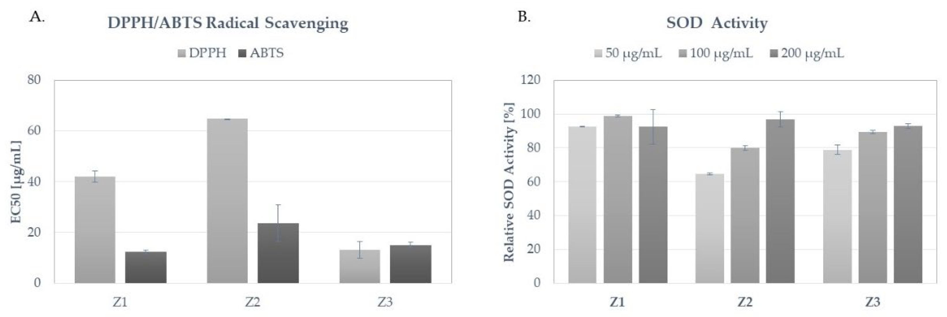
2.3. Antioxidant Activity Assessment
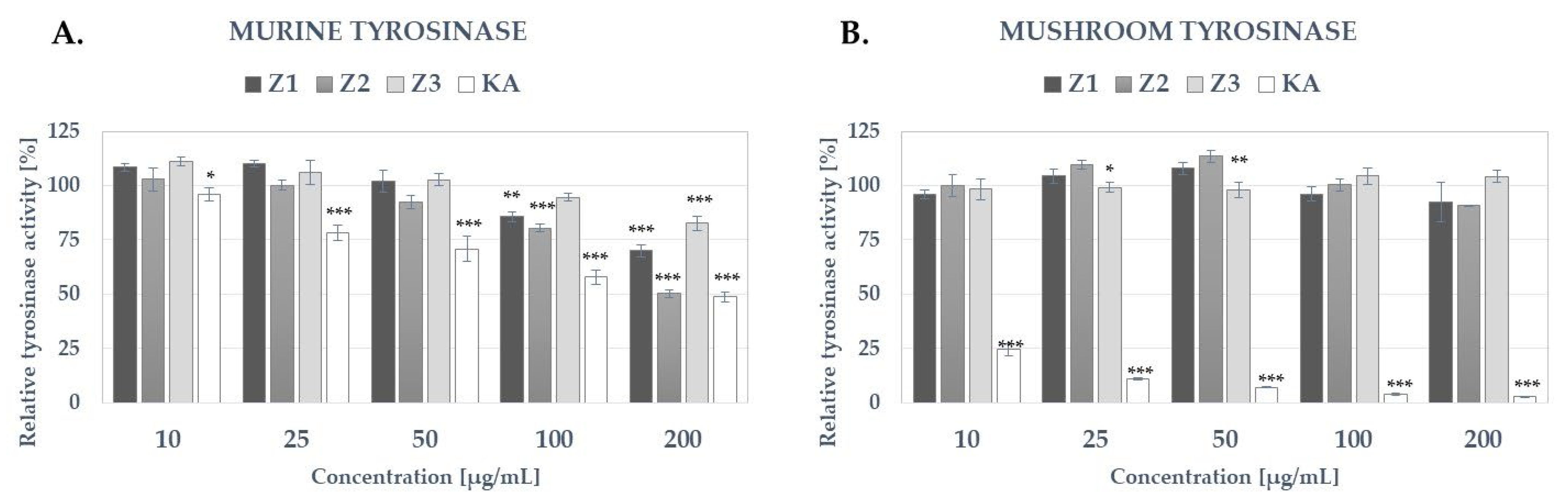
2.4. Tyrosinase Activity Assay
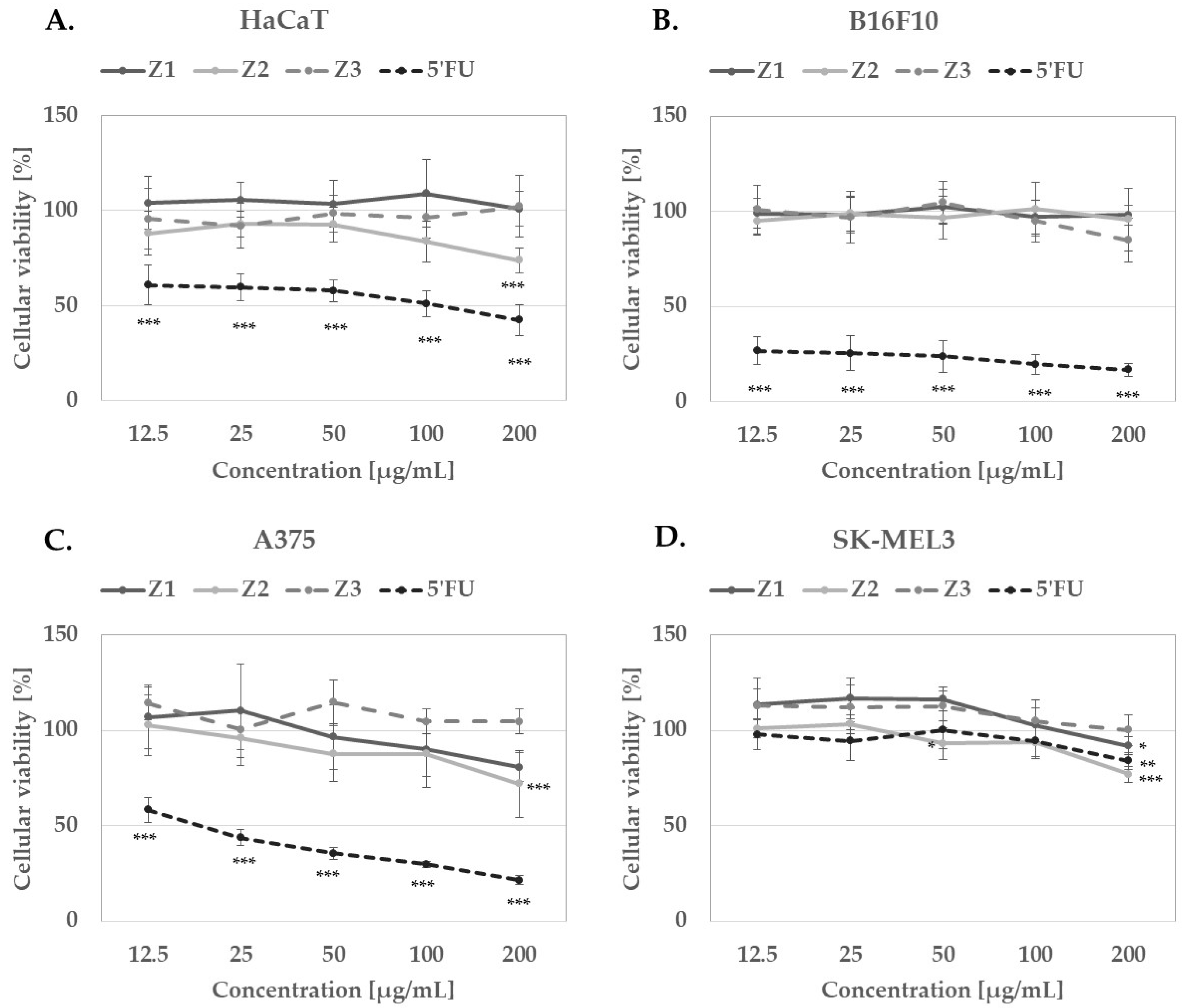
2.5. Cytotoxic Activity
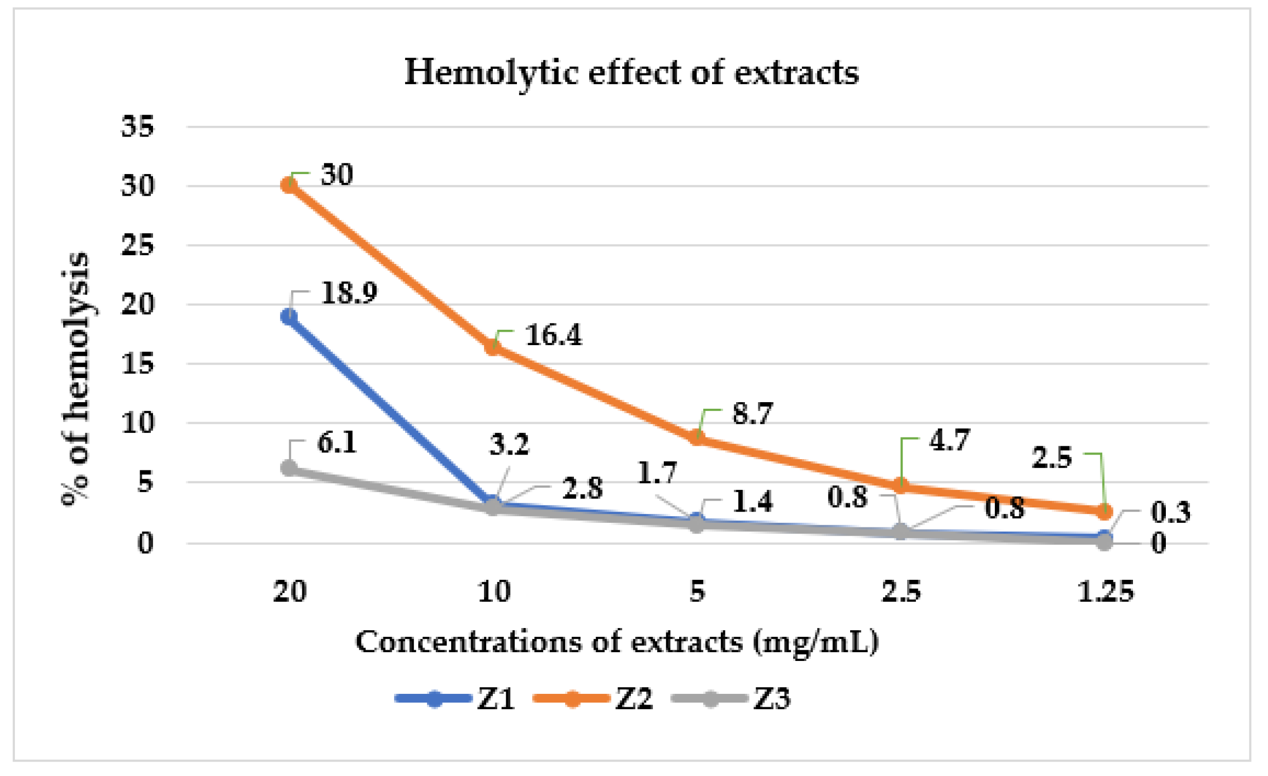
2.6. The Hemolytic Activity Assay (Toxicity towards Erythrocytes)
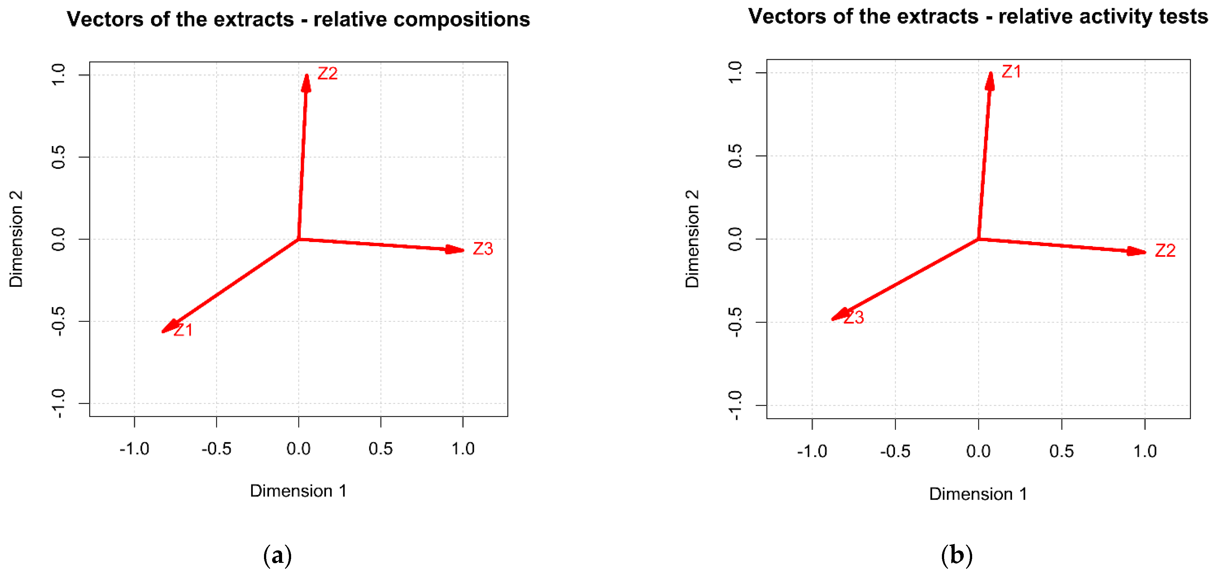
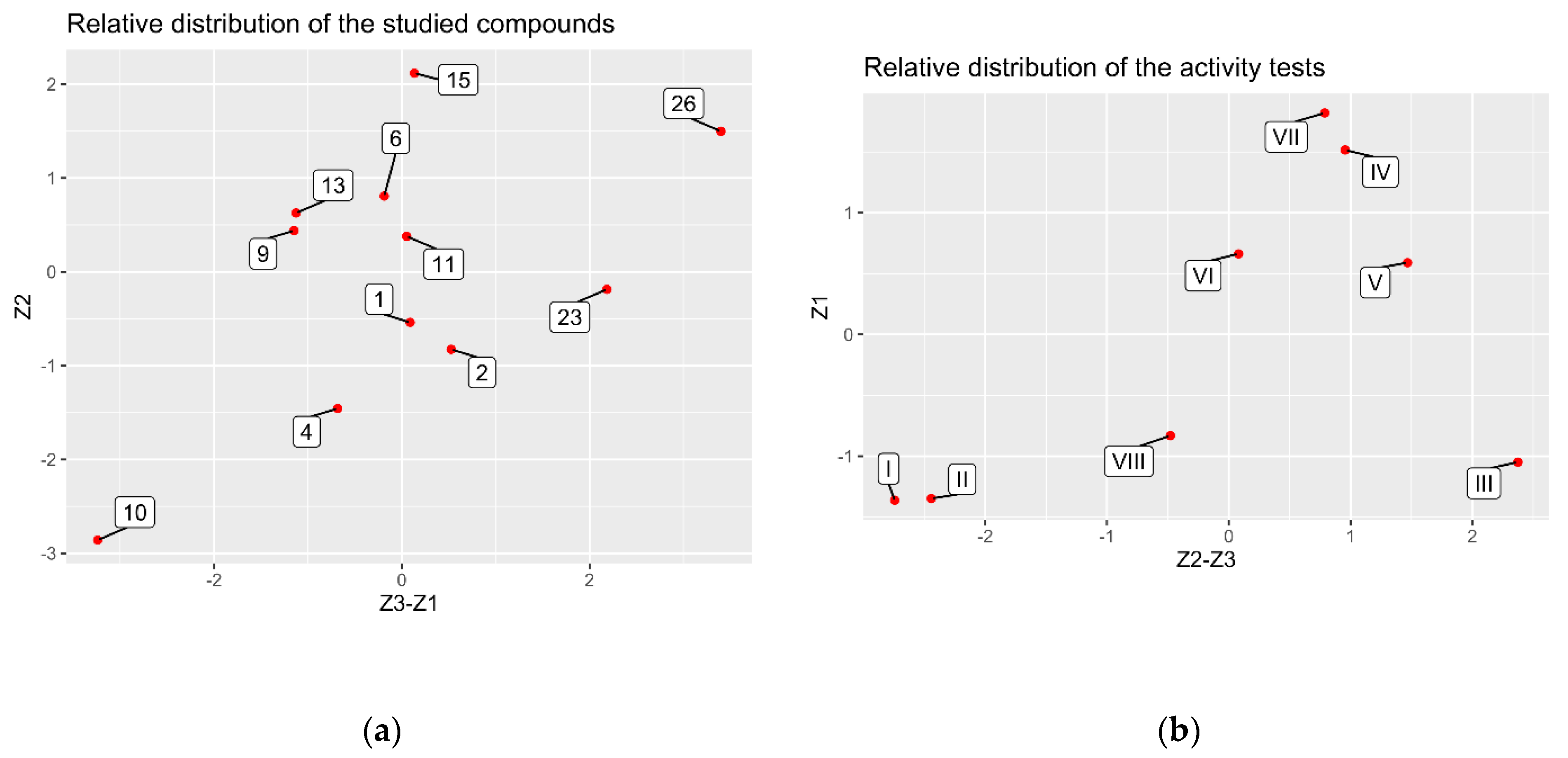
2.7. Chemometric Assessment
3. Material and Methods
3.1. Materials
3.1.1. Plant Material
3.1.2. Microorganisms
3.2. Methods
3.2.1. Extraction Procedure
3.2.2. The HPLC-ESI-QTOF-MS/MS Analysis
3.2.3. In Vitro Antimicrobial Activity Assay
3.2.4. Total Antimicrobial Activity (TAA) Assay
3.2.5. Antioxidant Activity
DPPH Scavenging Assay
ABTS Scavenging Assay
SOD Inhibitory Assay
3.2.6. Tyrosinase Inhibitory Activity
3.2.7. In Vitro Cytotoxicity Assay
3.2.8. Toxicity to Erythrocyte Assay
3.3. Chemometric Analysis
4. Conclusions
Supplementary Materials
Author Contributions
Funding
Institutional Review Board Statement
Informed Consent Statement
Data Availability Statement
Conflicts of Interest
Sample Availability
References
- Shahbazi, Y. Chemical compositions, antioxidant and antimicrobial properties of Ziziphora clinopodioides Lam. essential oils collected from different parts of Iran. J. Food Sci. Technol. 2017, 54, 3491–3503. [Google Scholar] [CrossRef] [PubMed]
- Hazrati, S.; Govahi, M.; Sedaghat, M.; Beyraghdar Kashkooli, A. A comparative study of essential oil profile, antibacterial and antioxidant activities of two cultivated Ziziphora species (Z. clinopodioides and Z. tenuior). Ind. Crops Prod. 2020, 157, 112942. [Google Scholar] [CrossRef]
- Eroglu Ozkan, E.; Boga, M.; Yilmaz, M.; Mataraci, E.; Yeşil, Y. LC-MS/MS analyses of Ziziphora clinopodioides Lam. from Turkey: Antioxidant, anticholinesterase, antimicrobial and, anticancer activities. İstanbul J. Pharm. 2020, 50, 33–41. [Google Scholar] [CrossRef]
- Ding, W.; Yang, T.; Liu, F.; Tian, S. Effect of different growth stages of Ziziphora clinopodioides Lam. on its chemical composition. Pharm. Mag. 2014, 10, S1–S5. [Google Scholar] [CrossRef] [Green Version]
- Hulya, D. Compositional diversity in essential oil of Ziziphora tenuior L. ecotypes. Genetika 2021, 53, 717–727. [Google Scholar] [CrossRef]
- Masrournia, M. Elemental Determination and Essential Oil Composition of Ziziphora clinopodioides and Consideration of its Antibacterial Effects. Asian J. Chem. 2013, 25, 6553. [Google Scholar] [CrossRef]
- Ozturk, Y.; Aydm, S.; Tecik, B.; Baser, K.H.C. Effects of essential oils from certain Ziziphora species on swimming performance in mice. Phytother. Res. 1995, 9, 225–227. [Google Scholar] [CrossRef]
- Tarakci, Z.; Coşkun, H.; Tuncturk, Y. Some Properties of Fresh and Ripened Herby Cheese, a Traditional Variety Produced in Turkey. Food Technol. Biotechnol 2004, 42, 47–50. [Google Scholar]
- Ozturk, S. Antibacterial activity and chemical constitutions of Ziziphora Clinopodioides. Food Control. 2007, 18, 535–540. [Google Scholar] [CrossRef]
- Šmejkal, K.; Malaník, M.; Zhaparkulova, K.; Sakipova, Z.; Ibragimova, L.; Ibadullaeva, G.; Žemlička, M. Kazakh Ziziphora Species as Sources of Bioactive Substances. Molecules 2016, 21, 826. [Google Scholar] [CrossRef] [Green Version]
- Hosseinzadeh, M.H.; Ebrahimzadeh, M.A. Antioxidant Potential of Ziziphora Clinopodioides Lam: A Narrative Review. mazu tbsrj 2020, 2, 1–7. [Google Scholar] [CrossRef]
- Srivedavyasasri, R.; Zhaparkulova, K.A.; Sakipova, Z.B.; Ibragimova, L.; Ross, S.A. Phytochemical and Biological Studies on Ziziphora bungeana. Chem. Nat. Compd. 2018, 54, 195–197. [Google Scholar] [CrossRef] [PubMed]
- Abdykerimova, S.; Sakipova, Z.; Nakonieczna, S.; Koch, W.; Biernasiuk, A.; Grabarska, A.; Malm, A.; Kozhanova, K.; Kukula-Koch, W. Superior Antioxidant Capacity of Berberis iliensis—HPLC-Q-TOF-MS Based Phytochemical Studies and Spectrophotometric Determinations. Antioxidants 2020, 9, 504. [Google Scholar] [CrossRef] [PubMed]
- Ahmadi, A.; Gandomi, H.; Derakhshandeh, A.; Misaghi, A.; Noori, N. Phytochemical composition and in vitro safety evaluation of Ziziphora clinopodioides Lam. ethanolic extract: Cytotoxicity, genotoxicity and mutagenicity assessment. J. Ethnopharmacol. 2021, 266, 113428. [Google Scholar] [CrossRef]
- Awwad, A.; Poucheret, P.; Idres, Y.; Tshibangu, D.; Servent, A.; Karine, F.; Lazennec, F.; Bidel, L.; Cazals, G.; Tousch, D. In Vitro Tests for a Rapid Evaluation of Antidiabetic Potential of Plant Species Containing Caffeic Acid Derivatives: A Validation by Two Well-Known Antidiabetic Plants, Ocimum gratissimum L. Leaf and Musanga cecropioides R. Br. ex Tedlie (Mu) Stem Bark. Mol. Cells 2021, 26, 5566. [Google Scholar] [CrossRef]
- Liu, C.; Wahefu, A.; Lu, X.; Abdulla, R.; Dou, J.; Zhao, H.; Aisa, H.A.; Xin, X.; Liu, Y. Chemical Profiling of Kaliziri Injection and Quantification of Six Caffeoyl Quinic Acids in Beagle Plasma by LC-MS/MS. Pharmaceuticals 2022, 15, 663. [Google Scholar] [CrossRef]
- de Oliveira, J.K.; Ronik, D.F.V.; Ascari, J.; Mainardes, R.M.; Khalil, N.M. A stability-indicating high performance liquid chromatography method to determine apocynin in nanoparticles. J. Pharm. Anal. 2017, 7, 129–133. [Google Scholar] [CrossRef]
- Zengin, G.; Mahomoodally, F.; Ibrahime, S.; Ak, G.; Etienne, O.; Jugreet, S.; Brunetti, L.; Leone, S.; Cristina, S.; Di Simone, S.; et al. Chemical Composition and Biological Properties of Two Jatropha Species: Different Parts and Different Extraction Methods. Antioxidants 2021, 10, 792. [Google Scholar] [CrossRef]
- Wang, X.; Qian, Y.; Li, X.; Jia, X.; Yan, Z.; Han, M.; Qiao, M.; Ma, X.; Chu, Y.; Zhou, S.; et al. Rapid determination of rosmarinic acid and its two bioactive metabolites in the plasma of rats by LC-MS/MS and application to a pharmacokinetics study. Biomed. Chromatogr 2021, 35, e4984. [Google Scholar] [CrossRef]
- Micucci, M.; Protti, M.; Aldini, R.; Frosini, M.; Corazza, I.; Marzetti, C.; Mattioli, L.; Tocci, G.; Chiarini, A.; Mercolini, L.; et al. Thymus vulgaris L. Essential Oil Solid Formulation: Chemical Profile and Spasmolytic and Antimicrobial Effects. Biomolecules 2020, 10, 860. [Google Scholar] [CrossRef]
- Huwait, E.; Mobashir, M. Potential and Therapeutic Roles of Diosmin in Human Diseases. Biomedicines 2022, 10, 1076. [Google Scholar] [CrossRef] [PubMed]
- Li, Y.; Guang, C.; Zhao, N.; Feng, X.; Qiu, F. LC-MS/MS Method for Simultaneous Determination of Linarin and Its Metabolites in Rat Plasma and Liver Tissue Samples: Application to Pharmacokinetic and Liver Tissue Distribution Study After Oral Administration of Linarin. Molecules 2019, 24, 3342. [Google Scholar] [CrossRef] [PubMed] [Green Version]
- Wianowska, D.; Dawidowicz, A.L.; Bernacik, K.; Typek, R. Determining the true content of quercetin and its derivatives in plants employing SSDM and LC–MS analysis. Eur. Food Res. Technol. 2016, 243, 27–40. [Google Scholar] [CrossRef] [Green Version]
- Shen, J.; Jia, Q.; Huang, X.; Yao, G.; Ma, W.; Zhang, H.; Ouyang, H.; He, J. Development of a HPLC-MS/MS Method to Determine the 13 Elements of Semen cuscutae and Application to a Pharmacokinetic Study in Rats. Evid. Based Complement. Altern. Med. 2019, 2019, 6518528. [Google Scholar] [CrossRef] [Green Version]
- Cheruvu, H.S.; Yadav, N.K.; Valicherla, G.R.; Arya, R.K.; Hussain, Z.; Sharma, C.; Arya, K.R.; Singh, R.K.; Datta, D.; Gayen, J.R. LC-MS/MS method for the simultaneous quantification of luteolin, wedelolactone and apigenin in mice plasma using hansen solubility parameters for liquid-liquid extraction: Application to pharmacokinetics of Eclipta alba chloroform fraction. J. Chromatogr. B 2018, 1081–1082, 76–86. [Google Scholar] [CrossRef]
- Kim, S.; Kim, J.; Kim, N.; Lee, D.; Lee, H.; Lee, D.Y.; Kim, K.H. Metabolomic Elucidation of the Effect of Sucrose on the Secondary Metabolite Profiles in Melissa officinalis by Ultraperformance Liquid Chromatography-Mass Spectrometry. ACS Omega 2020, 5, 33186–33195. [Google Scholar] [CrossRef]
- Silvestro, L.; Tarcomnicu, I.; Dulea, C.; Attili, N.R.; Ciuca, V.; Peru, D.; Rizea Savu, S. Confirmation of diosmetin 3-O-glucuronide as major metabolite of diosmin in humans, using micro-liquid-chromatography-mass spectrometry and ion mobility mass spectrometry. Anal. Bioanal Chem. 2013, 405, 8295–8310. [Google Scholar] [CrossRef] [Green Version]
- Knez Hrnčič, M.; Cör, D.; Simonovska, J.; Knez, Ž.; Kavrakovski, Z.; Rafajlovska, V. Extraction Techniques and Analytical Methods for Characterization of Active Compounds in Origanum Species. Molecules 2020, 25, 4735. [Google Scholar] [CrossRef]
- Stebounova, L.; Ebert, S.M.; Murry, L.T.; Adams, C.M.; Murry, D.J. Rapid and Sensitive Quantification of Ursolic Acid and Oleanolic Acid in Human Plasma Using Ultra-performance Liquid Chromatography–Mass Spectrometry. J. Chromatogr. Sci. 2018, 56, 644–649. [Google Scholar] [CrossRef] [Green Version]
- Castellano, J.M.; Ramos-Romero, S.; Perona, J.S. Oleanolic Acid: Extraction, Characterization and Biological Activity. Nutrients 2022, 14, 623. [Google Scholar] [CrossRef]
- Sánchez-González, M.; Lozano-Mena, G.; Juan, M.E.; García-Granados, A.; Planas, J.M. Liquid chromatography-mass spectrometry determination in plasma of maslinic acid, a bioactive compound from Olea europaea L. Food Chem 2013, 141, 4375–4381. [Google Scholar] [CrossRef] [PubMed]
- Sarikurkcu, C.; Kakouri, E.; Sarikurkcu, R.T.; Tarantilis, P.A. Study on the Chemical Composition, Enzyme Inhibition and Antioxidant Activity of Ziziphora taurica subsp. cleonioides. Appl. Sci. 2019, 9, 5515. [Google Scholar] [CrossRef] [Green Version]
- Zhu, Y.; Xiong, Y.; Wang, H.; Li, P. Pharmacognostical and phytochemical studies on Ziziphora clinopodioides Lam.—A Kazakh and Uygur ethnomedicinal plant. J. Pharm. Pharmacogn. Res. 2017, 5, 345–353. [Google Scholar]
- Koch, W. Dietary Polyphenols—Important Non-Nutrients in the Prevention of Chronic Noncommunicable Diseases. A Systematic Review. Nutrients 2019, 11, 1039. [Google Scholar] [CrossRef] [PubMed] [Green Version]
- Koch, W.; Baj, T.; Kukula-Koch, W.; Marzec, Z. Dietary intake of specific phenolic compounds and their effect on the antioxidant activity of daily food rations. Open Chem. 2015, 13, 869–876. [Google Scholar] [CrossRef]
- Eloff, J.N. Quantification the bioactivity of plant extracts during screening and bioassay guided fractionation. Phytomedicine 2004, 11, 370–371. [Google Scholar] [CrossRef]
- Henley-Smith, C.J.; Steffens, F.E.; Botha, F.S.; Lall, N. Predicting the influence of multiple components on microbial inhibition using a logistic response model—A novel approach. BMC Complement. Altern. Med. 2014, 14, 190. [Google Scholar] [CrossRef] [Green Version]
- Sonboli, A.; Mirjalili, M.H.; Hadian, J.; Ebrahimi, S.N.; Yousefzadi, M. Antibacterial activity and composition of the essential oil of Ziziphora clinopodioides subsp. bungeana (Juz.) Rech. f. from Iran. Z. Nat. C J. Biosci. 2006, 61, 677–680. [Google Scholar] [CrossRef]
- Ozturk, S.; Ercisli, S. The chemical composition of essential oil and in vitro antibacterial activities of essential oil and methanol extract of Ziziphora persica Bunge. J. Ethnopharmacol. 2006, 106, 372–376. [Google Scholar] [CrossRef]
- Anzabi, Y.; Khaki, A.; Rasoli, A.; Ebrahimpour, S.; Fallah Rostami, F. Antibacterial properties of essential oils and methanol extracts of Ziziphora tenuior Lam. (a native plant) in pre-flowering stage against isolated bacteria from urogenital tract infections. Bulg. Chem. Commun. 2016, 48, 120–125. [Google Scholar]
- Gursoy, N.; Sihoglu-Tepe, A.; Tepe, B. Determination of in vitro antioxidative and antimicrobial properties and total phenolic contents of Ziziphora clinopodioides, Cyclotrichium niveum, and Mentha longifolia ssp. typhoides var. typhoides. J. Med. Food 2009, 12, 684–689. [Google Scholar] [CrossRef] [PubMed]
- Salehi, P.; Sonboli, A.; Eftekhar, F.; Nejad-Ebrahimi, S.; Yousefzadi, M. Essential oil composition, antibacterial and antioxidant activity of the oil and various extracts of Ziziphora clinopodioides subsp. rigida (BOISS.) RECH. f. from Iran. Biol. Pharm. Bull. 2005, 28, 1892–1896. [Google Scholar] [CrossRef] [PubMed] [Green Version]
- Celik, C.; Tutar, U.; Karaman, I.; Hepokur, C.; Atas, M. Evaluation of the Antibiofilm and Antimicrobial Properties of Ziziphora tenuior L. Essential Oil Against Multidrug-resistant Acinetobacter baumannii. Int. J. Pharmacol. 2016, 12, 28–35. [Google Scholar] [CrossRef]
- Ydyrys, A.; Zhaparkulova, N.; Aralbaeva, A.; Mamataeva, A.; Seilkhan, A.; Syraiyl, S.; Murzakhmetova, M. Systematic Analysis of Combined Antioxidant and Membrane-Stabilizing Properties of Several Lamiaceae Family Kazakhstani Plants for Potential Production of Tea Beverages. Plants 2021, 10, 666. [Google Scholar] [CrossRef] [PubMed]
- Tomczyk, M.; Ceylan, O.; Locatelli, M.; Tartaglia, A.; Ferrone, V.; Sarikurkcu, C. Ziziphora taurica subsp. taurica: Analytical Characterization and Biological Activities. Biomolecules 2019, 9, 367. [Google Scholar] [CrossRef] [Green Version]
- Zolghadri, S.; Bahrami, A.; Hassan Khan, M.T.; Munoz-Munoz, J.; Garcia-Molina, F.; Garcia-Canovas, F.; Saboury, A.A. A comprehensive review on tyrosinase inhibitors. J. Enzym. Inhib. Med. Chem. 2019, 34, 279–309. [Google Scholar] [CrossRef] [Green Version]
- Niu, C.; Aisa, H.A. Upregulation of Melanogenesis and Tyrosinase Activity: Potential Agents for Vitiligo. Molecules 2017, 22, 1303. [Google Scholar] [CrossRef] [Green Version]
- Pillaiyar, T.; Manickam, M.; Namasivayam, V. Skin whitening agents: Medicinal chemistry perspective of tyrosinase inhibitors. J. Enzym. Inhib. Med. Chem. 2017, 32, 403–425. [Google Scholar] [CrossRef] [Green Version]
- Yoshimori, A.; Oyama, T.; Takahashi, S.; Abe, H.; Kamiya, T.; Abe, T.; Tanuma, S. Structure-activity relationships of the thujaplicins for inhibition of human tyrosinase. Bioorg. Med. Chem. 2014, 22, 6193–6200. [Google Scholar] [CrossRef]
- Strzępek-Gomółka, M.; Gaweł-Bęben, K.; Kukula-Koch, W. Achillea Species as Sources of Active Phytochemicals for Dermatological and Cosmetic Applications. Oxid. Med. Cell Longev. 2021, 2021, 6643827. [Google Scholar] [CrossRef]
- Zhu, H.; Ghoufi, A.; Szymczyk, A.; Balannec, B.; Morineau, D. Zhu et al. Reply. Phys. Rev. Lett. 2013, 111, 089802. [Google Scholar] [CrossRef] [PubMed] [Green Version]
- Pakniyat, A.; Caines, P.E. Time Optimal Hybrid Minimum Principle and the Gear Changing Problem for Electric Vehicles. IFAC Pap. 2015, 48, 187–192. [Google Scholar] [CrossRef]
- Nath, R.; Beyer, D.; Friedland, J.; Grimm, P.; Nag, S. Regarding, Kubo, Coursey, Hanson et al., IJROBP 1998; 40:697–702. Int. J. Radiat. Oncol. Biol. Phys. 1999, 44, 469–470. [Google Scholar] [PubMed]
- Salehi, T.; Mahzounieh, M.; Saeedzadeh, A. Detection of InvA Gene in Isolated Salmonella from Broilers by PCR Method. Int. J. Poult. Sci. 2005, 4, 557–559. [Google Scholar] [CrossRef] [Green Version]
- Nitoda, T.; Fan, M.D.; Kubo, I. Effects of cuminaldehyde on melanoma cells. Phytother. Res. 2008, 22, 809–813. [Google Scholar] [CrossRef]
- Azimi, M.; Mehrzad, J.; Ahmadi, A.; Ahmadi, E.; Ghorbani Ranjbary, A. Apoptosis Induced by Ziziphora tenuior Essential Oil in Human Colorectal Cancer Cells. BioMed. Res. Int. 2021, 2021, 5522964. [Google Scholar] [CrossRef]
- Taghizadeh, M.S.; Niazi, A.; Moghadam, A.; Afsharifar, A.R. The potential application of the protein hydrolysates of three medicinal plants: Cytotoxicity and functional properties. J. Food Sci. 2020, 85, 3160–3167. [Google Scholar] [CrossRef]
- Ghavam, M. A GC-MC analysis of chemical compounds and identification of the antibacterial characteristics of the essential oil of two species exclusive to Iranian habitats: New chemotypes. PLoS ONE 2022, 17, e0273987. [Google Scholar] [CrossRef]
- Yousefbeyk, F.; Ostad, S.; Sourmaghi, M.; Amin, G. Investigation of chemical composition and cytotoxic activity of aerial parts of Ziziphora clinopodioides Lam. Res. J. Pharmacogn. 2016, 3, 41–57. [Google Scholar]
- Zohra, M.; Fawzia, A. Hemolytic activity of different herbal extracts used in Algeria. Int. J. Pharm. Sci. Res. 2014, 5, 495–500. [Google Scholar]
- Luna-Vázquez-Gómez, R.; Arellano-García, M.E.; García-Ramos, J.C.; Radilla-Chávez, P.; Salas-Vargas, D.S.; Casillas-Figueroa, F.; Ruiz-Ruiz, B.; Bogdanchikova, N.; Pestryakov, A. Hemolysis of Human Erythrocytes by Argovit™ AgNPs from Healthy and Diabetic Donors: An In Vitro Study. Materials 2021, 14, 2792. [Google Scholar] [CrossRef] [PubMed]
- European Committee for Antimicrobial Susceptibility Testing (EUCAST) of the European Society of Clinical Microbiology and Infectious Diseases (ESCMID). EUCAST Definitive Document E.DEF 3.1, June 2000: Determination of minimum inhibitory concentrations (MICs) of antibacterial agents by agar dilution. Clin. Microbiol. Infect. 2000, 6, 509–515. [Google Scholar] [CrossRef] [PubMed] [Green Version]
- Wiegand, I.; Hilpert, K.; Hancock, R.E. Agar and broth dilution methods to determine the minimal inhibitory concentration (MIC) of antimicrobial substances. Nat. Protoc. 2008, 3, 163–175. [Google Scholar] [CrossRef] [PubMed]
- O’Donnell, F.; Smyth, T.J.; Ramachandran, V.N.; Smyth, W.F. A study of the antimicrobial activity of selected synthetic and naturally occurring quinolines. Int. J. Antimicrob. Agents 2010, 35, 30–38. [Google Scholar] [CrossRef] [Green Version]
- Wald-Dickler, N.; Holtom, P.; Spellberg, B. Busting the Myth of “Static vs. Cidal”: A Systemic Literature Review. Clin. Infect. Dis. 2018, 66, 1470–1474. [Google Scholar] [CrossRef] [PubMed]
- Wayne, P. Reference method for broth dilution antifungal susceptibility testing of yeasts. Clin. Lab. Stand. Inst. 2008, 3, M27-A2. [Google Scholar]
- Abdrahimov, R. Annual river runoff of the ile-balkash basin and prospects of its assessment due to climatic changes and water economy activities. Int. J. Geomate 2020, 18, 230–239. [Google Scholar] [CrossRef]
- Eloff, J.; McGaw, L. Using African Plant Biodiversity to Combat Microbial Infections. In Novel Plant Bioresources: Applications in Food, Medicine and Cosmetics; Wiley: Hoboken, NJ, USA, 2014; pp. 163–173. [Google Scholar] [CrossRef]
- Elisha, I.L.; Botha, F.S.; McGaw, L.J.; Eloff, J.N. The antibacterial activity of extracts of nine plant species with good activity against Escherichia coli against five other bacteria and cytotoxicity of extracts. BMC Complement. Altern. Med. 2017, 17, 133. [Google Scholar] [CrossRef] [Green Version]
- Famuyide, I.M.; Aro, A.O.; Fasina, F.O.; Eloff, J.N.; McGaw, L.J. Antibacterial and antibiofilm activity of acetone leaf extracts of nine under-investigated south African Eugenia and Syzygium (Myrtaceae) species and their selectivity indices. BMC Complement. Altern. Med. 2019, 19, 141. [Google Scholar] [CrossRef] [Green Version]
- Matejić, J.; Dzamic, A.; Mihajilov-Krstev, T.; Randjelovic, V.; Krivošej, Z.D.; Marin, P. Total phenolic and flavonoid content, antioxidant and antimicrobial activity of extracts from Tordylium maximum. J. Appl. Pharm. Sci. 2013, 3, 55–59. [Google Scholar]
- Re, R.; Pellegrini, N.; Proteggente, A.; Pannala, A.; Yang, M.; Rice-Evans, C. Antioxidant activity applying an improved ABTS radical cation decolorization assay. Free Radic. Biol. Med. 1999, 26, 1231–1237. [Google Scholar] [CrossRef] [PubMed]
- Strzępek-Gomółka, M.; Gaweł-Bęben, K.; Angelis, A.; Antosiewicz, B.; Sakipova, Z.; Kozhanova, K.; Głowniak, K.; Kukula-Koch, W. Identification of Mushroom and Murine Tyrosinase Inhibitors from Achillea biebersteinii Afan. Extract. Molecules 2021, 26, 964. [Google Scholar] [CrossRef] [PubMed]
- Uchida, R.; Ishikawa, S.; Tomoda, H. Inhibition of tyrosinase activity and melanine pigmentation by 2-hydroxytyrosol. Acta Pharm. Sin. B 2014, 4, 141–145. [Google Scholar] [CrossRef] [PubMed] [Green Version]
- Repetto, G.; del Peso, A.; Zurita, J.L. Neutral red uptake assay for the estimation of cell viability/cytotoxicity. Nat. Protoc. 2008, 3, 1125–1131. [Google Scholar] [CrossRef] [PubMed]
- Boukamp, P.; Petrussevska, R.T.; Breitkreutz, D.; Hornung, J.; Markham, A.; Fusenig, N.E. Normal keratinization in a spontaneously immortalized aneuploid human keratinocyte cell line. J. Cell Biol. 1988, 106, 761–771. [Google Scholar] [CrossRef] [Green Version]
- Biernasiuk, A.; Berecka-Rycerz, A.; Gumieniczek, A.; Malm, M.; Łączkowski, K.Z.; Szymańska, J.; Malm, A. The newly synthesized thiazole derivatives as potential antifungal compounds against Candida albicans. Appl. Microbiol. Biotechnol. 2021, 105, 6355–6367. [Google Scholar] [CrossRef]
- Turecka, K.; Chylewska, A.; Kawiak, A.; Waleron, K.F. Antifungal Activity and Mechanism of Action of the Co(III) Coordination Complexes With Diamine Chelate Ligands Against Reference and Clinical Strains of Candida spp. Front. Microbiol. 2018, 9, 1594. [Google Scholar] [CrossRef] [Green Version]
- Silva, S.; Rodrigues, C.F.; Araújo, D.; Rodrigues, M.E.; Henriques, M. Candida Species Biofilms’ Antifungal Resistance. J. Fungi 2017, 3, 8. [Google Scholar] [CrossRef]
- R Core Team. R: A Language and Environment for Statistical Computing; R Core Team: Vienna, Austria, 2020. [Google Scholar]
- RStudio Team. RStudio: Integrated Development Environment for R.; RStudio, Inc.: Boston, MA, USA, 2019. [Google Scholar]
- Borchers, H.W. Pracma: Practical Numerical Math Functions 2022, Version 2.3.8 from R-Forge; R Package Vignette: Madison, WI, USA, 2022. [Google Scholar]
- Kassambara, A.; Mundt, F. Factoextra: Extract and Visualize the Results of Multivariate Data Analyses, R Package Version 1.0.7; R Foundation for Statistical Computing: Vienna, Austria, 2020; pp. 337–354. [Google Scholar]
- Friendly, M.; Fox, J.; Chalmers, P. Matlib: Matrix Functions for Teaching and Learning Linear Algebra and Multivariate Statistics; R Package Vignette: Madison, WI, USA, 2021. [Google Scholar]
- Wei, T.; Simko, V. R Package “Corrplot”: Visualization of a Correlation Matrix; R Package Vignette: Madison, WI, USA, 2021. [Google Scholar]


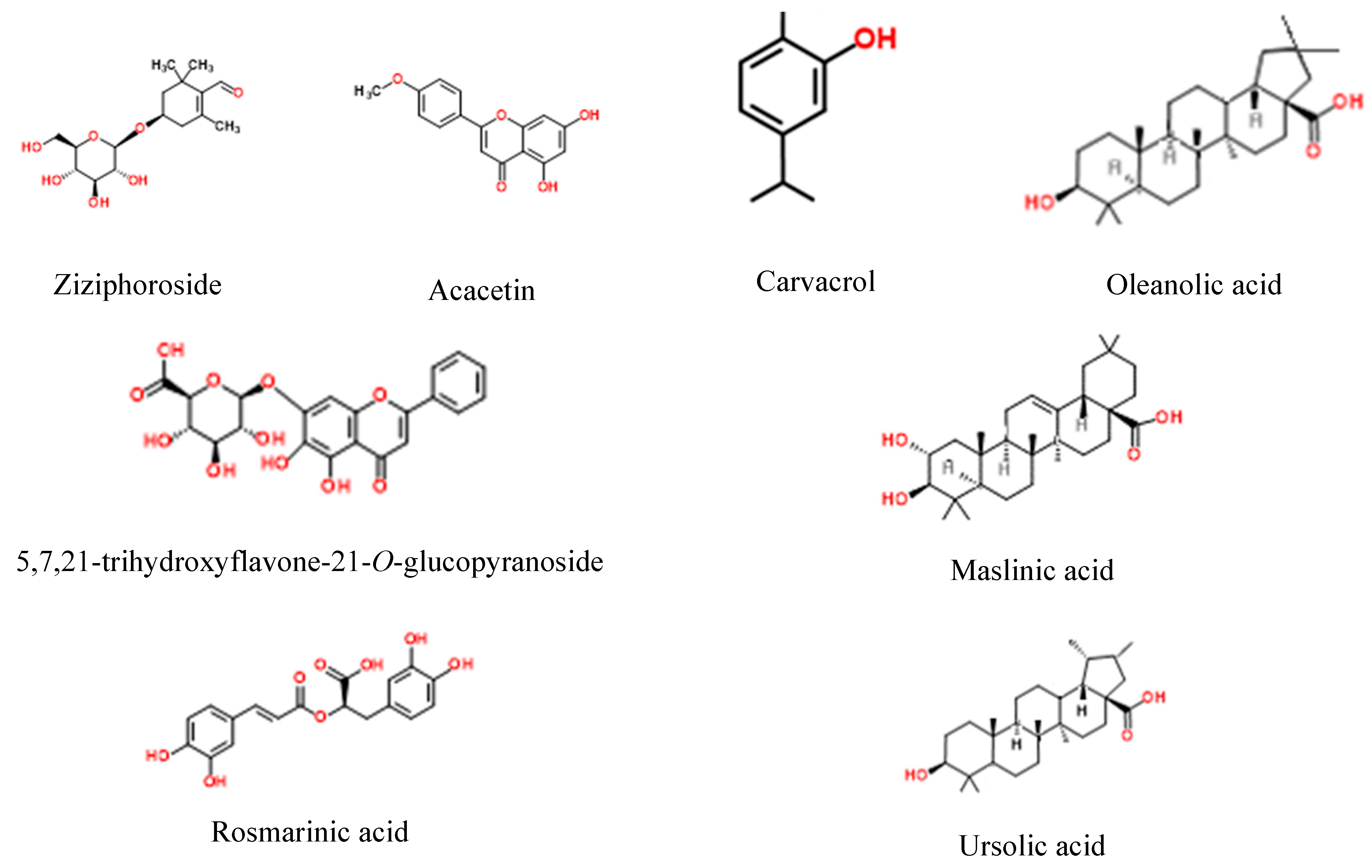
| No | Ion.+/− | Rt [min] | Molecular Formula | m/z Theoretical | m/z Experimental | Error | DBE | MS/MS Spectrum | Proposed Compound | Distribution | References |
|---|---|---|---|---|---|---|---|---|---|---|---|
| 1 | − | 3.03 | C16H18O9 | 353.0878 | 353.0884 | −1.68 | 8 | 191, 179, 173, 154 | Chlorogenic acid | Z1, Z2, Z3 | [13] |
| 2 | − | 3.9 | C16H18O9 | 353.0878 | 353.0880 | −0.55 | 8 | 179, 191 | Neochlorogenic acid | Z1, Z2, Z3 | [13] |
| 3 | + | 3.9 | C16H28O8 | 349.1857 | 349.1851 | 1.71 | 3 | 281, 163 | Schizonepetaside E | Z1, Z2, Z3 | |
| 4 | − | 4.4 | C16H18O9 | 353.0878 | 353.0884 | −1.68 | 8 | 191, 179, 173, 135, 155 | (Z)-Chlorogenic acid | Z1, Z2, Z3 | [13] |
| 5 | − | 5.2 | C14H18O7 | 297.098 | 297.1011 | −10.48 | 6 | ND | Picein | Traces Z3 | [14] |
| 6 | − | 6.5 | C9H8O4 | 179.0350 | 179.0357 | −3.99 | 6 | 135, 117, 107 | Caffeic acid | Z1, Z2, Z3 | [15] |
| 7 | − | 7.7 | C11H12O4 | 207.0663 | 207.0664 | −0.57 | 6 | 192, 179, 174, 163, 135 | Ethyl ester of caffeic acid | Z1, Z3 | [16] |
| 8 | − | 9.5 | C9H10O3 | 165.0557 | 165.0540 | 10.34 | 5 | ND | Apocynin | Traces: Z1, Z2, Z3 | [17] |
| 9 | + | 10.9 | C16H26O7 | 331.1751 353.1571 (+Na) | 331.1775 353.1607 (+Na) | −7.18 −10.3 | 4 | 201 | Ziziphoroside isomer 1 | Z1, Z2, Z3 | [14] |
| 10 | + | 12.9 | C16H26O7 | 331.1751 353.1571 (+Na) | 331.1761 353.1607 (+Na) | −2.94 −10.3 | 4 | 201, 147, 119 | Ziziphoroside isomer 2 | Z1, Z2, Z3 | [14] |
| 11 | + | 14.3 | C16H26O7 | 331.1751 353.1571 (+Na) | 331.1757 353.1609 (+Na) | −1.73 −10.86 | 4 | 201, 165, 147 | Ziziphoroside isomer 3 | Z1, Z2, Z3 | [14] |
| 12 | − | 18.5 | C21H18O11 | 445.0776 | 445.0767 | 2.1 | 13 | 269, 175, 113 | 5,7,21-trihydroxyflavone-21- O-glucopyranoside | Z1, Z3 | [18] |
| 13 | − | 19.1 | C18H16O8 | 359.0772 | 359.0780 | −2.11 | 11 | 197, 179, 161, 135 | Rosmarinic acid | Z1, Z2, Z3 | [19] |
| 14 | + | 19.8 | C10H14O | 151.1117 | 151.1129 | −7.72 | 4 | 133, 123, 109, 105 | Thymol | Z1, Z2, Z3 | [20] |
| 15 | − | 20.1 | C28H32O15 | 607.1668 | 607.1671 | −0.42 | 13 | 561, 253 | Diosmin | Z1, Z2, Z3 | [21] |
| 16 | − | 20.4 | C28H32O14 | 591.1719 | 591.1727 | −1.3 | 13 | ND | Linarin | Traces: Z1, Z2, Z3 | [22] |
| 17 | − | 20.9 | C15H10O7 | 301.0354 | 301.0363 | −3.06 | 11 | ND | Quercetin | Z1, Z2, Z3 (traces) | [23] |
| 18 | − | 21.1 | C15H10O6 | 285.0405 | 285.0396 | 3.01 | 11 | 241, 151, 133 | Luteolin | Z1, Z2, Z3 | [24] |
| 19 | − | 21.6 | C15H10O5 | 269.0455 | 269.0462 | 2.03 | 11 | 225, 151 | Apigenin | Z1, Z2, Z3 | [25] |
| 20 | 21.8 | C18H16O8 | 359.0772 | 359.0772 | 0.11 | 11 | 344, 329 | Thymonin | Z1, Z2, Z3 | [26] | |
| 21 | − | 22.3 | C16H12O6 | 299.0561 | 299.0555 | 2.04 | 11 | 284, 256, 165, 135 | Diosmetin | Z1, Z2, Z3 | [27] |
| 22 | + | 22.6 | C10H14O | 151.1117 | 151.1135 | −11.71 | 4 | 136, 123, 117, 105 | Carvacrol | Z1, Z2, Z3 | [28] |
| 23 | − | 22.7 | C16H12O5 | 283.0612 | 283.0620 | −2.83 | 11 | 268, 240 | Acacetin | Z1, Z2, Z3 | [22] |
| 24 | − | 23.0 | C30H48O3 | 455.3531 | 455.3538 | −1.6 | 7 | 455 | Oleanolic acid | Z1, Z3 | [22,29] |
| 25 | − | 23.3 | C30H48O4 | 471.3480 | 471.3479 | 0.18 | 7 | 337 | Maslinic acid | Z1, Z2, Z3 | [30] |
| 26 | − | 24.0 | C30H48O3 | 455.3531 | 455.3528 | 0.59 | 7 | 455 | Ursolic acid | Z1, Z3 | [31] |
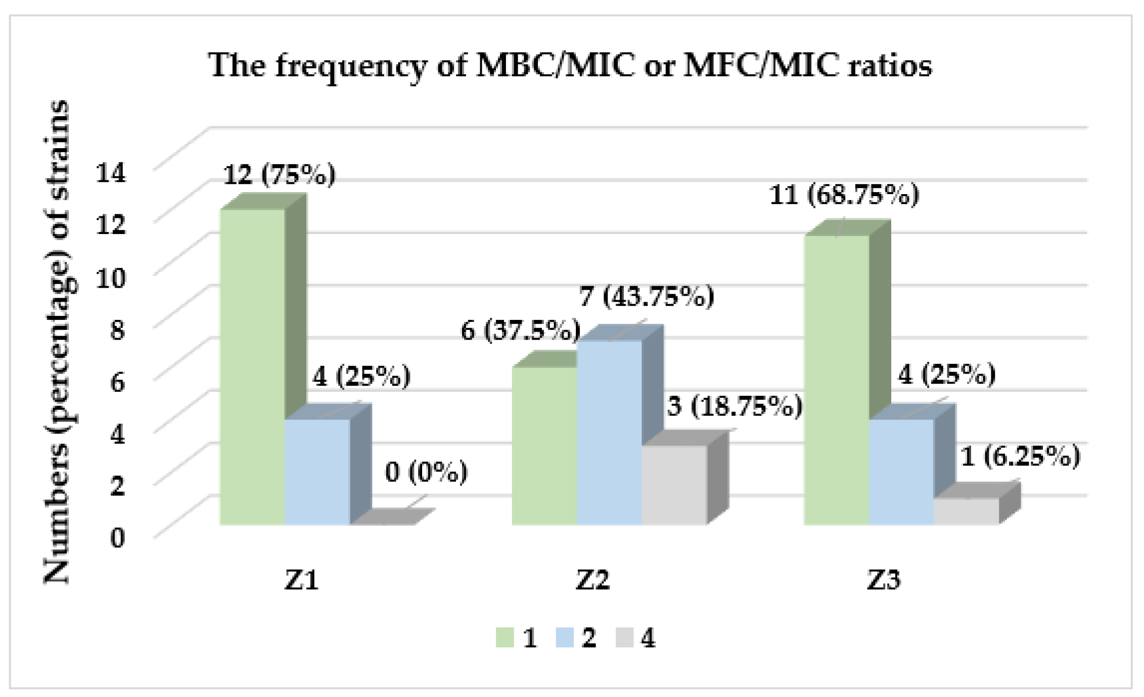
| Species of Microorganism | Z1 | Z2 | Z3 | CIP | |||||||||
|---|---|---|---|---|---|---|---|---|---|---|---|---|---|
| MIC | MBC | MBC /MIC | MIC | MBC | MBC /MIC | MIC | MBC | MBC /MIC | MIC | MBC | MBC /MIC | ||
| Gram-positive | Staphylococcus aureus ATCC 29213 | 2.5 | 2.5 | 1 | 5 | 5 | 1 | 2.5 | 2.5 | 1 | 0.24 | 0.24 | 1 |
| Staphylococcus aureus ATCC 43300 | 2.5 | 2.5 | 1 | 2.5 | 5 | 2 | 2.5 | 5 | 2 | 0.24 | 0.24 | 1 | |
| Staphylococcus epidermidis ATCC 12228 | 2.5 | 2.5 | 1 | 2.5 | 5 | 2 | 2.5 | 5 | 2 | 0.12 | 0.12 | 1 | |
| Micrococcus luteus ATCC 10240 | 2.5 | 5 | 2 | 1.25 | 5 | 4 | 5 | 10 | 2 | 0.98 | 1.96 | 2 | |
| Bacillus subtilis ATCC 6633 | 5 | 10 | 2 | 5 | 10 | 2 | 2.5 | 10 | 4 | 0.03 | 0.03 | 1 | |
| Bacillus cereus ATCC 10876 | 10 | 10 | 1 | 5 | 10 | 2 | 5 | 10 | 2 | 0.06 | 0.12 | 2 | |
| Gram-negative | Bordetella bronchiseptica ATCC 4617 | 10 | 10 | 1 | 5 | 20 | 4 | 20 | 20 | 1 | 0.98 | 0.98 | 1 |
| Klebsiella pneumoniae ATCC 13883 | 20 | 20 | 1 | 10 | 20 | 2 | 20 | 20 | 1 | 0.12 | 0.12 | 1 | |
| Salmonella typhimurium ATCC 14028 | 20 | 20 | 1 | 20 | 20 | 1 | 20 | 20 | 1 | 0.06 | 0.06 | 1 | |
| Escherichia coli ATCC 25922 | 20 | 20 | 1 | 20 | 20 | 1 | 20 | 20 | 1 | 0.004 | 0.004 | 1 | |
| Pseudomonas aeruginosa ATCC 9027 | 10 | 20 | 2 | 20 | 20 | 1 | 20 | 20 | 1 | 0.48 | 0.98 | 2 | |
| Species of Microorganism | Z1 | Z2 | Z3 | NYS | ||||||||
|---|---|---|---|---|---|---|---|---|---|---|---|---|
| MIC | MFC | MFC /MIC | MIC | MFC | MFC /MIC | MIC | MFC | MFC /MIC | MIC | MFC | MFC /MIC | |
| Candida albicans ATCC 10231 | 20 | 20 | 1 | 10 | 20 | 2 | 20 | 20 | 1 | 0.48 | 0.48 | 1 |
| Candida albicans ATCC 2091 | 20 | 20 | 1 | 10 | 20 | 2 | 20 | 20 | 1 | 0.24 | 0.24 | 1 |
| Candida parapsilosis ATCC 22019 | 10 | 20 | 1 | 5 | 20 | 4 | 20 | 20 | 1 | 0.24 | 0.48 | 2 |
| Candida glabrata ATCC 90030 | 20 | 20 | 1 | 20 | 20 | 1 | 20 | 20 | 1 | 0.24 | 0.48 | 2 |
| Candida krusei ATCC 14243 | 20 | 20 | 1 | 10 | 20 | 2 | 20 | 20 | 1 | 0.24 | 0.24 | 1 |
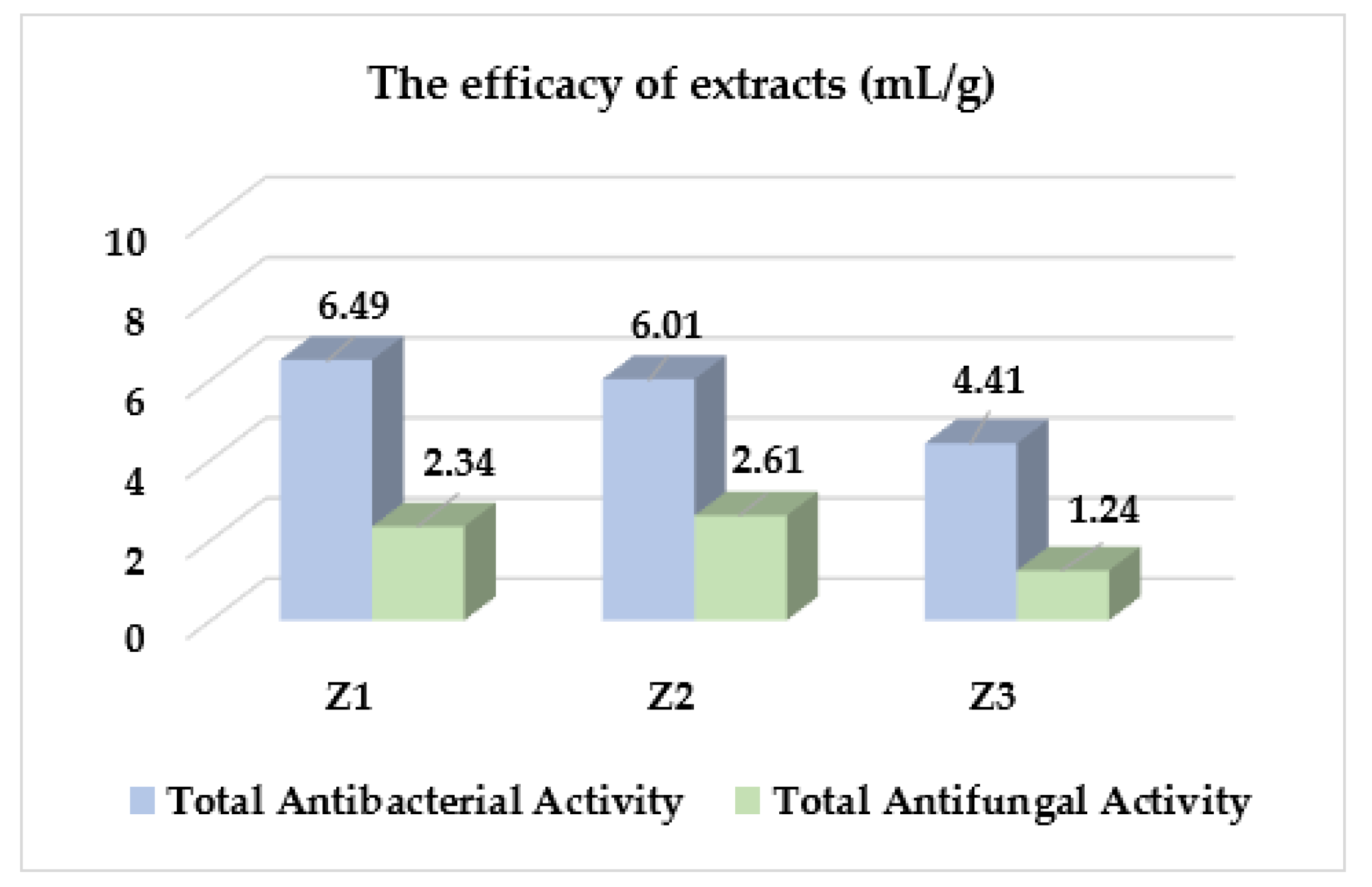
| Species of Microorganism | TAA (mL/g) | |||
|---|---|---|---|---|
| Z1 | Z2 | Z3 | ||
| Gram-positive bacteria | Staphylococcus aureus ATCC 29213 | 13.39 ± 0.0 | 6.74 ± 2.92 | 8.75 ± 0.0 |
| Staphylococcus aureus ATCC 43300 | 11.16 ± 3.86 | 8.42 ± 2.92 | 7.30 ± 2.53 | |
| Staphylococcus epidermidis ATCC 12228 | 13.39 ± 0.0 | 10.10 ± 0.0 | 8.75 ± 0.0 | |
| Micrococcus luteus ATCC 10240 | 13.39 ± 0.0 | 20.20 ± 0.0 | 5.84 ± 2.53 | |
| Bacillus subtilis ATCC 6633 | 5.58 ± 1.93 | 5.05 ± 0.0 | 7.30 ± 2.53 | |
| Bacillus cereus ATCC 10876 | 3.35 ± 0.0 | 4.21 ± 1.46 | 4.38 ± 0.0 | |
| Gram-negative bacteria | Bordetella bronchiseptica ATCC 4617 | 2.79 ± 0.97 | 4.21 ± 1.46 | 1.46 ± 0.63 |
| Klebsiella pneumoniae ATCC 13883 | 1.67 ± 0.0 | 2.53 ± 0.0 | 1.09 ± 0.0 | |
| Salmonella typhimurium ATCC 14028 | 1.67 ± 0.0 | 1.68 ± 0.73 | 1.09 ± 0.0 | |
| Escherichia coli ATCC 25922 | 2.23 ± 0.97 | 1.68 ± 0.73 | 1.46 ± 0.63 | |
| Pseudomonas aeruginosa ATCC 9027 | 2.79 ± 0.97 | 1.26 ± 0.0 | 1.09 ± 0.0 | |
| Fungi | Candida albicans ATCC 10231 | 2.23 ± 0.97 | 2.53 ± 0.0 | 1.46 ± 0.63 |
| Candida albicans ATCC 2091 | 1.67 ± 0.0 | 2.53 ± 0.0 | 1.09 ± 0.0 | |
| Candida parapsilosis ATCC 22019 | 3.35 ± 0.0 | 4.21 ± 1.46 | 1.46 ± 0.63 | |
| Candida glabrata ATCC 90030 | 2.23 ± 0.97 | 1.68 ± 0.73 | 1.09 ± 0.0 | |
| Candida krusei ATCC 14243 | 2.23 ± 0.97 | 2.10 ± 0.89 | 1.09 ± 0.0 | |
Publisher’s Note: MDPI stays neutral with regard to jurisdictional claims in published maps and institutional affiliations. |
© 2022 by the authors. Licensee MDPI, Basel, Switzerland. This article is an open access article distributed under the terms and conditions of the Creative Commons Attribution (CC BY) license (https://creativecommons.org/licenses/by/4.0/).
Share and Cite
Zhaparkulova, K.; Karaubayeva, A.; Sakipova, Z.; Biernasiuk, A.; Gaweł-Bęben, K.; Laskowski, T.; Kusniyeva, A.; Omargali, A.; Bekezhanova, T.; Ibragimova, L.; et al. Multidirectional Characterization of Phytochemical Profile and Health-Promoting Effects of Ziziphora bungeana Juz. Extracts. Molecules 2022, 27, 8994. https://doi.org/10.3390/molecules27248994
Zhaparkulova K, Karaubayeva A, Sakipova Z, Biernasiuk A, Gaweł-Bęben K, Laskowski T, Kusniyeva A, Omargali A, Bekezhanova T, Ibragimova L, et al. Multidirectional Characterization of Phytochemical Profile and Health-Promoting Effects of Ziziphora bungeana Juz. Extracts. Molecules. 2022; 27(24):8994. https://doi.org/10.3390/molecules27248994
Chicago/Turabian StyleZhaparkulova, Karlygash, Aigerim Karaubayeva, Zuriyadda Sakipova, Anna Biernasiuk, Katarzyna Gaweł-Bęben, Tomasz Laskowski, Aliya Kusniyeva, Azamat Omargali, Tolkyn Bekezhanova, Liliya Ibragimova, and et al. 2022. "Multidirectional Characterization of Phytochemical Profile and Health-Promoting Effects of Ziziphora bungeana Juz. Extracts" Molecules 27, no. 24: 8994. https://doi.org/10.3390/molecules27248994