Article

On the Origin of the Blue Color in The Iodine/Iodide/Starch Supramolecular Complex

by
Szilárd Pesek
,
Maria Lehene
,
Adrian M. V. Brânzanic
and
Radu Silaghi-Dumitrescu
*
Department of Chemistry, Faculty of Chemistry and Chemical Engineering, Babeş-Bolyai University, 11 Arany Janos Street, 400028 Cluj-Napoca, Romania
*
Author to whom correspondence should be addressed.
Molecules 2022, 27(24), 8974; https://doi.org/10.3390/molecules27248974
Submission received: 18 November 2022 / Revised: 5 December 2022 / Accepted: 12 December 2022 / Published: 16 December 2022

Abstract

:
The nature of the blue color in the iodine-starch reaction is still a matter of debate. Some textbooks still invoke charge-transfer bands within a chain of neutral I2 molecules inside the hydrophobic channel defined by the interior of the amylose helical structure. However, the consensus is that the interior of the helix is not altogether hydrophobic—and that a mixture of I2 molecules and iodide anions reside there and are responsible for the intense charge-transfer bands that yield the blue color of the “iodine-starch complex”. Indeed, iodide is a prerequisite of the reaction. However, some debate still exists regarding the nature of the iodine-iodine units inside the amylose helix. Species such as I3-, I5-, I7- etc. have been invoked. Here, we report UV-vis titration data and computational simulations using density functional theory (DFT) for the iodine/iodide chains as well as semiempirical (AM1, PM3) calculations of the amylose-iodine/iodide complexes, that (1) confirm that iodide is a pre-requisite for blue color formation in the iodine-starch system, (2) propose the nature of the complex to involve alternating sets of I2 and Ix- units, and (3) identify the nature of the charge-transfer bands as involving transfer from the Ix- σ* orbitals (HOMO) to I2 σ* LUMO orbitals. The best candidate for the “blue complex”, based on DFT geometry optimizations and TD-DFT spectral simulations, is an I2-I5-I2 unit, which is expected to occur in a repetitive manner inside the amylose helix.

1. Introduction

The nature of the blue complex of amylose with iodine (λmax = 615 nm) has been a focus of research for many decades [1]. The left-handed helix, with an outer diameter of ~13 Å and a pitch of 8 Å (six 1,4-glucose units per pitch), hosts an internal cavity of ~5 Å where the iodine alongside iodide is hosted with I–I distances of ~3.1 Å [2,3,4,5,6,7,8,9,10]. Slight differences in the blue color are seen depending on the size (and, implicitly, biological source) of amylose; similar complexes are seen with many related poly and oligo saccharides, or even with other organic polymers [11,12,13,14,15,16,17,18,19,20,21].
The stoichiometry and charge of the poly-iodine substructures of the starch-iodine complex is still disputed. The initial supposition was that the cavity of the starch helix serves as a nonpolar solvent [22], where I2 molecules then dissolve in the hydrophobic interior as discrete units connected by non-covalent interactions, and that these non-covalent interactions afford the intense blue color. Semiempirical intermediate neglect of differential overlap configuration interaction (INDO CI) simulations of UV-vis spectra have been used as an argument to support an entirely neutral character poly-I2 nature of the blue complex (more specifically, a (C6H10O5)16.5 · I6 formula), and to refute In- (n = 3, 5, or 7) as possible species in the blue amylose-iodine complex [23]. However, it is now generally accepted that iodide ions are also required in the process, so that the poly-iodine chains inside the amylose helix consist of a mixture/combination of I2 and I- [1,2,3,4,5,24] with an unusual metallic-like structure [25]. The manner in which contiguous chains of such polyanionic structures can be reconciled with a hydrophobic nature of the interior cavity of the helix, especially without including counterions, at any point throughout the cavity, may be argued to remain an interesting puzzle. Indeed, based on thermodynamic considerations it has been predicted that tri-iodide units would be unable to form long chains inside the amylose helix unless cooperativity with I2 molecules occurs; furthermore, the ends of the helix would in this scenario remain unoccupied by iodine atoms [26]. Also importantly, the secondary structure of amylose does vary depending on the environment (e.g., solvent, ionic strength, pH, temperature, surfactants, affecting among others the internal diameter of the helix)—to the extent that alcohol-precipitated amylose in the solid state can efficiently bind molecular I2 vapors (and, unlike in aqueous solution, no iodide is needed) [18,21,27,28,29]. Interestingly, however, this amylose-I2 complex, unlike the iodine-iodide one, is not stable when dissolved in water [30], though the preparation of an amylose-iodine complex in water at higher temperatures in the absence of iodide has also been reported [31]. Inclusion complexes of I2 with other oligosaccharides in the absence of iodide have also been described [32].
Various lengths of the poly-iodine chain have been proposed based on experimental data, from 3–4 to 14–15 to as high as 160 [1,2,3,4,5]. Based on potentiometric titration at low iodide concentrations, a 3/2 I2/I- ratio was proposed, leading to an I82- empirical formula. However, many other chain lengths have been proposed, such as I4-, I7-, I9-, I62-, I82-, I102-, I42-, I6-, and I242-—especially as the structure (as observable e.g., in UV-vis and circular dichroism spectra) appears to be distinctly dependent on the iodide and iodine concentrations [3,19,20,33,34,35,36]. Moreover, based on stopped-flow UV-vis and circular dichroism (CD) kinetic data, a dynamic nature of complex formation was shown, with shorter chains of iodine entering the amylose helix very fast (less than 1 millisecond), and then rearranging rapidly without further interaction with iodine/iodide from the exterior of the helix [2,37]. Interestingly, at conditions where the UV-vis spectrum of the blue complex is stable in time, its optical rotatory dispersion (ORD) signal is seen to still increase, suggesting more complex dynamics of the helix that do not affect the length of the poly-iodine chains [38]. Based on the structure of (benzamide)z H+I3-, a poly-I3 structure has been proposed, and a range of kinetic, spectroscopic (UV-vis, CD, Raman, X-ray absorption) and thermodynamic data have been interpreted within this framework [15,17,39,40]. However, X-ray diffractometric data has suggested that the exclusive occurrence of either of the single species I2 or I3- is unlikely [41,42,43]. Based on similarities between the Raman and Mössbauer spectra of the starch–iodine complex and those of polycrystalline (trimesic acid · H2O)10H+I5-, an I5- structure was proposed for the amylose-iodine complex [44,45]. In the Raman spectra of starch–iodine complexes, four principal peaks at 27, 55, 109, and 160 cm−1 were observed. Using theoretical calculations and far-infrared data, the peak at 109 cm−1 was assigned to I3- regarded as an impurity while I5- would be the dominant species [46,47,48,49,50]. A role for water molecules in controlling the structure of the iodine-amylose complex was also noted [51], as was the fact that that that iodine binding does to a certain degree affect the structure of amylose–both geometrically (in terms of the dimensions of the helix, as well as in terms of the helical vs. random coil secondary structure within the amylose polymer) and in terms of rigidity [13,52,53,54,55].
Reported here is an experimental and computational study of the amylose-iodine-iodide system, using UV-vis spectra, time-dependent density functional theory (TD-DFT), and semiempirical and molecular mechanics calculations in order to model the structure of the complex and to assess the viability of various candidates potentially responsible for the blue color. Poly-I2 as well as In- structures are examined. Poly-I2 structures are found to afford charge-transfer bands that amplify the intensity of the color compared with an isolated I2 molecule, but this is still significantly below the intensity observed experimentally for the amylose-iodine-iodide mixture. On the other hand, I2-In- mixtures afford significantly stronger charge-transfer bands, from the Ix- σ* orbitals (HOMO) to I2 σ* (LUMO) orbitals. These bands are most likely responsible for the well-known intense blue color of the amylose-iodine-iodide system.

2. Results and Discussion

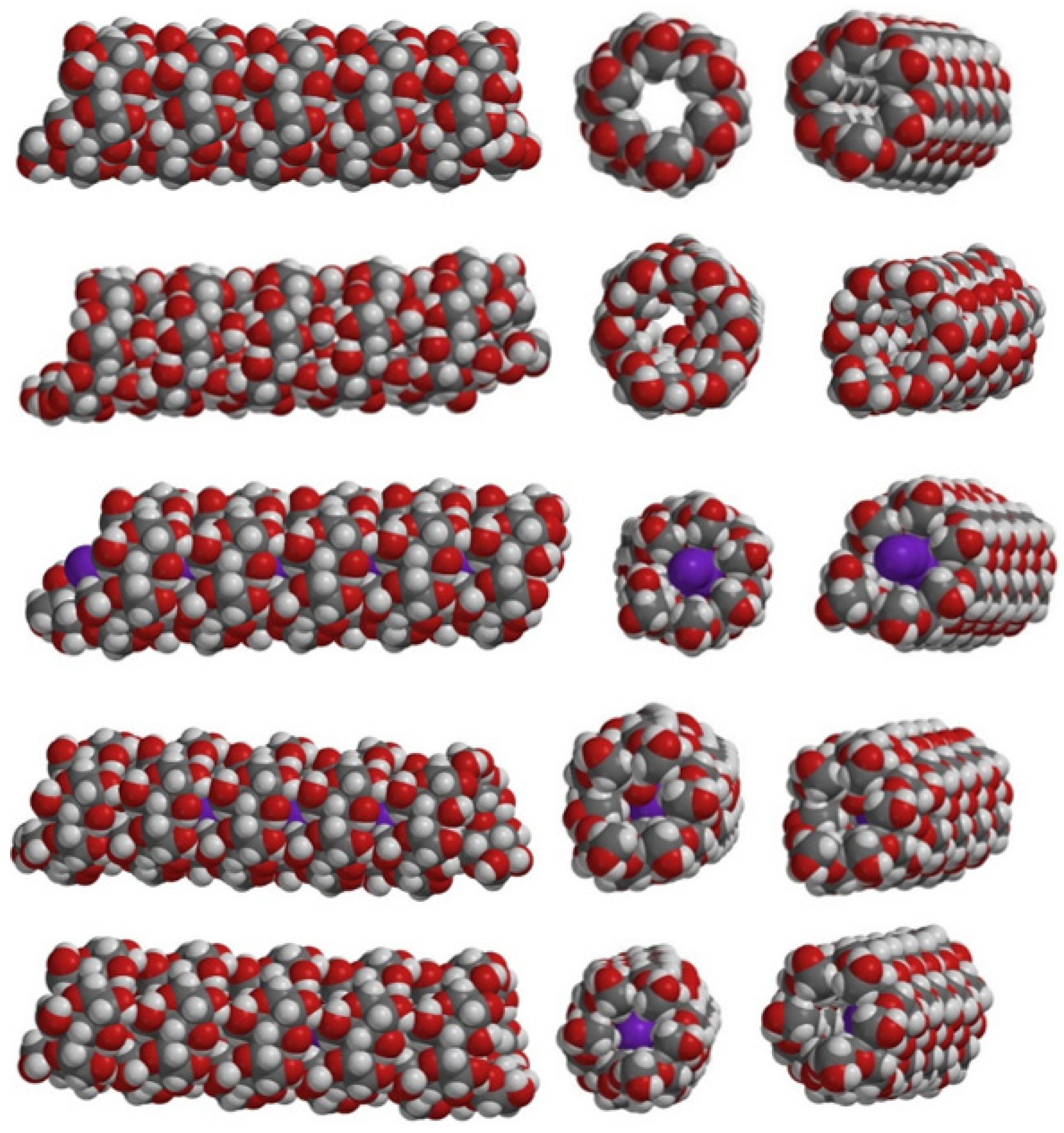
2.1. Amylose-Iodine/Iodide Complexes: Structural Considerations

As detailed in the Introduction and confirmed by experiments shown in Supporting Information, I2 alone, either in aqueous or in alcoholic solutions (methanol or ethanol) leads to barely detectable changes in absorbance at ~600 nm in reaction with starch. Iodine-iodide mixtures do yield a blue color, which is not observed with glucose instead of starch. The absorbance maximum of the iodine-iodide-starch complex in the experiments reported in Supporting Information is at ~580 nm. This is slightly different from the 615 nm reported for amylose but is in line with the fact that our starch samples also contain amylopectin, as well as with the fact that the precise position of the maximum depends on the source of the starch as well as on the relative concentrations of the reagents [3,17,35,36,39,56]. Indeed, slight concentration-dependent changes in the position of the maximum are observed, as seen in Supporting Information, when varying the iodide concentration as well as when varying the length of the amylose/amylopectin chains by employing an amylase.
Figure 1 and Supporting Information Table S1 show the results of geometry optimizations on helical amylose models where the internal cavity is either empty (model A), or filled with water or with iodine (I2, I3- or combinations thereof: models A-H2O, A-I2, A-I2-H2O, A-I3-).
In model A-H2O, although geometry optimizations were initiated with the water molecules aligned at the center of the internal cavity (in the same manner that the iodine would be), the final result was that the water adhered to the inside walls of the helix, cf. Figure 1. The presence of such molecules inside the helix has not been commented on or hypothesized experimentally, even though hydration of amylose in principle is known to entail structural changes [51].
The binding of I2 to the helix (model A-I2 in Table S1 and Figure 1) leads to negligible distortions of the helix. The binding of interspersed water and iodine molecules (model A-I2-H2O) results in the water molecules migrating to the internal walls of the helix, suggesting that an iodine-water mixed chain is unlikely to occur in amylose. Indeed, I2 has been shown to efficiently bind to amylose in a dry/solid state, but much less so in solution (unless special conditions, e.g., heating, are provided) [31].
Attempts to optimize an A-I3- model with several triiodide units inside the helix were unsuccessful, as all but one of the triiodide units migrated outside the helix upon geometry optimization. This may be taken as evidence against the hypothesis that the amylose-iodine/iodide blue color would be due to chains of I3- units (and possibly, by extension, of In- units) aligned inside the helix. This finding is in line with the relatively hydrophobic nature of the helix cavity and with the anionic character of the triiodide. As a consequence, the A-I3- model in Table S1 and Figure 1 only features a single I3- unit inside the helix.

2.2. UV-Vis Simulations of Linear I2 Chains

Simulation of the UV-vis spectrum would allow one to verify which of the possible iodine/iodide combinations are the most likely to yield the ~600 nm maximum. This can be reliably done with TD-DFT calculations; however, since the amylose-iodine models were too large for such methods, Table 1 and Table 2 report TD-DFT calculations on linear iodine chains without, including the surrounding saccharide.
As shown in Table 1, an isolated iodine molecule displays a weak maximum at ~600 nm, due to a π* (HOMO) -> σ* (LUMO) transition. In a linear I2 dimer with I-I bonds identical to those in the monomer and with a set I2-I2 distance equal to the sum of the van der Waals radii, this maximum sees a bathochromic shift and a slight increase in intensity, as expected since at such short intermolecular distance the frontier orbitals of the two I2 molecules mix. Geometry optimization of this dimer leads to dissociation, and hence the ~600 nm peak is seen almost exactly in the same position as for the monomer—though, interestingly, the intensity of the “blue” band is twice larger than the monomer.
As also shown in Table 1, longer chains of I2 molecules follow the same trends seen when going from monomeric I2 to (I2)2 (including the elongation of intermolecular distances upon geometry optimization). The bathochromic and hyperchromic shifts continue to the extent that in the heptamer the intensity of the 600 nm band has ~ tripled compared with isolated iodine.
These data confirm the experimental observations and previous semiempirical calculations according to which a linear chain of I2 molecules may under certain conditions be hosted inside the amylose helix and that such a chain would display a more intense color than free iodine molecules in solution. The band responsible for this enhanced blue color entails a HOMO -> LUMO transition, where HOMO is a combination of I2 π* orbitals and LUMO is a combination of I2 σ* orbitals. The longer the poly-I2 chain is, the more there is a tendency for these molecular orbitals to no longer have exactly the same distribution across the same atoms–hence allowing the 600 nm band to gain a charge-transfer character which may be responsible for the increased oscillator strength. In the optimized poly-I2 geometries, the frontier molecular orbitals responsible for the blue color are located mostly at the two ends of the chains, and it is at these ends, and at the I2 molecules preceding the ends, that the above-mentioned charge transfer will occur. However, if the I2 molecules are compacted together within van der Waals radii (and not further optimized), it is the center and not the edges of the poly-I2 chain that holds the most contribution to the 600 nm band—though, again, the more important charge-transfer part will impact more clearly the iodine molecules preceding the ends of the chain.

2.3. UV-Vis Simulations of Linear In- Systems

Although the above considerations may explain the amylose–I2 interactions under certain conditions, the enhancement of the 600 nm band upon elongation of the poly-I2 is still relatively small—not even one order of magnitude upon going from monomer to heptamer. Therefore, poly-iodine-iodide chains were considered next, as illustrated in Table 2.
As seen in Table 2, the mono-anions In- (n = 1, 5, 7, 9) tend to exhibit bands in the ~400 nm region. However, the nature and intensity of this band depend strongly on the geometry of the anion. When the anion is built with equivalent I-I distances, to mimic complete delocalization of the negative charge across the molecule, the predicted absorption band remains very weak and close to the 400 nm region. The nature of this transition remains very similar to the one seen in the poly-I2 chains of Table 2 (i.e., π* (HOMO) -> σ* (LUMO))—except, of course, that it is no longer in the 600 nm region. These results are in line with previous lower level (i.e., semiempirical) INDO configuration interaction (CI) simulations relying on similar models, which were interpreted as evidence that poly-I2 but not iodine-iodide mixtures are hosted inside the helix in the blue amylose-iodine complex. However, Table 2 now also shows that when the geometry of the In- chains is optimized, the symmetry of the molecule is lost as the I-I distances no longer remain degenerate; importantly, this asymmetry allows the HOMO-LUMO transition to gain significant charge-transfer character—so much so that its intensity increases by 3–4 orders of magnitude compared with the completely symmetrical models. These increases in intensity are also accompanied by small bathochromic shifts; however, these shifts appear confined below 500 nm and hence these polyanions of iodide do not appear to be reasonable candidates for the experimentally observed 600 nm species. Another notable change in these geometry-optimized In- species is that at n > 3 the HOMO orbital involved in the visible transition now also has σ* character.

2.4. UV-Vis Simulations of Linear In--I2 Systems

Table 3 also shows a set of models which yield excellent agreement with the experiment, insofar as featuring extremely intense bands at 600 nm. These models include any In- species surrounded by two neutral I2 molecules. In such models, the ~600 nm transition occurs from a σ* orbital of the central oligo-iodide anion to the σ * orbitals of the terminal I2 molecules. For the I2-I3-I2 system, the formal structure featuring the I2 molecules at van der Waals radii from the I3- anion does allow a very strong band at 680 nm. However, upon geometry optimization, this structure collapses into the I7- anion, already seen in Table 2 as an unlikely candidate for the blue-colored complex. No such collapse is seen for the I2-I5-I2 species, which is thus the first of those discussed in the present study that can be proposed as a strong candidate as a contributor to the amylose-iodine blue color. Moreover, the average iodine-iodine distances in the DFT-optimized geometry of I2-I5-I2 is 3.09 Å, remarkably close to the 3.1 Å distance measured experimentally for the iodine-iodide-amylose complex. As shown in Table 3, I2 combinations with longer In- species cannot be excluded.
The 600 nm band in Table 3 is predicted to shift by as much as 25 nm upon going from n = 3 to n = 9, while over the same interval its intensity is seen to double. Given the uncertainties regarding the exact formula of the species, reliable extinction coefficients for the amylose-iodine-iodide complex are not known; Table 3 now shows that they would be particularly difficult to measure, since poly-iodine chains of various lengths will have different extinction coefficients. On the other hand, having singled out the I2-I5-I2 unit as the most likely candidate of those examined in Table 1, Table 2 and Table 3 for the “blue complex”, one may consider how long chains of such units may behave. Table 3 therefore also shows that the homologous assembly I2-I5-I2-I5-I2—technically, a dimer of I2-I5-I2, features an absorption maximum essentially at the same wavelength as the monomer—albeit with a different extinction coefficient. As such, I2-I5-I2 itself, and no longer chains/polymers thereof, appears sufficient to justify the blue color of the iodine-iodide-amylose complex.

2.5. Solvent Effects

The above considerations on TD-DFT spectra are based on vacuum calculations (ε = 1). The magnitude of solvent effects on the positions of UV-vis maxima are generally within the same margins as the effects of changing functionals and basis sets [58,59]. These margins are typically in the range of at most tens of nm. This is confirmed, as seen in Supporting Information, by calculations on linear I2 and In- models. However, charge-transfer complexes may be distinctly more problematic to predict by DFT methods and can be distinctly more sensitive to the dielectric constants [59]. The linear I2 chains in Table 1 do show some charge transfer character as discussed above; hence, perhaps not surprisingly, Table S2 in Supporting Information shows that, while the solvent effect is ~ 20 nm for the isolated I2 molecule, it almost doubles for the (I2)2 dimer. The strong solvent effect on the UV-vis spectrum of iodine is indeed well-known [60]. However, as seen in Supporting Information for the representative case of I2-I3-I2 where charge transfer is even more dominant than in the neutral I2 homodimer, there is a very large ~exponential dependence on the position of the maximum (but not on the oscillator strength), so that between ε = 1 and ε = 9 a ~100 nm hypsochromic shift is observed—after which the changes are much smaller (i.e., only 25 nm from ε = 9 to ε~30, and only 4 nm further from ε ~ 30 to ε = 80.
The interior cavity/channel of the amylose helix has, as discussed above, been described as hydrophobic. Indeed, as also illustrated in Figure 1 and in Supporting Information, this channel is lined with carbon-bound hydrogen and with ether oxygen atoms. Such an environment is somewhat reminiscent of the interior of proteins, but it is also similar to that provided by solvents such as diethyl ether, acetone, or dymethylsulfoxide; these solvents are in a range of ε ~ 4–30–which, according to our TD-DFT calculations, should be able to measurably affect the positions of the maxima. As discussed in the Introduction, subtle structural changes induced by other solutes, or by the origin of the amylose and hence the length of its chains, are known experimentally–and are also known to affect the UV-vis properties of the respective iodine/iodide complexes. It is reasonable to assume that at least part of these experimentally observed differences is due to slight changes in the dielectric constant within the internal amylose cavity as a consequence of slight changes to solvent exposure either at the ends of the chain (i.e., dependence on the length of the chain) or throughout the chain (i.e., from interactions with other solutes).
The strong solvent effects described here for iodine-iodide charge transfer complexes do mirror previous observations on other I2 charge transfer complexes [61].

3. Materials and Methods

UV–vis spectra were performed on Lambda 25 (PerkinElmer Singapore) spectrophotometers, in the range of 200–1000 nm, in 1 mL quartz cuvettes. Sample preparation and data collection take no more than two hours. Stock solutions included saturated starch, iodine aqueous solution 1 mM, iodine-iodide aqueous solution, iodide aqueous solution (100 mM), glucose (50 mM), 0.5% starch in water, 0.1 N ethanol, 10 mM methanol.
Models of the amylose helix were constructed based on a canonical structure extracted from https://www.biotopics.co.uk/jsmol/amylose-iodide.html (accessed on 2 August 2021), with the internal cavity left empty or filled with water, iodine and/or I3-. The guest molecules were placed and aligned along the central axis of the helix, at distances equal to the respective sums of the van der Waals radii. These models were subjected to geometry optimizations using molecular mechanics (SYBYL [62], MMFF [63]) and semiempirical (PM3, [64,65] PM6 [66] and AM1 [67]) methods as implemented within the Spartan software package [68]. The AM1 data are reported here; the other methods were found to yield qualitatively similar results. AM1 (Austin Model 1) is a semiempirical method based on modified neglect of differential diatomic overlap approximation, developed by Dewar and related to the PM3 method (Parametric Method 3) of Stewart and to its subsequent version PM6 [64,67]. We have previously shown that secondary-type structural elements in short chains of biopolymers (e.g., helical structures in peptides, or polylactic acid) are extremely challenging for all computational methods–even for density functional methods corrected/parametrized especially for describing weak/non-covalent interactions; nevertheless, even in those cases, semiempirical methods (and especially AM1 and PM6) performed close to on par with the highest-level density functional (DFT) methods [69,70,71,72]. Unfortunately, the size of the amylose-iodine models has so far precluded us from completing DFT calculations that would explicitly include the amylose helix. With these considerations in mind, the semiempirical (AM1) results are presented here only summarily, with the focus remaining on the density functional calculations on smaller, iodine-only models.
For iodine-only models (no helix included), geometries were built and, in cases indicated in Tables, optimized at the B3LYP/6-31G* level of theory in the Spartan software package. Further TD-DFT calculations were carried out using the B3LYP [73,74,75] functional coupled with def2-SV(P) [76] as implemented in the Gaussian software package; the PCM (polarizable continuum model) solvation model was employed with solvents as indicated in the text and Supporting Information [77,78].

4. Conclusions

The amylose-iodine-iodide interaction has been modeled with electronic structure calculations. Changes in the structure of the helix upon iodine binding are predicted. Poly-I2 structures are shown to be responsible for the enhanced blue color under certain conditions (e.g., dry/solid amylose). Poly-In- structures are found unlikely to exist inside the amylose helix or to be responsible for the blue color. Instead, I2/In- pairs with charge transfer bands from the occupied In- (n > 3) σ* to the empty I2 σ * orbital are found to be reasonably responsible for the blue color. Of these, I2-I5-I2 associations are the smallest (and possibly–but not necessarily–repetitive) unit that represent local minima in DFT calculations, with average iodine-iodine distances essentially identical to the 3.1 Å value seen experimentally in the iodine-amylose complex. The distinct charge-transfer character of the UV-vis bands also brings about a strong dependence on the dielectric constant in the region ε ~ 1–30, which in turn may explain at least part of the dependence of the UV-vis properties of amylose-iodine/iodine complexes on various external factors that may subtly affect amylose architecture and hence exposure of the interior cavity to solvent (e.g., temperature, other solutes, solvents, chain length).

Supplementary Materials

The following supporting information can be downloaded at: https://www.mdpi.com/article/10.3390/molecules27248974/s1, Figure S1: UV-vis spectra of starch–I2 mixtures, Figure S2: Same data as in Figure S1, after subtracting the from each spectrum the corresponding spectrum of a starch-free I2 aqueous solution of the same concentration, Figure S3: UV-vis spectra of an I2 aqueous solution, to which indicated amounts of starch 0.5% solution were added, Figure S4: Corrections made to spectra from Figure S3 by subtracting the control spectra of starch at indicated concentrations, Figure S5: UV-vis spectra of starch 0.5%, to which different amounts of solution of I2 0.1N in alcohol were added, Figure S6: Corrections made to spectra from Figure S5 by subtracting the control spectra of I2 at indicated concentrations, Figure S7: UV-vis spectra of starch 0.5%, to which indicated amounts of solution I2 in methanol 10 mM were added, Figure S8: UV-vis spectra of water, to which I2 from a 10 mM stock solution was added, followed by indicated amounts of 0.5% starch solution, Figure S9: UV-vis spectra of starch solutions to which indicated amounts of I2 1 mM aqueous solution were added, Figure S10: Corrections made to spectra from Figure S9 by subtracting the control spectra of I2 at indicated concentrations, Figure S11: UV-vis spectra of a 1 mM I2 aqueous solution, to which indicated amounts of starch 0.5% solution were added, Figure S12: UV-vis spectra of starch 2% glucose 1 mM to which 5 µL solution of iodine-iodide aqueous solution were added, Figure S13: UV-vis spectra of starch 0.5%, to which iodine-iodide was added, Figure S14: Same spectra as in Figure S13, now corrected by subtracting the control spectra of iodine-iodide solutions collected in the absence of starch, Figure S15: UV-vis spectra of an iodine-iodide aqueous solution to which indicated amounts of starch 0.5% solution were added, Figure S16: Same spectra as in Figure S15, now corrected by subtracting the control spectra of starch solutions collected in the absence of iodine, Figure S17: Same spectra as in Figure S16, multiplied by arbitrary constants each, so that a clearer comparison of the shapes of the maxima can be offered, Figure S18: Time evolution of UV-vis spectra of an iodine-iodide mixture, to which starch and amylase were added, Figure S19: Same spectra as in Figure S18, now corrected by subtracting the control spectrum of the iodine-iodide solution, Figure S20: Structures of AM1-optimized amylose models, in order of rows: A, A-H2O, A-I2, A-I2-H2O, A-I3-, Figure S21: Structure of the amylose model helix, showing in yellow highlight the atoms employed for estimating the changes in helix length/diameter (as discussed in main text, Table 1), Table S1: Key distances (Å) derived computed for iodine-amylose models, Table S2: AM1 energies (kcal/mol) of amylose models employed in the present study, Table S3: Solvent effects on TD-DFT-predicted maxima and oscillator strengths for selected iodine/iodide models.

Author Contributions

Conceptualization, R.S.-D.; methodology, R.S.-D. and A.M.V.B.; software, R.S.-D. and A.M.V.B.; validation, R.S.-D. and A.M.V.B.; formal analysis, R.S.-D. and S.P.; investigation, R.S.-D., M.L. and S.P.; resources, R.S.-D. and S.P.; data curation, R.S.-D. and S.P.; writing—original draft preparation, R.S.-D. and S.P.; writing—review and editing, R.S.-D., A.M.V.B. and S.P.; visualization, R.S.-D. and S.P.; supervision, R.S.-D.; project administration, R.S.-D.; funding acquisition, R.S.-D. All authors have read and agreed to the published version of the manuscript.

Funding

This research was funded by the Romanian Ministry of Education and Research (projects PN-III-P4-ID-PCCF-2016-0142 and PN-III-P1-1.1-PD-2021-0279). The APC was funded by a waiver from the publisher.

Institutional Review Board Statement

Not applicable.

Informed Consent Statement

Not applicable.

Data Availability Statement

Not applicable.

Acknowledgments

Augustin C. Mot and Cezara Zagrean-Tuza (BBU) are thanked for helpful discussions.

Conflicts of Interest

The authors declare no conflict of interest.

References

  1. Stein, R.S.; Rundle, R.E. On the Nature of the Interaction between Starch and Iodine. J. Chem. Phys. 1948, 16, 195–207. [Google Scholar] [CrossRef]
  2. Hiromi, K.; Shibaoka, T.; Ono, S. Kinetic Studies of Amylose-iodine-iodide Reaction by Stopped-flow Method. J. Biochem. 1970, 68, 205–214. [Google Scholar] [CrossRef] [PubMed]
  3. Yajima, H.; Nishimura, T.; Ishii, T.; Handa, T. Effect of concentration of iodide on the bound species of I2/I−3 in the amylose-iodine complex. Carbohydr. Res. 1987, 163, 155–167. [Google Scholar] [CrossRef]
  4. Cronan, C.L.; Schneider, F.W. Cooperativity and composition of the linear amylose-iodine-iodide complex. J. Phys. Chem. 1969, 73, 3990–4004. [Google Scholar] [CrossRef]
  5. Cramer, F.; Herbst, W. Die Lichtabsorption von Jodkettenmolekeln. Naturwissenschaften 1952, 39, 256. [Google Scholar] [CrossRef]
  6. Barrett, A.J.; Barrett, K.L.; Khan, A. Effects of Acetone, Ethanol, Isopropanol, and Dimethyl Sulfoxide on Amylose-Iodine Complex. J. Macromol. Sci. Part A 1998, 35, 711–722. [Google Scholar] [CrossRef]
  7. Fonslick, J.; Khan, A. Thermal stability and composition of the amylose–iodine complex. J. Polym. Sci. Part A Polym. Chem. 1989, 27, 4161–4167. [Google Scholar] [CrossRef]
  8. Rundle, R.E. The Configuration of Starch in the Starch-Iodine Complex. V. Fourier Projections from X-Ray Diagrams 1. J. Am. Chem. Soc. 1947, 69, 1769–1772. [Google Scholar] [CrossRef]
  9. Rundle, R.E.; French, D. The Configuration of Starch and the Starch—Iodine Complex. II. Optical Properties of Crystalline Starch Fractions 1. J. Am. Chem. Soc. 1943, 65, 558–561. [Google Scholar] [CrossRef]
  10. Bluhm, T.L.; Zugenmaier, P. Detailed structure of the Vh-amylose-iodine complex: A linear polyiodine chain. Carbohydr. Res. 1981, 89, 1–10. [Google Scholar] [CrossRef]
  11. Séne, M.; Thévenot, C.; Prioul, J.L. Simultaneous Spectrophotometric Determination of Amylose and Amylopectin in Starch from Maize Kernel by Multi-wavelength Analysis. J. Cereal Sci. 1997, 26, 211–221. [Google Scholar] [CrossRef]
  12. Moulay, S. Molecular iodine/polymer complexes. J. Polym. Eng. 2013, 33, 389–443. [Google Scholar] [CrossRef]
  13. Hirai, M.; Hirai, T.; Ueki, T. Effect of branching of amylopectin on complexation with iodine as steric hindrance. Polymer 1994, 35, 2222–2225. [Google Scholar] [CrossRef]
  14. Tashiro, K.; Gakhutishvili, M. Crystal structure of cellulose-iodine complex. Polymer 2019, 171, 140–148. [Google Scholar] [CrossRef]
  15. Konishi, T.; Tanaka, W.; Kawai, T.; Fujikawa, T. Iodine L -edge XAFS study of linear polyiodide chains in amylose and α-cyclodextrin. J. Synchrotron Radiat. 2001, 8, 737–739. [Google Scholar] [CrossRef]
  16. Knutson, C.A. Evaluation of variations in amylose–iodine absorbance spectra. Carbohydr. Polym. 2000, 42, 65–72. [Google Scholar] [CrossRef]
  17. Nishimura, T.; Yajima, H.; Ishii, T.; Endo, R. Effect of molecular weight of amylose on the iodine coloring species responsible for the optical properties of amylose-iodine complexes. Kobunshi Ronbunshu 1989, 46, 537–544. [Google Scholar] [CrossRef] [Green Version]
  18. Sakajiri, T.; Kikuchi, T.; Simon, I.; Uchida, K.; Yamamura, T.; Ishii, T.; Yajima, H. Molecular dynamics approach to study the discrepancies in the thermal behavior of amylose and chitosan conformations. J. Mol. Struct. THEOCHEM 2006, 764, 133–140. [Google Scholar] [CrossRef]
  19. Szejtli, J.; Augustat, S.; Richter, M. Molecular configuration of amylose and its complexes in aqueous solutions. Part III. Investigation of the DP distribution of helical segments in amylose-iodine complexes. Biopolymers 1967, 5, 17–26. [Google Scholar] [CrossRef]
  20. Davis, H.; Khan, A. Determining the chromophore in the amylopectin–iodine complex by theoretical and experimental studies. J. Polym. Sci. Part A Polym. Chem. 1994, 32, 2257–2265. [Google Scholar] [CrossRef]
  21. Rendleman, J.A. The reaction of starch with iodine vapor. Determination of iodide-ion content of starch–iodine complexes. Carbohydr. Polym. 2003, 51, 191–202. [Google Scholar] [CrossRef]
  22. Immel, S.; Lichtenthaler, F.W. The Hydrophobic Topographies of Amylose and its Blue Iodine Complex. Starch—Stärke 2000, 52, 1–8. [Google Scholar] [CrossRef]
  23. Minick, M.; Fotta, K.; Khan, A. Polyiodine units in starch-iodine complex: INDO CI study of spectra and comparison with experiments. Biopolymers 1991, 31, 57–63. [Google Scholar] [CrossRef]
  24. Thoma, J.A.; French, D. The Starch-Iodine-Iodide Interaction. Part I. Spectrophotometric Investigations 1. J. Am. Chem. Soc. 1960, 82, 4144–4147. [Google Scholar] [CrossRef]
  25. Bersohn, R.; Isenberg, I. Metallic Nature of the Starch-Iodine Complex. J. Chem. Phys. 1961, 35, 1640–1643. [Google Scholar] [CrossRef]
  26. Cesaro, A.; Benegas, J.C.; Ripoll, D.R. Molecular model of the cooperative amylose-iodine-triiodide complex. J. Phys. Chem. 1986, 90, 2787–2791. [Google Scholar] [CrossRef]
  27. Benecky, M.J.; Frew, J.E.; Scowen, N.; Jones, P.; Hoffman, B.M. EPR and ENDOR detection of compound I from Micrococcus lysodeikticus catalase. Biochemistry 1993, 32, 11929–11933. [Google Scholar] [CrossRef]
  28. Szejtli, J.; Richter, M.; Augustat, S. Molecular configuration of amylose and its complexes in aqueous solutions. Part IV. Determination of DP of amylose by measuring the concentration of free iodine in solution of amylose-iodine complex. Biopolymers 1968, 6, 27–41. [Google Scholar] [CrossRef]
  29. Peng, Q.-J.; Perlin, A.S. Observations on N.M.R. spectra of starches in dimethyl sulfoxide, iodine-complexing, and solvation in water-di-methyl sulfoxide. Carbohydr. Res. 1987, 160, 57–72. [Google Scholar] [CrossRef]
  30. Murdoch, K.A. The amylose-iodine complex. Carbohydr. Res. 1992, 233, 161–174. [Google Scholar] [CrossRef]
  31. Calabrese, V.T.; Khan, A. Amylose-iodine complex formation without KI: Evidence for absence of iodide ions within the complex. J. Polym. Sci. Part A Polym. Chem. 1999, 37, 2711–2717. [Google Scholar] [CrossRef]
  32. McMullan, R.K.; Saenger, W.; Fayos, J.; Mootz, D. Topography of cyclodextrin inclusion complexes. Carbohydr. Res. 1973, 31, 211–227. [Google Scholar] [CrossRef]
  33. Gilbert, G.A.; Marriott, J.V.R. Starch-iodine complexes. Part I. Trans. Faraday Soc. 1948, 44, 84. [Google Scholar] [CrossRef]
  34. Knutson, C.A.; Cluskey, J.E.; Dintzis, F.R. Properties of amylose-iodine complexes prepared in the presence of excess iodine. Carbohydr. Res. 1982, 101, 117–128. [Google Scholar] [CrossRef]
  35. Nishimura, T.; Yajima, H.; Kubota, S.; Ishii, T.; Endo, R. Polymer effect on the iodine coloring species responsible for the spectroscopic properties of amylose-iodine complexes. Kobunshi Ronbunshu 1990, 47, 717–725. [Google Scholar] [CrossRef] [Green Version]
  36. Nishimura, T.; Yajima, H.; Kubota, S.; Ishii, T.; Endo, R. Effect of I- concentration on the optical properties of amylose-iodine complexes. Kobunshi Ronbunshu 1988, 45, 945–952. [Google Scholar] [CrossRef] [Green Version]
  37. Nishimura, T.; Yajima, H.; Ishii, T.; Endo, R. Study of the Bluing Mechanism of Amylose-Iodine Complexes by CD Stopped-Flow Method. Kobunshi Ronbunshu 1991, 48, 525–528. [Google Scholar] [CrossRef] [Green Version]
  38. Wolf, R.; Schulz, R.C. Optical Rotatory Dispersion of the Starch Iodine Complex. Part 2. J. Macromol. Sci. Part A Chem. 1968, 2, 821–832. [Google Scholar] [CrossRef]
  39. Agafonov, A.V.; Vladimirov, A.V.; Volkova, T.V. The concentration dependences of the stability constants of Iodine-Iodide-Amylose complexes in aqueous solutions of electrolytes. Russ. J. Phys. Chem. A 2004, 78, 1584–1587. [Google Scholar]
  40. Yamamoto, M.; Sano, T.; Harada, S.; Yasunaga, T. Interaction of Amylose with Iodine. II. Kinetic Studies of the Complex Formation by the Temperature-jump Method. Bull. Chem. Soc. Jpn. 1982, 55, 3702–3706. [Google Scholar] [CrossRef]
  41. Noltemeyer, M.; Saenger, W. Topography of cyclodextrin inclusion complexes. 12. Structural chemistry of linear.alpha.-cyclodextrin-polyiodide complexes. X-ray crystal structures of (.alpha.-cyclodextrin)2.LiI3.I2.8H2O and (.alpha.-cyclodextrin)2.Cd0.5).I5.27H2O. Models for the blue. J. Am. Chem. Soc. 1980, 102, 2710–2722. [Google Scholar] [CrossRef]
  42. Betzel, C.; Hingerty, B.; Noltemeyer, M.; Weber, G.; Saenger, W.; Hamilton, J.A. (?-Cyclodextrin)2 KI7 9 H2O. Spatial fitting of a polyiodide chain to a given matrix. J. Incl. Phenom. 1983, 1, 181–191. [Google Scholar] [CrossRef]
  43. Noltemeyer, M.; Saenger, W. X-ray studies of linear polyiodide chains in α-cyclodextrin channels and a model for the starch-iodine complex. Nature 1976, 259, 629–632. [Google Scholar] [CrossRef]
  44. Teitelbaum, R.C.; Ruby, S.L.; Marks, T.J. A resonance Raman/iodine Moessbauer investigation of the starch-iodine structure. Aqueous solution and iodine vapor preparations. J. Am. Chem. Soc. 1980, 102, 3322–3328. [Google Scholar] [CrossRef]
  45. Bowmaker, G. Bonding and nuclear quadrupole coupling in linear pentaiodide ions. Aust. J. Chem. 1978, 31, 2713. [Google Scholar] [CrossRef]
  46. Nimz, O.; Geßler, K.; Usón, I.; Laettig, S.; Welfle, H.; Sheldrick, G.; Saenger, W. X-ray structure of the cyclomaltohexaicosaose triiodide inclusion complex provides a model for amylose–iodine at atomic resolution. Carbohydr. Res. 2003, 338, 977–986. [Google Scholar] [CrossRef]
  47. Ziegast, G.; Pfannemüller, B. Resonance Raman studies of amaylose—iodine complexes. Int. J. Biol. Macromol. 1982, 4, 419–424. [Google Scholar] [CrossRef]
  48. Kazachenko, Y. Specific segments of the amino acid sequence of plant peroxidase. Biokhim 1996, 61, 82–84. [Google Scholar]
  49. Okuda, M.; Hiramatsu, T.; Yasuda, M.; Ishigaki, M.; Ozaki, Y.; Hayashi, M.; Tominaga, K.; Chatani, E. Theoretical Modeling of Electronic Structures of Polyiodide Species Included in α-Cyclodextrin. J. Phys. Chem. B 2020, 124, 4089–4096. [Google Scholar] [CrossRef]
  50. Mizuno, M.; Tanaka, J.; Harada, I. Electronic spectra and structures of polyiodide chain complexes. J. Phys. Chem. 1981, 85, 1789–1794. [Google Scholar] [CrossRef]
  51. Zaslow, B.; Miller, R.L. Hydration of the “V” Amylose Helix 1. J. Am. Chem. Soc. 1961, 83, 4378–4381. [Google Scholar] [CrossRef]
  52. Moulik, S.P.; Gupta, S. Effects of solvents on the spectrophotometric and hydrodynamic behavior of amylose and its iodine complex. Carbohydr. Res. 1980, 81, 131–143. [Google Scholar] [CrossRef]
  53. Vladimirov, A.V.; Volkova, T.V.; Agafonov, A.V. Temperature dependence of stability constants of the iodine-iodide-amylose complexes. Russ. J. Phys. Chem. A 2003, 77, 612–615. [Google Scholar]
  54. Zhang, Q.; Lu, Z.; Hu, H.; Yang, W.; Marszalek, P.E. Direct detection of the formation of V-amylose helix by single molecule force spectroscopy. J. Am. Chem. Soc. 2006, 128, 9387–9393. [Google Scholar] [CrossRef] [Green Version]
  55. Mikus, F.F.; Hixon, R.M.; Rundle, R.E. The Complexes of Fatty Acids with Amylose 1. J. Am. Chem. Soc. 1946, 68, 1115–1123. [Google Scholar] [CrossRef]
  56. Dintzis, F.R.; Tobin, R.; Beckwith, A.C. Amylose-Iodine Complex. II. Molecular Weight Estimates. Macromolecules 1976, 9, 478–482. [Google Scholar] [CrossRef]
  57. Li, W.-K.; Zhou, G.-D.; Mak, T. Advanced Structural Inorganic Chemistry; Oxford University Press: Oxford, UK, 2008; ISBN 0199216940. [Google Scholar]
  58. Stoean, B.; Gaina, L.; Cristea, C.; Silaghi-Dumitrescu, R.; Branzanic, A.M.V.; Focsan, M.; Fischer-Fodor, E.; Tigu, B.; Moldovan, C.; Cecan, A.D.; et al. New methylene blue analogues with N-piperidinyl-carbinol units: Synthesis, optical properties and in vitro internalization in human ovarian cancer cells. Dye. Pigment. 2022, 205, 110460. [Google Scholar] [CrossRef]
  59. Attia, A.A.A.; Cioloboc, D.; Lupan, A.; Silaghi-Dumitrescu, R. Multiconfigurational and DFT analyses of the electromeric formulation and UV–vis absorption spectra of the superoxide adduct of ferrous superoxide reductase. J. Inorg. Biochem. 2016, 165, 49–53. [Google Scholar] [CrossRef]
  60. Getman, F.H. The color of iodine solutions. J. Am. Chem. Soc. 1928, 50, 2883–2890. [Google Scholar] [CrossRef]
  61. Liu, Z.-B.; Tian, J.-G.; Zang, W.-P.; Zhou, W.-Y.; Song, F.; Zhang, C.-P.; Zheng, J.-Y.; Xu, H. Flexible alteration of optical nonlinearities of iodine charge-transfer complexes in solutions. Opt. Lett. 2004, 29, 1099. [Google Scholar] [CrossRef]
  62. Clark, M.; Cramer, R.D.; Van Opdenbosch, N. Validation of the general purpose tripos 5.2 force field. J. Comput. Chem. 1989, 10, 982–1012. [Google Scholar] [CrossRef]
  63. Halgren, T.A. Merck Molecular Force Field. V. Extension of MMFF94 Using Experimental Data, Additional Empirical Rules. J. Comput. Chem. 1996, 641, 616–641. [Google Scholar] [CrossRef]
  64. Stewart, J.J.P. Optimization of Parameters for Semiempirical Methods II. Applications. J. Comput. Chem. 1989, 10, 221–264. [Google Scholar] [CrossRef]
  65. Stewart, J.J.P. Optimization of parameters for semiempirical methods I. Method. J. Comput. Chem. 1989, 10, 209–220. [Google Scholar] [CrossRef] [Green Version]
  66. Stewart, J.J.P. Optimization of parameters for semiempirical methods V: Modification of NDDO approximations and application to 70 elements. J. Mol. Model. 2007, 13, 1173–1213. [Google Scholar] [CrossRef] [PubMed] [Green Version]
  67. Dewar, M.J.S.; Zoebisch, E.G.; Healy, E.F.; Stewart, J.J.P. AM1: A New General Purpose Quantum Mechanical Molecular Model1. J. Am. Chem. Soc. 1985, 107, 3902–3909. [Google Scholar] [CrossRef]
  68. Spartan 18. SPARTAN ’18 for Windows; Wavefunction Inc.: Irvine, CA, USA, 2018. [Google Scholar]
  69. Irsai, I.; Majdik, C.; Lupan, A.; Silaghi-Dumitrescu, R. Secondary structure elements in polylactic acid models. J. Math. Chem. 2012, 50, 703–733. [Google Scholar] [CrossRef]
  70. Lupan, A.; Kun, A.-Z.Z.; Carrascoza, F.; Silaghi-Dumitrescu, R. Performance comparison of computational methods for modeling alpha-helical structures. J. Mol. Model. 2013, 19, 193–203. [Google Scholar] [CrossRef]
  71. Silaghi-Dumitrescu, R. Computational description of peptide architectures based on hydrogen bonds. Stud. Univ. Babes-Bolyai Chem. 2010, LV, 31–36. [Google Scholar]
  72. Xie, Y.M.; Schaefer, H.F.; Silaghi-Dumitrescu, R.; Peng, B.; Li, Q.S.; Stearns, J.A.; Rizzo, T.R. Conformational Preferences of Gas-Phase Helices: Experiment and Theory Struggle to Agree: The Seven-Residue Peptide Ac-Phe-(Ala)5-Lys-H+. Chem. Eur. J. 2012, 18, 12941–12944. [Google Scholar] [CrossRef]
  73. Becke, A.D. Density-functional exchange-energy approximation with correct asymptotic behavior. Phys. Rev. A 1988, A38, 3098–3100. [Google Scholar] [CrossRef] [PubMed]
  74. Becke, A.D. Density-functional thermochemistry. III. The role of exact exchange. J. Chem. Phys. 1993, 98, 5648–5652. [Google Scholar] [CrossRef] [Green Version]
  75. Lee, C.; Yang, W.; Parr, R.G. Development of the Colle-Salvetti correlation-energy formula into a functional of the electron density. Phys. Rev. B 1988, 37, 785–789. [Google Scholar] [CrossRef] [PubMed] [Green Version]
  76. Schäfer, A.; Horn, H.; Ahlrichs, R. Fully optimized contracted Gaussian basis sets for atoms Li to Kr. J. Chem. Phys. 1992, 97, 2571–2577. [Google Scholar] [CrossRef] [Green Version]
  77. Dennington, R.; Keith, T.; Millam, J. GaussView, Version 5; Semichem Inc.: Shawnee, KS, USA, 2009. [Google Scholar]
  78. Frisch, M.J.; Trucks, G.W.; Schlegel, H.B.; Scuseria, G.E.; Robb, M.A.; Cheeseman, J.R.; Scalmani, G.; Barone, V.; Mennucci, B.; Petersson, G.A.; et al. Gaussian 09. In Gaussian 09 r A1; Gaussian, Inc.: Wallingford, CT, USA, 2009. [Google Scholar]
Figure 1. Structures of AM1-optimized amylose models, in order of rows: A, A-H2O, A-I2, A-I2-H2O, A-I3-.
Molecules 27 08974 g001
Table 1. I-I distances (Å), wavelengths (λ, nm), oscillator strengths (OS), and relevant molecular orbitals for the largest visible maxima of linear I2 models (TD-DFT, B3LYP/def2-SV(P)). Data in italics are from energy calculations on structures with 2.72 Å I-I distances (known experimentally for I2 in solid state, identical to the those obtained upon DFT optimization of a single I2 molecule) [57], and with I2-I2 distances arbitrarily set at the limit of the sum of van der Waals radii (4.05 Å). All other data are from optimized geometries.
ModelI-IλOSOrbitals
I22.726080.0014HOMO (25)
Molecules 27 08974 i001
HOMO-1 (24)
Molecules 27 08974 i002
LUMO (26)
Molecules 27 08974 i003
(I2)22.72, 4.056660.0016HOMO-2 (48)
Molecules 27 08974 i004
HOMO-3 (47)
Molecules 27 08974 i005
LUMO (51)
Molecules 27 08974 i006
(I2)22.72, 12.976070.0027HOMO-2 (48)
Molecules 27 08974 i007
HOMO-3 (47)
Molecules 27 08974 i008
LUMO (51)
Molecules 27 08974 i009
(I2)32.72, 4.056920.0019HOMO-5 (70)
Molecules 27 08974 i010
LUMO (76)
Molecules 27 08974 i011
(I2)32.72, 7.626090.0035HOMO-2 (73)
Molecules 27 08974 i012
HOMO-3 (72)
Molecules 27 08974 i013
LUMO (77)
Molecules 27 08974 i014
(I2)42.72, 4.057030.0022HOMO-7 (93)
Molecules 27 08974 i015
LUMO (101)
Molecules 27 08974 i016
(I2)42.72, 6.06–6.116120.0035HOMO-2 (98)
Molecules 27 08974 i017
HOMO-3 (97)
Molecules 27 08974 i018
LUMO+2 (103)
Molecules 27 08974 i019
(I2)52.72, 4.057080.0026HOMO-9 (116)
Molecules 27 08974 i020
LUMO (126)
Molecules 27 08974 i021
(I2)52.72, 6.48–6.566100.0028HOMO-2 (123)
Molecules 27 08974 i022
HOMO-3 (122)
Molecules 27 08974 i023
LUMO+3 (129)
Molecules 27 08974 i024
(I2)62.72, 4.057100.0028HOMO-11 (139)
Molecules 27 08974 i025
LUMO (151)
Molecules 27 08974 i026
(I2)72.72, 4.057110.0030HOMO-12 (163)
Molecules 27 08974 i027
HOMO-13 (162)
Molecules 27 08974 i028
LUMO (176)
Molecules 27 08974 i029
(I2)72.72, 6.20–6.296110.0045HOMO-1 (174)
Molecules 27 08974 i030
HOMO-3 (172)
Molecules 27 08974 i031
LUMO+5 (181)
Molecules 27 08974 i032
Table 2. I-I distances (Å), wavelengths for the major maximum in the visible region (nm), oscillator strengths (OS) and relevant molecular orbitals on linear In- models from TD-DFT (B3LYP/def2-SV(P)) calculations. Data shown in italics are from single-point calculations on structures built with intramolecular I-I distances known experimentally for I2 in solid state (2.72 Å). All other data are from optimized geometries.
ModelDistanceWaveLengthOSOrbitals
I3-2.723720.0000HOMO (38)
Molecules 27 08974 i033
HOMO-1 (37)
Molecules 27 08974 i034
LUMO (39)
Molecules 27 08974 i035
I3-3.034390.0018HOMO-3 (35)
Molecules 27 08974 i036
HOMO-4 (34)
Molecules 27 08974 i037
LUMO (39)
Molecules 27 08974 i038
I5-2.723650.0007HOMO-6 (58)
Molecules 27 08974 i039
HOMO-7 (57)
Molecules 27 08974 i040
LUMO (64)
Molecules 27 08974 i041
I5-2.88, 3.19, 3.19, 2.883872.9128HOMO-4 (59)
Molecules 27 08974 i042
LUMO (64)
Molecules 27 08974 i043
I7-2.723915.1665HOMO-5 (84)
Molecules 27 08974 i044
LUMO (89)
Molecules 27 08974 i045
I7-2.80, 3.37, 3.00, 3.00, 3.36, 2.804793.7345HOMO-5 (84)
Molecules 27 08974 i046
LUMO (89)
Molecules 27 08974 i047
I9-2.724583.6835HOMO (114)
Molecules 27 08974 i048
HOMO-2 (112)
Molecules 27 08974 i049
HOMO-5 (109)
Molecules 27 08974 i050
LUMO (115)
Molecules 27 08974 i051
I9-2.83, 3.29, 2.93, 3.07, 3.07, 2.93, 3.28, 2.834310.4008HOMO-3 (111)
Molecules 27 08974 i052
LUMO (115)
Molecules 27 08974 i053
Table 3. I-I distances (Å), wavelengths for the major maximum in the visible region (nm), oscillator strengths (OS) and relevant molecular orbitals on linear In-I2 models from TD-DFT (B3LYP/def2-SV(P)) calculations. Data shown in italics are for structures built with intramolecular I-I distances known experimentally for I2 in solid state (2.72 Å), and with intermolecular distances arbitrarily set at 4.05 Å (i.e., at the limit of the sum of van der Waals radii)–without any further geometry optimization. All other data are from fully optimized geometries.
ModelDistanceWaveLengthOSOrbitals
I2-I3-I22.72, 4.056801.5507HOMO-2 (86)
Molecules 27 08974 i054
LUMO (89)
Molecules 27 08974 i055
I2-I3-I22.80, 3.36, 3.00, 3.00, 3.36, 2.804793.7407HOMO-4 (84)
Molecules 27 08974 i056
LUMO (89)
Molecules 27 08974 i057
I2-I5-I22.72, 4.056811.9297HOMO-4 (109)
Molecules 27 08974 i058
LUMO (114)
Molecules 27 08974 i059
I2-I5-I22.76, 3.57, 2.89, 3.13, 3.13, 2.89, 3.57, 2.765763.8021HOMO-4 (109)
Molecules 27 08974 i060
LUMO (114)
Molecules 27 08974 i061
I2-I7-I22.72, 4.056662.6088HOMO-4 (134)
Molecules 27 08974 i062
LUMO (139)
Molecules 27 08974 i063
I2-I9-I22.72, 4.056553.9582HOMO-6 (157)
Molecules 27 08974 i064
LUMO (164)
Molecules 27 08974 i065
I2-I5-I2-I5-I22.72, 4.056812.3723HOMO-9 (192)
Molecules 27 08974 i066
LUMO+1 (203)
Molecules 27 08974 i067
Publisher’s Note: MDPI stays neutral with regard to jurisdictional claims in published maps and institutional affiliations.

