
Ferulic Acid: A Natural Phenol That Inhibits Neoplastic Events through Modulation of Oncogenic Signaling
 , , ,
, , , Manoj_Kumar.jpg) , , , and
, , , and 
Abstract
:1. Introduction
2. Sources, Chemistry, and Structural Activity Relationship of Ferulic Acid
3. In Vivo Pharmacokinetics of Ferulic Acid
4. Cellular Mechanism of Ferulic Acid in Cancer
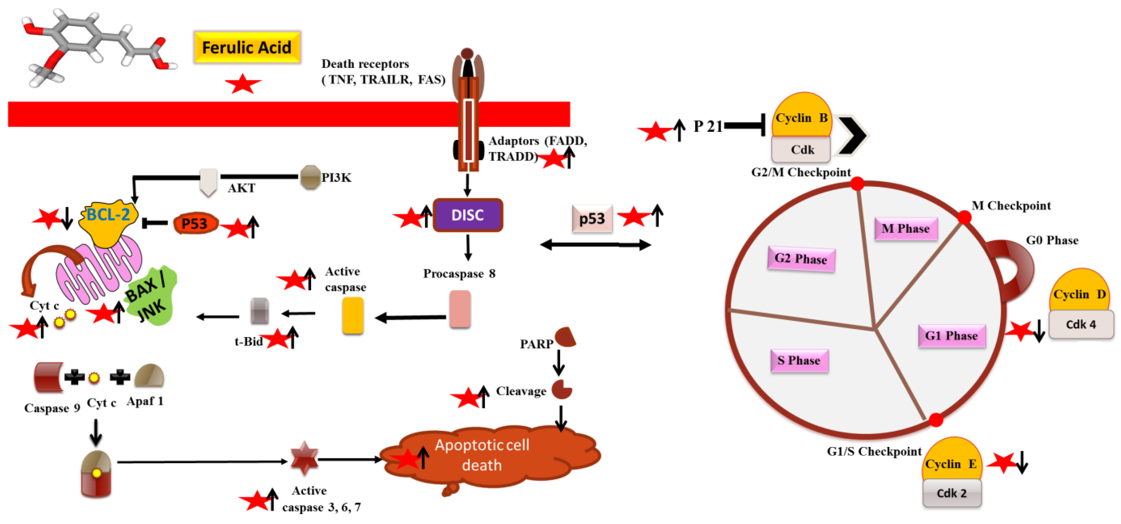
4.1. Induction of Apoptosis and Cell Cycle Arrest
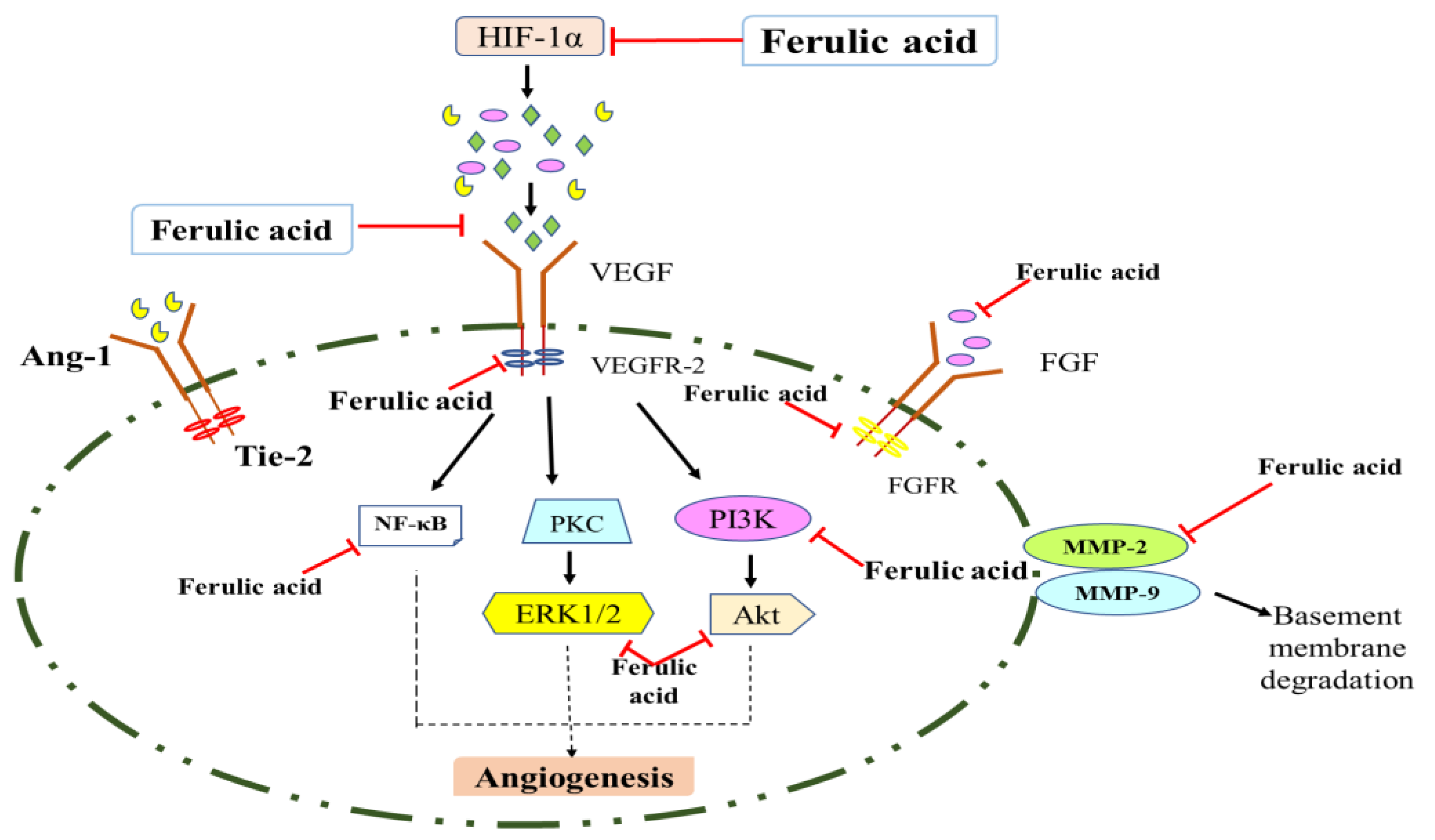
4.2. Antiangiogenic Action of Ferulic Acid
4.3. Inhibition of Metastasis and Invasion
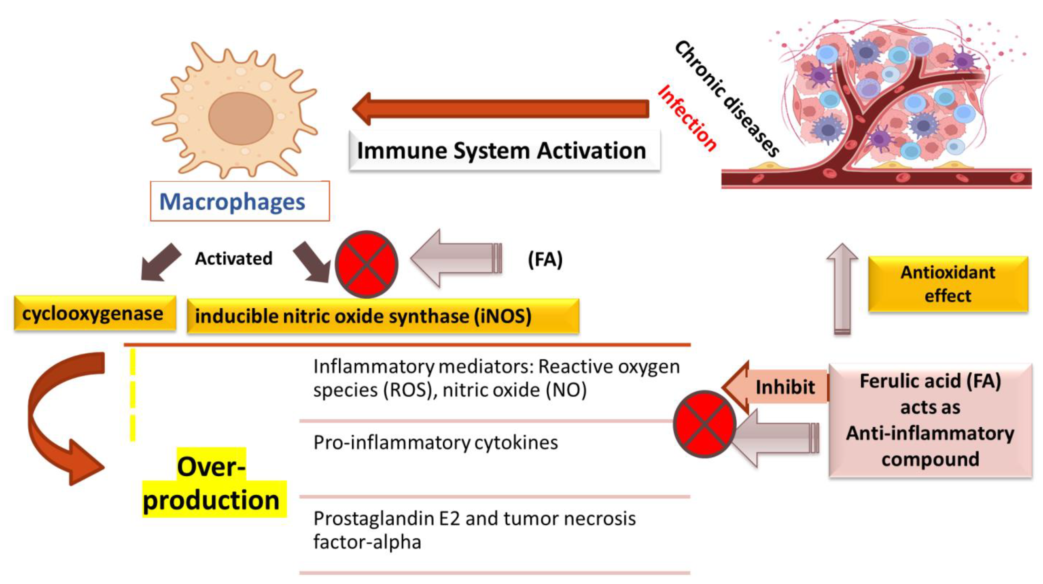
4.4. Anti-Inflammatory Mechanisms
5. Synergistic Interactions of Ferulic Acid in Cancer
6. Safety Studies
7. Conclusions and Future Perspectives
Author Contributions
Funding
Institutional Review Board Statement
Informed Consent Statement
Data Availability Statement
Acknowledgments
Conflicts of Interest
References
- Samtiya, M.; Aluko, R.E.; Dhewa, T.; Moreno-Rojas, J.M. Potential Health Benefits of Plant Food-Derived Bioactive Components: An Overview. Foods 2021, 10, 839. [Google Scholar] [CrossRef] [PubMed]
- Sung, H.; Ferlay, J.; Siegel, R.L.; Laversanne, M.; Soerjomataram, I.; Jemal, A.; Bray, F. Global Cancer Statistics 2020: GLOBOCAN Estimates of Incidence and Mortality Worldwide for 36 Cancers in 185 Countries. CA Cancer J. Clin. 2021, 71, 209–249. [Google Scholar] [CrossRef] [PubMed]
- Sak, K. Anticancer Action of Plant Products: Changing Stereotyped Attitudes. Explor Target. Antitumor Ther. 2022, 3, 423–427. [Google Scholar] [CrossRef] [PubMed]
- Alara, O.R.; Abdurahman, N.H.; Ukaegbu, C.I. Extraction of Phenolic Compounds: A Review. Curr. Res. Food Sci. 2021, 4, 200–214. [Google Scholar] [CrossRef] [PubMed]
- Sova, M.; Saso, L. Natural Sources, Pharmacokinetics, Biological Activities and Health Benefits of Hydroxycinnamic Acids and Their Metabolites. Nutrients 2020, 12, 2190. [Google Scholar] [CrossRef]
- Choudhari, A.S.; Mandave, P.C.; Deshpande, M.; Ranjekar, P.; Prakash, O. Phytochemicals in Cancer Treatment: From Preclinical Studies to Clinical Practice. Front. Pharmacol. 2020, 10, 1614. [Google Scholar] [CrossRef] [Green Version]
- Haque, A.; Brazeau, D.; Amin, A.R. Perspectives on Natural Compounds in Chemoprevention and Treatment of Cancer: An Update with New Promising Compounds. Eur. J. Cancer 2021, 149, 165–183. [Google Scholar] [CrossRef]
- Ramasamy, K.; Agarwal, R. Multitargeted Therapy of Cancer by Silymarin. Cancer Lett. 2008, 269, 352–362. [Google Scholar] [CrossRef] [Green Version]
- Helmy, S.A.; El-Mofty, S.; el Gayar, A.M.; El-Sherbiny, I.M.; El-Far, Y.M. Novel Doxorubicin / Folate-Targeted Trans-Ferulic Acid-Loaded PLGA Nanoparticles Combination: In-Vivo Superiority over Standard Chemotherapeutic Regimen for Breast Cancer Treatment. Biomed. Pharmacother. 2022, 145, 112376. [Google Scholar] [CrossRef]
- Abotaleb, M.; Liskova, A.; Kubatka, P.; Büsselberg, D. Therapeutic Potential of Plant Phenolic Acids in the Treatment of Cancer. Biomolecules 2020, 10, 221. [Google Scholar] [CrossRef]
- Ceci, C.; Lacal, P.; Tentori, L.; de Martino, M.; Miano, R.; Graziani, G. Experimental Evidence of the Antitumor, Antimetastatic and Antiangiogenic Activity of Ellagic Acid. Nutrients 2018, 10, 1756. [Google Scholar] [CrossRef] [PubMed] [Green Version]
- Vengoji, R.; Macha, M.A.; Batra, S.K.; Shonka, N.A. Natural Products: A Hope for Glioblastoma Patients. Oncotarget 2018, 9, 22194–22219. [Google Scholar] [CrossRef] [PubMed] [Green Version]
- Talib, W.H.; Awajan, D.; Hamed, R.A.; Azzam, A.O.; Mahmod, A.I.; AL-Yasari, I.H. Combination Anticancer Therapies Using Selected Phytochemicals. Molecules 2022, 27, 5452. [Google Scholar] [CrossRef]
- Tilay, A.; Bule, M.; Kishenkumar, J.; Annapure, U. Preparation of Ferulic Acid from Agricultural Wastes: Its Improved Extraction and Purification. J. Agric. Food Chem. 2008, 56, 7644–7648. [Google Scholar] [CrossRef] [PubMed]
- Ou, S.; Kwok, K.-C. Ferulic Acid: Pharmaceutical Functions, Preparation and Applications in Foods. J. Sci. Food Agric. 2004, 84, 1261–1269. [Google Scholar] [CrossRef]
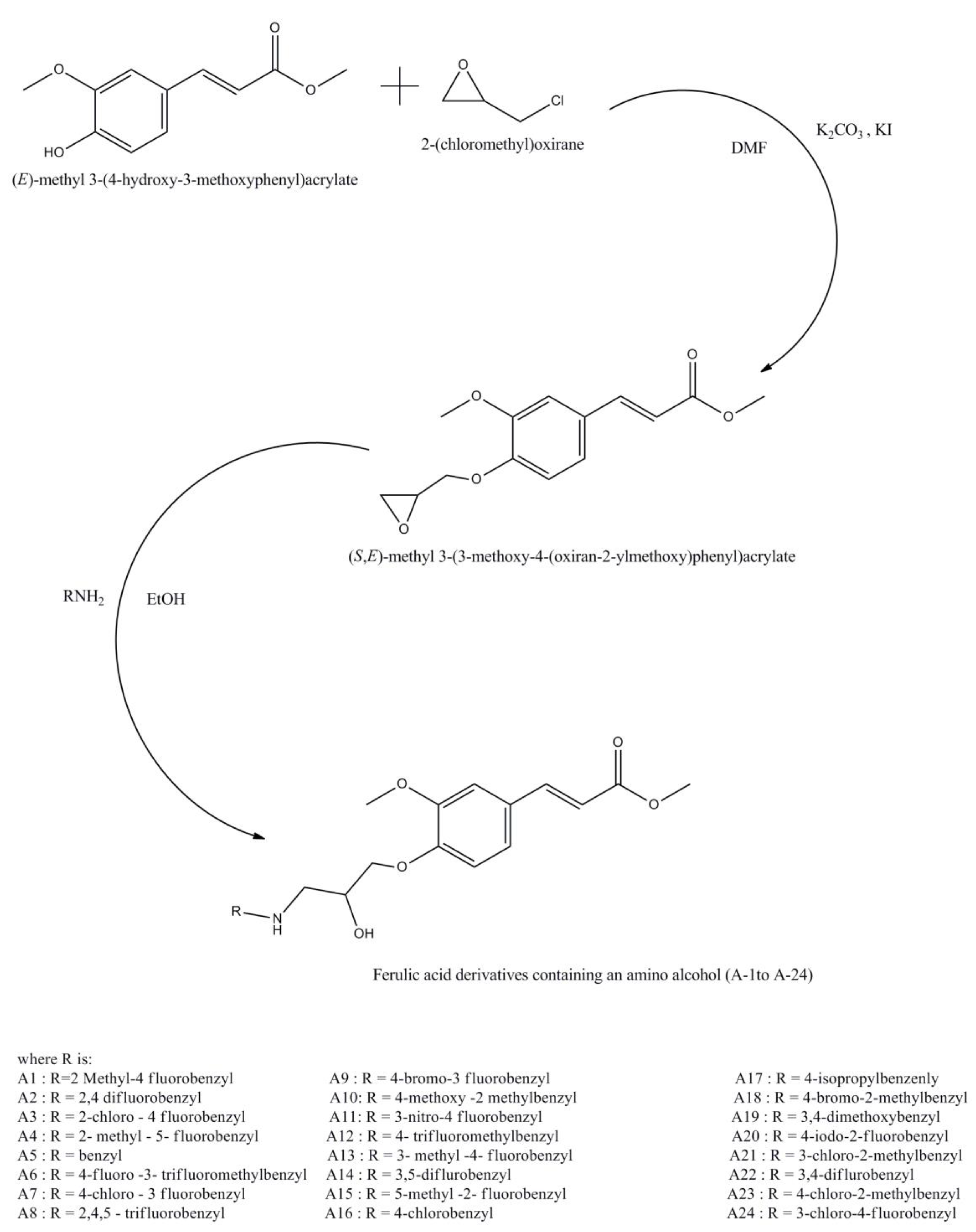
- Dai, A.; Huang, Y.; Yu, L.; Zheng, Z.; Wu, J. Design, Synthesis, and Bioactivity of Ferulic Acid Derivatives Containing an β-Amino Alcohol. BMC Chem. 2022, 16, 34. [Google Scholar] [CrossRef]
- Das, A.; Baidya, R.; Chakraborty, T.; Samanta, A.K.; Roy, S. Pharmacological Basis and New Insights of Taxifolin: A Comprehensive Review. Biomed. Pharmacother. 2021, 142, 112004. [Google Scholar] [CrossRef]
- Zhao, Z.; Moghadasian, M.H. Chemistry, Natural Sources, Dietary Intake and Pharmacokinetic Properties of Ferulic Acid: A Review. Food Chem. 2008, 109, 691–702. [Google Scholar] [CrossRef]
- Alam, M.A. Anti-Hypertensive Effect of Cereal Antioxidant Ferulic Acid and Its Mechanism of Action. Front. Nutr. 2019, 6, 121. [Google Scholar] [CrossRef]
- Clifford, M.N.; King, L.J.; Kerimi, A.; Pereira-Caro, M.G.; Williamson, G. Metabolism of Phenolics in Coffee and Plant-Based Foods by Canonical Pathways: An Assessment of the Role of Fatty Acid β-Oxidation to Generate Biologically-Active and -Inactive Intermediates. Crit. Rev. Food Sci. Nutr. 2022, 1–58. [Google Scholar] [CrossRef]
- Tada, Y.; Tayama, K.; Aoki, N. Acute Oral Toxicity of Ferulic Acid, Natural Food Additive, in Rats. Ann. Rep. Tokyo Metr. Lab. PH 1999, 50, 311–313. [Google Scholar]
- Ramadan, M.A.; Shawkey, A.E.; Rabeh, M.A.; Abdellatif, A.O. Expression of P53, BAX, and BCL-2 in Human Malignant Melanoma and Squamous Cell Carcinoma Cells after Tea Tree Oil Treatment in Vitro. Cytotechnology 2019, 71, 461–473. [Google Scholar] [CrossRef] [PubMed]
- Zhang, X.; Wu, Q.; Yang, S. Ferulic Acid Promoting Apoptosis in Human Osteosarcoma Cell Lines. Pak. J. Med. Sci. 2017, 33, 127–131. [Google Scholar] [CrossRef] [PubMed]
- Kampa, M.; Alexaki, V.-I.; Notas, G.; Nifli, A.-P.; Nistikaki, A.; Hatzoglou, A.; Bakogeorgou, E.; Kouimtzoglou, E.; Blekas, G.; Boskou, D.; et al. Antiproliferative and Apoptotic Effects of Selective Phenolic Acids on T47D Human Breast Cancer Cells: Potential Mechanisms of Action. Breast Cancer Res. 2004, 6, R63. [Google Scholar] [CrossRef] [Green Version]
- Eroğlu, C.; Seçme, M.; Bağcı, G.; Dodurga, Y. Assessment of the Anticancer Mechanism of Ferulic Acid via Cell Cycle and Apoptotic Pathways in Human Prostate Cancer Cell Lines. Tumor Biology 2015, 36, 9437–9446. [Google Scholar] [CrossRef]
- Bandugula, V.R. 2-Deoxy-d-Glucose and Ferulic Acid Modulates Radiation Response Signaling in Non-Small Cell Lung Cancer Cells. Tumor Biol. 2013, 34, 251–259. [Google Scholar] [CrossRef]
- Grasso, R.; Dell’Albani, P.; Carbone, C.; Spatuzza, M.; Bonfanti, R.; Sposito, G.; Puglisi, G.; Musumeci, F.; Scordino, A.; Campisi, A. Synergic Pro-Apoptotic Effects of Ferulic Acid and Nanostructured Lipid Carrier in Glioblastoma Cells Assessed through Molecular and Delayed Luminescence Studies. Sci. Rep. 2020, 10, 4680. [Google Scholar] [CrossRef] [Green Version]
- Luo, L.; Zhu, S.; Tong, Y.; Peng, S. Ferulic Acid Induces Apoptosis of HeLa and Caski Cervical Carcinoma Cells by Down-Regulating the Phosphatidylinositol 3-Kinase (PI3K)/Akt Signaling Pathway. Med. Sci. Monit. 2020, 26, e920095. [Google Scholar] [CrossRef]
- Hou, Y.; Yang, J.; Zhao, G.; Yuan, Y. Ferulic Acid Inhibits Endothelial Cell Proliferation through NO Down-Regulating ERK1/2 Pathway. J. Cell Biochem. 2004, 93, 1203–1209. [Google Scholar] [CrossRef]
- Wu, X.; Hu, Z.; Zhou, J.; Liu, J.; Ren, P.; Huang, X. Ferulic Acid Alleviates Atherosclerotic Plaques by Inhibiting VSMC Proliferation Through the NO/P21 Signaling Pathway. J. Cardiovasc. Transl. Res. 2022, 15, 865–875. [Google Scholar] [CrossRef]
- Gao, J.; Yu, H.; Guo, W.; Kong, Y.; Gu, L.; Li, Q.; Yang, S.; Zhang, Y.; Wang, Y. The Anticancer Effects of Ferulic Acid Is Associated with Induction of Cell Cycle Arrest and Autophagy in Cervical Cancer Cells. Cancer Cell Int. 2018, 18, 102. [Google Scholar] [CrossRef] [PubMed]
- Janicke, B.; Hegardt, C.; Krogh, M.; Önning, G.; Åkesson, B.; Cirenajwis, H.M.; Oredsson, S.M. The Antiproliferative Effect of Dietary Fiber Phenolic Compounds Ferulic Acid and p -Coumaric Acid on the Cell Cycle of Caco-2 Cells. Nutr. Cancer 2011, 63, 611–622. [Google Scholar] [CrossRef] [PubMed]
- Wang, J.; Lai, X.; Yuan, D.; Liu, Y.; Wang, J.; Liang, Y. Effects of Ferulic Acid, a Major Component of Rice Bran, on Proliferation, Apoptosis, and Autophagy of HepG2 Cells. Food Res. Int. 2022, 161, 111816. [Google Scholar] [CrossRef] [PubMed]
- Pellerito, C.; Emanuele, S.; Ferrante, F.; Celesia, A.; Giuliano, M.; Fiore, T. Tributyltin(IV) Ferulate, a Novel Synthetic Ferulic Acid Derivative, Induces Autophagic Cell Death in Colon Cancer Cells: From Chemical Synthesis to Biochemical Effects. J. Inorg. Biochem. 2020, 205, 110999. [Google Scholar] [CrossRef]
- McDaniel, J.T.; Nuhu, K.; Ruiz, J.; Alorbi, G. Social Determinants of Cancer Incidence and Mortality around the World: An Ecological Study. Glob. Health Promot. 2019, 26, 41–49. [Google Scholar] [CrossRef]
- Nishida, N.; Yano, H.; Nishida, T.; Kamura, T.; Kojiro, M. Angiogenesis in Cancer. Vasc. Health Risk Manag. 2006, 2, 213–219. [Google Scholar] [CrossRef]
- Lopes-Coelho, F.; Martins, F.; Pereira, S.A.; Serpa, J. Anti-Angiogenic Therapy: Current Challenges and Future Perspectives. Int. J. Mol. Sci. 2021, 22, 3765. [Google Scholar] [CrossRef]
- Waltham, M.; Burnand, K.G.; Collins, M.; Smith, A. Vascular Endothelial Growth Factor and Basic Fibroblast Growth Factor Are Found in Resolving Venous Thrombi. J. Vasc. Surg. 2000, 32, 988–996. [Google Scholar] [CrossRef] [Green Version]
- Chen, L.-T.; Oh, D.-Y.; Ryu, M.-H.; Yeh, K.-H.; Yeo, W.; Carlesi, R.; Cheng, R.; Kim, J.; Orlando, M.; Kang, Y.-K. Anti-Angiogenic Therapy in Patients with Advanced Gastric and Gastroesophageal Junction Cancer: A Systematic Review. Cancer Res. Treat. 2017, 49, 851–868. [Google Scholar] [CrossRef] [Green Version]
- Sagar, S.M.; Yance, D.; Wong, R.K. Natural Health Products That Inhibit Angiogenesis: A Potential Source for Investigational New Agents to Treat Cancer-Part 1. Curr. Oncol. 2006, 13, 14–26. [Google Scholar] [CrossRef]
- Srinivasan, M.; Sudheer, A.R.; Menon, V.P. Ferulic Acid: Therapeutic Potential Through Its Antioxidant Property. J. Clin. Biochem. Nutr. 2007, 40, 92–100. [Google Scholar] [CrossRef] [PubMed]
- Lin, C.-M.; Chiu, J.-H.; Wu, I.-H.; Wang, B.-W.; Pan, C.-M.; Chen, Y.-H. Ferulic Acid Augments Angiogenesis via VEGF, PDGF and HIF-1α. J. Nutr. Biochem. 2010, 21, 627–633. [Google Scholar] [CrossRef] [PubMed]
- Yang, G.-W.; Jiang, J.-S.; Lu, W.-Q. Ferulic Acid Exerts Anti-Angiogenic and Anti-Tumor Activity by Targeting Fibroblast Growth Factor Receptor 1-Mediated Angiogenesis. Int. J. Mol. Sci. 2015, 16, 24011–24031. [Google Scholar] [CrossRef]
- EKOWATI, J.; HAMID, I.S.; DIYAH, N.W.; SISWANDONO, S. Ferulic Acid Prevents Angiogenesis Through Cyclooxygenase-2 and Vascular Endothelial Growth Factor in the Chick Embryo Chorioallantoic Membrane Model. Turk. J. Pharm. Sci. 2020, 17, 424–431. [Google Scholar] [CrossRef] [PubMed]
- Fares, J.; Fares, M.Y.; Khachfe, H.H.; Salhab, H.A.; Fares, Y. Molecular Principles of Metastasis: A Hallmark of Cancer Revisited. Signal. Transduct. Target Ther. 2020, 5, 28. [Google Scholar] [CrossRef] [PubMed] [Green Version]
- Jiang, Y.-L.; Liu, Z.-P. Natural Products as Anti-Invasive and Anti-Metastatic Agents. Curr. Med. Chem. 2011, 18, 808–829. [Google Scholar] [CrossRef] [PubMed]
- Groblewska, M.; Siewko, M.; Mroczko, B.; Szmitkowski, M. The Role of Matrix Metalloproteinases (MMPs) and Their Inhibitors (TIMPs) in the Development of Esophageal Cancer. Folia Histochem. Cytobiol. 2012, 50, 12–19. [Google Scholar] [CrossRef] [PubMed] [Green Version]
- Bauvois, B. New Facets of Matrix Metalloproteinases MMP-2 and MMP-9 as Cell Surface Transducers: Outside-in Signaling and Relationship to Tumor Progression. Biochim. Biophys. Acta 2012, 1825, 29–36. [Google Scholar] [CrossRef]
- Zhang, X.; Lin, D.; Jiang, R.; Li, H.; Wan, J.; Li, H. Ferulic Acid Exerts Antitumor Activity and Inhibits Metastasis in Breast Cancer Cells by Regulating Epithelial to Mesenchymal Transition. Oncol. Rep. 2016, 36, 271–278. [Google Scholar] [CrossRef] [Green Version]
- El-Gogary, R.I.; Nasr, M.; Rahsed, L.A.; Hamzawy, M.A. Ferulic Acid Nanocapsules as a Promising Treatment Modality for Colorectal Cancer: Preparation and in Vitro/in Vivo Appraisal. Life Sci. 2022, 298, 120500. [Google Scholar] [CrossRef]
- Walsh, L.J. Mast Cells and Oral Inflammation. Crit. Rev. Oral Biol. Med. 2003, 14, 188–198. [Google Scholar] [CrossRef] [PubMed]
- Sakai, S.; Kawamata, H.; Kogure, T.; Mantani, N.; Terasawa, K.; Umatake, M.; Ochiai, H. Inhibitory Effect of Ferulic Acid and Isoferulic Acid on the Production of Macrophage Inflammatory Protein-2 in Response to Respiratory Syncytial Virus Infection in RAW264.7 Cells. Mediat. Inflamm. 1999, 8, 173–175. [Google Scholar] [CrossRef] [PubMed]
- Tetsuka, T.; Baier, L.D.; Morrison, A.R. Antioxidants Inhibit Interleukin-1-Induced Cyclooxygenase and Nitric-Oxide Synthase Expression in Rat Mesangial Cells. Evidence for Post-Transcriptional Regulation. J. Biol. Chem. 1996, 271, 11689–11693. [Google Scholar] [CrossRef] [PubMed] [Green Version]
- Ou, L.; Kong, L.-Y.; Zhang, X.-M.; Niwa, M. Oxidation of Ferulic Acid by Momordica charantia Peroxidase and Related Anti-Inflammation Activity Changes. Biol. Pharm. Bull. 2003, 26, 1511–1516. [Google Scholar] [CrossRef] [PubMed] [Green Version]
- Ohnishi, M.; Matuo, T.; Tsuno, T.; Hosoda, A.; Nomura, E.; Taniguchi, H.; Sasaki, H.; Morishita, H. Antioxidant Activity and Hypoglycemic Effect of Ferulic Acid in STZ-Induced Diabetic Mice and KK-Ay Mice. Biofactors 2015, 21, 315–319. [Google Scholar] [CrossRef]
- Kim, E.O.; Min, K.J.; Kwon, T.K.; Um, B.H.; Moreau, R.A.; Choi, S.W. Anti-Inflammatory Activity of Hydroxycinnamic Acid Derivatives Isolated from Corn Bran in Lipopolysaccharide-Stimulated Raw 264.7 Macrophages. Food Chem. Toxicol. 2012, 50, 1309–1316. [Google Scholar] [CrossRef]
- Hosoda, A.; Ozaki, Y.; Kashiwada, A.; Mutoh, M.; Wakabayashi, K.; Mizuno, K.; Nomura, E.; Taniguchi, H. Syntheses of Ferulic Acid Derivatives and Their Suppressive Effects on Cyclooxygenase-2 Promoter Activity. Bioorg. Med. Chem. 2002, 10, 1189–1196. [Google Scholar] [CrossRef]
- Sudheer, A.R.; Muthukumaran, S.; Devipriya, N.; Devaraj, H.; Menon, V.P. Influence of Ferulic Acid on Nicotine-Induced Lipid Peroxidation, DNA Damage and Inflammation in Experimental Rats as Compared to N-Acetylcysteine. Toxicology 2008, 243, 317–329. [Google Scholar] [CrossRef]
- Tuli, H.S.; Kashyap, D.; Sharma, A.K.; Sandhu, S.S. Molecular Aspects of Melatonin (MLT)-Mediated Therapeutic Effects. Life Sci. 2015, 135, 147–157. [Google Scholar] [CrossRef]
- Eitsuka, T.; Tatewaki, N.; Nishida, H.; Nakagawa, K.; Miyazawa, T. A Combination of δ-Tocotrienol and Ferulic Acid Synergistically Inhibits Telomerase Activity in DLD-1 Human Colorectal Adenocarcinoma Cells. J. Nutr. Sci. Vitaminol. 2016, 62, 281–287. [Google Scholar] [CrossRef] [Green Version]
- Eitsuka, T.; Tatewaki, N.; Nishida, H.; Kurata, T.; Nakagawa, K.; Miyazawa, T. Synergistic Inhibition of Cancer Cell Proliferation with a Combination of δ-Tocotrienol and Ferulic Acid. Biochem. Biophys. Res. Commun. 2014, 453, 606–611. [Google Scholar] [CrossRef] [PubMed]
- Montagnani Marelli, M.; Marzagalli, M.; Fontana, F.; Raimondi, M.; Moretti, R.M.; Limonta, P. Anticancer Properties of Tocotrienols: A Review of Cellular Mechanisms and Molecular Targets. J. Cell Physiol. 2019, 234, 1147–1164. [Google Scholar] [CrossRef] [PubMed] [Green Version]
- Ju, J.; Picinich, S.C.; Yang, Z.; Zhao, Y.; Suh, N.; Kong, A.-N.; Yang, C.S. Cancer-Preventive Activities of Tocopherols and Tocotrienols. Carcinogenesis 2010, 31, 533–542. [Google Scholar] [CrossRef] [PubMed] [Green Version]
- Indap, M.A.; Radhika, S.; Motiwale, L.; Rao, K.V.K. Inhibitory Effect of Cinnamoyl Compounds against Human Malignant Cell Line. Indian J. Exp. Biol. 2006, 44, 216–220. [Google Scholar]
- Eroglu, C.; Avci, E.; Secme, M.; Dodurga, Y. The Combination Effect of Ferulic Acid and Gemcitabine on Expression of Genes Related Apoptosis and Metastasis in PC-3 Prostate Cancer Cells. Eur. J. Biol. 2018, 77, 32–37. [Google Scholar] [CrossRef]
- Choi, Y.E.; Park, E. Ferulic Acid in Combination with PARP Inhibitor Sensitizes Breast Cancer Cells as Chemotherapeutic Strategy. Biochem. Biophys. Res. Commun. 2015, 458, 520–524. [Google Scholar] [CrossRef]
- Sadoughi, F.; Mansournia, M.A.; Mirhashemi, S.M. The Potential Role of Chitosan-based Nanoparticles as Drug Delivery Systems in Pancreatic Cancer. IUBMB Life 2020, 72, 872–883. [Google Scholar] [CrossRef]
- Paciello, F.; Fetoni, A.R.; Mezzogori, D.; Rolesi, R.; di Pino, A.; Paludetti, G.; Grassi, C.; Troiani, D. The Dual Role of Curcumin and Ferulic Acid in Counteracting Chemoresistance and Cisplatin-Induced Ototoxicity. Sci. Rep. 2020, 10, 1063. [Google Scholar] [CrossRef] [Green Version]
- Predarska, I.; Saoud, M.; Drača, D.; Morgan, I.; Komazec, T.; Eichhorn, T.; Mihajlović, E.; Dunđerović, D.; Mijatović, S.; Maksimović-Ivanić, D.; et al. Mesoporous Silica Nanoparticles Enhance the Anticancer Efficacy of Platinum(IV)-Phenolate Conjugates in Breast Cancer Cell Lines. Nanomaterials 2022, 12, 3767. [Google Scholar] [CrossRef]
- Raina, K.; Agarwal, R. Combinatorial Strategies for Cancer Eradication by Silibinin and Cytotoxic Agents: Efficacy and Mechanisms. Acta Pharmacol. Sin. 2007, 28, 1466–1475. [Google Scholar] [CrossRef] [Green Version]
- Mancuso, C.; Santangelo, R. Ferulic Acid: Pharmacological and Toxicological Aspects. Food Chem. Toxicol. 2014, 65, 185–195. [Google Scholar] [CrossRef] [PubMed]
- Xue, C.; Lu, H.; Liu, Y.; Zhang, J.; Wang, J.; Luo, W.; Zhang, W.; Chen, J. Trans-Ferulic Acid-4-β-Glucoside Alleviates Cold-Induced Oxidative Stress and Promotes Cold Tolerance. Int. J. Mol. Sci. 2018, 19, 2321. [Google Scholar] [CrossRef] [PubMed]
- Zhang, L.-W.; Al-Suwayeh, S.A.; Hsieh, P.-W.; Fang, J.-Y. A Comparison of Skin Delivery of Ferulic Acid and Its Derivatives: Evaluation of Their Efficacy and Safety. Int. J. Pharm. 2010, 399, 44–51. [Google Scholar] [CrossRef] [PubMed]
- Peng, C.-C.; Hsieh, C.-L.; Wang, H.-E.; Chung, J.-Y.; Chen, K.-C.; Peng, R.Y. Ferulic Acid Is Nephrodamaging While Gallic Acid Is Renal Protective in Long Term Treatment of Chronic Kidney Disease. Clin. Nutr. 2012, 31, 405–414. [Google Scholar] [CrossRef] [PubMed]
- Maruyama, H.; Kawakami, F.; Lwin, T.-T.; Imai, M.; Shamsa, F. Biochemical Characterization of Ferulic Acid and Caffeic Acid Which Effectively Inhibit Melanin Synthesis via Different Mechanisms in B16 Melanoma Cells. Biol. Pharm. Bull. 2018, 41, 806–810. [Google Scholar] [CrossRef] [PubMed] [Green Version]
- Pandi, A.; Raghu, M.H.; Chandrashekar, N.; Kalappan, V.M. Cardioprotective Effects of Ferulic Acid against Various Drugs and Toxic Agents. Beni Suef Univ. J. Basic Appl. Sci. 2022, 11, 92. [Google Scholar] [CrossRef]
- Wang, T.; Gong, X.; Jiang, R.; Li, H.; Du, W.; Kuang, G. Ferulic Acid Inhibits Proliferation and Promotes Apoptosis via Blockage of PI3K/Akt Pathway in Osteosarcoma Cell. Am. J. Transl. Res. 2016, 8, 968–980. [Google Scholar]
- Dodurga, Y.; Eroğlu, C.; Seçme, M.; Elmas, L.; Avcı, Ç.B.; Şatıroğlu-Tufan, N.L. Anti-Proliferative and Anti-Invasive Effects of Ferulic Acid in TT Medullary Thyroid Cancer Cells Interacting with URG4/URGCP. Tumour Biol. 2016, 37, 1933–1940. [Google Scholar] [CrossRef]
- Al-Mutairi, A.; Rahman, A.; Rao, M.S. Low Doses of Thymoquinone and Ferulic Acid in Combination Effectively Inhibit Proliferation of Cultured MDA-MB 231 Breast Adenocarcinoma Cells. Nutr. Cancer 2021, 73, 282–289. [Google Scholar] [CrossRef]
- Rezaei, A.; Varshosaz, J.; Fesharaki, M.; Farhang, A.; Jafari, S.M. Improving the Solubility and in Vitro Cytotoxicity (Anticancer Activity) of Ferulic Acid by Loading It into Cyclodextrin Nanosponges. Int. J. Nanomed. 2019, 14, 4589–4599. [Google Scholar] [CrossRef] [Green Version]
- Park, E. Data on Cell Cycle in Breast Cancer Cell Line, MDA-MB-231 with Ferulic Acid Treatment. Data Brief. 2016, 7, 107–110. [Google Scholar] [CrossRef] [PubMed] [Green Version]
- Serafim, T.L.; Carvalho, F.S.; Marques, M.P.M.; Calheiros, R.; Silva, T.; Garrido, J.; Milhazes, N.; Borges, F.; Roleira, F.; Silva, E.T.; et al. Lipophilic Caffeic and Ferulic Acid Derivatives Presenting Cytotoxicity against Human Breast Cancer Cells. Chem. Res. Toxicol. 2011, 24, 763–774. [Google Scholar] [CrossRef] [PubMed] [Green Version]
- Das, U.; Manna, K.; Adhikary, A.; Mishra, S.; das Saha, K.; Sharma, R.D.; Majumder, B.; Dey, S. Ferulic Acid Enhances the Radiation Sensitivity of Lung and Liver Carcinoma Cells by Collapsing Redox Homeostasis: Mechanistic Involvement of Akt/P38 MAPK Signalling Pathway. Free Radic. Res. 2019, 53, 944–967. [Google Scholar] [CrossRef]
- Cao, Y.; Zhang, H.; Tang, J.; Wang, R. Ferulic Acid Mitigates Growth and Invasion of Esophageal Squamous Cell Carcinoma through Inducing Ferroptotic Cell Death. Dis. Markers 2022, 2022, 1–19. [Google Scholar] [CrossRef] [PubMed]
- Ezhuthupurakkal, P.B.; Ariraman, S.; Arumugam, S.; Subramaniyan, N.; Muthuvel, S.K.; Kumpati, P.; Rajamani, B.; Chinnasamy, T. Anticancer Potential of ZnO Nanoparticle-Ferulic Acid Conjugate on Huh-7 and HepG2 Cells and Diethyl Nitrosamine Induced Hepatocellular Cancer on Wistar Albino Rat. Nanomedicine 2018, 14, 415–428. [Google Scholar] [CrossRef] [PubMed]
- Fahrioğlu, U.; Dodurga, Y.; Elmas, L.; Seçme, M. Ferulic Acid Decreases Cell Viability and Colony Formation While Inhibiting Migration of MIA PaCa-2 Human Pancreatic Cancer Cells in Vitro. Gene 2016, 576, 476–482. [Google Scholar] [CrossRef] [PubMed]
- Kim, H.-J.; Ryu, K.; Kang, J.-H.; Choi, A.-J.; Kim, T.; Oh, J.-M. Anticancer Activity of Ferulic Acid-Inorganic Nanohybrids Synthesized via Two Different Hybridization Routes, Reconstruction and Exfoliation-Reassembly. Sci. World J. 2013, 2013, 421967. [Google Scholar] [CrossRef] [Green Version]
- Karthikeyan, S.; Kanimozhi, G.; Prasad, N.R.; Mahalakshmi, R. Radiosensitizing Effect of Ferulic Acid on Human Cervical Carcinoma Cells in Vitro. Toxicol. In Vitro 2011, 25, 1366–1375. [Google Scholar] [CrossRef]
- Luo, Y.; Wang, C.-Z.; Sawadogo, R.; Yuan, J.; Zeng, J.; Xu, M.; Tan, T.; Yuan, C.-S. 4-Vinylguaiacol, an Active Metabolite of Ferulic Acid by Enteric Microbiota and Probiotics, Possesses Significant Activities against Drug-Resistant Human Colorectal Cancer Cells. ACS Omega 2021, 6, 4551–4561. [Google Scholar] [CrossRef]
- Sawata, Y.; Matsukawa, T.; Doi, S.; Tsunoda, T.; Arikawa, N.; Matsunaga, N.; Ohnuki, K.; Shirasawa, S.; Kotake, Y. A Novel Compound, Ferulic Acid-Bound Resveratrol, Induces the Tumor Suppressor Gene P15 and Inhibits the Three-Dimensional Proliferation of Colorectal Cancer Cells. Mol. Cell Biochem. 2019, 462, 25–31. [Google Scholar] [CrossRef]
- Potez, M.; Trappetti, V.; Bouchet, A.; Fernandez-Palomo, C.; Güç, E.; Kilarski, W.W.; Hlushchuk, R.; Laissue, J.; Djonov, V. Characterization of a B16-F10 Melanoma Model Locally Implanted into the Ear Pinnae of C57BL/6 Mice. PLoS ONE 2018, 13, e0206693. [Google Scholar] [CrossRef]
- Wang, J.; Sheng, Y.; Ji, L.; Wang, Z. Ferulic Acid Prevents Liver Injury and Increases the Anti-Tumor Effect of Diosbulbin B in Vivo. J. Zhejiang Univ. Sci. B 2014, 15, 540–547. [Google Scholar] [CrossRef] [PubMed] [Green Version]
- Yue, S.-J.; Zhang, P.-X.; Zhu, Y.; Li, N.-G.; Chen, Y.-Y.; Li, J.-J.; Zhang, S.; Jin, R.-Y.; Yan, H.; Shi, X.-Q.; et al. A Ferulic Acid Derivative FXS-3 Inhibits Proliferation and Metastasis of Human Lung Cancer A549 Cells via Positive JNK Signaling Pathway and Negative ERK/P38, AKT/MTOR and MEK/ERK Signaling Pathways. Molecules 2019, 24, 2165. [Google Scholar] [CrossRef] [PubMed]
- Thakkar, A.; Chenreddy, S.; Wang, J.; Prabhu, S. Ferulic Acid Combined with Aspirin Demonstrates Chemopreventive Potential towards Pancreatic Cancer When Delivered Using Chitosan-Coated Solid-Lipid Nanoparticles. Cell Biosci. 2015, 5, 46. [Google Scholar] [CrossRef] [PubMed]


| Type of Cancer | Cell Lines | Effects | Mechanisms | Concentration | References |
|---|---|---|---|---|---|
| Melanoma | Murine B16 | - | ↓ melanin production, ↓ tyrosinase activity, ↓ casein kinase 2 (CK2), ↑ p- tyrosinase | 25 and 50 µM | [75] |
| A375, CHL-1, SK-MEL-2, B16F10 | Anti-angiogenic | ↓ proliferation, migration and tube formation, ↓ fibroblast growth factor 1 (FGF1), ↓ FGFR1, ↓ PI3K, ↓ protein kinase B (Akt) signaling, ↓ PI3K-Akt pathway, ↑ (HUVEC) Growth, ↓ VEGF-A, FGF1, FGF2, PDGF-α, PDGF-β and phosphatidylinositol-glycan biosynthesis class f protein (PIGF) | 0, 2.5, 5, 10, 20, 30, 40 μM | [43] | |
| Sarcoma | S180 | Ameliorating oxidative stress injury | ↓ diosbulbin B-induced liver injury, ↓ ALT and AST activities, Ferulic acid reverses diosbulbin B-decreased CuZn-SOD and CAT enzymatic activities and mRNA expression | - | [76] |
| Osteosarcoma | 143B and MG63 | Induces apoptosis | ↓ proliferation, ↑ G0/G1 phase arrest, ↓ CDK 2, CDK 4, CDK 6, ↑ Bax, ↓ Bcl-2, ↑ caspase-3 activity, ↓ PI3K/Akt activation | 0,10,30,100 and 150 µM | [77] |
| Thyroid | TT cells | Induces apoptosis | ↓ invasion, migration and colony formation, ↓ URG4/URGCP (upregulated gene-4/upregulator of cell proliferation), ↓ CCND1, CDK4, CDK6, BCL2, MMP2, and MMP9, ↑ p53, PARP, PUMA, NOXA, BAX, BID, CASP3, CASP9 and TIMP1 | 50, 75, 100, 150, 200, 300, 400, 500, 750 μM and 1 mM | [78] |
| Breast | MDA-MB 231 | Induces apoptosis | ↓ proliferation, ↑ apoptotic cells, ↓ percentages of cells in G0/G1 phases by TQ, ↓ in %ages of cells in the S phase by FA | Thymoquinone (TQ) and Ferulic Acid (FA) 25 μM TQ + 250 μM FA, 50 μM TQ + 350 μM FA, 50 μM TQ + 450 μM FA, 100 μM TQ + 350 μM FA, 100 μM TQ + 450 μM FA) | [79] |
| MCF7 and 4T1 | Induces apoptosis | ↓ viability, structural changes in cancer cells as compared to normal cells, ↑ apoptosis, ↑ lipid peroxidation, ↑ mitochondrial damage, ↑ cell death | FA-Nanosponges 5, 10, 20, 40, 80, 125, 250, 500, 750, and 1000 µM | [80] | |
| MDA-MB-231 | - | ↓ S phase, ↑ antiproliferative effects, ↑ sensitivity to UV treatment | 0–10 µM | [81] | |
| MDA-MB-231 | Induces apoptosis and inhibits metastasis | ↓ viability, ↑ apoptosis, ↓ metastatic potential, reversal of epithelial-mesenchymal transition (EMT), ↑ caspase-3, ↓ migration across the wound edges, ↓ migration, ↓ vimentin, ↑ E-cadherin | 3, 10, 30 and 100 µM | [49] | |
| MCF-7, MDAMB-231 and HS578T | Induces apoptosis | ↓ proliferation, ↑ cytotoxicity, ↑ p53, ↑ Bax, ↑ caspase-9 | 0–75 μM | [82] | |
| Lung | A549 | Induces apoptosis | ↓ proliferation, ↓ oxidative stress, ↓ GSH, ↑ Keap1, ↓ Nrf2 nuclear level, ↑ apoptotic population, ↓ p-p38 MAPK level, ↓ activation of Akt/MAPK, ↓ p-STAT3, ↓ Cox-2, ↓ MMP-9 and VEGF, ↓ PECAM1, ↑ arrest at at G2/M phase, ↑ p53 and p21 protein, ↓ Cdc25C, ↑ active caspase 9,3, ↑ Bax, ↓ Bcl-2, ↑ radiation sensitivity | ferulic acid −10–400 μM, Gamma radiation 5, 7.5, 10 and 15 Gy (60 Co) | [83] |
| A549 | Inhibits metastasis | ↓ Proliferation, ↑ G0/G1 phase (cell cycle arrest), ↓ migration and invasion, ↓ Bcl-2, ↑ Bax, ↑ Bax/Bcl-2 ratio, ↓ MMP-2 and MMP-9, ↓p- ERK and p-p38, it increased JNK expression, ↓ p-AKT, p-mTOR, p-MEK, and p-ERK | Ferulic acid derivative FXS-3 0.2–50 µM | [84] | |
| Hepatocellular | HepG2 | Induces apoptosis | ↓ proliferation, ↓ oxidative stress, ↓ GSH, ↑ Keap1, ↓ Nrf2 nuclear level, ↑ apoptotic population, ↓ p-p38 MAPK level, ↓ activation of Akt/MAPK, ↓ p-STAT3, ↓ Cox-2, ↓ MMP-9 and VEGF, ↓ PECAM1, ↑ arrest at at G2/M phase, ↑ p53 and p21 protein, ↓ Cdc25C, ↑ active caspase 9,3, ↑ Bax, ↓ Bcl-2, ↑ radiation sensitivity | ferulic acid -10–400 μM, Gamma radiation 5, 7.5, 10 and 15 Gy (60 Co) | [83] |
| Huh-7 and HepG2 | Induces apoptosis | ↓ viability, ↑ structural changes, ↑ROS, ↓ MMP, ↑ DNA damage, ↓ percent of cells in G0/G and G2/M, ↑ S phase, ↑ γH2AX, ↑ Bax, Bad, cleaved caspase 3 | ZnONPs with ferulic acid (ZnONPs-FAC) 0.05, 0.1, 1, 5, 10 and 20 µg/ml | [85] | |
| Pancreatic | MIA PaCa-2 | Induces apoptosis | ↓ cell viability and colony formation, ↑ p53, Bax, PTEN caspase 3 and 9 | 150 μM, 200 μM, 300 μM, 400 μM, 500 μM, 750 μM and 1 mM | [86] |
| Cervical | HeLa and Caski | Induces apoptosis | ↓ viability, ↑ DNA condensation, ↑ apoptosis, ↑ pro-caspase-3, pro-caspase-8, pro-caspase-9 and PARP, ↓ Bcl-2 and Mcl-1, ↑ Bax and ROS, ↓p-Akt and p-PI3K | 4–20 µM | [28] |
| Hela and Caski | Induction of cell cycle arrest and autophagy | ↓ invasion, ↓ MMP-9, ↑ arrest in G0/G1 phase, ↑ p53 and p21, ↓ Cyclin D1 and Cyclin E, ↓ LC3-II, Beclin1 and Atg12-Atg5 | 0, 0.5, 1.0,1.5 and 2.0 mM | [32] | |
| HeLa | - | ↓ Cell viability | ferulic acid nanohybrids 1, 5, 10, 20, 30, 40, and 50 μM | [87] | |
| HeLa and ME-180 | Enhances radiation effects by increasing lipid peroxidative markers | ↓ viability ↓ GSH, ↑ TBARS, CD and LHO, ↓ SOD, CAT and GPx, ↑ DNA damage, ↑ intracellular ROS levels (results by ferulic acid + irradiation in comparison with radiation or ferulic acid treatment alone) | ferulic acid (1, 5, 10, 20, 30 and 40 µg/mL) + radiation (2, 4, 6, 8, 10, 12 and 15 Gy) | [88] | |
| Prostate | PC-3 | Induces apoptosis | ↓ proliferation, ↑ ATR, ATM, CDKN1A, CDKN1B, E2F4, RB1, and TP53 (Gene expression), ↓ CCND1, CCND2, CCND3, CDK2, CDK4, and CDK6 (gene expression) ↓ CDK4 and BCL2 (protein expression), ↓ invasion and colony formation | 20, 30, 50, 75, 100, 150, 200, 250, 300, 350, 500, 750, 900 µM, 1, 2 mM | [25] |
| LNCaP | Induces apoptosis | ↓ proliferation, ↑ CASP1, CASP2, CASP8, CYCS, FAS, FASLG, and TRADD (gene expression), ↓ BCL2 and XIAP (gene expression), ↓CDK4 and BCL2 (protein expression), ↓ invasion and colony formation | 20, 30, 50, 75, 100, 150, 200, 250, 300, 350, 500, 750, 900, 1000 and 2000 µM | [25] | |
| Colorectal | HCT- 116 and HT-29 | Induces apoptosis | ↑ antiproliferative effects, ↑ arrest at the G1 phase, ↓ S phase, ↑ Early apoptotic cells, ↑ Caspase 3, 8, and 9 activity | 0,0.25,0.5,1.0 and 1.5 mM | [89] |
| HCT116 | Induces apoptosis | ↓ proliferation, ↑ p15 (mRNA level) | Ferulic acid-bound resveratrol- 0, 0.625, 1.25, 2.5, 5, 10 and 20 µM | [90] |
| Type of Cancer | Animal Models | Effects | Mechanisms | Dosage | Duration | References |
|---|---|---|---|---|---|---|
| Melanoma | Female C57BL/6 mice xenografted with B16F10 cells | Inhibited tumor angiogenesis | ↓ tumor volume and weight, ↓ p-FGFR1Y1 positive cells, ↓ FGFR1, ↓p-Akt, ↓ p-PI3K | 0, 10, 30, 50 mg/kg | 30 days | [91] |
| Sarcoma | ICR male mice transplanted with S180 cells | - | ↑ diosbulbin B-induced anti-tumor activity | ferulic acid 8 mg/kg + DB 32 mg/kg | - | [92] |
| Colon | Male BALB/c mice xenografted with CT 26 cells | Inhibited tumor growth | ↑ tumor regression, ↑ cleaved caspase 3, ↑ tumor shrinkage, ↑ damage in tumor cell parenchyma, ↑ shrinkage in tissues, ↑ nuclear fragmentation, ↑ apoptotic body formation at the neoplastic region | ferulic acid 50 mg/kg + 2 Gy dose of radiation | 27 days | [86] |
| Breast | Female BALB/c nude xenografted with MDA-MB-231 | Inhibited tumor metastasis | ↓ toxicity, ↓ tumor volumes and weights, ↓ proliferation (Ki67 staining), ↑ apoptosis (active caspase-3 staining), ↓ tumor nodules on the surface of the lungs and liver | 100 mg/kg | 28 days | [84] |
| Lung | C57BL/6 mice transplanted with A549 cells | Inhibited tumor metastasis | ↓ tumor volume, ↓ pulmonary metastatic nodules, ↓ pulmonary tumor metastasis | FXS-3 at 25–100 mg/kg | 27 days | [93] |
| Hepatocellular | Wistar albino rat | Inhibited tumor metastasis | ↓ nodular formation, ↓ GST-P + ive, ↓ Ki67 and 8-OHdG positivity, ↓ ALT, AST, ALP, γ-GT and TBARS (liver marker enzymes) | ZnONPs with ferulic acid (ZnONPs-FAC) 3.6 µg/mL µg/ml | - | [88] |
| Pancreatic | SCID mice | Inhibited tumor growth ↓↑ | ↓ tumor volume, ↓ PCNA and MKI67, and ↑ p-RB, ↑ p21, ↑ p-ERK1/2 | 75 mg/kg | 35 days | [94] |
Publisher’s Note: MDPI stays neutral with regard to jurisdictional claims in published maps and institutional affiliations. |
© 2022 by the authors. Licensee MDPI, Basel, Switzerland. This article is an open access article distributed under the terms and conditions of the Creative Commons Attribution (CC BY) license (https://creativecommons.org/licenses/by/4.0/).
Share and Cite
Singh Tuli, H.; Kumar, A.; Ramniwas, S.; Coudhary, R.; Aggarwal, D.; Kumar, M.; Sharma, U.; Chaturvedi Parashar, N.; Haque, S.; Sak, K. Ferulic Acid: A Natural Phenol That Inhibits Neoplastic Events through Modulation of Oncogenic Signaling. Molecules 2022, 27, 7653. https://doi.org/10.3390/molecules27217653
Singh Tuli H, Kumar A, Ramniwas S, Coudhary R, Aggarwal D, Kumar M, Sharma U, Chaturvedi Parashar N, Haque S, Sak K. Ferulic Acid: A Natural Phenol That Inhibits Neoplastic Events through Modulation of Oncogenic Signaling. Molecules. 2022; 27(21):7653. https://doi.org/10.3390/molecules27217653
Chicago/Turabian StyleSingh Tuli, Hardeep, Ajay Kumar, Seema Ramniwas, Renuka Coudhary, Diwakar Aggarwal, Manoj Kumar, Ujjawal Sharma, Nidarshana Chaturvedi Parashar, Shafiul Haque, and Katrin Sak. 2022. "Ferulic Acid: A Natural Phenol That Inhibits Neoplastic Events through Modulation of Oncogenic Signaling" Molecules 27, no. 21: 7653. https://doi.org/10.3390/molecules27217653