Article

Metabolic Profiling by GC-MS, In Vitro Biological Potential, and In Silico Molecular Docking Studies of Verbena officinalis

1
Department of Pharmaceutical Chemistry, Faculty of Pharmacy, The Islamia University of Bahawalpur, Bahawalpur 63100, Pakistan
2
Department of Chemistry and Physics, College of Sciences and Mathematics, Arkansas State University, Jonesboro, AR 72404, USA
3
Department of Pharmacognosy, College of Pharmacy, Prince Sattam bin Abdul Aziz University, Alkharj 11942, Saudi Arabia
4
Department of Pharmacognosy, Faculty of Pharmacy, Mansoura University, Mansoura 35516, Egypt
5
Department of Pharmacology and Toxicology, College of Pharmacy, King Saud University, Riyadh 11451, Saudi Arabia
6
Department of Pharmaceutical Chemistry, College of Pharmacy, University of Hafr Al-Batin, Hafr Al-Batin 31991, Saudi Arabia
7
Department of Pharmaceutical Chemistry, Faculty of Pharmaceutical Sciences, Prince of Songkla University, Hat Yai 90110, Thailand
8
Department of Food Science and Engineering, College of Chemistry and Engineering, Shenzhen University, Shenzhen 518060, China
9
Department of Food, Dairy Science and Technology, Faculty of Agriculture, Damanhour University, Damanhour 22516, Egypt
10
Department of Food Science, Faculty of Agriculture, Zagazig University, Zagazig 44519, Egypt
11
School of Food Science and Engineering, South China University of Technology, Guangzhou 510641, China
*
Authors to whom correspondence should be addressed.
Molecules 2022, 27(19), 6685; https://doi.org/10.3390/molecules27196685
Submission received: 29 August 2022 / Revised: 3 October 2022 / Accepted: 5 October 2022 / Published: 8 October 2022

Abstract

:
Verbena officinalis L. is a traditionally important medicinal herb that has a rich source of bioactive phytoconstituents with biological benefits. The objective of this study was to assess the metabolic profile and in vitro biological potential of V. officinalis. The bioactive phytoconstituents were evaluated by preliminary phytochemical studies, estimation of polyphenolic contents, and gas chromatography-mass spectrometry (GC-MS) analysis of all fractions (crude methanolic, n-hexane, ethyl acetate, and n-butanol) of V. officinalis. The biological investigation was performed by different assays including antioxidant assays (DPPH, ABTS, CUPRAC, and FRAP), enzyme inhibition assays (urease and α-glucosidase), and hemolytic activity. The ethyl acetate extract had the maximum concentration of total phenolic and total flavonoid contents (394.30 ± 1.09 mg GAE·g−1 DE and 137.35 ± 0.94 mg QE·g−1 DE, respectively). Significant antioxidant potential was observed in all fractions by all four antioxidant methods. Maximum urease inhibitory activity in terms of IC50 value was shown by ethyl acetate fraction (10 ± 1.60 µg mL−1) in comparison to standard hydroxy urea (9.8 ± 1.20 µg·mL−1). The n-hexane extract showed good α-glucosidase inhibitory efficacy (420 ± 20 µg·mL−1) as compared to other extract/fractions. Minimum hemolytic activity was found in crude methanolic fraction (6.5 ± 0.94%) in comparison to positive standard Triton X-100 (93.5 ± 0.48%). The GC-MS analysis of all extract/fractions of V. officinalis including crude methanolic, n-hexane, ethyl acetate, and n-butanol fractions, resulted in the identification of 24, 56, 25, and 9 bioactive compounds, respectively, with 80% quality index. Furthermore, the bioactive compounds identified by GC-MS were analyzed using in silico molecular docking studies to determine the binding affinity between ligands and enzymes (urease and α-glucosidase). In conclusion, V. officinalis possesses multiple therapeutical potentials, and further research is needed to explore its use in the treatment of chronic diseases.

1. Introduction

The existence of a wide range of secondary metabolites in medicinal plants has led to extensive investigation of these plants in recent years to identify the lead compound that can contribute to the management of chronic diseases and therapeutic effectiveness [1]. There has been an increase in scientific interest in medicinal plants [2]. Natural products have been used therapeutically to cure many diseases since ancient times. According to the World Health Organization, 80% of people around the world use plant-based treatments to cover their basic health needs [3]. Approximately 52% of approved molecules from 1981–2014 were natural products or derived directly from them [4]. According to multiple studies, secondary metabolites that are separated from medicinal plants are responsible for a variety of therapeutic uses, including antioxidant, antibacterial, anti-inflammatory, antiviral, antifungal, and anticancer [5]. Phytomedicines have been used largely due to their safety, accessibility, low cost, and sociological acceptance when compared to synthetic drugs [6,7].
Research on enzyme inhibition has expanded significantly over the last two decades [8]. Urease is a metalloenzyme containing nickel that facilitates the rapid conversion of urea into ammonia along with carbon dioxide. Urease is abundantly found in numerous plants, a variety of bacteria, and selected fungi [9,10]. One of the virulent elements in the pathogenesis of the gram-negative, microaerophilic, stomach-found Helicobacter pylori is ureases. The H. pylori infection can cause gastrointestinal inflammation, which raises the risk of chronic disorders, such as duodenal and gastric ulcers, gastric adenocarcinoma, and gastric lymphoma [11,12,13]. Researchers are being encouraged to find new urease inhibitory compounds from natural resources because the blocking of ureases is thought to be the most successful treatment for urease-dependent bacterial infections [14]. So, the discovery of safe and effective urease inhibitors is a demand nowadays due to the release of urease by microorganisms in different pathological disorders [15].
The enzyme α-Glucosidase, found on the intestinal cell membrane surface, catalyzes the breakage of α-glycosidic linkage present in oligosaccharides to make monosaccharides. Hence, inhibitors of α-glucosidase can postpone the generation of d-glucose from complex carbohydrates thus slowing down glucose absorption, and lowering the level of postprandial plasma glucose [16]. To decrease disorders associated with diabetes, regulating the concentration of glucose is a primary technique [17]. The incidence of postprandial hyperglycemia among diabetic individuals is reduced by the inhibitory activity of α-glucosidase, which is thought to interfere with the digestive process of carbohydrates. Several inhibitors of α-glucosidase, such as miglitol and acarbose, have been discovered [18]. However, acarbose use has been associated with gastrointestinal disturbances [19]. The ability of natural products to block the activity of digestive enzymes, and hence lower hyperglycemia in the management of chronic diabetes, had been successfully demonstrated by numerous researchers [20].
Verbena officinalis L. (Verbenaceae) is known as the herb of grace, pigeons’ grass and vervain. It is primarily found in North Africa, Asia and all over Europe. It is mostly distributed in wastelands and near water in cultivated fields in the northern as well as western regions of Pakistan. V. officinalis is a perennial erect small herb that grows up to 25–100 cm in height with lobed and serrated leaves. Pink or purple is the color of flowers [21]. V. officinalis has been utilized to alleviate several ailments in the folk medicinal system, including rheumatic pain, thyroid problem and wounds [22], gastric diseases, skin burns, abrasion [23], cough and asthma [24], depression, amenorrhea, and acute dysentery enteritis [25]. V. officinalis has been studied for its important bioactivities such as antioxidant [26], diuretics and expectorant analgesic and anti-inflammatory [27,28], anticonvulsant [21], antifungal [29], antibacterial [30,31], anticancer [32,33], antidepressant [34], neuroprotective [25], urolithiasis [35] antiproliferative [36] and antitumor [37] effects. The abundance of bioactive metabolites in V. officinalis, including flavonoids [38], phenylethanoid glycosides [36], sterols and triterpenoids [39], and ursolic acid [40] explains the folklore use of V. officinalis [21].
In account of this, a crude methanolic extract of V. officinalis (CRVO) was prepared, then fractionated using various solvents in ascending order of their polarity to produce different fractions; n-hexane (NHVO), ethyl acetate (EAVO) and n-butanol (NBVO). The methanolic crude extract along with its various fractions was evaluated for its total phenolic content (TPC) and total flavonoid content (TFC), urease and α-glucosidase inhibition assays, and antioxidant assays by different methods (DPPH, ABTS, CUPRAC, and FRAP). Metabolic profiles of all fractions of the whole plant of V. officinalis were performed by gas chromatography-mass spectrometry (GC-MS) to identify the tentative secondary metabolites in the respective fractions. In silico molecular docking studies were also conducted for the bioactive compounds identified in all fractions by GC-MS.

2. Results

2.1. Phytochemical Profile of V. officinalis

2.1.1. Preliminary Phytochemical Assessments

Preliminary phytochemical testing of CRVO, NHVO, EAVO, and NBVO fractions of the whole plant of V. officinalis were performed. This analysis confirmed the presence of many bioactive primary and secondary metabolites, as shown in Table 1. The plant showed the presence of primary and secondary plant bioactive metabolites, including carbohydrates, saponins, tannins, phenols, flavonoids, starch, alkaloids, glycosides and resins.

2.1.2. Polyphenolic Contents Estimation

  • Total phenolic content (TPC)
The maximum amount of TPC was observed in EAVO (394.30 ± 2.50 mg GAE·g−1 DE) and the minimum amount was observed in CRVO (89.07 ± 1.88 mg GAE·g−1 DE) (milligram gallic acid equivalent per gram weight of dry extract) (Figure 1).
  • Total flavonoid content (TFC)
The EAVO sample showed the highest amount of TFC with a value of 137.35 ± 0.94 mg QE·g−1 DE (milligram quercetin equivalent per gram weight of dry extract) and the CRVO sample exhibited the lowest amount of TFC with a value of 66.26 ± 1.42 mg QE·g−1 DE (Figure 2).

2.1.3. Detection of Bioactive Compounds by GC-MS

CRVO, NHVO, EAVO, and NBVO fractions of the whole plant of V. officinalis were subjected to GC-MS analysis. The mass spectra of each plant metabolite at different retention times were checked with databases of the mass spectra of the National Institute Standard and Technology (NIST-14). Tentatively identified compounds in CRVO, NHVO, EAVO and NBVO fractions of V. officinalis were 112, 112, 90, and 46, respectively. Compounds with a quality index of more than 80% were finally selected and represented in Table S1. The retention time in minutes (RT), peak area (%, calculated by dividing each compound peak area by the sum of all compounds’ peak areas within the sample), name of the compound, molecular formula, and molecular weight of the metabolites identified in CRVO, NHVO, EAVO, and NBVO fractions of V. officinalis using GC-MS were shown in Table S1. The GC-MS chromatogram of CRVO, NHVO, EAVO, and NBVO fractions of V. officinalis were exhibited in Figure S1.

2.2. In Vitro Biological Investigation of V. officinalis

CRVO, NHVO, EAVO, and NBVO fractions of the whole plant of V. officinalis were evaluated for their biological potential using different approaches such as antioxidant assay, enzyme inhibition and hemolytic activities.

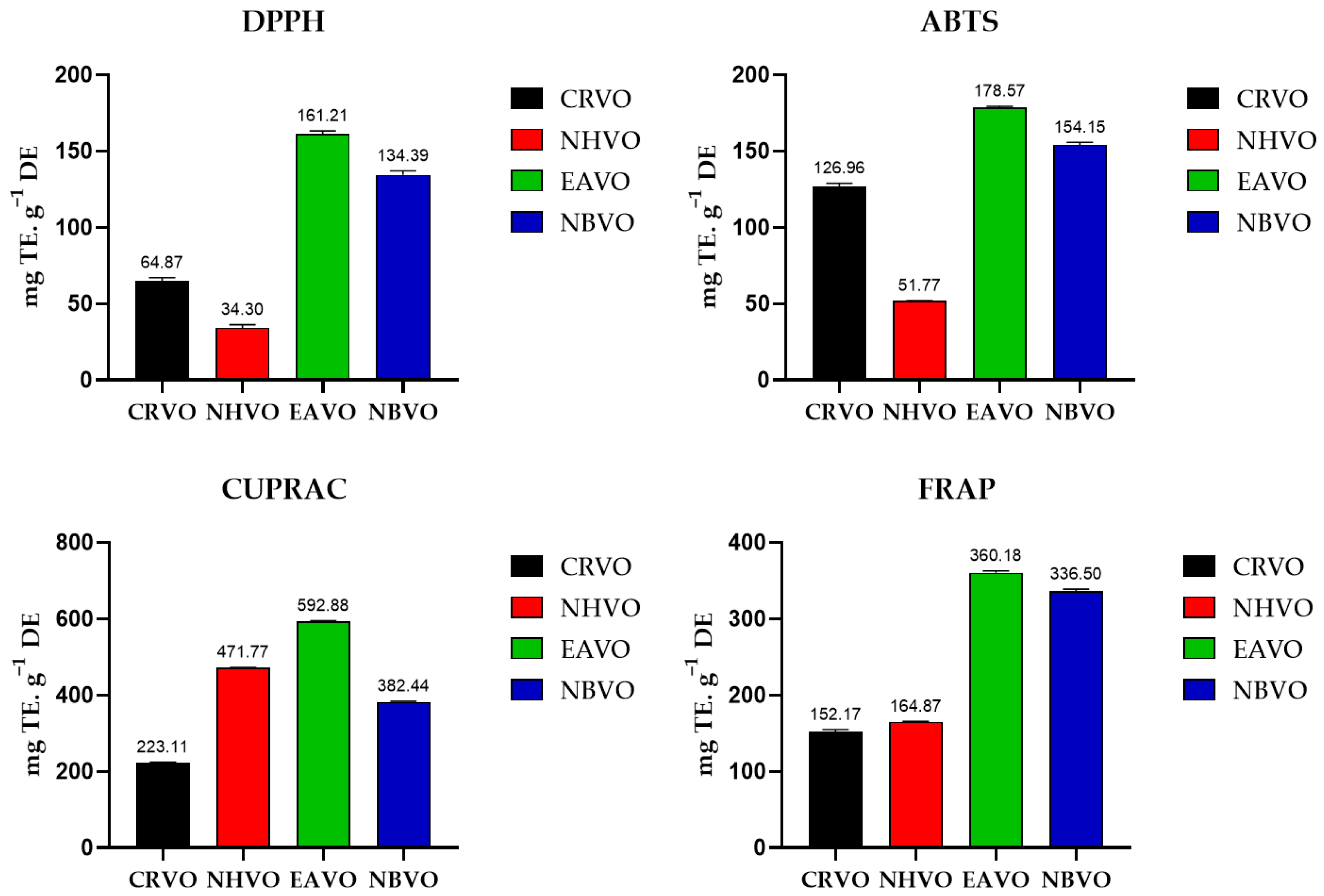
2.2.1. Antioxidant Assays

  • Radical scavenging potential
The radical scavenging potential of the whole plant of V. officinalis was determined using DPPH and ABTS methods. The order of activity of different fractions was as follows; EAVO > NBVO > CRVO > NHVO for DPPH and EAVO > NBVO > CRVO > NHVO for ABTS. The highest scavenging potential estimated by the DPPH method was shown by EAVO (161.21 ± 2.02 mg TE·g−1 DE), and the minimum value was shown by NHVO (34.30 ± 2.02 mg TE·g−1 DE). The maximum free radical scavenging activity calculated by the ABTS method was shown by EAVO (178.57 ± 0.83 mg TE·g−1 DE), while the lowest value was observed in NHVO (51.77 ± 0.36 mg TE·g−1 DE) (Figure 3).
  • Reducing power antioxidant assay
The reducing antioxidant potential of CRVO, NHVO, EAVO and NBVO fractions of the whole plant of V. officinalis was evaluated by two methods namely CUPRAC and FRAP assays. The results were as follows: EAVO > NHVO > NBVO > CRVO for CUPRAC assay and EAVO > NBVO > NHVO > CRVO for FRAP assay. EAVO showed the maximum reducing potential with a value of 592.88 ± 2.44 mg TE·g−1 DE and CRVO showed the minimum reducing capacity value of 223.11 ± 1.55 mg TE·g−1 DE for CUPRAC assay. The highest reducing activity was calculated for FRAP assay for EAVO with a value of 360.18 ± 2.68 mg TE·g−1 DE, and the lowest was determined for CRVO, with a value of 152.171 ± 2.68 mg TE·g−1 DE (Figure 3).

2.2.2. In vitro Enzyme Inhibition Assay

  • Urease inhibition assay
The urease inhibitory potential of CRVO, NHVO, EAVO, and NBVO fractions of V. officinalis whole plant was evaluated using a previously modified method [41]. Results were presented as IC50 values. Lower IC50 values indicate the highest enzyme inhibition. The order of inhibition of the urease enzyme of crude methanolic extract and different fractions of the whole plant of V. officinalis were as follows; EAVO < NBVO < NHVO < CRVO. The IC50 values of EAVO and NBVO were found to be 10 ± 1.60 µg·mL−1 and 30 ± 2.40 µg·mL−1 in comparison to IC50 value of hydroxy urea 9.8 ± 1.20 µg·mL−1. The results of urease inhibitory activity of different fractions of the whole plant of V. officinalis showed the plant as a potent inhibitor of the urease enzyme (Table 2).
  • α-Glucosidase inhibition assay
α-Glucosidase enzyme inhibition assay results were expressed as IC50 values. NHVO showed the best IC50 value of 420 ± 20 µg·mL−1 with good antidiabetic potential while EAVO showed a moderate IC50 value of 685 ± 31 µg mL−1 and IC50 for quercetin was 10 ± 1.30 µg·mL−1 (Table 2).

2.2.3. Hemolytic Activity

The hemolytic potential of CRVO, NHVO, EAVO, and NBVO fractions of the whole plant of V. officinalis were exhibited (Table 3). The percentage of hemolytic activity was found in the order: NBVO > EAVO > NHVO > CRVO. The maximum hemolytic value was 14.5 ± 1.20% for NBVO and the minimum hemolytic value was 6.5 ± 0.94% for CRVO. Results confirmed that V. officinalis is a safe and non-toxic plant due to less than 30% of hemolysis activity [1].

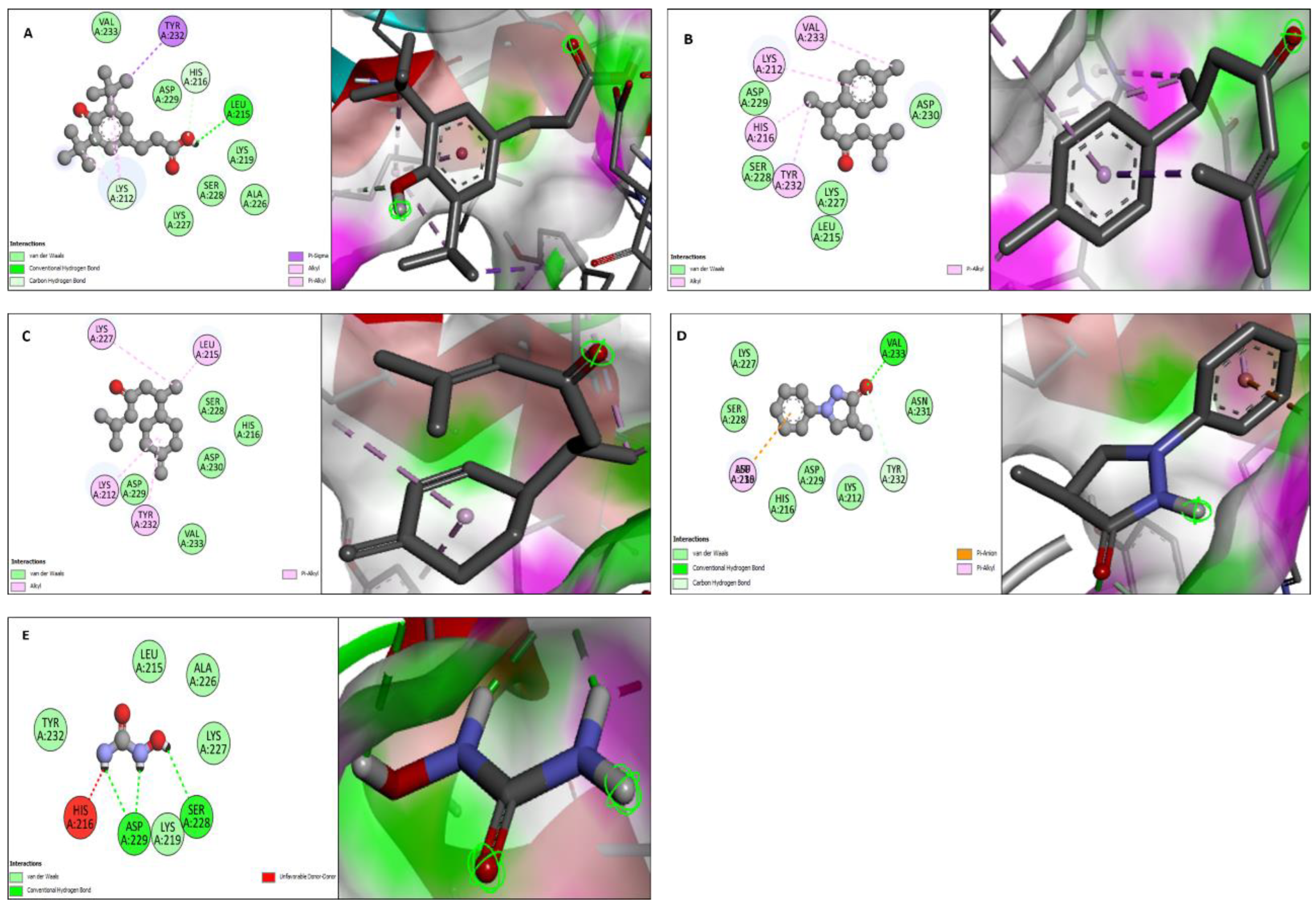
2.3. In Silico Molecular Docking Studies

Molecular docking studies were performed for both urease (PDB DOI: 10.2210/pdb1E9Z/pdb) and α-glucosidase (PDB DOI: 10.2210/pdb5ZCB/pdb). All the compounds from GC-MS profiles of methanolic, n-hexane, ethyl acetate, and n-butanol fractions were docked against urease and α-glucosidase enzymes. Four compounds showed the best binding affinity against both enzymes. Benzenepropanoic acid, 3,5-bis(1,1-dimethylethyl)-4-hydroxy-, methyl ester showed the maximum binding affinity i.e., −6.8 Kcal·mol−1 against urease and α-glucosidase. ar-Turmerone showed a −5.8 Kcal·mol−1 binding affinity against urease, and it showed a −6.5 Kcal·mol−1 binding affinity against α-glucosidase. Curlone showed a −5.6 Kcal·mol−1 binding affinity against urease while it showed a −5.9 Kcal·mol−1 binding affinity against α-glucosidase. 3-pyrazolidinone, 4,4-dimethyl-1-phenyl has a binding affinity of −5.7 Kcal·mol−1 against urease and −5.8 Kcal·mol−1 against α-glucosidase while the binding affinity of hydroxy urea and quercetin (standards) for these enzymes (urease and α-glucosidase) was −4.1 and −7.9 Kcal·mol−1, respectively.
The molecular docking study was validated by redocking of urease, α-glucosidase, and selected ligands with Autodock-1.5.6. Additionally, the same results were found in terms of the binding affinity and RMSD values. The docking results of the four ligands with both enzymes are depicted in Table 4 and Figure 4 and Figure 5.

3. Discussion

Plants naturally contain substances called phytochemicals, which can have beneficial or harmful effects on human health [42]. The most abundant biological reservoirs of different phytochemicals are medicinal plants, which are used to treat various diseases and conditions. The biological potential of plants may be due to the presence of metabolites [43]. Alkaloids, phenolics, flavonoids, saponins, tannins, steroids, terpenoids, terpenes, glycosides, coumarins, polysaccharides, and other significant secondary metabolites can be found in plants [43]. Major phytochemicals including alkaloids having antimicrobial and analgesic potential, flavonoids and tannins demonstrated antioxidant and antibacterial activity [44], and saponins have anticancer, anti-inflammatory, antibacterial, and anti-diabetic potential [45]. The phytochemical analysis of V. officinalis extracts exhibited plant has a variety of phytochemicals such as phenols, flavonoids, glycosides, alkaloids tannins, saponins, resins, and terpenes. Previous studies showed the presence of these chemicals in the leaves of V. officinalis [46]. The results suggested that the medicinal activity of V. officinalis may be attributable to the presence of these phytoconstituents.
Polyphenols are bioactive substances that are frequently found in food products made from plants, such as fruits, seeds, and cereals. Polyphenols help to prevent the possibility of chronic diseases including cancer, neurodegenerative and cardiovascular diseases [47]. Several important flavonoids were reported in V. officinalis previously [48]. Flavonoids were reported to possess different biological activities, including antioxidant, anticancer, anti-inflammatory, antiviral, hepatoprotective, anti-fungal, and antibacterial [49]. The ethyl acetate fraction showed the highest values of both phenolic and flavonoid contents with values of 394.30 ± 1.09 mg GAE·g−1 DE and 137.35 ± 0.94 mg QE·g−1 DE, respectively while the crude methanolic fraction showed the minimum values of both phenolic and flavonoid contents having values of 89.07 ± 1.88 mg GAE·g−1 DE and 66.26 ± 1.42 mg QE·g−1 DE, respectively. Previous studies reported the presence of flavonoids (in hydroalcoholic and aqueous extracts, 0.76 and 0.79 g/100 g, respectively) and phenolic constituents (in hydroalcoholic and aqueous extracts, 1.25 and 1.75 g/100 g, respectively) in V. officinalis aerial parts contributing their role in the antioxidant potential of V. officinalis [50].
Oxidation processes are essential to provide the energy to support biological activity in living organisms. As a result, the uncontrolled production of oxygen reactive species (ROS) is associated with many chronic diseases, including cancer, atherosclerosis, rheumatoid disease, and degenerative processes linked with aging [51]. Synthetic as well as semi-synthetic antioxidants are frequently employed to reduce ROS damage, however, they have also been linked to cancers and damage to cells or entire organs (such as the liver) [51]. As a result, there is a substantial need for natural and functional antioxidants that can lower ROS overproduction and stop the progression of many chronic diseases. Natural antioxidants are considered safer as compared to synthetic antioxidants [52]. By neutralizing ROS, natural antioxidants obtained from plants are particularly effective at preventing the oxidation process. Bioactive compounds from plants exert their antioxidant activity via multiple mechanisms, including activation of Nrf2/ARE (Nuclear factor erythroid 2-related factor 2/Antioxidant response element) and deactivation of the NF-кB (Nuclear factor kappa B) pathways, directly involved in the inflammatory reaction [53]. Additionally, drugs made from plant sources are thought to be safer than synthetic ones [54].
The antioxidant activity of V. officinalis plant extracts has been established in various scientific studies, which is essential in the prevention of heart disease and cancer [55]. A study from the Faculty of Pharmacy of the University of Navarra in Spain on the antioxidant potential of 50% ethanolic as well as an aqueous extract of the plant proved beneficial in the removal of free radicals. The DPPH assays revealed that both extracts had substantial antiradical activity. The IC50 was 21.04 ± 1.61 µg·mL−1 and 33.8 ± 0.43 µg·mL−1 for ethanolic and aqueous extract, respectively. Xanthan oxidase is an enzyme that induces the production of oxygen radicals and was likewise inhibited by the solutions. The fraction including verbascoside and small quantities of luteolin 7-glucoside, isoverbascoside, and 1,5- and 4,5-dicaffeoylquinic acid had the highest antioxidant activity [50]. Another study performed in the College of Pharmacy; Woosuk University (Korea) revealed that methylene chloride fraction showed strong scavenging potential on DPPH radical, nitric oxide radical, superoxide radical, and 2,2’-azino-bis(3-ethylbenzthiazoline-6-sulphonic acid) radical exhibiting its potent reducing effect [55]. Research conducted in Spain exhibited the antioxidant effect of different fractions obtained from 50% methanolic extract of V. officinalis as well as some compounds isolated from this plant [29]. The amount of polyphenols have been found to directly correlate with scavenging capability [56], and EAVO showed a greater TPC and TFC (394.30 ± 2.50 mg GAE·g−1 DE and 137.35 ± 0.94 mg QE·g−1 DE), respectively. As a result, EAVO may be a source of free radical scavengers that naturally combat high ROS burdens. Phenolic compounds demonstrate redox characteristics, with the ability to act as antioxidants [57]. There was no comprehensive study reported on the antioxidant activity of the different solvents i.e., methanolic extract, n-hexane fraction, ethyl acetate fraction and n-butanol fraction of the whole plant of V. officinalis.
Helicobacter pylori is one of the causes of dyspepsia and extra-digestive problems linked with peptic ulcers worldwide [58,59]. The World Health Organization also validated this, stating that H. pylori is a class one carcinogen for gastric carcinoma and that it was determined that carcinogenic infections, including H. pylori, were responsible for 12% of malignancies detected in 2012. The success of synthetic drugs to cure gastric ulcers is overshadowed due to the toxicity risks associated with such drugs. Additionally, H. pylori resistance to antibiotics was among the list of antibiotic-resistant major priority diseases that encouraged researchers to find new antibiotics to eradicate this pathogen [60,61]. Given this, the goal of this study was to evaluate the urease inhibitory potential of V. officinalis as there is no comprehensive literature found on the urease inhibitory activity of the whole plant of V. officinalis. EAVO and NBVO fractions exhibited the highest urease inhibitory potential with IC50 of 10 ± 1.60 µg·mL−1 and 30 ± 2.40 µg·mL−1, respectively as compared to hydroxy urea IC50 value of 9.8 ± 1.20 µg·mL−1. The NHVO and CRVO fractions showed moderate results with IC50 values of 324 ± 16.40 µg·mL−1 and 465 ± 20.20 µg·mL−1, respectively (Table 2). The IC50 value for Urease inhibition of Terminalia neotaliala different extract/fractions is 1.79−3.54 mg·mL−1 [62]. The different extract/fractions of Rondeletia odorata at a concentration of 5 mg·mL−1 revealed urease inhibition of 45.69–73.39% [1]. The significant urease inhibitory activity of EAVO may be validated due to some bioactive constituents found in GC-MS of ethyl acetate fraction such as 9,12-Octadecadienoic acid (Z,Z)-, methyl ester [63], 2-Cyclopenten-1-one, 3-methyl- [64]. Several naturally available flavonoids including (quercetin), flavones, isoflavone, and polyphenolic compounds showed promising urease inhibitory activity [65,66,67]. Polyphenols and flavonoids exhibited antioxidant activity associated with anti-ulcer activity due to the production of free radicals in gastric mucosal abrasions. Histological data confirmed that the highest flavonoid contents in fraction might be involved in significant inhibition of the generation of reactive radical species indicating their role in gastric protection with anti-oxidant potential [68,69]. The current study revealed that V. officinalis is rich in flavonoids and phenols. Until now, there has been no comprehensive research on the urease inhibitory activity of the whole plant of V. officinalis. Further research on this plant might result in its use as a potent inhibitor of urease.
Diabetes mellitus is accompanied by hyperglycemia which has other consequences, including retinopathy, neuropathy, nephropathy, atherosclerosis, and cardiac dysfunction etc. Additionally, the glycation of several proteins may be brought on by hyperglycemia and result in chronic dysfunctions. Around 28,000 plant species have been documented for their therapeutic properties throughout the world, and approximately 3000 plant species, have the ethnopharmacological potential to manage diabetes and other problems [70]. The NHVO showed the promising inhibition of α-glucosidase with an IC50 value of 420 ± 20 µg·mL−1 when compared to quercetin with an IC50 value of 10 ± 1.30 µg·mL−1. EAVO showed moderate results of α-glucosidase inhibition with an IC50 value of 685 ± 31 µg mL−1 (Table 2). The IC50 value for α-glucosidase inhibition of Terminalia neotaliala different extract/fractions is 210−730 µg·mL−1 [62]. The α-glucosidase inhibition potential of NHVO was verified by GC-MS analysis by the presence of thymol [71], Neophytadiene [72], and ar-Turmerone [73]. This was the first time to study the different fractions of the whole plant of V. officinalis for antidiabetic potential. So, it’s important to perform further testing to determine which compounds are safe and efficient for managing diabetes.
Hemolysis, which results in the release of hemoglobin from red blood cells (RBCs), is the dissolution or breakage of the integrity of the RBC membrane [74]. The prolonged usage of some traditional plants can cause a potential toxic effect [75]. Many plants possess chemical constituents that could either hemolyze or anti-hemolyze activity on human RBCs. Plant extracts have the potential to disrupt red blood cell membranes resulting in harmful adverse effects, including the development of hemolytic anemia. Therefore, it is necessary to assess the potential hemolytic activity of several of the regularly utilized plants [76]. The plant extracts are considered dangerous to erythrocytes if there is more than 30% hemolysis [1].
The hemolytic activity of different fractions of the whole plant of V. officinalis was presented in Table 3. Results showed that CRVO possesses the minimum hemolytic percentage (6.5 ± 0.94%), whereas NBVO possesses maximum hemolytic activity (14.5 ± 1.20%). All fractions have hemolysis activity of less than 30% so all the fractions are safe and non-toxic to humans. This is the first time to report hemolytic activity of the whole plant of V. officinalis.
Additionally, it is possible to make significant advancements to in vitro research techniques for the quick screening of enzyme inhibitors utilizing molecular modeling. Therefore, to assess the biological activities of the extract and fraction, a combination of bioinformatics simulation and in vitro study will be helpful. Docking is a method of molecular modeling used to foretell how proteins (enzymes) will interact with small molecules (binders or ligands) [77]. Therefore, a thorough comprehension of protein-ligand interactions is essential to comprehending biology at the molecular level. Additionally, understanding the mechanisms underlying the interactions and binding between proteins and ligands can help in the discovery, design, and creation of pharmaceuticals. The binding affinity plays a crucial role in the interaction between ligands and enzymes. The better the interaction between the ligands and enzyme, the lower the binding affinity. The absence of contact between the ligand and the enzyme is represented by the binding affinity’s positive (+) sign. To gain a better understanding of the inhibition capacity of the examined compounds to inhibit the enzymes and their correlation to the inhibition results of experimental enzymes, all the compounds from GC-MS profiles of methanolic, n-hexane, ethyl acetate, and n-butanol fractions were docked against urease and α-glucosidase enzymes, along with hydroxy urea and quercetin (standards) docked against urease and α-glucosidase enzymes. The in silico molecular docking results depict the interaction of urease and α-glucosidase with the ligands benzenepropanoic acid, 3,5-bis(1,1-dimethylethyl)-4-hydroxy-, methyl ester, ar-turmerone, curlone, 3-pyrazolidinone, and 4,4-dimethyl-1-phenyl detected in GC-MS analysis, which conclusively supports our observation of the plant extract in terms of urease and alpha-glucosidase inhibitory assays. The favorable in vitro potential of any extract should always be followed by toxicological experiments to determine the safety level and beneficial effects on animal models and it will be included in future studies [78].

4. Materials and Methods

4.1. Plant Collection and Identification, and Chemicals

The whole plant of V. officinalis was collected during the flowering season from 31°10′35″ N 72°42′13″ E Chak NO. 363 JB, Tehsil Gojra, District Toba Tek Singh, Punjab, Pakistan from November 2017 to March 2018. The taxonomic status of the plant was verified by Botanist Government College University, Lahore, Pakistan. The plant specimen with a voucher number of 3514 was deposited in the Botany department of the university. The solvents and chemicals of methanol, n-hexane, ethyl acetate, n-butanol, Folin-Ciocalteu reagent, sodium carbonate, gallic acid, aluminum chloride, quercetin, DPPH solution, Trolox, 2,2-azinobis(3-ethylbenothiazoline) 6-sulfonic acid, potassium persulfate, CuCl2, Neocuprion, ferric chloride, 2,4,6-tris(2-pyridyl)-s-triazine TPTZ, urease solution, α-glucosidase, and p-nitro-α-D-glucopyranoside were purchased from Sigma-Aldrich Chemical Co Ltd. (Darmstadt, Germany). All other reagents used in the study were of analytical and chromatographic grade. Deionized water was used to prepare all solutions.

4.2. Extraction and Fractionation

The whole plant was shade dried and then pulverized into a coarse powder. The pulverized powdered material (10 kg) was then macerated in 80% methanol (20 L) for 2 weeks with frequent shaking at room temperature. Filtration of the methanolic extract was completed using Whatman filter paper and dried in a vacuum under reduced pressure at 40 °C by a rotary evaporator to produce a dry crude methanolic extract. The dry crude methanolic extract (460 g) of V. officinalis was suspended in 1000 mL of distilled water. n-Hexane (20 g), ethyl acetate (100 g), and n-butanol (95 g) were used as extraction solvents to obtain different solvent fractions. Each fraction was then concentrated by using a rotary evaporator, followed by a 45 °C oven dry extraction. All extracts are kept in the refrigerator in air-tight containers for future assessment [77].

4.3. Phytochemical Assessment of V. officinalis

4.3.1. Preliminary Phytochemical Assessment

Various phytochemical tests were performed for the phytochemical analysis of V. officinalis to evaluate the primary and secondary groups of metabolites in its methanolic extract along with its different fractions. The identification of primary metabolites including carbohydrates, amino acids, proteins, and starch was completed using Molish’s test, Ninhydrin, Biuret test, and Iodine test, respectively. The screening of secondary metabolites such as saponins (Frothing test), tannins (Ferric-Chloride test), phenols (Lead acetate test), flavonoids (Amyl Alcohol test), alkaloids (Dragendroff’s test), glycosides (Erdmann’s test, Borntrager’s Test, Keller-killani test), resin (Acetic-anhydride test) and Steroids and Terpenes (Salkowski’s test) were also done according to standard methods [79].

4.3.2. Estimation of Polyphenolic Contents

  • Determination of TPC
The TPC of the crude methanolic sample and its different fractions were determined using the Folin-Ciocalteu reagent as reported previously with some modifications [62]. The sample solution of concentration 1 mg·mL−1 was made in methanol. The volume of 200 µL of sample solution was mixed with 200 µL of Folin Ciocalteu reagent in a 2 mL test tube and was vigorously mixed by the vortex. Then 0.8 mL of sodium carbonate solution (700 µM) was added to the mixture. The mixture was incubated for 2 h at ambient temperature, followed by the transfer of 200 µL of assay sample mixture to a 96-microtiter plate. The absorbance of each sample was recorded at λ 765 nm by using the instrument Biotek-Synergy HT. The same procedure was completed by producing aliquots of gallic acid’s at various concentrations including 10, 20, 40, 60, 80, 100, and 200 µg·mL−1 in methanol and the calibration curve was drawn by recording the absorbance of each aliquot of gallic acid at λ 765 nm. The methanol was used as a negative control. Total phenolic content was expressed in milligrams of gallic acid per gram of dry extract (mg·GAE·g−1 DE).
  • Determination of TFC
The TFC of each sample extract solution, including methanolic and its fractions, was assessed using a modified aluminum chloride method as reported in previous literature [80]. The stock solution for each extract solution had a concentration of 1 mg·mL−1, prepared in methanol. A solution mixture was prepared by combining 1 mL of sample extract (1 mg·mL−1), 4 mL of deionized water, 300 µL of sodium nitrite solution (5%), and 300 µL of AlCl3 solution (10%). Two mL of sodium hydroxide solution (1 M) was added, incubated for 6 min, then 2.4 mL of deionized water was added. The absorbance of each sample mixture solution was measured at λ 510 nm using an instrument UV-visible spectrophotometer IRMECO U2020. The same procedure was repeated by preparing the solution of quercetin’s different aliquots including 50, 100, 200, 300, 400, 500, 600, 800, and 1000 µg·mL−1 in methanol. The methanol (solvent) was used as the negative control. The calibration curve of quercetin was acquired by recording the absorbance of each aliquot of quercetin at λ 510 nm. The result of TFC of each sample extract was expressed as milligrams of quercetin per gram of dry extract (mg QE·g−1 DE).

4.3.3. GC-MS Analysis

CRVO, NHVO, EAVO, and NBVO fractions of the whole plant of V. officinalis were studied by employing GC-MS. GC-MS was conducted using a gas chromatograph (Agilent 7890B) combined with an Agilent 5977B MSD equipped with mass hunter acquisition software. The system consisted of an HP-5ms ultra inert column with dimensions (30 m × 250 µm, 0.25 μm). The carrier gas was helium at a flow rate of 1.3 mL/min in constant flow mode. The temperature at the front inlet was adjusted to 250 °C. The initial oven temperature was held at 50 °C for 2 min, and then the oven temperature steadily increased from 50 °C to 200 °C at a rate of 15 °C/min. The sample extract was prepared in one microliter solution strength and was injected. MS source and MS Quad temperature were set at 230 °C and 151 °C, respectively. The identification was made using a scanning ranging from 50 to 1000 m/z and metabolites were identified by a comparison of the mass spectrum of each separated metabolite on specific retention time with mass spectrum data stored in the NIST-14 library [81].

4.4. Antioxidant Assays

4.4.1. Radical Scavenging Potential

The radical scavenging potential of different extracts of V. officinalis was assessed by using DPPH and ABTS assays with minor modifications described in the literature [5].
  • 2,2-diphenyl-1-picrylhydrazyl (DPPH) assay
Each extract was added into a sufficient amount of methanol to acquire the desired concentration of each sample (0.3127 mg·mL−1). The 50 µL of each extract solution was added to a 96-microtiter plate followed by the addition of 150 µL 200 mM DPPH solution. The mixture was incubated at room temperature in dark for 30 min. The same procedure was repeated for the different concentrations of Trolox between 5–100 µg·mL−1 (positive control) to generate a calibration curve for the calculation of scavenging potential. The same procedure was conducted for the blank (negative control) by adding 50 µL methanol instead of Trolox or sample. The absorbance was measured at λ 517 nm using an instrument Bio Tek Synergy HT reader. The results of antioxidant potential were exhibited in milligram Trolox equivalent per gram of dry extract (mg TE·g−1 DE).
  • 2-azino-bis(3-ethylbenzothiazoline-6-sulfonic acid (ABTS) assay
An equal volume of 2,2-azinobis(3-ethylbenothiazoline) 6-sulfonic acid (2.5 mM) and potassium persulfate (2.45 mM) was mixed, and 2 mL of this mixture was added to a sample solution of 1 mL (0.3127 mg·mL−1) in a glass test tube. The test tube was incubated for 30 min in the dark and absorbance was noted at 734 nm. To generate a calibration curve for Trolox, the same procedure was repeated using 5–80 µg·mL−1 solution of Trolox, with methanol as the negative control. The results were presented in milligram Trolox per gram of dry extract (mg TE·g−1 DE).

4.4.2. Reducing Power Antioxidant Assay

The reducing antioxidant potential of different fractions and methanolic extract of V. officinalis was evaluated by two methods namely cupric ion reducing antioxidant capacity (CUPRAC) and ferric reducing antioxidant power (FRAP) assays in accordance with the modified procedure reported in the literature [82].
  • CUPRAC assay
A reaction mixture was prepared by taking the equal volume (1:1:1) of CuCl2 10 mM, neocuprion 7.5 mM, ammonium acetate buffer 1M pH 7, and then 3 mL of this reaction mixture was mixed with a sample solution of the 0.5 mL (0.3127 mg·mL−1). The mixture was then incubated at room temperature for 30 min. The solution’s absorbance was noted at λ 450 nm. For blank, methanol was used. The calibration curve was drawn for Trolox by using the concentrations of Trolox between 2.5–100 µg·mL−1. The results were presented in mg Trolox per gram of dry extract (mg·TE·g−1 of DE).
  • FRAP assay
A reaction mixture was made by taking acetate buffer (0.3 M, pH 3.6), ferric chloride (20 mM), 2,4,6-tris(2-pyridyl)-s-triazine TPTZ (10 mM) in HCl (40 mM) (10:1:1). Then 2 mL of this reaction mixture was mixed with each extract solution of 30 µL (0.3127 mg·mL−1). The resulting mixture was incubated for 30 min at room temperature. The absorbance was determined at 593 nm. For blank, methanol was used. The calibration curve for Trolox was drawn to calculate the antioxidant potential. The results were exhibited in mg Trolox per gram of dry extract (mg·TE·g−1 of DE).

4.5. In Vitro Enzyme Inhibition Assay

The in vitro biological potential of different extracts of V. officinalis was assessed using two significant enzymes i.e., urease and α-glucosidase enzymes.

4.5.1. Urease Enzyme Inhibition Assay

The anti-urease potential of methanolic crude extract and different fractions of V. officinalis were evaluated by using the reported method [41]. A mixture of 20 µL of urease solution (0.025%) prepared in phosphate buffer (1 M, pH 7.0) and 20 μL of extract sample was added in a microtiter plate and then kept for incubation for 15 min at room temperature. Then 60 µL of aqueous urea solution (2.25%) was mixed with the resultant reaction mixture and kept for incubation for 15 min at room temperature and absorbance was recorded at 630 nm (pre-read). Then 60 µL of phenol reagent and 100 µL of solution of sodium hypochlorite (prepared in alkali) were added to the above reaction mixture which was then incubated at room temperature for 30 min. The absorbance was noted at 630 nm (after read). For positive control, hydroxy urea and for negative control phosphate buffer was used. The % inhibition of the urease enzyme was determined using the following Equation (1).
Inhibition activity (%) = 1 − (Asample/Acontrol) × 100
Asample—absorbance of sample; Acontrol—absorbance of control.

4.5.2. α-Glucosidase Enzyme Inhibition Assay

The α-glucosidase enzyme inhibition activity has been conducted in accordance with a previously reported modified method [81]. A mixture of a solution of enzyme 10 µL (1 U/mL), 50 µL of phosphate buffer (50 mM, pH 6.8) and 20 µL of the sample solution was added in 96 well microtiter plate and incubated for 15 min at room temperature. The absorbance was taken at 405 nm (pre-read). The volume of 20 µL of substrate solution of p-nitro-α-D-glucopyranoside (0.5 mM) was added to the above reaction mixture solution and then kept in incubation again for 15 min at room temperature. The absorbance was measured at 405 nm (after read). The method was repeated with quercetin (positive control) and methanol (negative control). The % inhibition of the enzyme was computed using Equation (1).

4.6. Hemolytic Activity

Using the method previously described, the hemolytic activity of extract/fractions obtained from plants was assessed [83]. In a sterile screw top EDTA tube, 10 mL of human blood from volunteers was added. The tube was then centrifuged at 850 g for 5 min. The top portion was removed, and erythrocytes were then repeatedly washed using 10 mL of cold, sterile, isotonic PBS (Phosphate-Buffered Saline) at a pH of 7.4. In 20 mL of sterile, cold PBS, the washed cells were once again suspended. Erythrocyte solution was mixed with the extracts (1000 µg/1 mL) and incubated at 37 °C for 1 h. The hemolysis rate was calculated using the hemoglobin absorbance in the supernatant at 540 nm. PBS was employed as the negative control, and the positive control was 0.1 percent Triton X-100. The following Equation (2) was used to assess the hemolysis percentage.
Hemolysis (%) = (Asample − Anegative control)/Apositive control × 100
Asample—absorbance of sample; Anegative control—absorbance of negative control; Apositive control—absorbance of positive control

4.7. Molecular Docking

Several tools, including Auto Dock vina software, MGL Tools, Discovery Studio, PyRx, and Babel, were utilized for molecular docking. Using the Discovery Studio, the receptor molecule that was downloaded from the protein data library [84] was further prepared for increasing the efficacy of enzymes [85]. The Babel was used to prepare ligand compounds. These produced ligands and receptors were uploaded into Vina, which was built into PyRx. Finally, Vina was used for docking. Discovery Studio was used to visualize the results [77].

4.8. Statistical Analysis

The tests were presented in triplicates. The findings were exhibited as a mean of triplicate ± standard deviation. One-way ANOVA was performed with IBM SPSS statistics 23 by applying Post Hoc Tukey’s Test. p ≤ 0.05 values remained set as a significant value.

5. Conclusions

The current study analyzed in-vitro antioxidant potential, urease and α-glucosidase enzyme inhibition activity and hemolytic potential of the whole plant of V. officinalis fractions. Ethyl acetate fraction in comparison to other fractions showed the maximum polyphenolic contents (TFC and TPC) which correlate with the current results of antioxidant, urease and α-glucosidase activities of this plant. Moreover, bioactive compounds identified by GC-MS in all fractions of V. officinalis also validated the results of this study. The urease and α-glucosidase inhibition activities of V. officinalis were further justified by in silico molecular docking studies of GC-MS-identified ligands, Benzenepropanoic acid and 3,5-bis(1,1-dimethylethyl)-4-hydroxy-, methyl ester, ar-Turmerone, Curlone, 3-pyrazolidinone, 4,4-dimethyl-1-phenyl with these enzymes. The biological and phytochemical potential of this plant demonstrated its importance for the ongoing process of further isolating bioactive compounds.

Supplementary Materials

The following supporting information can be downloaded at: https://www.mdpi.com/article/10.3390/molecules27196685/s1, Figure S1: GC-MS chromatogram of (A) methanolic crude extract (CRVO), (B) n-hexane extract (NHVO), (C) ethyl acetate extract (EAVO) and (D) n-butanol extract (NBVO) of Verbena officinalis; Table S1: GC-MS study of a methanolic extract (CRVO), n-hexane extract (NHVO), ethyl acetate extract (EAVO) and n-butanol extract (NBVO) of Verbena officinalis.

Author Contributions

Conceptualization, R.N. and K.-u.-R.K.; methodology, A.E.S. and C.O.; software, S.A.K. and U.K.; validation, M.A.K., S.A.K. and R.D.; formal analysis, M.U.; investigation, S.A.K. and R.N.; resources, S.A.K. and M.U.; data curation, A.F.A., F.A. and B.A.G.; writing—original draft preparation, R.N. and H.R.; writing—review and editing, A.F.A., F.A., K.S.N., S.A.K., B.A.G. and M.U.; visualization, S.A.K.; supervision, S.A.; project administration, S.A.K. and A.E.S.; funding acquisition, A.F.A., F.A., S.A.K. and M.U. All authors have read and agreed to the published version of the manuscript.

Funding

This work was supported by the Special Fund of the Researchers supporting project number (RSP2022R235), King Saud University, Riyadh, Saudi Arabia.

Institutional Review Board Statement

All the trials were carried out following the NIH guidelines and were approved by the Department of Pharmaceutical chemistry’s concerned committee (1009/AS & RB/12/07/2021).

Informed Consent Statement

Not applicable.

Data Availability Statement

All data are available in the text.

Acknowledgments

The authors thank the Special Fund for Development of Strategic Emerging Industries in Shenzhen (JCYJ20190808145613154, KQJSCX20180328100801771). The Authors want to acknowledge ORIC (Offices of research, innovation and commercialization), The Islamia University of Bahawalpur for providing research grant under project number 4170/ORIC/IUB/2021.

Conflicts of Interest

The authors certify that the contents of this manuscript do not contradict their interests. The authors are not affiliated with any organizations or businesses that could pose a conflict of interest in the writing of this research.

References

  1. Khursheed, A.; Ahmad, S.; Khan, K.-u.-R.; Tousif, M.I.; Aati, H.Y.; Ovatlarnporn, C.; Rao, H.; Khurshid, U.; Ghalloo, B.A.; Tabassum, S. Efficacy of phytochemicals derived from roots of Rondeletia odorata as antioxidant, antiulcer, diuretic, skin brightening and hemolytic agents-a comprehensive biochemical and in silico study. Molecules 2022, 27, 4204. [Google Scholar] [CrossRef] [PubMed]
  2. Shams ul Hassan, S.; Abbas, S.Q.; Hassan, M.; Jin, H.-Z. Computational exploration of anti-cancer potential of guaiane dimers from Xylopia vielana by targeting B-RAF kinase using chemo-informatics, molecular docking, and MD simulation studies. Anti-Cancer Agents Med. Chem. 2022, 22, 731–746. [Google Scholar] [CrossRef] [PubMed]
  3. Dias, D.A.; Urban, S.; Roessner, U. A historical overview of natural products in drug discovery. Metabolites 2012, 2, 303–336. [Google Scholar] [CrossRef] [PubMed] [Green Version]
  4. Majolo, F.; Delwing, L.K.d.O.B.; Marmitt, D.J.; Bustamante-Filho, I.C.; Goettert, M.I. Medicinal plants and bioactive natural compounds for cancer treatment: Important advances for drug discovery. Phytochem. Lett. 2019, 31, 196–207. [Google Scholar] [CrossRef]
  5. Ghalloo, B.A.; Khan, K.-u.-R.; Ahmad, S.; Aati, H.Y.; Al-Qahtani, J.H.; Ali, B.; Mukhtar, I.; Hussain, M.; Shahzad, M.N.; Ahmed, I. Phytochemical profiling, in vitro biological activities, and in silico molecular docking studies of Dracaena reflexa. Molecules 2022, 27, 913. [Google Scholar] [CrossRef] [PubMed]
  6. Chikezie, P.; Ibegbulem, C.; Mbagwu, F. Medicinal potentials and toxicity concerns of bioactive principles. Med. Aromat. Plant Sci. Biotechnol. 2015, 4, 1–15. [Google Scholar]
  7. Hassan, S.S.u.; Muhammad, I.; Abbas, S.Q.; Hassan, M.; Majid, M.; Jin, H.-Z.; Bungau, S. Stress driven discovery of natural products from actinobacteria with anti-oxidant and cytotoxic activities including docking and admet properties. Int. J. Mol. Sci. 2021, 22, 11432. [Google Scholar] [CrossRef] [PubMed]
  8. Zahid, H.; Rizwani, G.H.; Kamil, A.; Shareef, H.; Tasleem, S.; Khan, A. Anti-urease activity of Mimusops elengi Linn (Sapotaceae). Eur. J. Med. Plants. 2015, 6, 223–230. [Google Scholar] [CrossRef]
  9. Zulfiqar, I.; Asif, H.; Sultana, S.; Akram, M.; Qayoom, I. In-vitro antiurease activity of aqueous-ethanol extract of some medicinal plants. Pak. J. Life. Soc. Sci. 2017, 69, 5. [Google Scholar]
  10. Wu, D.-W.; Yu, X.-D.; Xie, J.-H.; Su, Z.-Q.; Su, J.-Y.; Tan, L.-R.; Huang, X.-Q.; Chen, J.-N.; Su, Z.-R. Inactivation of jack bean urease by scutellarin: Elucidation of inhibitory efficacy, kinetics and mechanism. Fitoterapia 2013, 91, 60–67. [Google Scholar] [CrossRef] [PubMed]
  11. Algood, H.M.S.; Cover, T.L. Helicobacter pylori persistence: An overview of interactions between H. pylori and host immune defenses. Clin. Microbiol. Rev. 2006, 19, 597–613. [Google Scholar] [CrossRef] [PubMed] [Green Version]
  12. Maroney, M.J.; Ciurli, S. Nonredox nickel enzymes. Chem. Rev. 2014, 114, 4206–4228. [Google Scholar] [CrossRef] [PubMed]
  13. Mahernia, S.; Bagherzadeh, K.; Mojab, F.; Amanlou, M. Urease inhibitory activities of some commonly consumed herbal medicines. Iran. J. Pharm. Res. 2015, 14, 943. [Google Scholar]
  14. Abu-Izneid, T.; Rauf, A.; Saleem, M.; Mansour, N.; Abdelhady, M.I.; Ibrahim, M.M.; Patel, S. Urease inhibitory potential of extracts and active phytochemicals of Hypochaeris radicata (Asteraceae). Nat. Prod. Res. 2020, 34, 553–557. [Google Scholar] [CrossRef] [PubMed]
  15. Ibrar, A.; Khan, I.; Abbas, N. Structurally diversified heterocycles and related privileged scaffolds as potential urease inhibitors: A brief overview. Arch. Pharm. 2013, 346, 423–446. [Google Scholar] [CrossRef]
  16. Lawag, I.L.; Aguinaldo, A.M.; Naheed, S.; Mosihuzzaman, M. α-Glucosidase inhibitory activity of selected Philippine plants. J. Ethnopharmacol. 2012, 144, 217–219. [Google Scholar] [CrossRef]
  17. Ahmed, N. Advanced glycation endproducts-role in pathology of diabetic complications. Diabetes Res. Clin. Pract. 2005, 67, 3–21. [Google Scholar] [CrossRef] [PubMed]
  18. Gao, H.; Kawabata, J. α-Glucosidase inhibition of 6-hydroxyflavones. Part 3: Synthesis and evaluation of 2, 3, 4-trihydroxybenzoyl-containing flavonoid analogs and 6-aminoflavones as α-glucosidase inhibitors. Bioorg. Med. Chem. 2005, 13, 1661–1671. [Google Scholar] [CrossRef] [PubMed] [Green Version]
  19. Balfour, J.A.; McTavish, D. Acarbose. Drugs 1993, 46, 1025–1054. [Google Scholar] [CrossRef] [PubMed]
  20. Munhoz, A.; Frode, T.S. Isolated compounds from natural products with potential antidiabetic activity-a systematic review. Curr. Diabetes Rev. 2018, 14, 36–106. [Google Scholar] [CrossRef] [PubMed]
  21. Khan, A.W.; Khan, A.-u.; Ahmed, T. Anticonvulsant, anxiolytic, and sedative activities of Verbena officinalis. Front. Pharmacol. 2016, 7, 499. [Google Scholar] [CrossRef] [PubMed] [Green Version]
  22. Guarrera, P.M.; Forti, G.; Marignoli, S. Ethnobotanical and ethnomedicinal uses of plants in the district of Acquapendente (Latium, Central Italy). J. Ethnopharmacol. 2005, 96, 429–444. [Google Scholar] [CrossRef] [PubMed]
  23. Speroni, E.; Cervellati, R.; Costa, S.; Guerra, M.; Utan, A.; Govoni, P.; Berger, A.; Müller, A.; Stuppner, H. Effects of differential extraction of Verbena officinalis on rat models of inflammation, cicatrization and gastric damage. Planta Med. 2007, 73, 227–235. [Google Scholar] [CrossRef] [PubMed]
  24. Vitalini, S.; Tomè, F.; Fico, G. Traditional uses of medicinal plants in Valvestino (Italy). J. Ethnopharmacol. 2009, 121, 106–116. [Google Scholar] [CrossRef]
  25. Lai, S.-W.; Yu, M.-S.; Yuen, W.-H.; Chang, R.C.-C. Novel neuroprotective effects of the aqueous extracts from Verbena officinalis Linn. Neuropharmacology 2006, 50, 641–650. [Google Scholar] [CrossRef] [PubMed]
  26. Bilia, A.; Giomi, M.; Innocenti, M.; Gallori, S.; Vincieri, F. HPLC-DAD-ESI-MS analysis of the constituents of aqueous preparations of verbena and lemon verbena and evaluation of the antioxidant activity. J. Pharm. Biomed. Anal. 2008, 46, 463–470. [Google Scholar] [CrossRef] [PubMed]
  27. Calvo, M. Anti-inflammatory and analgesic activity of the topical preparation of Verbena officinalis L. J. Ethnopharmacol. 2006, 107, 380–382. [Google Scholar] [CrossRef]
  28. Calvo, M.; Vilalta, N.; San Julian, A.; Fernandez, M. Anti-inflammatory activity of leaf extract of Verbena officinalis L. Phytomedicine 1998, 5, 465–467. [Google Scholar] [CrossRef]
  29. Casanova, E.; GarcAa-Mina, J.; Calvo, M. Antioxidant and antifungal activity of Verbena officinalis L. leaves. Plant Foods Hum. Nutr. 2008, 63, 93–97. [Google Scholar] [CrossRef] [PubMed]
  30. Hernández, N.E.; Tereschuk, M.; Abdala, L. Antimicrobial activity of flavonoids in medicinal plants from Tafı del Valle (Tucuman, Argentina). J. Ethnopharmacol. 2000, 73, 317–322. [Google Scholar] [CrossRef]
  31. Mengiste, B.; Lulie, S.; Getachew, B.; Gebrelibanos, M.; Mekuria, A.; Masresha, B. In vitro antibacterial activity of extracts from aerial parts of Verbena officinalis. Adv. Biol. Res. 2015, 9, 53–57. [Google Scholar]
  32. Martino, L.D.; D’Arena, G.; Minervini, M.M.; Deaglio, S.; Sinisi, N.P.; Cascavilla, N.; Feo, V.D. Active caspase-3 detection to evaluate apoptosis induced by Verbena officinalis essential oil and citral in chronic lymphocytic leukaemia cells. Adv. Biol. Res. 2011, 21, 869–873. [Google Scholar] [CrossRef]
  33. De Martino, L.; D’Arena, G.; Minervini, M.; Deaglio, S.; Fusco, B.; Cascavilla, N.; Feo, V.d. Verbena officinalis essential oil and its component citral as apoptotic-inducing agent in chronic lymphocytic leukemia. Int. J. Immunopath. Pharmacol. 2009, 22, 1097–1104. [Google Scholar] [CrossRef]
  34. Bekara, A.; Amazouz, A.; Douma, T.B. Evaluating the antidepressant Effect of Verbena officinalis L. (Vervain) aqueous extract in adult rats. Basic Clin. Neurosci. 2020, 11, 91. [Google Scholar] [CrossRef] [PubMed]
  35. Grases, F.; Melero, G.; Costa-Bauza, A.; Prieto, R.; March, J. Urolithiasis and phytotherapy. Int. Urol. Nephrol. 1994, 26, 507–511. [Google Scholar] [CrossRef]
  36. Encalada, M.A.; Rehecho, S.; Ansorena, D.; Astiasaran, I.; Cavero, R.Y.; Calvo, M.I. Antiproliferative effect of phenylethanoid glycosides from Verbena officinalis L. on colon cancer cell lines. LWT-Food Sci. Technol. 2015, 63, 1016–1022. [Google Scholar] [CrossRef]
  37. Kou, W.-Z.; Yang, J.; Yang, Q.-H.; Wang, Y.; Wang, Z.-F.; Xu, S.-L.; Liu, J. Study on in-vivo anti-tumor activity of Verbena officinalis extract. Afr. J. Tradit. Complement. Altern. Med. 2013, 10, 512–517. [Google Scholar] [CrossRef] [Green Version]
  38. Calvo, M.; San Julian, A.; Fernandez, M. Identification of the major compounds in extracts of Verbena officinalis L. (Verbenaceae) by HPLC with post-column derivatization. Chromatographia 1997, 46, 241–244. [Google Scholar] [CrossRef]
  39. Deepak, M.; Handa, S.S. Antiinflammatory activity and chemical composition of extracts of Verbena officinalis. Phytother. Res. 2000, 14, 463–465. [Google Scholar] [CrossRef]
  40. Kaur, J.; Kumar, D.; Madaan, R.; Kumar, S. Estimation of isolated triterpenoid-ursolic acid in Verbena officinalis L. aerial parts using TLC densitometry. J. Pharm. Technol. Res. Manag. 2014, 29, 121–135. [Google Scholar] [CrossRef]
  41. Khan, K.M.; Naz, F.; Taha, M.; Khan, A.; Perveen, S.; Choudhary, M.; Voelter, W. Synthesis and in vitro urease inhibitory activity of N, N′-disubstituted thioureas. Eur. J. Med. Chem. 2014, 74, 314–323. [Google Scholar] [CrossRef] [PubMed]
  42. De Silva, G.O.; Abeysundara, A.T.; Aponso, M.M.W. Extraction methods, qualitative and quantitative techniques for screening of phytochemicals from plants. Am. J. Essent. Oil. Nat. Prod. 2017, 5, 29–32. [Google Scholar]
  43. Ezeonu, C.S.; Ejikeme, C.M. Qualitative and quantitative determination of phytochemical contents of indigenous Nigerian softwoods. New J. Sci. 2016, 2016, 5601327. [Google Scholar] [CrossRef] [Green Version]
  44. Atanassova, M.; Georgieva, S.; Ivancheva, K. Total phenolic and total flavonoid contents, antioxidant capacity and biological contaminants in medicinal herbs. J. Chem. Technol. Metall. 2011, 46, 81–88. [Google Scholar]
  45. Urzúa, A.; Rezende, M.C.; Mascayano, C.; Vásquez, L. A structure-activity study of antibacterial diterpenoids. Molecules 2008, 13, 882–891. [Google Scholar] [CrossRef] [Green Version]
  46. Mengiste, B.; Yesufn, J.M.; Getachew, B. In-vitro antibacterial activity and phytochemical analysis of leaf extract of Verbena officinalis. Int. J. Pharmacogn. 2014, 1, 744–779. [Google Scholar]
  47. Tomas-Barberan, F.A.; Andres-Lacueva, C. Polyphenols and health: Current state and progress. J. Agric. Food Chem. 2012, 60, 8773–8775. [Google Scholar] [CrossRef]
  48. Duan, K.; Yuan, Z.; Guo, W.; Meng, Y.; Cui, Y.; Kong, D.; Zhang, L.; Wang, N. LC–MS/MS determination and pharmacokinetic study of five flavone components after solvent extraction/acid hydrolysis in rat plasma after oral administration of Verbena officinalis L. extract. J. Ethnopharmacol. 2011, 135, 201–208. [Google Scholar] [CrossRef]
  49. Batiha, G.E.-S.; Beshbishy, A.M.; Ikram, M.; Mulla, Z.S.; El-Hack, M.E.A.; Taha, A.E.; Algammal, A.M.; Elewa, Y.H.A. The pharmacological activity, biochemical properties, and pharmacokinetics of the major natural polyphenolic flavonoid: Quercetin. Foods 2020, 9, 374. [Google Scholar] [CrossRef] [Green Version]
  50. Rehecho, S.; Hidalgo, O.; de Cirano, M.G.-I.; Navarro, I.; Astiasarán, I.; Ansorena, D.; Cavero, R.Y.; Calvo, M.I. Chemical composition, mineral content and antioxidant activity of Verbena officinalis L. LWT-Food Sci. Technol. 2011, 44, 875–882. [Google Scholar] [CrossRef]
  51. Aati, H.Y.; Anwar, M.; Al-Qahtani, J.; Al-Taweel, A.; Khan, K.-u.-R.; Aati, S.; Usman, F.; Ghalloo, B.A.; Asif, H.M.; Shirazi, J.H.; et al. Phytochemical profiling, in vitro biological activities, and in-silico studies of Ficus vasta Forssk.: An unexplored plant. Antibiotics 2022, 11, 1155. [Google Scholar] [CrossRef] [PubMed]
  52. Tasneem, R.; Khan, H.M.S.; Rasool, F.; Khan, K.-u.-R.; Umair, M.; Esatbeyoglu, T.; Korma, S.A. Development of Phytocosmeceutical microemulgel containing flaxseed extract and its in vitro and in vivo characterization. Pharmaceutics 2022, 14, 1656. [Google Scholar] [CrossRef] [PubMed]
  53. Mateș, L.; Popa, D.-S.; Rusu, M.E.; Fizeșan, I.; Leucuța, D. Walnut intake interventions targeting biomarkers of metabolic syndrome and inflammation in middle-aged and older adults: A systematic review and meta-analysis of randomized controlled trials. Antioxidants. 2022, 11, 1412. [Google Scholar] [CrossRef] [PubMed]
  54. Dziurka, M.; Kubica, P.; Kwiecień, I.; Biesaga-Kościelniak, J.; Ekiert, H.; Abdelmohsen, S.A.; Al-Harbi, F.F.; El-Ansary, D.O.; Elansary, H.O.; Szopa, A. In vitro cultures of some medicinal plant species (cistus × incanus, Verbena officinalis, Scutellaria lateriflora, and Scutellaria baicalensis) as a rich potential source of antioxidants-evaluation by cuprac and quencher-cuprac assays. Plants 2021, 10, 454. [Google Scholar] [CrossRef] [PubMed]
  55. Shim, H.-K.; Kim, S.-Y.; Kim, B.-R.; Cho, J.-P.; Park, Y.-J.; Ji, W.-G.; Cha, D.-S.; Jeon, H. Anti-inflammatory and radical scavenging properties of Verbena officinalis. Orient. Pharm. Exp. Med. 2010, 10, 310–318. [Google Scholar]
  56. Sethi, S.; Joshi, A.; Arora, B.; Bhowmik, A.; Sharma, R.; Kumar, P. Significance of FRAP, DPPH, and CUPRAC assays for antioxidant activity determination in apple fruit extracts. Eur. Food Res. Technol. 2020, 246, 591–598. [Google Scholar] [CrossRef]
  57. Basit, A.; Ahmad, S.; Naeem, A.; Usman, M.; Ahmed, I.; Shahzad, M.N. Chemical profiling of Justicia vahlii Roth. (Acanthaceae) using UPLC-QTOF-MS and GC-MS analysis and evaluation of acute oral toxicity, antineuropathic and antioxidant activities. J. Ethnopharmacol. 2022, 287, 114942. [Google Scholar] [CrossRef]
  58. Lin, Y.; Kwon, Y.; Labbe, R.; Shetty, K. Inhibition of Helicobacter pylori and associated urease by oregano and cranberry phytochemical synergies. Appl. Environ. Microbiol. 2005, 71, 8558–8564. [Google Scholar] [CrossRef]
  59. Korona-Glowniak, I.; Glowniak-Lipa, A.; Ludwiczuk, A.; Baj, T.; Malm, A. The in vitro activity of essential oils against Helicobacter pylori growth and urease activity. Molecules 2020, 25, 586. [Google Scholar] [CrossRef] [Green Version]
  60. Hassan, S.T.; Švajdlenka, E.; Rengasamy, K.R.; Melichárková, R.; Pandian, S.K. The metabolic profile of essential oils and assessment of anti-urease activity by ESI-mass spectrometry of Salvia officinalis L. S. Afr. J. Bot. 2019, 120, 175–178. [Google Scholar] [CrossRef]
  61. Amin, M.; Anwar, F.; Naz, F.; Mehmood, T.; Saari, N. Anti-Helicobacter pylori and urease inhibition activities of some traditional medicinal plants. Molecules 2013, 18, 2135–2149. [Google Scholar] [CrossRef]
  62. Shahzad, M.N.; Ahmad, S.; Tousif, M.I.; Ahmad, I.; Rao, H.; Ahmad, B.; Basit, A. Profiling of phytochemicals from aerial parts of Terminalia neotaliala using LC-ESI-MS2 and determination of antioxidant and enzyme inhibition activities. PLoS ONE 2022, 17, e0266094. [Google Scholar] [CrossRef] [PubMed]
  63. Bruce, S.; Nwafor, O.; Omoirri, M.; Adione, N.; Onyeka, I.; Ezeoru, V. GC-MS, FTIR and antiulcer screening of aqueous seed extract and oil of Nigella sativa in Wistar rats. J. Drug Deliv. Ther. 2021, 11, 48–60. [Google Scholar] [CrossRef]
  64. Tanaka, T.; Kawase, M.; Tani, S. Urease inhibitory activity of simple α, β-unsaturated ketones. Life Sci. 2003, 73, 2985–2990. [Google Scholar] [CrossRef]
  65. Xiao, Z.-P.; Ma, T.-W.; Fu, W.-C.; Peng, X.-C.; Zhang, A.-H.; Zhu, H.-L. The synthesis, structure and activity evaluation of pyrogallol and catechol derivatives as Helicobacter pylori urease inhibitors. Eur. J. Med. Chem. 2010, 45, 5064–5070. [Google Scholar] [CrossRef] [PubMed]
  66. Xiao, Z.-P.; Shi, D.-H.; Li, H.-Q.; Zhang, L.-N.; Xu, C.; Zhu, H.-L. Polyphenols based on isoflavones as inhibitors of Helicobacter pylori urease. Bioorg. Med. Chem. 2007, 15, 3703–3710. [Google Scholar] [CrossRef] [PubMed]
  67. Wotherspoon, A.; Ortiz-Hidalgo, C.; Falzon, M.; Isaacson, P. Helicobacter pylori-associated gastritis and primary B-cell gastric lymphoma. Lancet 1991, 338, 1175–1176. [Google Scholar] [CrossRef]
  68. Kwiecien, S.; Brzozowski, T.; Konturek, S. Effects of reactive oxygen species action on gastric mucosa in various models of mucosal injury. J. Physiol. Pharmacol. 2002, 53, 39–50. [Google Scholar] [PubMed]
  69. Umesha, S.; Marahel, S.; Aberomand, M. Antioxidant and antidiabetic activities of medicinal plants: A short review. Int. J. Res. Phytochem. Pharmacol. 2013, 3, 40–53. [Google Scholar]
  70. Kızıltaş, H.; Bingol, Z.; Gören, A.C.; Kose, L.P.; Durmaz, L.; Topal, F.; Alwasel, S.H.; Gulcin, İ. LC-HRMS profiling and antidiabetic, anticholinergic, and antioxidant activities of aerial parts of Kınkor (Ferulago stellata). Molecules 2021, 26, 2469. [Google Scholar] [CrossRef] [PubMed]
  71. Zengin, M.; Genc, H.; Taslimi, P.; Kestane, A.; Guclu, E.; Ogutlu, A.; Karabay, O.; Gulcin, I. Novel thymol bearing oxypropanolamine derivatives as potent some metabolic enzyme inhibitors-their antidiabetic, anticholinergic and antibacterial potentials. Bioorg. Chem. 2018, 81, 119–126. [Google Scholar] [CrossRef] [PubMed]
  72. Sadiq, A.; Rashid, U.; Ahmad, S.; Zahoor, M.; AlAjmi, M.F.; Ullah, R.; Noman, O.M.; Ullah, F.; Ayaz, M.; Khan, I. Treating hyperglycemia from Eryngium caeruleum M. Bieb: In-vitro α-glucosidase, antioxidant, in-vivo antidiabetic and molecular docking-based approaches. Front. Chem. 2020, 8, 558641. [Google Scholar] [CrossRef] [PubMed]
  73. Lekshmi, P.; Arimboor, R.; Indulekha, P.; Nirmala Menon, A. Turmeric (Curcuma longa L.) volatile oil inhibits key enzymes linked to type 2 diabetes. Int. J. Food Sci. Nutr. 2012, 63, 832–834. [Google Scholar] [CrossRef] [PubMed]
  74. Sowemimo-Coker, S.O. Red blood cell hemolysis during processing. Transfus. Med. Rev. 2002, 16, 46–60. [Google Scholar] [CrossRef]
  75. Fennell, C.; Lindsey, K.; McGaw, L.; Sparg, S.; Stafford, G.; Elgorashi, E.; Grace, O.; Van Staden, J. Assessing African medicinal plants for efficacy and safety: Pharmacological screening and toxicology. J. Ethnopharmacol. 2004, 94, 205–217. [Google Scholar] [CrossRef] [PubMed]
  76. Zohra, M.; Fawzia, A. Hemolytic activity of different herbal extracts used in Algeria. Int. J. Pharm. Sci. Res. 2014, 5, 495–500. [Google Scholar]
  77. Dilshad, R.; Ahmad, S.; Aati, H.Y.; Al-qahtani, J.H.; Sherif, A.E.; Hussain, M.; Ghalloo, B.A.; Tahir, H.; Basit, A.; Ahmed, M. Phytochemical profiling, in vitro biological activities, and in-silico molecular docking studies of Typha domingensis. Arab. J. Chem. 2022, 15, 104133. [Google Scholar] [CrossRef]
  78. Vedeanu, N.; Voica, C.; Magdas, D.A.; Kiss, B.; Stefan, M.-G.; Simedrea, R.; Georgiu, C.; Berce, C.; Vostinaru, O.; Boros, R. Subacute co-exposure to low doses of ruthenium (III) changes the distribution, excretion and biological effects of silver ions in rats. Environ. Chem. 2019, 17, 163–172. [Google Scholar] [CrossRef]
  79. Hemthanon, T.; Ungcharoenwiwat, P. Antibacterial activity, stability, and hemolytic activity of heartwood extract from Caesalpinia sappan for application on nonwoven fabric. Electron. J. Biotech. 2022, 55, 9–17. [Google Scholar] [CrossRef]
  80. Basit, A.; Ahmad, S.; Sherif, A.E.; Aati, H.Y.; Ovatlarnporn, C.; Khan, M.A.; Rao, H.; Ahmad, I.; Shahzad, M.N.; Ghalloo, B.A. New mechanistic insights on Justicia vahlii Roth: UPLC-Q-TOF-MS and GC–MS based metabolomics, in-vivo, in-silico toxicological, antioxidant based anti-inflammatory and enzyme inhibition evaluation. Arab. J. Chem. 2022, 15, 104135. [Google Scholar] [CrossRef]
  81. Ahmed, M.; Khan, K.-u.-R.; Ahmad, S.; Aati, H.Y.; Ovatlarnporn, C.; Rehman, M.S.-u.; Javed, T.; Khursheed, A.; Ghalloo, B.A.; Dilshad, R. Comprehensive phytochemical profiling, biological activities, and molecular docking studies of Pleurospermum candollei: An insight into potential for natural products development. Molecules 2022, 27, 4113. [Google Scholar] [CrossRef]
  82. Dilshad, R.; Khan, K.-u.-R.; Saeed, L.; Sherif, A.E.; Ahmad, S.; Ovatlarnporn, C.; Nasim, J.; Hussain, M.; Ghalloo, B.A.; Basit, A.; et al. Chemical composition and biological evaluation of Typha domingensis Pers. to ameliorate health pathologies: In vitro and in silico approaches. BioMed Res. Int. 2022, 2022, 8010395. [Google Scholar] [CrossRef] [PubMed]
  83. Tabassum, S.; Ahmad, S.; Rehman Khan, K.u.; Tabassum, F.; Khursheed, A.; Zaman, Q.u.; Bukhari, N.A.; Alfagham, A.; Hatamleh, A.A.; Chen, Y. Phytochemical profiling, antioxidant, anti-inflammatory, thrombolytic, hemolytic activity in vitro and in silico potential of Portulacaria afra. Molecules 2022, 27, 2377. [Google Scholar] [CrossRef] [PubMed]
  84. Majid, M.; Farhan, A.; Asad, M.I.; Khan, M.R.; Hassan, S.S.u.; Haq, I.-u.; Bungau, S. An extensive pharmacological evaluation of new anti-cancer triterpenoid (nummularic acid) from ipomoea batatas through in vitro, in silico, and in vivo studies. Molecules 2022, 27, 2474. [Google Scholar] [CrossRef]
  85. Hassan, S.S.u.; Abbas, S.Q.; Ali, F.; Ishaq, M.; Bano, I.; Hassan, M.; Jin, H.-Z.; Bungau, S.G. A Comprehensive in silico exploration of pharmacological properties, bioactivities, molecular docking, and anticancer potential of vieloplain F from Xylopia vielana targeting B-Raf Kinase. Molecules 2022, 27, 917. [Google Scholar] [CrossRef] [PubMed]
Figure 1. Total phenolic content (TPC) of the whole plant of Verbena officinalis fractions (All experiments were performed in triplicates and the error bar represents standard deviation). CRVO; crude methanol fraction, NHVO; n-hexane fraction, EAVO; ethyl acetate fraction, NBVO; n-butanol fraction, GAE; gallic acid equivalent and DE; dry extract.
Molecules 27 06685 g001
Figure 2. Total flavonoid content (TFC) of the whole plant of Verbena officinalis fractions (All experiments were performed in triplicates and the error bar represents standard deviation). CRVO; crude methanol fraction, NHVO; n-hexane fraction, EAVO; ethyl acetate fraction, NBVO; n-butanol fraction, QE; quercetin equivalent and DE; dry extract.
Molecules 27 06685 g002
Figure 3. Antioxidant activity of crude methanolic extract (CRVO) and different fractions (n-hexane extract (NHVO), ethyl acetate extract (EAVO), and n-butanol extract (NBVO)) of the whole plant of Verbena officinalis by DPPH ABTS, CUPRAC, and FRAP assays. (All experiments were performed in triplicates, and the error bar represents the standard deviation).
Molecules 27 06685 g003
Figure 4. 2D and 3D interaction of urease with ligands. (A) Benzenepropanoic acid, 3,5-bis(1,1-dimethylethyl)-4-hydroxy-, methyl ester, (B) ar-Turmerone, (C) Curlone, (D) 3-pyrazolidinone, 4,4-dimethyl-1-phenyl and (E) Hydroxy urea.
Molecules 27 06685 g004
Figure 5. 2D and 3D interaction of α-glucosidase with ligands. (A) Benzenepropanoic acid, 3,5-bis(1,1-dimethylethyl)-4-hydroxy-, methyl ester, (B) ar-Turmerone, (C) Curlone, (D) 3-pyrazolidinone, 4,4-dimethyl-1-phenyl and (E) Quercetin.
Molecules 27 06685 g005
Table 1. Preliminary phytochemical assessment of the methanolic crude extract of Verbena officinalis and its different fractions.
No.Class of MetabolitesTest NameCRVONHVOEAVONBVO
1CarbohydrateMolish’s test+++
2Amino acidNinhydrin test
3ProteinBiuret test
4SaponinFrothing test++++
5TanninFerric-chloride test+++
6PhenolLead acetate test++++
7FlavonoidsAmyl alcohol test++++
8StarchIodine test++++
9AlkaloidDragendroff’s test++++
10GlycosidesErdmann’s test+
Borntrager’s test
Keller-killani test+++
11ResinsAcetic-anhydride test++++
CRVO; crude methanol fraction, NHVO; n-hexane fraction, EAVO; ethyl acetate fraction, NBVO; n-butanol fraction, +; present and −; absent.
Table 2. Urease and α-glucosidase inhibition values of the methanolic crude extract of Verbena officinalis and its different fractions.
Sample FractionUrease IC50 (µg·mL−1)α-Glucosidase IC50 (µg·mL−1)
CRVO465 ± 20.20 ANA
NHVO324 ± 16.40 B420 ± 20 B
EAVO10 ± 1.60 D685 ± 31 A
NBVO30 ± 2.40 CNA
Standard9.8 ± 1.20 * D10 ± 1.30 ** C
All tests were conducted in triplicates and results were expressed as mean ± S.D (The results of all samples significantly vary p ≤ 0.05). A,B,C,D Values with the different superscript letters (within a column) are significantly different. *; Hydroxyurea, **; Quercetin, CRVO; crude methanol extract, NHVO; n-hexane extract, EAVO; ethyl acetate extract, NBVO; n-butanol extract, and NA; no activity.
Table 3. Hemolytic potential of crude methanolic extract (CRVO) and different fractions (n-hexane extract (NHVO), ethyl acetate extract (EAVO) and n-butanol extract (NBVO)) of Verbena officinalis.
Sample FractionHemolytic Activity (%)
CRVO6.5 ± 0.94 E
NHVO7.2 ± 0.85 D
EAVO10.1 ± 1.30 C
NBVO14.5 ± 1.20 B
Triton X-10093.5 ± 0.48 A
All tests were conducted in triplicates and results were expressed as mean ± S.D. (The results of all samples significantly vary by p ≤ 0.05). A,B,C,D,E Values with the different superscript letters (within a column) are significantly different. CRVO; crude methanol extract, NHVO; n-hexane extract, EAVO; ethyl acetate extract, and NBVO; n-butanol extract.
Table 4. Molecular docking of urease and α-glucosidase with different ligands representing binding affinity and interacting amino acids.
No.Name of CompoundsUrease (Binding Affinity Kcal.·mol−1)Interacting Amino Acid Residuesα-Glucosidase (Binding Affinity Kcal.·mol−1)Interacting Amino Acid Residues
1Benzenepropanoic acid, 3,5-bis(1,1-dimethylethyl)-4-hydroxy-, methyl ester−6.8Lys212, Leu215, His216, Lys219, Ala226, Lys227, Ser228, Asp229, Tyr232, Val233−6.8Asp124, Tyr126, Ile127, Trp128, Leu170, Asn171, Trp172, Glu173, Ile204, Lys205, Lys206, Ala208, Gly209, Phe210, His237
2ar-Turmerone−5.8Lys212, Leu215, His2016, Lys227, Ser228, Asp229, Asp230, Tyr232, Val233−6.5Trp6, Lys7, Lys242, Ile251, Thr253, Val269, Ala270, Glu271, Gly274, Asn275, Phe276, Asn277, Asn316, Gly317, Trp318
3Curlone−5.6Lys212, Leu215, His216, Lys227, Ser228, Asp229, Asp230, Tyr232, Val233−5.9Ile524, Val526, Leu533, Asp534, Glu537, Thr538, Leu539, Cys542, Arg550, Tyr552
43-pyrazolidinone, 4,4-dimethyl-1-phenyl−5.7Lys212, His216, Lys227, Ser228, Asp229, Asp230, Asn231, Tyr232, Val233−5.8Glu141, Ile143, Ser145, Pro223, Phe225, Trp288, Lys90, Tyr388, Ile391, Gln392
5(Standard)−4.1 *Leu215, His216, Lys219, Ala226, Lys227, Ser228, Asp229, Tyr232−7.9 **Trp6, Lys7, Lys242, Ala247, Tyr249, Asp250, Ile251, Val269, Ala270, Glu271, Phe276, Asn277, Asn316, Gly317, Trp318
* Hydroxy urea and ** Quercetin.
Publisher’s Note: MDPI stays neutral with regard to jurisdictional claims in published maps and institutional affiliations.

