
Multinuclear MRI in Drug Discovery

 , ,
, , 
Abstract
:1. Introduction
2. The Physical Phenomenon of Magnetic Resonance
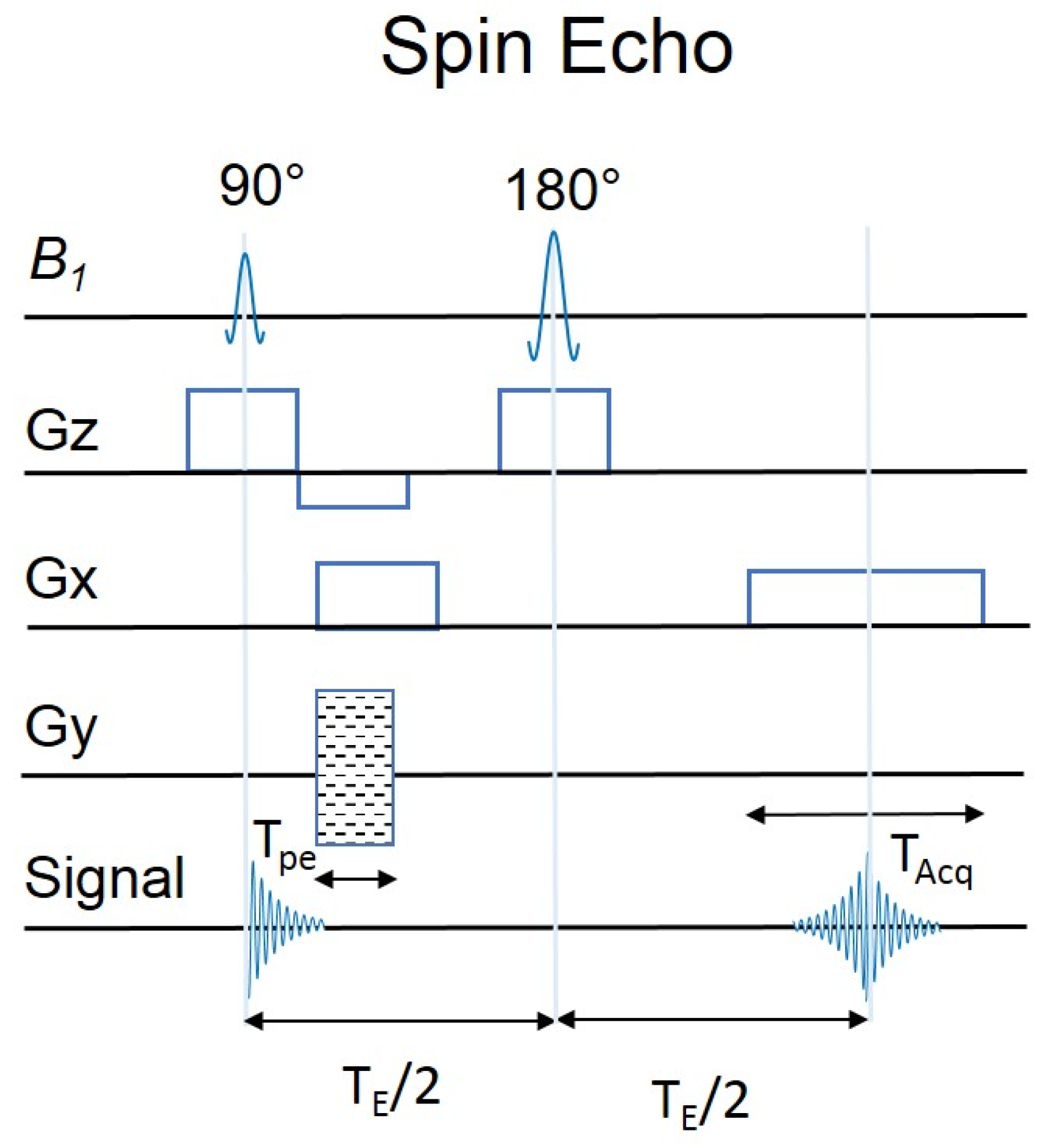
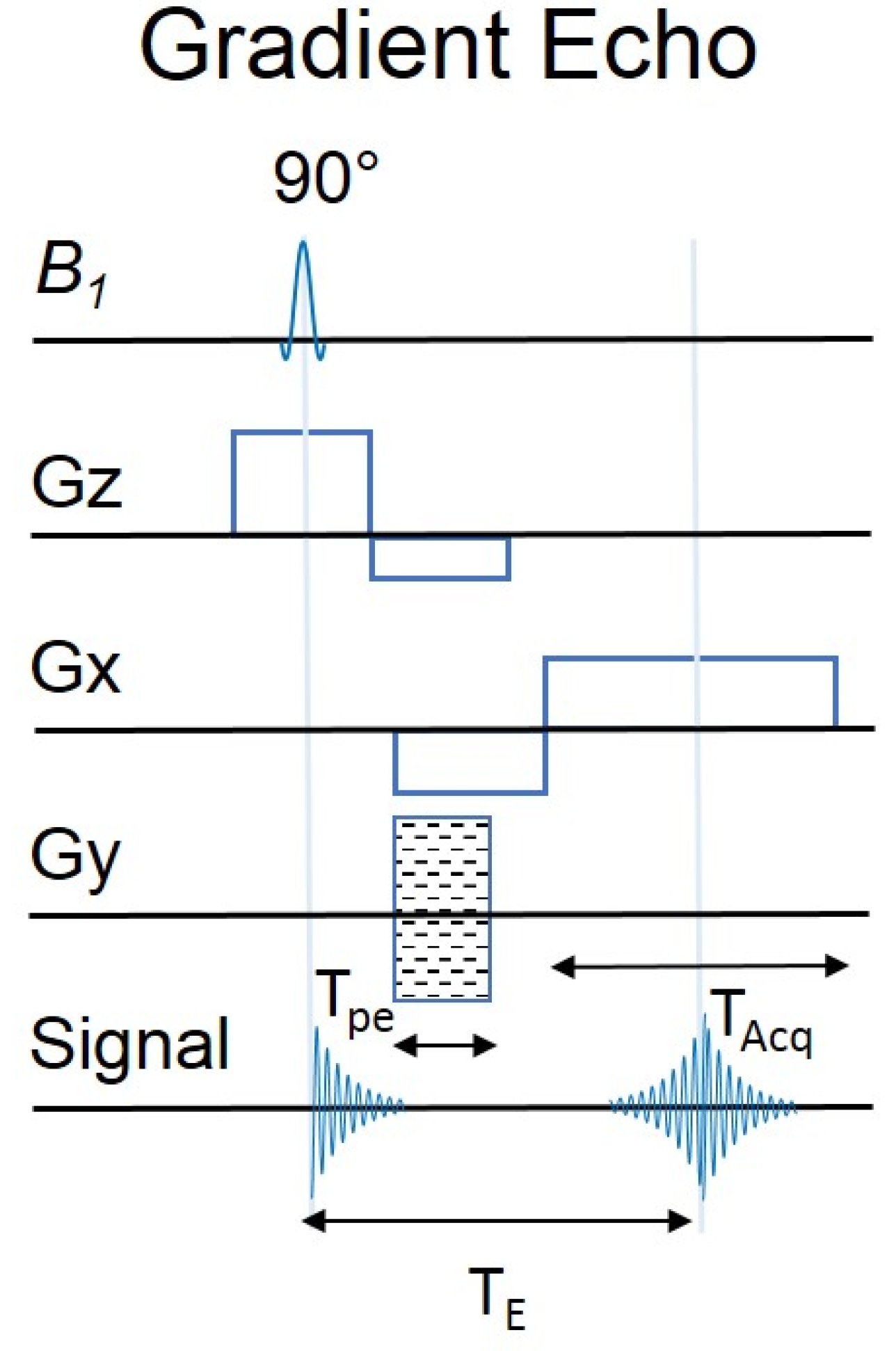
2.1. MRI Sequences
2.2. T1 and T2 MRI Relaxation Times
2.3. Variable MRS Methods
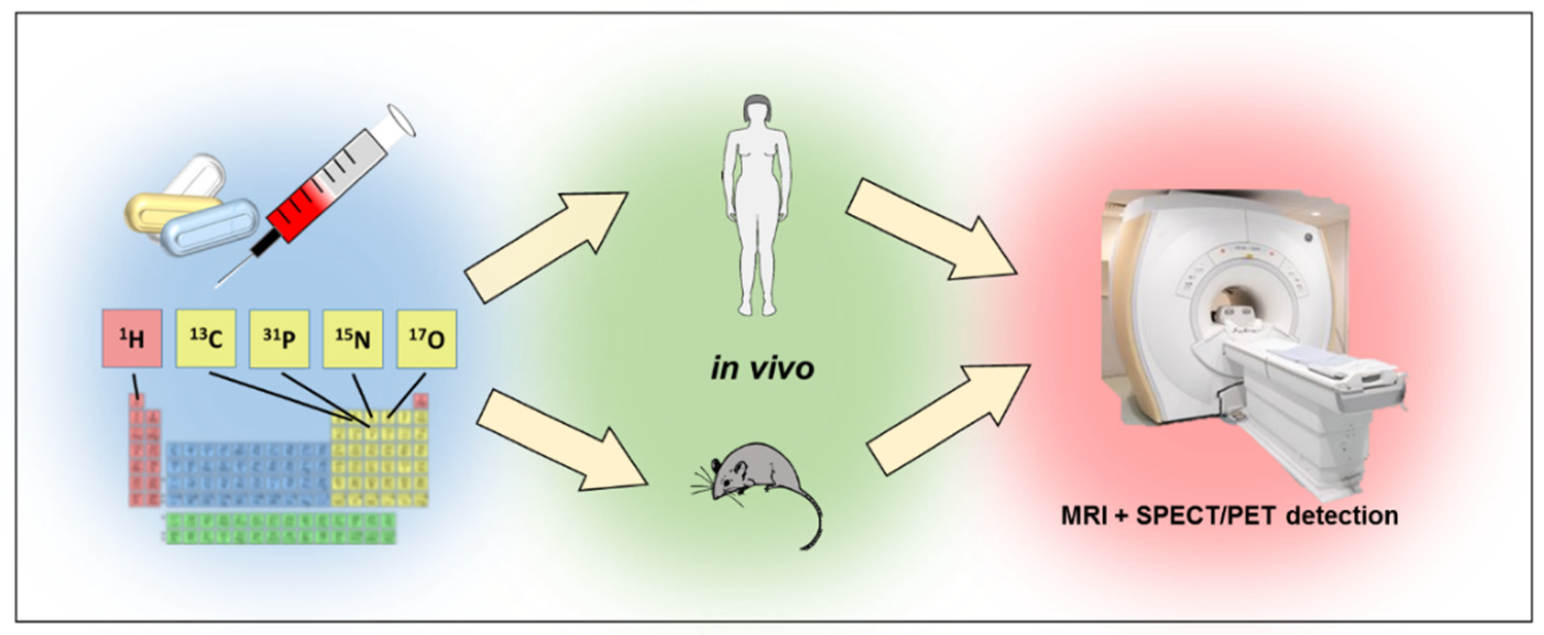
2.4. Multinuclear MRI
2.5. Hyperpolarized MRI/MRS
3. Compound Tracking In Vitro
3.1. 1H MRI
3.2. 19F MRI
3.3. 31P MRI
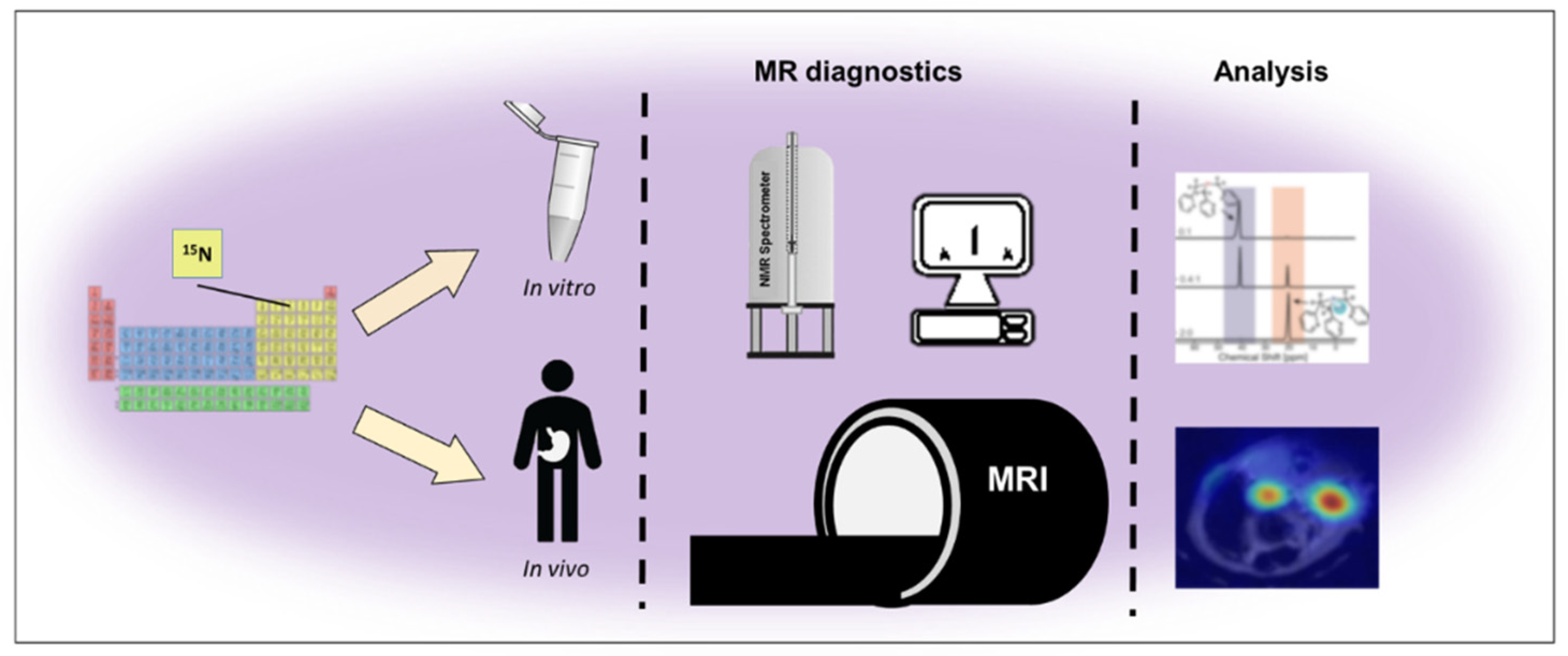
3.4. 15N MRI
4. Tracking In Vivo
4.1. 1H MRI
4.2. 19F MRI
4.3. 13C MRI
4.4. 31P MRI
4.5. 17O MRI
5. 23Na MRI
6. The Future Opportunities of Multinuclear MRI
7. Conclusions
Author Contributions
Funding
Institutional Review Board Statement
Informed Consent Statement
Data Availability Statement
Conflicts of Interest
Sample Availability
References
- McKinney, J.R.; Sussman, M.S.; Moineddin, R.; Amirabadi, A.; Rayner, T.; Doria, A.S. Accuracy of magnetic resonance imaging for measuring maturing cartilage: A phantom study. Clinics 2016, 71, 404–411. [Google Scholar] [CrossRef]
- Sadler, P.J.; Muncie, C.; Shipman, M.A. Biological Inorganic Chemistry: Structure & Reactivity; Bertini, I., Gray, H.B., Stiefel, E.I., Valentine, J.S., Eds.; University Science Books: Mill Valley, CA, USA, 2007; Volume 95. [Google Scholar]
- Korbecki, A.; Zimny, A.; Podgórski, P.; Sąsiadek, M.; Bladowska, J. Imaging of cerebrospinal fluid flow: Fundamentals, techniques, and clinical applications of phase-contrast magnetic resonance imaging. Pol. J. Radiol. 2019, 84, 240–250. [Google Scholar] [CrossRef] [PubMed]
- Arnold, J.R.; McCann, G.P. Cardiovascular magnetic resonance: Applications and practical considerations for the general cardiologist. Heart 2020, 106, 174–181. [Google Scholar] [CrossRef] [PubMed]
- Oudeman, J.; Nederveen, A.J.; Strijkers, G.J.; Maas, M.; Luijten, P.R.; Froeling, M. Techniques and applications of skeletal muscle diffusion tensor imaging: A review. J. Magn. Reson. Imaging 2016, 43, 773–788. [Google Scholar] [CrossRef] [PubMed]
- Mastrogiacomo, S.; Dou, W.; Jansen, J.A.; Walboomers, X.F. Magnetic resonance imaging of hard tissues and hard tissue engineered bio-substitutes. Mol. Imaging Biol. 2019, 21, 1003–1019. [Google Scholar] [CrossRef] [Green Version]
- Sanjari Moghaddam, H.; Dolatshahi, M.; Mohebi, F.; Aarabi, M.H. Structural white matter alterations as compensatory mechanisms in Parkinson’s disease: A systematic review of diffusion tensor imaging studies. J. Neurosci. Res. 2020, 98, 1398–1416. [Google Scholar] [CrossRef]
- Touska, P.; Connor, S.E.J. Recent advances in MRI of the head and neck, skull base and cranial nerves: New and evolving sequences, analyses and clinical applications. Br. J. Radiol. 2019, 92, 20190513. [Google Scholar] [CrossRef]
- Niesporek, S.C.; Nagel, A.M.; Platt, T. Multinuclear MRI at ultrahigh fields. Top. Magn. Reson. Imaging 2019, 28, 173–188. [Google Scholar] [CrossRef]
- Kupče, Ē.; Mote, K.R.; Webb, A.; Madhu, P.K.; Claridge, T.D.W. Multiplexing experiments in NMR and multi-nuclear MRI. Prog. Nucl. Magn. Reson. Spectrosc. 2021, 124–125, 1–56. [Google Scholar] [CrossRef]
- Gilad, A.A.; McMahon, M.T.; Walczak, P.; Winnard, P.T., Jr.; Raman, V.; van Laarhoven, H.W.M.; Skoglund, S.M.; Bulte, J.W.F.; van Zijl, P.C.M. Artificial reporter gene providing MRI contrast based on proton exchange. Nat. Biotechnol. 2007, 25, 217–219. [Google Scholar] [CrossRef]
- Huisman, M.; van den Bosch, M.A. MR-guided high-intensity focused ultrasound for noninvasive cancer treatment. Cancer Imaging 2011, 11, S161–S166. [Google Scholar] [CrossRef] [PubMed] [Green Version]
- Choi, C.H.; Ha, Y.H.; Veeraiah, P.; Felder, J.; Möllenhoff, K.; Shah, N.J. Design and implementation, of a simple multinuclear MRI system for ultra high-field imaging of animals. J. Magn. Reson. 2016, 273, 28–32. [Google Scholar] [CrossRef] [PubMed]
- Bottomley, P.A.; Griffiths, J.R. Handbook of Magnetic Resonance Spectroscopy In Vivo: MRS Theory, Practice and Applications; John Wiley & Sons: Chichester, UK, 2014. [Google Scholar]
- Henkelman, R.M.; Hardy, P.; Poon, P.Y.; Bronskill, M.J. Optimal pulse sequence for imaging hepatic metastases. Radiology 1986, 161, 727–734. [Google Scholar] [CrossRef] [PubMed]
- Bohenzky, R.A.; LeFebvre, R.B.; Berns, K.I. Sequence and symmetry requirements within the internal palindromic sequences of the adeno-associated virus terminal repeat. Virology 1988, 166, 316–327. [Google Scholar] [CrossRef]
- Kjos, B.O.; Ehman, R.L.; Brant-Zawadzki, M. Reproducibility of T1 and T2 relaxation times calculated from routine MR imaging sequences: Phantom study. AJR Am. J. Roentgenol. 1985, 144, 1157–1163. [Google Scholar] [CrossRef] [Green Version]
- Scheffler, K. Fast frequency mapping with balanced SSFP: Theory and application to proton-resonance frequency shift thermometry. Magn. Reson. Med. 2004, 51, 1205–1211. [Google Scholar] [CrossRef]
- Figueroa-Villar, J.D.; Tinoco, L.W. Spin-lattice relaxation time in drug discovery and design. Curr. Top. Med. Chem. 2009, 9, 811–823. [Google Scholar] [CrossRef]
- Bertoldo, D.; Watcharakorn, A.; Castillo, M. Brain proton magnetic resonance spectroscopy. Introduction and overview. Neuroimaging Clin. 2013, 23, 359–380. [Google Scholar] [CrossRef]
- Drake-Pérez, M.; Boto, J.; Fitsiori, A.; Lovblad, K.; Vargas, M.I. Clinical applications of diffusion weighted imaging in neuroradiology. Insights Imaging 2018, 9, 535–547. [Google Scholar] [CrossRef] [Green Version]
- Di Ieva, A.; Lam, T.; Alcaide-Leon, P.; Bharatha, A.; Montanera, W.; Cusimano, M.D. Magnetic resonance susceptibility weighted imaging in neurosurgery: Current applications and future perspectives. J. Neurosurg. 2015, 123, 1463–1475. [Google Scholar] [CrossRef] [Green Version]
- Saliou, G.; Krings, T.; Rutgers, D.R.; Toulgoat, F.; Ozanne, A.; Lasjaunias, P.; Ducreux, D. PWI-MRI and contrast extravasation in brain AVM help to estimate angiogenic activity. Neuroradiology 2011, 53, 793–800. [Google Scholar] [CrossRef] [PubMed]
- Logothetis, N.K. What we can do and what we cannot do with fMRI. Nature 2008, 453, 869–878. [Google Scholar] [CrossRef] [PubMed]
- Arbab, A.S.; Yocum, G.T.; Kalish, H.; Jordan, E.K.; Anderson, S.A.; Khakoo, A.Y.; Read, E.J.; Frank, J.A. Efficient magnetic cell labeling with protamine sulfate complexed to ferumoxides for cellular MRI. Blood 2004, 104, 1217–1223. [Google Scholar] [CrossRef] [PubMed] [Green Version]
- Apps, A.; Lau, J.; Peterzan, M.; Neubauer, S.; Tyler, D.; Rider, O. Hyperpolarised magnetic resonance for in vivo real-time metabolic imaging. Heart 2018, 104, 1484–1491. [Google Scholar] [CrossRef] [PubMed] [Green Version]
- Elkenhans, B.; Protti, A.; Shah, A.; Onthank, D.; Botnar, R. Visualization of elastin using cardiac magnetic resonance imaging after myocardial infarction as inflammatory response. Sci. Rep. 2021, 11, 11004. [Google Scholar] [CrossRef]
- Leaker, B.R.; Scadding, G.; Jones, C.R.; Barnes, P.J. Using magnetic resonance imaging to quantify the inflammatory response following allergen challenge in allergic rhinitis. Immun. Inflamm. Dis. 2015, 3, 445–454. [Google Scholar] [CrossRef] [Green Version]
- Lee, P.; Adany, P.; Choi, I.Y. Imaging based magnetic resonance spectroscopy (MRS) localization for quantitative neurochemical analysis and cerebral metabolism studies. Anal. Biochem. 2017, 529, 40–47. [Google Scholar] [CrossRef]
- Cudalbu, C.; Cooper, A.J.L. Editorial for the special issue on introduction to in vivo magnetic resonance spectroscopy (MRS): A method to non-invasively study metabolism. Anal. Biochem. 2017, 529, 1–3. [Google Scholar] [CrossRef]
- Capati, A.; Ijare, O.B.; Bezabeh, T. Diagnostic applications of nuclear magnetic resonance-based urinary metabolomics. Magn. Reson. Insights 2017, 10, 1178623X17694346. [Google Scholar] [CrossRef] [Green Version]
- Gillinder, L.; Yi Goo, S.; Cowin, G.; Strudwick, M.; van der Geest, R.; Wang, W.; Ng, A. Quantification of intramyocardial metabolites by proton magnetic resonance spectroscopy. Front. Cardiovasc. Med. 2015, 2, 24. [Google Scholar] [CrossRef] [Green Version]
- Mandal, P.; Shukla, D.; Govind, V.; Boulard, Y.; Ersland, L. Glutathione conformations and its implications for in vivo magnetic resonance spectroscopy. J. Alzheimer Dis. 2017, 59, 537–541. [Google Scholar] [CrossRef] [PubMed] [Green Version]
- Zhang, Y.; An, L.; Shen, J. Fast computation of full density matrix of multispin systems for spatially localized in vivo magnetic resonance spectroscopy. Med. Phys. 2017, 44, 4169–4178. [Google Scholar] [CrossRef] [PubMed] [Green Version]
- Pirogov, Y.A. Multinuclear MRI research. Appl. Magn. Reson. 2021, 52, 1695–1705. [Google Scholar] [CrossRef]
- Bober, Z.; Aebisher, D.; Bartusik-Aebisher, D. The use of 19F in Medicine in Poland and in the world. Biointerface Res. Appl. Chem. 2022, 12, 8561–8572. [Google Scholar]
- Bober, Z.; Aebisher, D.; Ożóg, Ł.; Tabarkiewicz, J.; Tutka, P.; Bartusik-Aebisher, D. 19F MRI as a tool for imaging drug delivery to tissue and individual cells. Eur. J. Clin. Exp. Med. 2017, 2, 99–109. [Google Scholar] [CrossRef]
- Bartusik, D.; Aebisher, D. 19F applications in drug development and imaging—A review. Biomed. Pharmacother. 2014, 68, 813–817. [Google Scholar] [CrossRef]
- Ogier, S.E.; Wilcox, M.; Cheshkov, S.; Dimitrov, I.E.; Malloy, C.R.; McDougall, M.P.; Wright, S.M. A Frequency Translation System for Multi-Channel, Multi-Nuclear MR Spectroscopy. IEEE Trans Biomed Eng. 2021, 68, 109–118. [Google Scholar] [CrossRef]
- Lewis, D.; McHugh, D.J.; Li, K.L.; Zhu, X.; Mcbain, C.; Lloyd, S.K.; Jackson, A.; Pathmanaban, O.N.; King, A.T.; Coope, D.J. Detection of early changes in the post-radiosurgery vestibular schwannoma microenvironment using multinuclear MRI. Sci. Rep. 2021, 11, 15712. [Google Scholar] [CrossRef]
- Ianniello, C.; Moy, L.; Fogarty, J.; Schnabel, F.; Adams, S.; Axelrod, D.; Axel, L.; Brown, R.; Madelin, G. Multinuclear MRI to disentangle intracellular sodium concentration and extracellular volume fraction in breast cancer. Sci. Rep. 2021, 11, 5156. [Google Scholar] [CrossRef]
- Golman, K.; Petersson, J.S.; Magnusson, P.; Johansson, E.; Akeson, P.; Chai, C.M.; Hansson, G.; Månsson, S. Cardiac metabolism measured noninvasively by hyperpolarized 13C MRI. Magn. Reson. Med. Off. J. Int. Soc. Magn. Reson. Med. 2008, 59, 1005–1013. [Google Scholar] [CrossRef]
- Nelson, S.J.; Vigneron, D.; Kurhanewicz, J.; Chen, A.; Bok, R.; Hurd, R. DNP-hyperpolarized C magnetic resonance metabolic imaging for cancer applications. Appl. Magn. Reson. 2008, 34, 533–544. [Google Scholar] [CrossRef] [PubMed] [Green Version]
- Schroeder, M.A.; Cochlin, L.E.; Heather, L.C.; Clarke, K.; Radda, G.K.; Tyler, D.J. In vivo assessment of pyruvate dehydrogenase flux in the heart using hyperpolarized carbon-13 magnetic resonance. Proc. Natl. Acad. Sci. USA 2008, 105, 12051–12056. [Google Scholar] [CrossRef] [PubMed] [Green Version]
- Day, S.E.; Kettunen, M.I.; Gallagher, F.A.; Hu, D.E.; Lerche, M.; Wolber, J.; Golman, K.; Ardenkjaer-Larsen, J.H.; Brindle, K.M. Detecting tumor response to treatment using hyperpolarized 13C magnetic resonance imaging and spectroscopy. Nat. Med. 2007, 13, 1382–1387. [Google Scholar] [CrossRef]
- Merritt, M.E.; Harrison, C.; Storey, C.; Sherry, A.D.; Malloy, C.R. Inhibition of carbohydrate oxidation during the first minute of reperfusion after brief ischemia: NMR detection of hyperpolarized 13CO2 and H13CO3−. Magn. Reson. Med. 2008, 60, 1029–1036. [Google Scholar] [CrossRef] [PubMed] [Green Version]
- Hyodo, F.; Eto, H.; Naganuma, T.; Koyasu, N.; Elhelaly, A.E.; Noda, Y.; Kato, H.; Murata, M.; Akahoshi, T.; Hashizume, M.; et al. In vivo dynamic nuclear polarization magnetic resonance imaging for the evaluation of redox-related diseases and theranostics. Antioxid. Redox. Signal. 2022, 36, 172–184. [Google Scholar] [CrossRef]
- Jørgensen, S.H.; Bøgh, N.; Hansen, E.; Væggemose, M.; Wiggers, H.; Laustsen, C. Hyperpolarized MRI—An update and future perspectives. Semin. Nucl. Med. 2022, 52, 374–381. [Google Scholar] [CrossRef]
- Ye, Z.; Song, B.; Lee, P.M.; Ohliger, M.A.; Laustsen, C. Hyperpolarized carbon 13 MRI in liver diseases: Recent advances and future opportunities. Liver Int. 2022, 42, 973–983. [Google Scholar] [CrossRef]
- Wei, Y.; Yang, C.; Jiang, H.; Li, Q.; Che, F.; Wan, S.; Yao, S.; Gao, F.; Zhang, T.; Wang, J.; et al. Multi-nuclear magnetic resonance spectroscopy: State of the art and future directions. Insights Imaging 2022, 13, 135. [Google Scholar] [CrossRef]
- Khan, A.S.; Harvey, R.L.; Birchall, J.R.; Irwin, R.K.; Nikolaou, P.; Schrank, G.; Emami, K.; Dummer, A.; Barlow, M.J.; Goodson, B.M.; et al. Enabling Clinical Technologies for Hyperpolarized 129Xenon Magnetic Resonance Imaging and Spectroscopy. Angew. Chem. Int. Ed. 2021, 60, 22126–22147. [Google Scholar] [CrossRef]
- Hirsch, M.L.; Smith, B.A.; Mattingly, M.; Goloshevsky, A.G.; Rosay, M.; Kempf, J.G. Transport and imaging of brute-force 13C hyperpolarization. J. Magn. Reson. 2015, 261, 87–94. [Google Scholar] [CrossRef]
- Sharma, U.; Jagannathan, N.R. Magnetic resonance imaging (MRI) and MR spectroscopic methods in understanding breast cancer biology and metabolism. Metabolites 2022, 12, 295. [Google Scholar] [CrossRef] [PubMed]
- Esmaeili, M.; Vettukattil, R. In vivo magnetic resonance spectroscopy methods for investigating cardiac metabolism. Metabolites 2022, 12, 189. [Google Scholar] [CrossRef] [PubMed]
- Stewart, N.J.; Sato, T.; Takeda, N.; Hirata, H.; Matsumoto, S. Hyperpolarized 13C magnetic resonance imaging as a tool for imaging tissue redox state, oxidative stress, inflammation, and cellular metabolism. Antioxid. Redox. Signal. 2022, 36, 81–94. [Google Scholar] [CrossRef] [PubMed]
- Pourfathi, M.; Kadlecek, S.J.; Chatterjee, S.; Rizi, R.R. Metabolic imaging and biological assessment: Platforms to evaluate acute lung injury and inflammation. Front. Physiol. 2020, 11, 937. [Google Scholar] [CrossRef] [PubMed]
- Le Page, L.M.; Guglielmetti, C.; Taglang, C.; Chaumeil, M.M. Imaging brain metabolism using hyperpolarized 13C magnetic resonance spectroscopy. Trends Neurosci. 2020, 43, 343–354. [Google Scholar] [CrossRef] [PubMed]
- Grist, J.T.; Miller, J.J.; Zaccagna, F.; McLean, M.A.; Riemer, F.; Matys, T.; Tyler, D.J.; Laustsen, C.; Coles, A.J.; Gallagher, F.A. Hyperpolarized 13C MRI: A novel approach for probing cerebral metabolism in health and neurological disease. J. Cereb. Blood Flow Metab. 2020, 40, 1137–1147. [Google Scholar] [CrossRef]
- Seelen, L.W.F.; van den Wildenberg, L.; van der Kemp, W.J.M.; Mohamed Hoesein, F.A.A.; Mohammad, N.H.; Molenaar, I.Q.; van Santvoort, H.C.; Prompers, J.J.; Klomp, D.W.J. Prospective of 31P MR spectroscopy in hepatopancreatobiliary cancer: A systematic review of the literature. J. Magn. Reson. Imaging 2022. [Google Scholar] [CrossRef]
- Giesel, F.L.; Stroick, M.; Griebe, M.; Tröster, H.; von der Lieth, C.W.; Requardt, M.; Rius, M.; Essig, M.; Kauczor, H.U.; Hennerici, M.G.; et al. Gadofluorine m uptake in stem cells as a new magnetic resonance imaging tracking method: An in vitro and in vivo study. Investig. Radiol. 2006, 41, 868–873. [Google Scholar] [CrossRef]
- Tang, K.X.; Shen, Y.F.; Yang, X.B.; Gu, H.M.; Duan, H.J.; Yan, Z.X.; Weng, J.P. A study of tracking the superparamagnetic iron oxide and enhanced green fluorescent protein labeled miniature porcine bone marrow stem cells by in vitro MRI. Zhonghua Nei Ke Za Zhi 2011, 50, 322–327. [Google Scholar]
- Feng, Y.; Jin, X.; Dai, G.; Liu, J.; Chen, J.; Yang, L. In vitro targeted magnetic delivery and tracking of superparamagnetic iron oxide particles labeled stem cells for articular cartilage defect repair. J. Huazhong Univ. Sci. Technol. Med. Sci. 2011, 31, 204–209. [Google Scholar] [CrossRef]
- Addicott, B.; Willman, M.; Rodriguez, J.; Padgett, K.; Han, D.; Berman, D.; Hare, J.M.; Kenyon, N.S. Mesenchymal stem cell labeling and in vitro MR characterization at 1.5 T of new SPIO contrast agent: Molday ION Rhodamine-B™. Contrast Media Mol. Imaging 2011, 6, 7–18. [Google Scholar] [CrossRef] [PubMed] [Green Version]
- Freichels, H.; Danhier, F.; Préat, V.; Lecomte, P.; Jérôme, C. Fluorescent labeling of degradable poly (lactide-co-glycolide) for cellular nanoparticles tracking in living cells. Int. J. Artif. Organs 2011, 34, 152–160. [Google Scholar] [CrossRef] [PubMed]
- Henning, T.D.; Gawande, R.; Khurana, A.; Tavri, S.; Mandrussow, L.; Golovko, D.; Horvai, A.; Sennino, B.; McDonald, D.; Meier, R.; et al. Magnetic resonance imaging of ferumoxide-labeled mesenchymal stem cells in cartilage defects: In vitro and in vivo investigations. Mol. Imaging 2012, 11, 197–209. [Google Scholar] [CrossRef] [PubMed]
- Lu, X.; Nie, Y.; Zhao, Z.; He, X.; Liu, Y.; Pulati, T.; Wu, J. SPIO-labeled rat bone marrow mesenchymal stem cells: Alterations of biological activity and labeling efficiency assay in vitro. Sheng Wu Yi Xue Gong Cheng Xue Za Zhi J. Biomed. Eng. Shengwu Yixue Gongchengxue Zazhi 2014, 31, 365–372. [Google Scholar]
- Zhang, R.; Li, J.; Li, J.; Xie, J. Efficient In vitro labeling rabbit bone marrow-derived mesenchymal stem cells with SPIO and differentiating into neural-like cells. Mol. Cells 2014, 37, 650–655. [Google Scholar] [CrossRef]
- Shuai, H.L.; Yan, R.L.; Song, H.; Chen, D.L.; Luo, X. Analysis of feasibility of in vitro nuclear magnetic resonance tracking human umbilical cord mesenchymal stem cells by Gd-DTPA labeled. Magn. Reson. Imaging 2014, 32, 934–940. [Google Scholar] [CrossRef]
- Tang, K.X.; Yan, J.H.; Shen, Y.F.; Li, B.Y.; Chen, Y.M.; Liu, D.Y.; Ma, D.D.; Li, J.; Liang, H.; Weng, J.P. Tracing type 1 diabetic Tibet miniature pig’s bone marrow mesenchymal stem cells in vitro by magnetic resonance imaging (1). J. Diabetes 2014, 6, 123–131. [Google Scholar] [CrossRef]
- Li, Y.Q.; Tang, Y.; Fu, R.; Meng, Q.H.; Zhou, X.; Ling, Z.M.; Cheng, X.; Tian, S.W.; Wang, G.J.; Liu, X.G.; et al. Efficient labeling in vitro with non-ionic gadolinium magnetic resonance imaging contrast agent and fluorescent transfection agent in bone marrow stromal cells of neonatal rats. Mol. Med. Rep. 2015, 12, 913–920. [Google Scholar] [CrossRef] [Green Version]
- Geng, K.; Yang, Z.X.; Huang, D.; Yi, M.; Jia, Y.; Yan, G.; Cheng, X.; Wu, R. Tracking of mesenchymal stem cells labeled with gadolinium diethylenetriamine pentaacetic acid by 7T magnetic resonance imaging in a model of cerebral ischemia. Mol. Med. Rep. 2015, 11, 954–960. [Google Scholar] [CrossRef] [Green Version]
- Liu, S.; Tay, L.M.; Anggara, R.; Chuah, Y.J.; Kang, Y. Long-term tracking mesenchymal stem cell differentiation with photostable fluorescent nanoparticles. ACS Appl. Mater. Interfaces 2016, 8, 11925–11933. [Google Scholar] [CrossRef]
- Zhang, M.; Liu, X.; Huang, J.; Wang, L.; Shen, H.; Luo, Y.; Li, Z.; Zhang, H.; Deng, Z.; Zhang, Z. Ultrasmall graphene oxide based T1 MRI contrast agent for in vitro and in vivo labeling of human mesenchymal stem cells. Nanomedicine 2018, 14, 2475–2483. [Google Scholar] [CrossRef] [PubMed]
- Lu, M.; Cheng, X.; Jiang, J.; Li, T.; Zhang, Z.; Tsauo, C.; Liu, Y.; Wang, Z. Dual-modal photoacoustic and magnetic resonance tracking of tendon stem cells with PLGA/iron oxide microparticles in vitro. PLoS ONE 2018, 13, e0193362. [Google Scholar] [CrossRef] [PubMed]
- Mathiasen, A.B.; Qayyum, A.A.; Jørgensen, E.; Helqvist, S.; Ekblond, A.; Ng, M.; Bhakoo, K.; Kastrup, J. In vivo MRI tracking of mesenchymal stromal cells labeled with ultrasmall paramagnetic iron oxide particles after intramyocardial transplantation in patients with chronic ischemic heart disease. Stem Cells Int. 2019, 2019, 2754927. [Google Scholar] [CrossRef] [PubMed] [Green Version]
- de Mello Costa, M.F.; Weiner, A.I.; Vaughan, A.E. Basal-like progenitor cells: A review of dysplastic alveolar regeneration and remodeling in lung repair. Stem Cell Rep. 2020, 15, 1015–1025. [Google Scholar] [CrossRef] [PubMed]
- Kim, S.H.; Djaja, Y.P.; Park, Y.B.; Park, J.G.; Ko, Y.B.; Ha, C.W. Intra-articular injection of culture-expanded mesenchymal stem cells without adjuvant surgery in knee osteoarthritis: A systematic review and meta-analysis. Am. J. Sport Med. 2019, 48, 2839–2849. [Google Scholar] [CrossRef]
- Hu, L.; Pan, H.; Wickline, S.A. Fluorine (19F) MRI to measure renal oxygen tension and blood volume: Experimental Protocol. In Preclinical MRI of the Kidney; Pohlmann, A., Niendorf, T., Eds.; Humana: New York, NY, USA, 2021; p. 2216. [Google Scholar]
- Herynek, V.; Martinisková, M.; Bobrova, Y.; Gálisová, A.; Kotek, J.; Hermann, P.; Koucký, F.; Jirák, D.; Hájek, M. Low-molecular-weight paramagnetic 19F contrast agents for fluorine magnetic resonance imaging. Magn. Reson. Mater. Phys. Biol. Med. 2019, 32, 115–122. [Google Scholar] [CrossRef] [Green Version]
- Zare, S.; Mehrabani, D.; Jalli, R.; Saeedi Moghadam, M.; Manafi, N.; Mehrabani, G.; Jamhiri, I.; Ahadian, S. MRI-tracking of dental pulp stem cells in vitro and in vivo using dextran-coated superparamagnetic iron oxide nanoparticles. J. Clin. Med. 2019, 8, 1418. [Google Scholar] [CrossRef] [Green Version]
- Chirizzi, C.; De Battista, D.; Tirotta, I.; Metrangolo, P.; Comi, G.; Bombelli, F.B.; Chaabane, L. Multispectral MRI with dual fluorinated probes to track mononuclear cell activity in mice. Radiology 2019, 291, 351–357. [Google Scholar] [CrossRef]
- Wang, G.; Fu, Y.; Shea, S.M.; Hegde, S.S.; Kraitchman, D.L. Quantitative CT and 19F-MRI tracking of perfluorinated encapsulated mesenchymal stem cells to assess graft immunorejection. Magn. Reson. Mater. Phys. Biol. Med. 2019, 32, 147–156. [Google Scholar] [CrossRef]
- Richard, J.P.; Hussain, U.; Grossm, S.; Tagam, A.; Kouser, M.; Almad, A.; Campanelli, J.T.; Bulte, J.W.M.; Maragakis, N.J. Perfluorocarbon labeling of human glial-restricted progenitors for 19F magnetic resonance imaging. Stem Cells Transl. Med. 2019, 8, 355–365. [Google Scholar] [CrossRef]
- Szczęch, M.; Łopuszyńska, N.; Tomal, W.; Jasiński, K.; Węglarz, W.P.; Warszyński, P.; Szczepanowicz, K. Nafion-based nanocarriers for fluorine magnetic resonance imaging. Langmuir 2020, 36, 9534–9539. [Google Scholar] [CrossRef] [PubMed]
- Wyszogrodzka, G.; Dorożyński, P.; Gil, B.; Roth, W.J.; Strzempek, M.; Marszałek, B.; Węglarz, W.P.; Menaszek, E.; Strzempek, W.; Kulinowski, P. Iron-based metal-organic frameworks as a theranostic carrier for local tuberculosis therapy. Pharm. Res. 2018, 35, 144. [Google Scholar] [CrossRef] [PubMed] [Green Version]
- Wyszogrodzka-Gaweł, G.; Dorożyński, P.; Giovagnoli, S.; Strzempek, W.; Pesta, E.; Węglarz, W.P.; Gil, B.; Menaszek, E.; Kulinowski, P. An inhalable theranostic system for local tuberculosis treatment containing an isoniazid loaded metal organic framework Fe-MIL-101-NH2—From raw MOF to drug delivery system. Pharmaceutics 2019, 11, 687. [Google Scholar] [CrossRef] [Green Version]
- Maximenko, A.; Depciuch, J.; Łopuszyńska, N.; Stec, M.; Światkowska-Warkocka, Ż.; Bayev, V.; Zieliński, P.M.; Baran, J.; Fedotova, J.; Węglarz, W.P.; et al. Fe3O4@SiO2@Au nanoparticles for MRI-guided chemo/NIR photothermal therapy of cancer cells. RSC Adv. 2020, 10, 26508–26520. [Google Scholar] [CrossRef] [PubMed]
- Robinson, S.P.; van den Boogaart, A.; Maxwell, R.J.; Griffiths, J.R.; Hamilton, E.; Waterton, J.C. 31P-magnetic resonance spectroscopy and 2H-magnetic resonance imaging studies of a panel of early-generation transplanted murine tumour models. Br. J. Cancer 1998, 77, 1752–1760. [Google Scholar] [CrossRef] [PubMed] [Green Version]
- Ouwerkerk, R.; Bottomley, P.A. On neglecting chemical exchange effects when correcting in vivo 31P MRS data for partial saturation. J. Magn. Reson. 2001, 148, 425–435. [Google Scholar] [CrossRef] [PubMed] [Green Version]
- Martino, R.; Gilard, V.; Desmoulin, F.; Malet-Martino, M. Fluorine-19 or phosphorus-31 NMR spectroscopy: A suitable analytical technique for quantitative in vitro metabolic studies of fluorinated or phosphorylated drugs. J. Pharm. Biomed. Anal. 2005, 10, 871–891. [Google Scholar] [CrossRef]
- Landis, C.S.; Yamanouchi, K.; Zhou, H.; Roy-Chowdhury, J.; Hetherington, H.P.; Guha, C. Non-invasive tracking of transplanted hepatocytes in irradiated livers by 31P MRSI. Radiat. Oncol. 2006, 66, 19–20. [Google Scholar]
- Zhang, Z.; Hancock, B.; Leen, S.; Ramaswamy, S.; Sollott, S.J.; Boheler, K.R.; Juhaszova, M.; Lakatta, E.G.; Spencer, R.G.; Fishbein, K.W. Compatibility of superparamagnetic iron oxide nanoparticle labeling for ¹H MRI cell tracking with ³¹P MRS for bioenergetic measurements. NMR Biomed. 2010, 23, 1166–1172. [Google Scholar] [CrossRef] [Green Version]
- Cameron, D.; Welch, A.A.; Adelnia, F.; Bergeron, C.M.; Reiter, D.A.; Dominguez, L.J.; Brennan, N.A.; Fishbein, K.W.; Spencer, R.G.; Ferrucci, L. Age and muscle function are more closely associated with intracellular magnesium, as assessed by 31P Magnetic resonance spectroscopy, than with serum magnesium. Front. Physiol. 2019, 10, 1454. [Google Scholar] [CrossRef]
- Gabellier, C.; Reynolds, S.; Lavie, A.; Payne, G.S.; Leach, M.O.; Eykyn, T.R. Therapeutic Target Metabolism Observed Using Hyperpolarized 15N Choline. Am. Chem. Soc. 2008, 130, 4598–7599. [Google Scholar] [CrossRef] [PubMed]
- Chiavazza, E.; Viale, A.; Karlsson, M.; Aime, S. 15N-permethylated amino acids as efficient probes for MRI-DNP applications. Contrast Media Mol. Imaging 2013, 8, 417–421. [Google Scholar] [CrossRef] [PubMed]
- Jagtap, A.P.; Kaltschnee, L.; Glöggler, S. Hyperpolarization of 15N-pyridinium and 15N-aniline derivatives by using parahydrogen: New opportunities to store nuclear spin polarization in aqueous media. Chem. Sci. 2019, 10, 8577–8582. [Google Scholar] [CrossRef] [PubMed] [Green Version]
- Cao, J.; Wang, Y.N.; Shi, X.L.; Ma, G.T.; Kong, L.Y.; Xue, H.D.; Lei, J.; He, Y.L.; Jin, Z.Y. In vivo and in vitro imaging tracing of dual-labeled bone mesenchymal stem cells transplanted into myocardium of F344 rats. Zhongguo Yi Xue Ke Xue Yuan Xue Bao 2012, 34, 474–479. [Google Scholar] [PubMed]
- Xu, Q.; Zhang, H.T.; Liu, K.; Rao, J.H.; Liu, X.M.; Wu, L.; Xu, B.N. in vitro and in vivo magnetic resonance tracking of sinerem-labeled human umbilical mesenchymal stromal cell-derived schwann cells. Cell Mol. Neurobiol. 2011, 31, 365–375. [Google Scholar] [CrossRef]
- Agudelo, C.A.; Tachibana, Y.; Noboru, T.; Iida, H.; Yamaoka, T. Long-term in vivo magnetic resonance imaging tracking of endothelial progenitor cells transplanted in rat ischemic limbs and their angiogenic potential. Tissue Eng. Part A 2011, 17, 2079–2089. [Google Scholar] [CrossRef]
- Laughney, A.M.; Kim, E.; Sprachman, M.M.; Sprachman, M.M.; Miller, M.A.; Kohler, R.H.; Yang, K.S.; Orth, J.D.; Mitchison, T.J.; Weissleder, R. Single-cell pharmacokinetic imaging reveals a therapeutic strategy to overcome drug resistance to the microtubule inhibitor eribulin. Sci. Transl. Med. 2014, 6, 261ra152. [Google Scholar] [CrossRef] [Green Version]
- Constantinides, C.; Basnett, P.; Lukasiewicz, B.; Carnicer, R.; Swider, E.; Majid, Q.A.; Srinivas, M.; Carr, C.A.; Roy, I. In vivo tracking and 1H/19F magnetic resonance imaging of biodegradable polyhydroxyalkanoate/polycaprolactone blend scaffolds seeded with labeled cardiac stem cells. ACS Appl. Mater. Interfaces 2018, 10, 25056–25068. [Google Scholar] [CrossRef] [Green Version]
- Shahror, R.A.; Wu, C.C.; Chiang, Y.H.; Chen, K.Y. Tracking superparamagnetic iron oxide-labeled mesenchymal stem cells using MRI after intranasal delivery in a traumatic brain injury murine model. J. Vis. Exp. 2019, 153, e60450. [Google Scholar] [CrossRef] [Green Version]
- Bardhan, R.; Chen, W.; Bartels, M.; Perez-Torres, C.; Botero, M.F.; McAninch, R.W.; Contreras, A.; Schiff, R.; Pautler, R.G.; Halas, N.J.; et al. Tracking of multimodal therapeutic nanocomplexes targeting breast cancer in vivo. Nano Lett. 2010, 10, 4920–4928. [Google Scholar] [CrossRef] [Green Version]
- Shan, L. Imaging and Contrast Agent Database (MICAD) [Internet]; National Center for Biotechnology Information: Bethesda, MD, USA, 2009; pp. 2004–2013. [Google Scholar]
- Al Faraj, A.; Shaik, A.S.; Al Sayed, B. Preferential magnetic targeting of carbon nanotubes to cancer sites: Noninvasive tracking using MRI in a murine breast cancer model. Nanomedicine 2015, 10, 931–948. [Google Scholar] [CrossRef] [PubMed]
- Danhier, P.; Magat, J.; Levêque, P.; De Preter, G.; Porporato, P.E.; Bouzin, C.; Jordan, B.F.; Demeur, G.; Haufroid, V.; Feron, O.; et al. In vivo visualization and ex vivo quantification of murine breast cancer cells in the mouse brain using MRI cell tracking and electron paramagnetic resonance. NMR Biomed. 2015, 28, 367–375. [Google Scholar] [CrossRef] [PubMed]
- Hong, W.; Lee, S.; Chang, H.J.; Lee, E.S.; Cho, Y. Multifunctional magnetic nanowires: A novel breakthrough for ultrasensitive detection and isolation of rare cancer cells from non-metastatic early breast cancer patients using small volumes of blood. Biomaterials 2016, 106, 78–86. [Google Scholar] [CrossRef] [PubMed]
- Makela, A.V.; Gaudet, J.M.; Foster, P.J. Quantifying tumor associated macrophages in breast cancer: A comparison of iron and fluorine-based MRI cell tracking. Sci. Rep. 2017, 7, 42109. [Google Scholar] [CrossRef] [Green Version]
- Rammohan, N.; Holbrook, R.J.; Rotz, M.W.; MacRenaris, K.W.; Preslar, A.T.; Carney, C.E.; Reichova, V.; Meade, T.J. Gd (III)-gold nanoconjugates provide remarkable cell labeling for high field magnetic resonance imaging. Bioconjugate Chem. 2017, 28, 153–160. [Google Scholar] [CrossRef] [Green Version]
- Murrell, D.H.; Zarghami, N.; Jensen, M.D.; Dickson, F.; Chambers, A.F.; Wong, E.; Foster, P.J. MRI surveillance of cancer cell fate in a brain metastasis model after early radiotherapy. Magn. Reson. Med. 2017, 78, 1506–1512. [Google Scholar] [CrossRef] [PubMed]
- Brewer, K.D.; Spitler, R.; Lee, K.R.; Chan, A.C.; Barrozo, J.C.; Wakeel, A.; Foote, C.S.; Machtaler, S.; Rioux, J.; Willmann, J.K.; et al. Characterization of magneto-endosymbionts as MRI cell labeling and tracking agents. Mol. Imaging Biol. 2018, 20, 65–73. [Google Scholar] [CrossRef] [Green Version]
- Martínez-Banderas, A.I.; Aires, A.; Plaza-García, S.; Colás, L.; Moreno, J.A.; Ravasi, T.; Merzaban, J.S.; Ramos-Cabrer, P.; Cortajarena, A.L.; Kosel, J. Magnetic core-shell nanowires as MRI contrast agents for cell tracking. J. Nanobiotechnol. 2020, 18, 42. [Google Scholar] [CrossRef] [Green Version]
- Ramm, P.; Bettscheider, M.; Beier, D.; Kalbitzer, H.R.; Kremer, W.; Bogdahn, U.; Hau, P.; Aigner, L.; Beier, C.P. 1H-nuclear magnetic resonance spectroscopy of glioblastoma cancer stem cells. Stem Cells Dev. 2011, 20, 2189–2195. [Google Scholar] [CrossRef]
- Chen, H.Z.; Guo, Y.K.; Li, Z.L.; Xia, R.; Zhang, L.Z.; Hou, J.L.; Gao, F.B.; Ai, H.; Ning, G. MR imaging of polyethylenimine-superparamagnetic iron oxide nanoparticle labeled bone marrow mesenchymal stem cells in vitro. Sichuan Da Xue Xue Bao Yi Xue Ban 2012, 43, 578–583. [Google Scholar]
- Xu, C.; Miranda-Nieves, D.; Ankrum, J.A.; Matthiesen, M.E.; Phillips, J.A.; Roes, I.; Wojtkiewicz, G.R.; Juneja, V.; Kultima, J.R.; Zhao, W.; et al. Tracking mesenchymal stem cells with iron oxide nanoparticle loaded poly (lactide-co-glycolide) microparticles. Nano Lett. 2012, 12, 4131–4139. [Google Scholar] [CrossRef] [PubMed] [Green Version]
- Herea, D.D.; Labusca, L.; Radu, E.; Chiriac, H.; Grigoras, M.; Panzaru, O.D.; Lupu, N. Human adipose-derived stem cells loaded with drug-coated magnetic nanoparticles for in-vitro tumor cells targeting. Mater. Sci. Eng. C 2019, 94, 666–676. [Google Scholar] [CrossRef] [PubMed]
- Struys, T.; Ketkar-Atre, A.; Gervois, P.; Leten, C.; Hilkens, P.; Martens, W.; Bronckaers, A.; Dresselaers, T.; Politis, C.; Lambrichts, I.; et al. Magnetic resonance imaging of human dental pulp stem cells in vitro and in vivo. Cell Transpl. 2013, 22, 1813–1829. [Google Scholar] [CrossRef] [PubMed] [Green Version]
- Ferrauto, G.; Castelli, D.D.; Terreno, E.; Aime, S. In vivo MRI visualization of different cell populations labeled with PARACEST agents. Magn. Reson. Med. 2013, 69, 1703–1711. [Google Scholar] [CrossRef] [PubMed]
- Charbe, N.B.; Castillo, F.; Tambuwala, M.M.; Prasher, P.; Chellappan, D.K.; Carreño, A.; Satija, S.; Singh, S.K.; Gulati, M.; Dua, K.; et al. A new era in oxygen therapeutics? From perfluorocarbon systems to haemoglobin-based oxygen carriers. Blood Rev. 2022, 54, 100927. [Google Scholar] [CrossRef]
- Spiess, B.D. Oxygen therapeutic agents to target hypoxia in cancer treatment. Curr. Opin. Pharmacol. 2020, 53, 146–151. [Google Scholar] [CrossRef]
- Krafft, M.P. Alleviating tumor hypoxia with perfluorocarbon-based oxygen carriers. Curr. Opin. Pharmacol. 2020, 53, 117–125. [Google Scholar] [CrossRef]
- Guo, R.; Xu, N.; Liu, Y.; Ling, G.; Yu, J.; Zhang, P. Functional ultrasound-triggered phase-shift perfluorocarbon nanodroplets for cancer therapy. Ultrasound Med. Biol. 2021, 47, 2064–2079. [Google Scholar] [CrossRef]
- Constantinides, C.; Maguire, M.; McNeill, E.; Carnicer, R.; Swider, E.; Srinivas, M.; Carr, C.A.; Schneider, J.E. Fast, quantitative, murine cardiac 19F MRI/MRS of PFCE-labeled progenitor stem cells and macrophages at 9.4T. PLoS ONE 2018, 13, e0190558. [Google Scholar] [CrossRef]
- Jahromi, A.H.; Wang, C.; Adams, S.R.; Zhu, W.; Narsinh, K.; Xu, H.; Gray, D.L.; Tsien, R.Y.; Ahrens, E.T. Fluorous-soluble metal chelate for sensitive fluorine-19 magnetic resonance imaging nanoemulsion probes. ACS Nano 2019, 13, 143–151. [Google Scholar] [CrossRef]
- Kislukhin, A.A.; Xu, H.; Adams, S.R.; Narsinh, K.H.; Tsien, R.Y.; Ahrens, E.T. Paramagnetic fluorinated nanoemulsions for sensitive cellular fluorine-19 magnetic resonance imaging. Nat. Mater. 2016, 15, 662–668. [Google Scholar] [CrossRef] [PubMed]
- Chapelin, F.; Capitini, C.M.; Ahrens, E.T. Fluorine-19 MRI for detection and quantification of immune cell therapy for cancer. J. Immunother. Cancer 2018, 6, 105. [Google Scholar] [CrossRef] [PubMed]
- Makela, A.V.; Foster, P.J. Imaging macrophage distribution and density in mammary tumors and lung metastases using fluorine-19 MRI cell tracking. Magn. Reson. Med. 2018, 80, 1138–1147. [Google Scholar] [CrossRef] [PubMed]
- Czyzynska-Cichon, I.; Janik-Hazuka, M.; Szafraniec-Szczęsny, J.; Jasinski, K.; Węglarz, W.P.; Zapotoczny, S.; Chlopicki, S. Low dose curcumin administered in hyaluronic acid-based nanocapsules induces hypotensive effect in hypertensive rats. Int. J. Nanomed. 2021, 16, 1377–1390. [Google Scholar] [CrossRef]
- Johansson, E.; Månsson, S.; Wirestam, R.; Svensson, J.; Petersson, J.S.; Golman, K.; Ståhlberg, F. Cerebral perfusion assessment by bolus tracking using hyperpolarized 13C. Magn. Reson. Med. Off. J. Int. Soc. Magn. Reson. Med. 2004, 51, 464–472. [Google Scholar] [CrossRef]
- Magnusson, P.; Johansson, E.; Månsson, S.; Petersson, J.S.; Chai, C.M.; Hansson, G.; Axelsson, O.; Golman, K. Passive catheter tracking during interventional MRI using hyperpolarized 13C. Magn. Reson. Med. 2007, 57, 1140–1147. [Google Scholar] [CrossRef]
- Albers, M.J.; Bok, R.; Chen, A.P.; Cunningham, C.H.; Zierhut, M.L.; Zhang, V.Y.; Kohler, S.J.; Tropp, J.; Hurd, R.E.; Yen, Y.F.; et al. Hyperpolarized 13C lactate, pyruvate, and alanine: Noninvasive biomarkers for prostate cancer detection and grading. Cancer Res. 2008, 15, 8607–8615. [Google Scholar] [CrossRef] [Green Version]
- Dafni, H.; Larson, P.E.; Hu, S.; Yoshihara, H.A.; Ward, C.S.; Venkatesh, H.S.; Wang, C.; Zhang, X.; Vigneron, D.B.; Ronen, S.M. Hyperpolarized 13C spectroscopic imaging informs on hypoxia-inducible factor-1 and Myc activity downstream of platelet-derived growth factor receptor. Cancer Res. 2010, 70, 7400–7410. [Google Scholar] [CrossRef] [Green Version]
- Lupo, J.M.; Chen, A.P.; Zierhut, M.L.; Bok, R.A.; Cunningham, C.H.; Kurhanewicz, J.; Vigneron, D.B.; Nelson, S.J. Analysis of hyperpolarized dynamic 13C lactate imaging in a transgenic mouse model of prostate cancer. Magn. Reson. Imaging 2010, 8, 153–162. [Google Scholar] [CrossRef] [Green Version]
- Marjańska, M.; Iltis, I.; Shestov, A.A.; Deelchand, D.K.; Nelson, C.; Uğurbil, K.; Henrya, P.G. In vivo 13C spectroscopy in the rat brain using hyperpolarized [1-13C] pyruvate and [2-13C] pyruvate. J. Magn. Reson. 2010, 206, 210–218. [Google Scholar] [CrossRef] [Green Version]
- Bhattacharya, P.; Chekmenev, E.Y.; Reynolds, W.F.; Wagner, S.; Zacharias, N.; Chan, H.R.; Bünger, R.; Ross, B.D. Parahydrogen-induced polarization (PHIP) hyperpolarized MR receptor imaging in vivo: A pilot study of 13C imaging of atheroma in mice. NMR Biomed. 2011, 24, 1023–1028. [Google Scholar] [CrossRef] [PubMed] [Green Version]
- Hu, S.; Balakrishnan, A.; Bok, R.A.; Anderton, B.; Larson, P.E.; Nelson, S.J.; Kurhanewicz, J.; Vigneron, D.B.; Goga, A. 13C-pyruvate imaging reveals alterations in glycolysis that precede c-Myc-induced tumor formation and regression. Cell Metab. 2011, 6, 131–142. [Google Scholar] [CrossRef] [PubMed] [Green Version]
- Hu, S.; Zhu, M.; Yoshihara, H.A.; Wilson, D.M.; Keshari, K.R.; Shin, P.; Reed, G.; von Morze, C.; Bok, R.; Larson, P.E.; et al. In vivo measurement of normal rat intracellular pyruvate and lactate levels after injection of hyperpolarized [1-13C] alanine. Magn. Reson. Imaging. 2011, 29, 1035–1040. [Google Scholar] [CrossRef] [PubMed] [Green Version]
- Bohndiek, S.E.; Kettunen, M.I.; Hu, D.E.; Kennedy, B.W.; Boren, J.; Gallagher, F.A.; Brindle, K.M. Hyperpolarized [1-13C]-ascorbic and dehydroascorbic acid: Vitamin C as a probe for imaging redox status in vivo. J. Am. Chem. Soc. 2011, 130, 11795–11801. [Google Scholar] [CrossRef]
- Chaumeil, M.M.; Ozawa, T.; Park, I.W.; Scott, K.; James, C.; Nelson, S.J.; Ronen, S.M. Hyperpolarized 13C MR spectroscopic imaging can be used to monitor everolimus treatment in vivo in an orthotopic rodent model of glioblastoma. Neuroimage 2012, 2, 193–201. [Google Scholar] [CrossRef] [PubMed] [Green Version]
- Keshari, K.R.; Sriram, R.; Van Criekinge, M.; Wilson, D.M.; Wang, Z.J.; Vigneron, D.B.; Peehl, D.M.; Kurhanewicz, J. Metabolic reprogramming and validation of hyperpolarized 13C lactate as a prostate cancer biomarker using a human prostate tissue slice culture bioreactor. Prostate 2013, 73, 1171–1181. [Google Scholar] [CrossRef] [PubMed] [Green Version]
- Chen, A.P.; Chu, W.; Gu, Y.P.; Cunningham, C.H. Probing early tumor response to radiation therapy using hyperpolarized [1-13C] pyruvate in MDA-MB-231 xenografts. PLoS ONE 2013, 8, e56551. [Google Scholar]
- Schroeder, M.A.; Lau, A.Z.; Chen, A.P.; Gu, Y.; Nagendran, J.; Barry, J.; Hu, X.; Dyck, J.R.B.; Tyler, D.J.; Clarke, K.; et al. Hyperpolarized 13C magnetic resonance reveals early- and late-onset changes to in vivo pyruvate metabolism in the failing heart. Eur. J. Heart Fail. 2013, 15, 130–140. [Google Scholar] [CrossRef] [Green Version]
- Zhang, H. The Potential of hyperpolarized 13C MRI in assessing signaling pathways in cancer. Acad. Radiol. 2014, 21, 215–222. [Google Scholar] [CrossRef]
- Durst, M.; Koellisch, U.; Gringeri, C.; Janich, M.A.; Rancan, G.; Frank, A.; Wiesinger, F.; Menzel, M.I.; Haase, A.; Schult, R.F. Bolus tracking for improved metabolic imaging of hyperpolarised compounds. J. Magn. Reson. 2014, 243, 40–46. [Google Scholar] [CrossRef]
- Dzien, P.; Tee, S.S.; Kettunen, M.I.; Lyons, S.K.; Larkin, T.J.; Timm, K.N.; Hu, D.E.; Wright, A.; Rodrigues, T.B.; Serrao, E.M.; et al. 13C magnetic resonance spectroscopy measurements with hyperpolarized [1-13C] pyruvate can be used to detect the expression of transgenic pyruvate decarboxylase activity in vivo. Magn. Reson. Med. 2016, 76, 391–401. [Google Scholar] [CrossRef] [PubMed] [Green Version]
- Gordon, J.W.; Fain, S.B.; Niles, D.J.; Ludwig, K.D.; Johnson, K.M.; Peterson, E.T. Simultaneous imaging of 13C metabolism and 1H structure: Technical considerations and potential applications. NMR Biomed. 2015, 28, 576–582. [Google Scholar] [CrossRef] [PubMed] [Green Version]
- Tang, S.; Jiang, W.; Chen, H.Y.; Bok, R.; Vigneron, D.B.; Larson, P.E. A 2DRF pulse sequence for bolus tracking in hyperpolarized 13C imaging. Magn. Reson. Med. 2015, 74, 506–512. [Google Scholar] [CrossRef] [PubMed] [Green Version]
- Flori, A.; Liserani, M.; Frijia, F.; Giovannetti, G.; Lionetti, V.; Casieri, V.; Positano, V.; Aquaro, G.D.; Recchia, F.A.; Santarelli, M.F.; et al. Real-time cardiac metabolism assessed with hyperpolarized [1-13C] acetate in a large-animal model. Contrast Media Mol. Imaging 2015, 10, 194–202. [Google Scholar] [CrossRef] [Green Version]
- Fuchs, J.; Melkus, G.; Borisjuk, L.; Jakob, P. Tracking metabolite dynamics in plants via indirect 13C chemical shift imaging with an interleaved variable density acquisition weighted sampling pattern. Magn. Reson. Mater. Phys. 2015, 28, 127–134. [Google Scholar] [CrossRef]
- Sriram, R.; Van Criekinge, M.; DeLos Santos, J.; Keshari, K.R.; Wilson, D.M.; Peehl, D.; Kurhanewicz, J.; Wang, Z.J. Non-invasive differentiation of benign renal tumors from clear cell renal cell carcinomas using clinically translatable hyperpolarized 13C pyruvate magnetic resonance. Tomography 2016, 2, 35–42. [Google Scholar] [CrossRef]
- Park, J.M.; Spielman, D.M.; Josa, S.; Jang, T.; Merchant, M.; Hurd, R.E.; Mayer, D.; Recht, L.D. Hyperpolarized 13C-lactate to 13C-bicarbonate ratio as a biomarker for monitoring the acute response of anti-vascular endothelial growth factor (anti-VEGF) treatment. NMR Biomed. 2016, 29, 650–659. [Google Scholar] [CrossRef] [Green Version]
- Serrao, E.M.; Kettunen, M.I.; Rodrigues, T.B.; Dzien, P.; Wright, A.J.; Gopinathan, A.; Gallagher, F.A.; Lewis, D.Y.; Frese, K.K.; Almeida, J.; et al. MRI With hyperpolarised [1-13C] pyruvate detects advanced pancreatic preneoplasia prior to invasive disease in a mouse model. Gut 2016, 65, 465–475. [Google Scholar] [CrossRef] [Green Version]
- Stovell, M.G.; Yan, J.L.; Sleigh, A.; Mada, M.O.; Carpenter, T.A.; Hutchinson, P.J.A.; Carpenter, K.L.H. Assessing metabolism and injury in acute human traumatic brain injury with magnetic resonance spectroscopy: Current and future applications. Front. Neurol. 2017, 8, 426. [Google Scholar] [CrossRef] [Green Version]
- Siddiqui, S.; Kadlecek, S.; Pourfathi, M.; Xin, Y.; Mannherz, W.; Hamedani, H.; Drachman, N.; Ruppert, K.; Clapp, J.; Rizi, R. The use of hyperpolarized carbon-1. The use of hyperpolarized carbon-13 magnetic resonance for molecular imaging. Adv. Drug Deliv. Rev. 2017, 113, 3–23. [Google Scholar] [CrossRef] [Green Version]
- Faarkrog Høyer, K.; Laustsen, C.; Ringgaard, S.; Qi, H.; Mariager, C.Ø.; Nielsen, T.S.; Sundekilde, U.K.; Treebak, J.T.; Jessen, N.; Stødkilde-Jørgensen, H. Assessment of mouse liver [1-13C] pyruvate metabolism by dynamic hyperpolarized MRS. J. Endocrinol. 2019, 242, 251–260. [Google Scholar] [CrossRef] [PubMed]
- Tang, S.; Milshteyn, E.; Reed, G.; Gordon, J.; Bok, R.; Zhu, X.; Zhu, Z.; Vigneron, D.B.; Larson, P.E.Z. A regional bolus tracking and real-time B1 calibration method for hyperpolarized 13C MRI. Magn. Reson. Med. 2019, 81, 839–851. [Google Scholar] [CrossRef] [PubMed]
- Bhujwalla, Z.M.; McCoy, C.L.; Glickson, J.D.; Gillies, R.J.; Stubbs, M. Estimations of intra- and extracellular volume and pH by 31P magnetic resonance spectroscopy: Effect of therapy on RIF-1 tumours. Br. J Cancer 1998, 78, 606–611. [Google Scholar] [CrossRef] [PubMed]
- de Roos, A.; van der Wall, E.E. Magnetic resonance imaging and spectroscopy of the heart. Curr. Opin. Cardiol. 1991, 6, 946–952. [Google Scholar] [CrossRef] [PubMed]
- Kemp, G.J.; Meyerspeer, M.; Moser, E. Absolute Quantification of Phosphorus Metabolite Concentrations in Human Muscle in Vivo by 31P MRS: A Quantitative Review. NMR Biomed. 2007, 20, 555–565. [Google Scholar] [CrossRef] [PubMed]
- Kozerke, S.; Schär, M.; Lamb, H.J.; Boesiger, P. Volume tracking cardiac 31P spectroscopy. Magn. Reson. Med. 2002, 48, 380–384. [Google Scholar] [CrossRef]
- Schneider-Gold, C.; Beer, M.; Köstler, H.; Buchner, S.; Sandstede, J.; Hahn, D.; Toyka, K.V. Cardiac and skeletal muscle involvement in myotonic dystrophy type 2 (DM2): A quantitative 31P-MRS and MRI study. Muscle Nerve Off. J. Am. Assoc. Electrodiagn. Med. 2004, 30, 636–644. [Google Scholar] [CrossRef]
- Lee, I.H.; Bulte, J.W.; Schweinhardt, P.; Douglas, T.; Trifunovski, A.; Hofstetter, C.; Olson, L.; Spenger, C. In vivo magnetic resonance tracking of olfactory ensheathing glia grafted into the rat spinal cord. Exp. Neurol. 2004, 187, 509–516. [Google Scholar] [CrossRef]
- Landis, C.S.; Yamanouchi, K.; Zhou, H.; Mohan, S.; Roy-Chowdhury, N.; Shafritz, D.A.; Koretsky, A.; Roy-Chowdhury, J.; Hetherington, H.P. Noninvasive evaluation of liver repopulation by transplanted hepatocytes using 31P MRS imaging in mice. Hepatology 2006, 44, 1250–1258. [Google Scholar] [CrossRef]
- Wijnen, J.P.; Jiang, L.; Greenwood, T.R.; van der Kemp, W.J.M.; Klomp, D.W.J.; Glunde, K. 1H/31P Polarization Transfer at 9.4 tesla for improved specificity of detecting phosphomonoesters and phosphodiesters in breast tumor models. PLoS ONE 2014, 9, 102256. [Google Scholar] [CrossRef] [Green Version]
- Li, M.; Chen, F.; Wang, H.; Wu, W.; Zhang, X.; Tian, C.; Yu, H.; Liu, R.; Zhu, B.; Zhang, B.; et al. Non-invasive assessment of phosphate metabolism and oxidative capacity in working skeletal muscle in healthy young Chinese volunteers using 31P Magnetic Resonance Spectroscopy. PeerJ 2016, 4, 2259. [Google Scholar] [CrossRef] [PubMed] [Green Version]
- Layec, G.; Gifford, J.R.; Trinity, J.D.; Hart, C.R.; Garten, R.S.; Park, S.Y.; Le Fur, Y.; Jeong, E.K.; Richardson, R.S. Accuracy and precision of quantitative 31P-MRS measurements of human skeletal muscle mitochondrial function. Am. J. Physiol.-Endocrinol. Metab. 2016, 311, E358–E366. [Google Scholar] [CrossRef] [PubMed] [Green Version]
- Liu, Y.; Gu, Y.; Yu, X. Assessing tissue metabolism by phosphorous-31 magnetic resonance spectroscopy and imaging: A methodology review. Quant. Imaging Med. Surg. 2017, 7, 707–726. [Google Scholar] [CrossRef] [Green Version]
- Chouinard, V.A.; Kim, S.Y.; Valeri, L.; Yuksel, C.; Ryan, K.P.; Chouinard, G.; Cohen, B.M.; Du, F.; Öngür, D. Brain bioenergetics and redox state measured by 31P magnetic resonance spectroscopy in unaffected siblings of patients with psychotic disorders. Schizophr. Res. 2017, 187, 11–16. [Google Scholar] [CrossRef] [PubMed]
- Ren, J.; Sherry, A.D.; Malloy, C.R. Efficient 31P band inversion transfer approach for measuring creatine kinase activity, ATP synthesis, and molecular dynamics in the human brain at 7 T. Magn. Reson. Med. 2017, 78, 1657–1666. [Google Scholar] [CrossRef]
- Philips, B.W.J.; van Uden, M.J.; Rietsch, S.H.G.; Orzada, S.; Scheenen, T.W.J. A multitransmit external body array combined with a 1H and 31P endorectal coil to enable a multiparametric and multimetabolic MRI examination of the prostate at 7T. Med. Phys. 2019, 46, 3893–3905. [Google Scholar] [CrossRef] [PubMed] [Green Version]
- Zhang, J.; Zhan, Z.; Li, X.; Xing, A.; Jiang, C.; Chen, Y.; Shi, W.; An, L. Intermittent fasting protects against alzheimer’s disease possible through restoring aquaporin-4 polarity. Front. Mol. Neurosci. 2017, 10, 395. [Google Scholar] [CrossRef] [Green Version]
- Ouwerkerk, R.; Jacobs, M.A.; Macura, K.J.; Wolf, A.C.; Stearns, V.; Mezban, S.D.; Khouri, N.F.; Bluemke, D.A.; Bottomley, P.A. Elevated tissue sodium concentration in malignant breast lesions detected with non-invasive 23Na MRI. Breast Cancer Res. Treat. 2007, 106, 151–160. [Google Scholar] [CrossRef]
- Bottomley, P.A. Sodium MRI in human heart: A review. NMR Biomed. 2016, 29, 187–196. [Google Scholar] [CrossRef] [Green Version]
- Huhn, K.; Engelhorn, T.; Linker, R.A.; Nagel, A.M. Potential of sodium MRI as a biomarker for neurodegeneration and neuroinflammation in multiple sclerosis. Front. Neurol. 2019, 10, 84. [Google Scholar] [CrossRef] [Green Version]


| Nucleus | Spin (I) | (γ) MHz/T | Abundance % | Comments |
|---|---|---|---|---|
| 1H | 42.58 | 99.99 | 1H occurs in nearly all biological molecules Primary nucleus of interest for MRI and MRS | |
| 3He | 32.43 | 0.0001 | Hyperpolarized 3He is used as a gaseous contrast agent for pulmonary MRI | |
| 13C | 10.71 | 1.108 | Well resolved peak but week signal. Labeled substrates uses to measure metabolism | |
| 19F | 40.06 | 100 | Strong signal, but does not naturally occur in biological tissues, used to label drugs | |
| 23Na | 11.26 | 100 | Strong signal, no natural chemical shifts so only MRI (no MRS) | |
| 31P | 17.24 | 100 | Strong signal, important in monitoring of energy in metabolism | |
| 129Xe | 11.78 | 26.44 | Hyperpolarized 129Xe serves as a gaseous contrast agent for MRI |
| No. | Magnetic Field | Protocol | Reference |
|---|---|---|---|
| 1 | 1.5 T | 55 degree flip angle, TR=1.3 s, TE=29.7 ms | [42] |
| 2 | 3 T | 10 degree flip angle, TR=80 ms, TE =30 ms | [43] |
| 3 | 7 T | 5 degree flip angle, TR=1s, TE =30 ms, | [44] |
| 4 | 9.4 T | 6 degree flip angle, TR=1.5s, TE =30 ms | [45] |
| 5 | 14.1 T | 66 degree flip angle, TR=83, TE =30 ms | [46] |
Publisher’s Note: MDPI stays neutral with regard to jurisdictional claims in published maps and institutional affiliations. |
© 2022 by the authors. Licensee MDPI, Basel, Switzerland. This article is an open access article distributed under the terms and conditions of the Creative Commons Attribution (CC BY) license (https://creativecommons.org/licenses/by/4.0/).
Share and Cite
Bartusik-Aebisher, D.; Bober, Z.; Zalejska-Fiolka, J.; Kawczyk-Krupka, A.; Aebisher, D. Multinuclear MRI in Drug Discovery. Molecules 2022, 27, 6493. https://doi.org/10.3390/molecules27196493
Bartusik-Aebisher D, Bober Z, Zalejska-Fiolka J, Kawczyk-Krupka A, Aebisher D. Multinuclear MRI in Drug Discovery. Molecules. 2022; 27(19):6493. https://doi.org/10.3390/molecules27196493
Chicago/Turabian StyleBartusik-Aebisher, Dorota, Zuzanna Bober, Jolanta Zalejska-Fiolka, Aleksandra Kawczyk-Krupka, and David Aebisher. 2022. "Multinuclear MRI in Drug Discovery" Molecules 27, no. 19: 6493. https://doi.org/10.3390/molecules27196493