
Identification of Coumarins and Antimicrobial Potential of Ethanolic Extracts of Dipteryx odorata and Dipteryx punctata

 ,
,  , and
, and
Abstract
:1. Introduction
2. Results
3. Discussion
4. Materials and Methods
4.1. Collection and Extraction of Plant Materials
4.2. Phytochemical Analysis of the Extracts
4.3. Antifungal Activity of the Extracts
4.4. Antibacterial Activity of Extracts
4.5. Statistical Analysis
5. Conclusions
Author Contributions
Funding
Institutional Review Board Statement
Informed Consent Statement
Data Availability Statement
Conflicts of Interest
Sample Availability
Appendix A


References
- The Plant List: A Working List of All Plant Species. 2010. Available online: http://www.theplantlist.org (accessed on 20 January 2021).
- Hoult, J.R.; Payá, M. Pharmacological and biochemical actions of simple coumarins: Natural products with therapeutic potential. Gen. Pharm. 1996, 27, 13–22. [Google Scholar] [CrossRef]
- Barata, L.E.S. Empirismo e ciência: Fonte de novos fitomedicamentos. Cienc. Cult. 2005, 57, 4–5. [Google Scholar]
- Fernandes, O.C.C.; Carneiro, A.L.B.; Silva, A.B.; Feitosa, K.B.; Lemos, R.A.; Filho, R.F.C.; Silva, J.C. Compostos Naturais e Atividade Antimicrobiana. In Fungos da Amazônia: Uma Riqueza Inexplorada (Aplicações Biotecnológicas); Teixeira, M.F.S., Silva, T.A., Palheta, R.A., Carneiro, A.L.B., Atayde, H.M., Eds.; da Universidade Federal do Amazonas: Manaus, Brazil, 2011; pp. 82–103. [Google Scholar]
- Ourhzif, E.; Ricelli, A.; Stagni, V.; Cirigliano, A.; Rinaldi, T.; Bouissane, L.; Saso, L.; Chalard, P.; Troin, Y.; Khouili, M.; et al. Antifungal and Cytotoxic Activity of Diterpenes and Bisnorsesquiterpenoides from the Latex of Euphorbia resinifera Berg. Molecules 2022, 27, 5234. [Google Scholar] [CrossRef]
- Morais, L.A.S. Óleos essenciais no controle fitossanitário. In Biocontrole de Doenças de Plantas: Uso e Perspectivas; Bettiol, W., Morandi, M.A.B., Eds.; Embrapa Meio Ambiente: Jaguariuna, Brazil, 2009; pp. 137–150. [Google Scholar]
- Carmello, C.R.; Cardoso, J.C. Effects of plant extracts and sodium hypochlorite on lettuce germination and inhibition of Cercospora longissima in vitro. Sci. Hortic. 2018, 234, 245–249. [Google Scholar] [CrossRef]
- Tofiño-Rivera, A.P.; Castro-Amaris, G.; Casierra-Posada, F. Effectiveness of Cymbopogon citratus Oil Encapsulated in Chitosan on Colletotrichum gloeosporioides Isolated from Capsicum annuum. Molecules 2020, 25, 4447. [Google Scholar] [CrossRef] [PubMed]
- Martins, M.K. Variabilidade Genética de Isolados de Fusarium spp. e estudo da Interação com a Planta Hospedeira. Ph.D. Thesis, Agronomy, Escola Superior de Agricultura Luiz de Queiroz, Piracicaba, Brazil, 2005; 110p. [Google Scholar]
- Seepe, H.A.; Nxumalo, W.; Amoo, S.O. Natural Products from Medicinal Plants against Phytopathogenic Fusarium Species: Current Research Endeavours, Challenges and Prospects. Molecules 2021, 26, 6539. [Google Scholar] [CrossRef] [PubMed]
- Xie, C.; Huang, C.H.; Vallad, G.E. Compatibilidade micelial e diversidade patogênica entre isolados de Sclerotium rolfsii no sudeste dos Estados Unidos. Plant Des. 2014, 98, 1685–1694. [Google Scholar]
- Tokeshi, H. Doenças e pragas agrícolas geradas e multiplicadas pelos agrotóxicos. Fitopatol. Bras. 2000, 25, 264–271. [Google Scholar]
- Venturoso, L.R.; Bacchi, L.M.A.; Gavassoni, W.L. Atividade antifúngica de extratos vegetais sobre o desenvolvimento de fitopatógenos. Summa Phytopathol. 2011, 37, 18–23. [Google Scholar] [CrossRef]
- Celoto, M.I.B.; Papa, M.F.S.; Sacramento, L.V.S.; Celoto, F.J. Atividade antifúngica de extratos de plantas a Colletotrichum gloeosporioides. Acta Sci. 2008, 30, 285–291. [Google Scholar]
- Shah, M.; Murad, W.; Ur Rehman, N.; Halim, S.A.; Ahmed, M.; Rehman, H.; Zahoor, M.; Mubin, S.; Khan, A.; Nassan, M.A. Biomedical applications of Scutellaria edelbergii Rech. f.: In vitro and in vivo approach. Molecules 2021, 26, 3740. [Google Scholar] [CrossRef] [PubMed]
- Mehta, J.; Utkarsh, K.; Fuloria, S.; Singh, T.; Sekar, M.; Salaria, D.; Rolta, R.; Begum, M.Y.; Gan, S.H.; Rani, N.N.I.M.; et al. Antibacterial Potential of Bacopa monnieri (L.) Wettst. and Its Bioactive Molecules against Uropathogens—An In Silico Study to Identify Potential Lead Molecule(s) for the Development of New Drugs to Treat Urinary Tract Infections. Molecules 2022, 27, 4971. [Google Scholar] [CrossRef] [PubMed]
- Ostrosky, E.A.; Mizumoto, M.K.; Lima, M.E.L.; Kaneko, T.M.; Nishikawa, S.O.; Freitas, B.R. Métodos para avaliação da atividade antimicrobiana e determinação da concentração mínima inibitória (CMI) de plantas medicinais. Rev. Bras. Farmacogn. 2008, 18, 301–307. [Google Scholar] [CrossRef]
- Penna, C.; Marino, S.; Vivot, E.; Cruanes, M.C.; Munoz, J.D.; Cruanes, J.; Ferraro, G.; Gutkind, G.; Martino, V. Antimicrobial activity of Argentine plants used in the treatment of infectious diseases. Isolation of active compounds from Sebastiania brasiliensis. J. Ethnopharmacol. 2001, 77, 37–40. [Google Scholar] [CrossRef]
- Wright, C.; Leyden, R.; Murphy, P.V.; Callaghan, M.; Velasco-Torrijos, T.; McClean, S. Inhibition of Burkholderia multivorans Adhesion to Lung Epithelial Cells by Bivalent Lactosides. Molecules 2012, 17, 10065–10071. [Google Scholar] [CrossRef]
- Masota, N.E.; Ohlsen, K.; Schollmayer, C.; Meinel, L.; Holzgrabe, U. Isolation and Characterization of Galloylglucoses Effective against Multidrug-Resistant Strains of Escherichia coli and Klebsiella pneumoniae. Molecules 2022, 27, 5045. [Google Scholar] [CrossRef]
- Brożyna, M.; Paleczny, J.; Kozłowska, W.; Ciecholewska-Juśko, D.; Parfieńczyk, A.; Chodaczek, G.; Junka, A. Chemical Composition and Antibacterial Activity of Liquid and Volatile Phase of Essential Oils against Planktonic and Biofilm-Forming Cells of Pseudomonas aeruginosa. Molecules 2022, 27, 4096. [Google Scholar] [CrossRef]
- Wu, X.; Tang, Y.; Osman, E.E.A.; Wan, J.; Jiang, W.; Yang, G.; Xiong, J.; Zhu, Q.; Hu, J. Bioassay-Guided Isolation of New Flavonoid Glycosides from Platanus × acerifolia Leaves and Their Staphylococcus aureus Inhibitory Effects. Molecules 2022, 27, 5357. [Google Scholar] [CrossRef]
- Lima, J.C.; Pinto, L.F.; Giufrida, W.M.; Freitas, L.S.; Cardozo-Filho, L. Extração supercrítica com utilização de modificadores e caracterização a partir da semente de cumaru (Dipteryx odorata). In Proceedings of the XX Congresso Brasileiro de Engenharia Química, Florianopolis, Brazil, 19–22 October 2014. [Google Scholar]
- Benelli, P.; Riehl, C.A.S.; Smania Junior, A.; Smânia, E.F.A.; Ferreira, S.R.S. Bioactive extracts of orange (Citrus sinensis L. Osbeck) pomace obtained by SFE and low-pressure techniques: Mathematical modeling and extract composition. J. Supercrit. Fluids. 2010, 55, 132–141. [Google Scholar] [CrossRef]
- Rodrigues, T.S.; Guimarães, S.F.; Rodrigues-das-Dôres, R.G.; Gabriel, J.V. Métodos de secagem e rendimento dos extratos de folhas de Plectranthus barbatus (boldo-da-terra) e P. ornatos (boldo-miúdo). Rev. Bras. Plantas Med. 2011, 13, 587–590. [Google Scholar] [CrossRef]
- Egan, D.; O’Kennedy, R.; Moran, E.; Cox, D.; Prosser, E.; Thornes, R.D. The Pharmacology, Metabolism, Analysis, and Applications of coumarin and coumarin-related compounds. Drug Metab. Rev. 1990, 22, 503–529. [Google Scholar]
- Ojala, T. Biological Screening of Plant Coumarins. Master’s Thesis, Pharmacognosy, Faculty of Science, University of Helsinki, Helsinki, Finland, 2001; 62p. [Google Scholar]
- Sharma, K.; Zafar, R. Ocorrência de taraxerol e taraxasterol em plantas medicinais. Farmacogn. Rev. 2015, 9, 19–23. [Google Scholar]
- Yamai, H.; Sawada, N.; Yoshida, K.; Seike, J.; Takizawa, H.; Kenzaki, K.; Miyoshi, T.; Kondo, K.; Bando, Y.; Ohnishi, Y.; et al. Triterpenos aumentam os efeitos inibidores das drogas anti-cancro no crescimento de células de carcinoma esofágico humanos in vitro e suprimir a metástase experimental in vivo. Int. J. Cancer 2009, 125, 952–960. [Google Scholar] [CrossRef] [PubMed]
- Jiang, S.; Ping, L.; Sun, F.; Wang, X.; Sun, Z. Protective effect of taraxasterol against rheumatoid arthritis by the modulation of inflammatory responses in mice. Exp. Ther. Med. 2016, 12, 4035–4040. [Google Scholar] [CrossRef]
- Singh, B.; Sahu, P.M.; Sharma, M.K. Anti-inflamatórias e antimicrobianas atividades de triterpenóides de Stribolanthes callosus Nees. Phytomedicine 2002, 9, 355–359. [Google Scholar] [CrossRef] [PubMed]
- Lorenzi, H.; Matos, F.J.A. Plantas Medicinais No Brasil: Nativas e Exóticas; Instituto Plantarum: Nova Odessa, Brazil, 2002; 512p. [Google Scholar]
- Vuorela, P.; Leinonen, M.; Saikku, P.; Tammela, P.; Rauha, J.P.; Wennberg, T.; Vuorela, H. Natural Products in the Process of Finding New Drug Candidates. Curr. Med. Chem. 2004, 11, 1375–1389. [Google Scholar] [CrossRef]
- Ambrósio, S.R.; Tirapelli, C.R.; Costa, F.B.; Oliveira, A.M. Kaurane and pimarane-type diterpenes from the Viguiera species inhibit vascular smooth muscle contractility. Life Sci. 2006, 79, 925–933. [Google Scholar] [CrossRef]
- Tirapelli, C.R.; Ambrosio, S.R.; Costa, F.B.; Oliveira, A.M. Diterpenes: A therapeutic promise for cardiovascular diseases. Recent Pat. Cardiovasc. Drug Discov. 2008, 3, 1–8. [Google Scholar]
- Silva, G.S. Substâncias Naturais: Uma Alternativa Para o Controle de Doenças; Fitopatologia Brasileira: Brasília, Brazil, 2006; 259p. [Google Scholar]
- Castro, H.G.; Ferreira, F.A.; da Silva, D.J.H.; Mosquim, P.R. Contribuição ao Estudo das Plantas Medicinais: Metabólitos Secundários; Viçosa: Minas Gerais, Brazil, 2001; 101p. [Google Scholar]
- Garcia, R.A.; Juliatti, F.C.; Barbosa, K.A.G.; Cassemiro, T.A. Atividade antifúngica de óleo e extratos vegetais sobre Sclerotinia sclerotiorum. Biosci. J. 2012, 28, 48–57. [Google Scholar]
- Sampaio, P.T.B. Cumaru (Dipteryx odorata). In Biodiversidade Amazônica: Exemplos e Estratégias de Utilização; Clay, J.W., Sampaio, P.T.B., Clement, C.R., Eds.; Programa de Desenvolvimento Empresarial e Tecnológico: Manaus, Brazil, 2000; pp. 281–287. [Google Scholar]
- Cowan, M.M. Plant Products as Antimicrobial Agents. Clin. Microbiol. Rev. 1999, 12, 564–582. [Google Scholar]
- Santos, A.C.A.; Rossato, M.; Serafini, L.A.; Bueno, M.; Crippa, L.B.; Sartori, V.C.; Dellacassa, E.; Moyna, P. Efeito fungicida dos óleos essenciais de Schinus molle L. e Schinus terebinthifolius Raddi, Anacardiaceae, do Rio Grande do Sul. Rev. Bras. Farmacogn. 2010, 20, 154–159. [Google Scholar] [CrossRef]
- Ferreira, A.G.; Aquila, M.E.A. Alelopatia: Uma área emergente na ecofisiologia. Rev. Bras. Fisiol. Veg. 2000, 12, 175–204. [Google Scholar]
- Benkeblia, N. Antimicrobial activity of essential oil extracts of various onions (Allium cepa) and garlic (Allium sativum). Food Sci. Technol. 2004, 37, 263–268. [Google Scholar] [CrossRef]
- Medice, R.; Alves, E.; Assis, R.T.; Júnior, R.G.M.; Lopes, E.A.G.L. Óleos essenciais no controle da ferrugem asiática da soja (Phakopsora pachyrhizi Syd. & P. Syd.). Cienc. Agrotec. 2007, 31, 83–90. [Google Scholar]
- Aligianis, N.; Kalpoutzakis, E.; Mitaku, S.; Chinou, I.B. Composition and antimicrobial activity of the essential oil of two Origanum species. J. Agric. Food Chem. 2001, 49, 4168–4170. [Google Scholar] [CrossRef]
- Souza, S.M. Atividade Antibacteriana de Cumarinas Naturais e Derivados. Master’s Thesis, Biotechnology, Universidade Federal de Santa Catarina, Florianopolis, Brazil, 2005; 94p. [Google Scholar]
- Holetz, F.B.; Pessini, G.L.; Sanches, N.R.; Cortez, D.A.; Nakamura, C.V.; Dias Filho, B.P. Screening of some plants used in the Brazilian folk medicine for the treatment of infectious diseases. Mem. Inst. Oswaldo Cruz. 2002, 97, 1027–1031. [Google Scholar] [CrossRef]
- Amaral, R.R.; Arcenio Neto, F.; Carvalho, E.S.; Teixeira, L.A.; Araújo, G.L.; Sharapin, N.; Testa, B.; Gnerre, C.; Rocha, L. Avaliação da atividade IMAO e antibacteriana de extratos de Mikania glomerata Sprengel. Rev. Bras. Farmacogn. 2003, 13, 24–27. [Google Scholar] [CrossRef]
- Duarte, M.C.T.; Leme, E.E.; Delarmelina, C.; Soares, A.A.; Figueira, G.M.; Sartoratto, A. Activity of essential oils from Brazilian medicinal plants on Escherichia coli. J. Ethnopharmacol. 2007, 111, 197–201. [Google Scholar] [CrossRef]
- Taube, P.S., Jr.; Castro, K.C.F.; Barata, L.E.S. Experimentos de Química; UFOPA: Santarem, Brazil, 2014; 242p. [Google Scholar]
- Wagner, H.; Bladt, S. Plant Drug Analysis: A Thin Layer Chromatography Atlas, 2nd ed.; Springer: New York, NY, USA, 2001; 384p. [Google Scholar]
- Pinto, T.J.A.; Kaneko, T.M.; Ohara, M.T. Controle Biológico de Qualidade de Produtos Farmacêuticos, Correlatos e Cosméticos, 2nd ed.; Atheneu Editora: São Paulo, Brazil, 2003; 325p. [Google Scholar]
- NCCLS. Methods for Dilution Antimicrobial Susceptibility Tests for Bacteria that Grow Aerobically, 6th ed.; NCCLS: Wayne, PA, USA, 2003. [Google Scholar]
- Silva, F.A.S. ASSISTAT Versão 7.7 Beta; Universidade Federal de Campina Grande: Campina Grande, Brazil, 2016; Available online: http://www.assistat.com (accessed on 20 January 2021).

| Materials | Average Yield (%) | |
|---|---|---|
| Dipteryx odorata | Dipteryx punctata | |
| Leaves | 26.36 b | 20.87 bc |
| Branches | 26.88 b | 26.88 b |
| Husks | 44.15 a | 54.21 a |
| Endocarps | 13.72 c | 15.49 c |
| Seeds | 48.89 a | 44.52 a |
| CV (%) 1 | 10.94 | 12.26 |
| Identified Substances | ||||||||||
|---|---|---|---|---|---|---|---|---|---|---|
| RT (min) | Leaves | Branches | Husks | Endocarps | Seeds | |||||
| M | % | M | % | M | % | M | % | M | % | |
| 11.94 | - | - | - | - | - | - | Copaene | 0.9 | - | - |
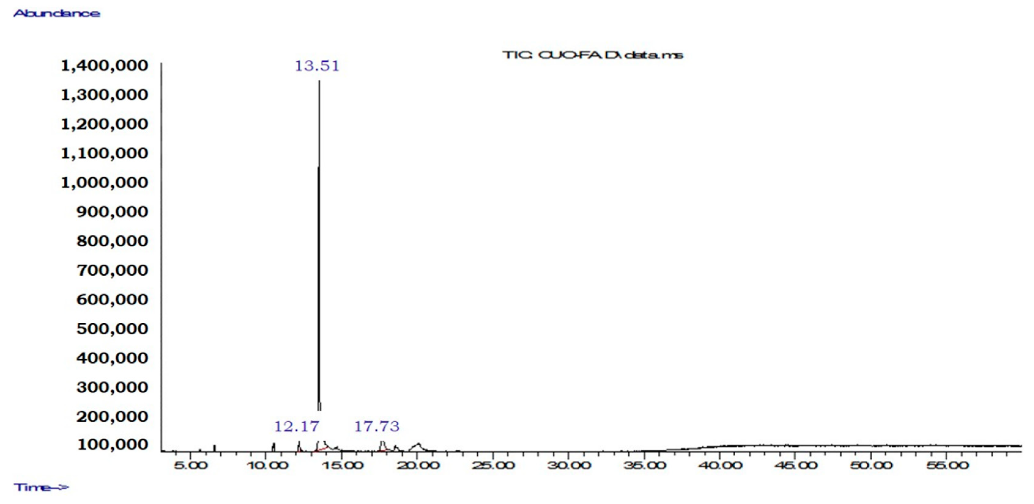
| 12.17 | - | - | - | - | - | - | - | - | 148 | 2.3 |
| 12.30 | - | - | - | - | - | - | 189 | 1.1 | - | - |
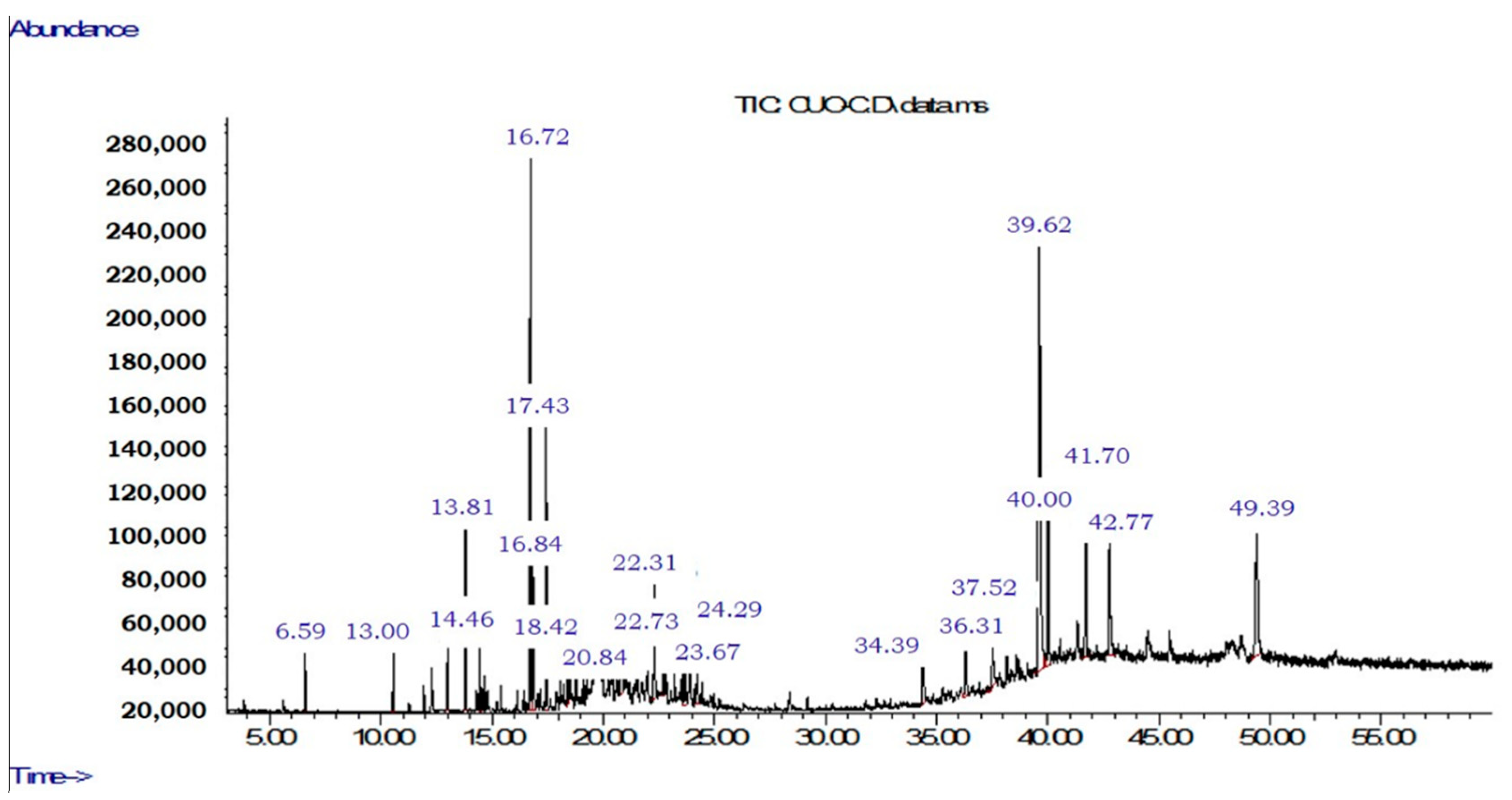
| 13.00 | - | - | - | - | 189 | 1.4 | Caryophyllene | 1.6 | - | - |
| 13.51 | - | - | - | - | - | - | - | - | 2H-1-Benzopyran-2- one (coumarin) | 87.3 |
| 13.81 | - | - | - | - | Alpha-caryophyllene | 4.0 | Alpha-caryophyllene | 3.9 | - | - |
| 14.32 | - | - | - | - | - | - | Gama-muurolene | 1.0 | - | - |
| 14.46 | - | - | - | - | Germacrene D | 1.5 | Beta-cubebene | 2.3 | - | - |
| 15.41 | - | - | - | - | - | - | Gama-cadinene | 1.3 | - | - |
| 16.71 | - | - | 205 | 5.0 | - | - | - | - | - | - |
| 16.72 | - | - | - | - | Spathulenol | 14.0 | Aromadendrene | 9.2 | - | - |
| 16.84 | - | - | - | - | Caryophyllene oxide | 4.6 | 205 | 2.5 | - | - |
| 17.43 | - | - | - | - | 220 | 7.5 | 220 | 4.0 | - | - |
| 17.73 | - | - | - | - | - | - | - | - | 182 | 10.4 |
| 18.06 | - | - | - | - | - | - | 220 | 1.0 | - | - |
| 18.42 | - | - | - | - | 220 | 1.8 | 220 | 1.6 | - | - |
| 19.84 | - | - | 129 | 2.7 | - | - | 220 | 0.7 | - | - |
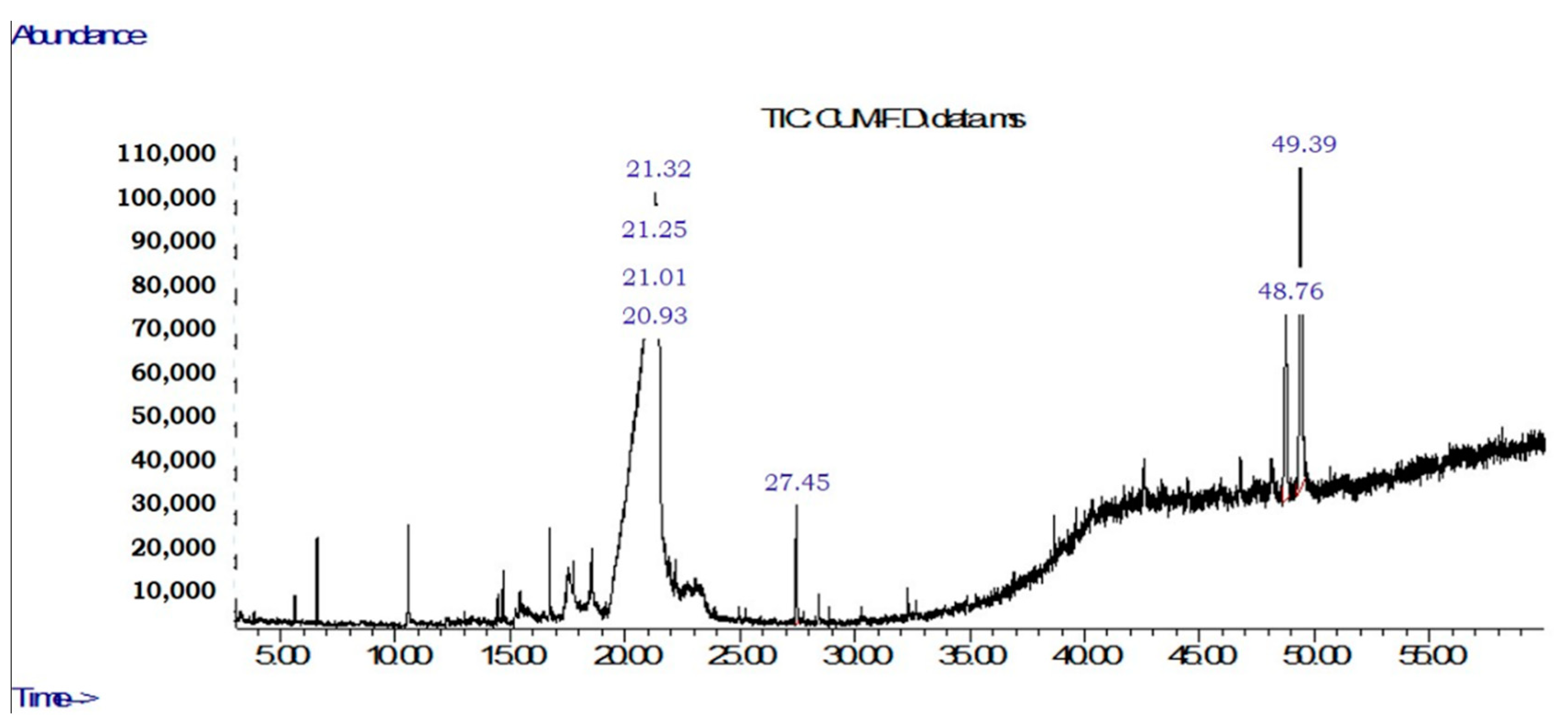
| 19.86 | 151 | 10,4 | - | - | - | - | - | - | - | - |
| 19.90 | 179 | 2.4 | - | - | - | - | - | - | - | - |
| 19.98 | 182 | 1.4 | - | - | - | - | - | - | - | - |
| 20.23 | - | - | - | - | 238 | 1.5 | - | - | - | - |
| 20.45 | 180 | 4.8 | - | - | - | - | - | - | - | - |
| 20.84 | - | - | - | - | 218 | 2.4 | - | - | - | - |
| 22.30 | - | - | - | - | - | - | 234 | 2.1 | - | - |
| 22.31 | - | - | - | - | 234 | 3.2 | - | - | - | - |
| 22.73 | - | - | - | - | 220 | 1.7 | 220 | 1.5 | - | - |
| 23.67 | - | - | - | - | 218 | 1.8 | 218 | 1.2 | - | - |
| 24.23 | - | - | - | - | - | - | 218 | 1.2 | - | - |
| 24.29 | - | - | - | - | 236 | 2.0 | - | - | - | - |
| 34.39 | - | - | - | - | Fenanthrene [3,2]furan-4- methanol-1,2,3,4,4a,5,6,6a,7,11,11a, 11b, dodecahydro-4,7,11-trimethyl | 2.3 | Fenanthrene [3,2]furan-4- methanol-1,2,3,4,4a,5,6,6a,7,11,11a, 11b, dodecahydro-4,7,11-trimethyl | 2.7 | - | - |
| 36.31 | - | - | - | - | 341 | 2.0 | 331 | 1.4 | - | - |
| 37.52 | - | - | - | - | 374 | 2.8 | 374 | 3.5 | - | - |
| 38.15 | - | - | - | - | - | - | 429 | 1.3 | - | - |
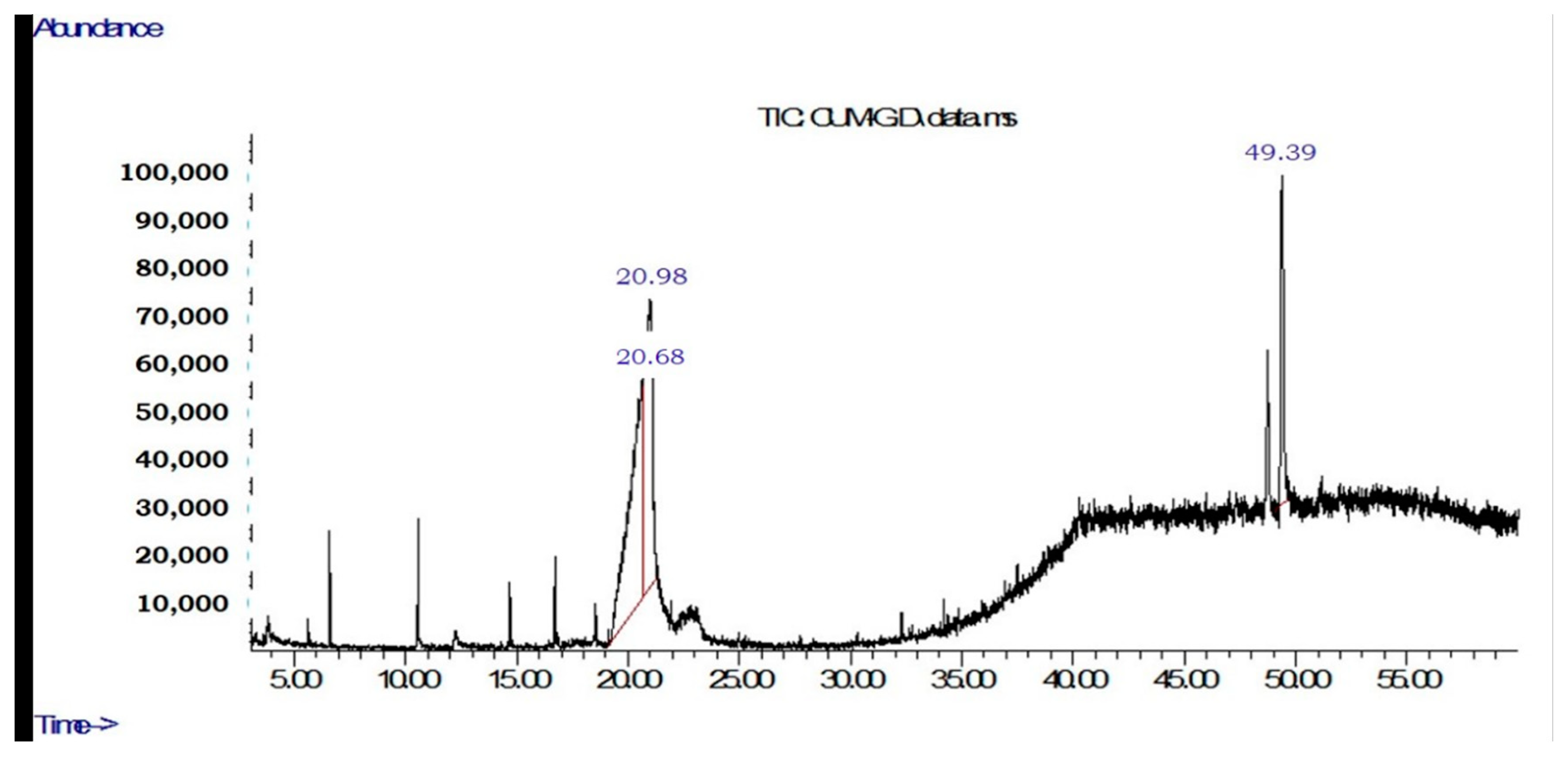
| 39.60 | - | - | 405 | 16.7 | - | - | - | - | - | - |
| 39.62 | - | - | - | - | 429 | 21.4 | - | - | - | - |
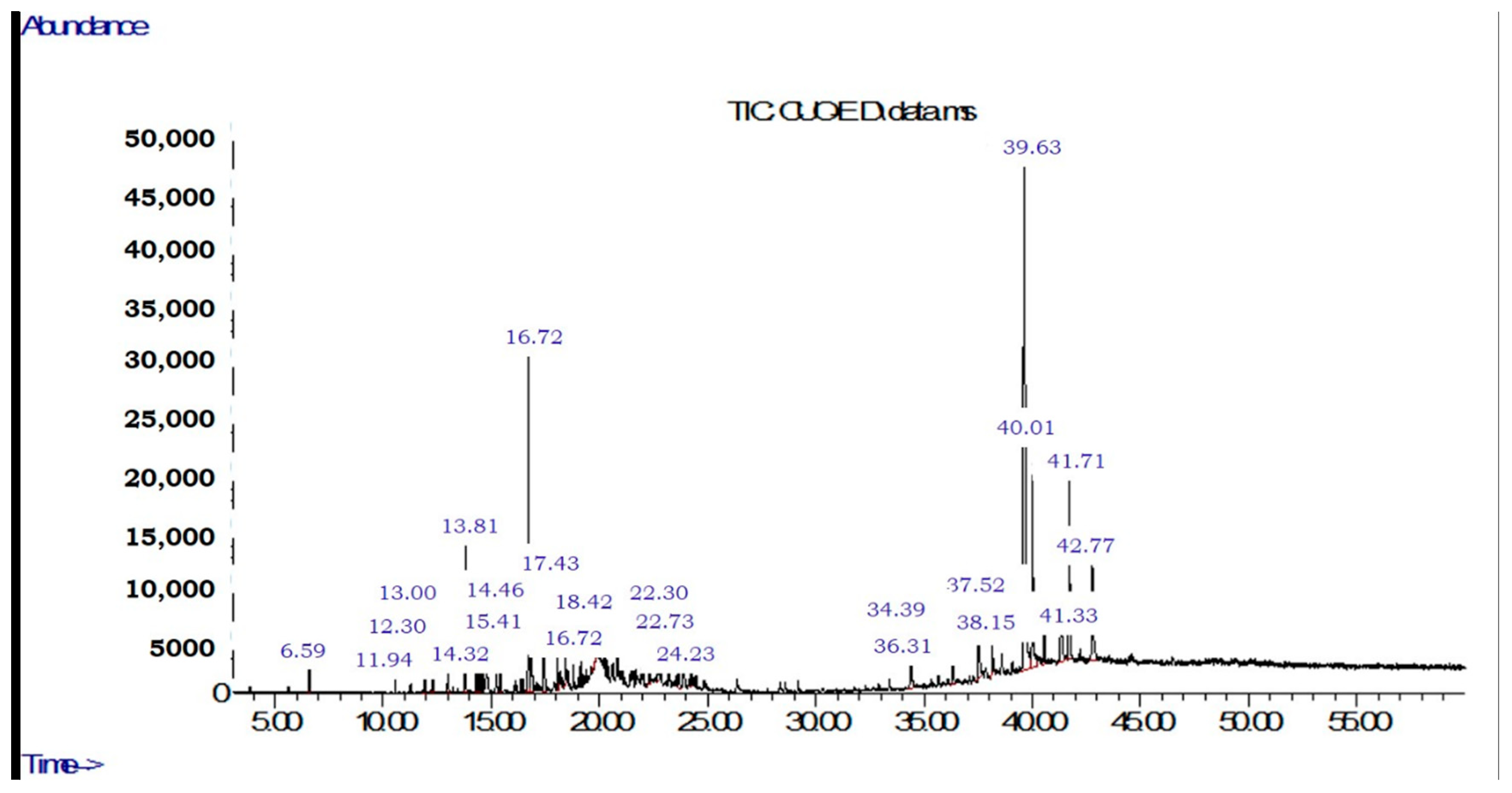
| 39.63 | - | - | - | - | - | - | 400 | 31.6 | - | - |
| 40.00 | - | - | - | - | 402 | 5.0 | - | - | - | - |
| 40.01 | - | - | - | - | - | - | 429 | 6.9 | - | - |
| 41.33 | - | - | - | - | - | - | 405 | 2.4 | - | - |
| 41.70 | - | - | - | - | 400 | 6.0 | - | - | - | - |
| 41.71 | - | - | - | - | - | - | 429 | 7.3 | - | - |
| 42.58 | - | - | 405 | 11.7 | - | - | - | - | - | - |
| 42.77 | - | - | - | - | 429 | 5.1 | 429 | 5.8 | - | - |
| 48.75 | 424 | 15.7 | - | - | - | - | - | - | - | - |
| 49.38 | 426 | 65.3 | - | - | - | - | - | - | - | - |
| 49.39 | - | - | Taraxas-terol | 63.9 | 426 | 8.0 | - | - | - | - |
| Total | 100 | 100 | 100 | 100 | 100 | |||||
| Identified Substances | ||||||||||
|---|---|---|---|---|---|---|---|---|---|---|
| RT (min) | Leaves | Branches | Husks | Endocarps | Seeds | |||||
| M | % | M | % | M | % | M | % | M | % | |
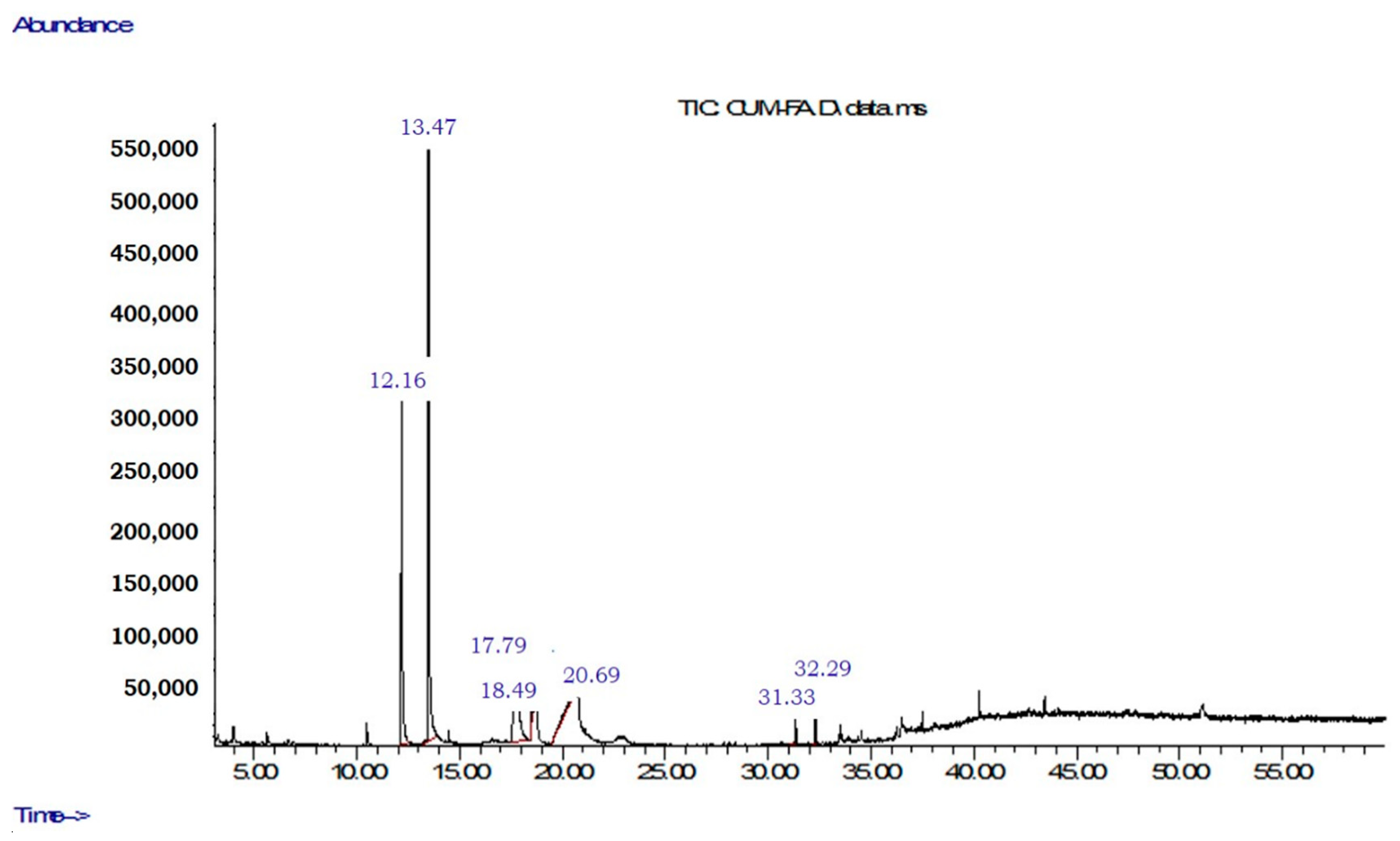
| 12.16 | - | - | - | - | - | - | - | - | 2H-1-Benzopyran-2-one,3,4- dihydro (3,4-Dihydrocoumarin) | 25.2 |
| 13.47 | - | - | - | - | - | - | - | - | 2H-1-Benzopyran-2- one (coumarin) | 43.0 |
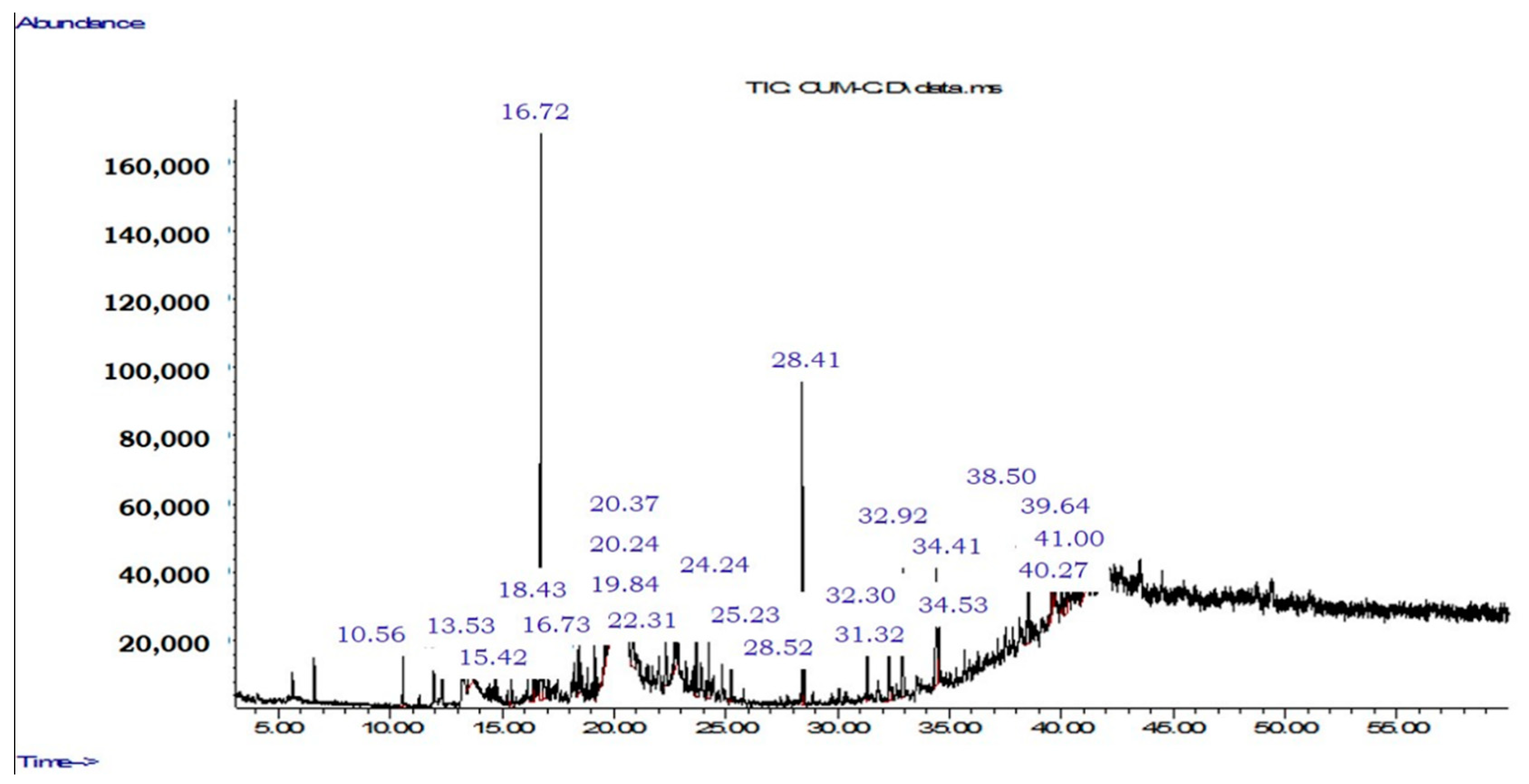
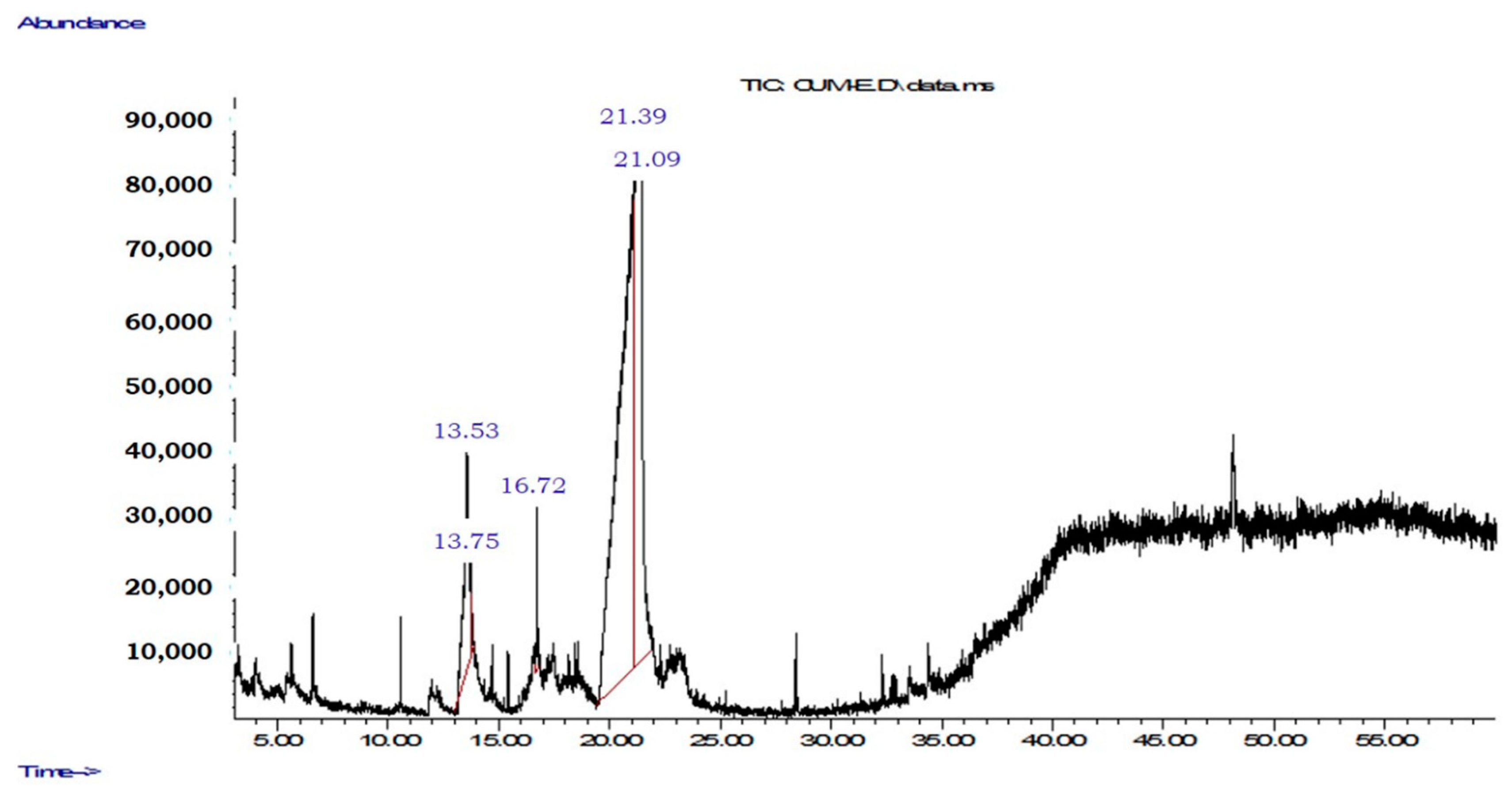
| 13.53 | - | - | - | - | 2H-1-Benzopyran-2-one (coumarin) | 2.8 | 2H-1-Benzopyran-2-one (coumarin) | 8.6 | - | - |
| 13.75 | - | - | - | - | - | - | 161 | 1.0 | - | - |
| 15.42 | - | - | - | - | Gama-cadinene | 1.2 | - | - | - | - |
| 16.39 | - | - | - | - | 235 | 1.5 | - | - | - | - |
| 16.72 | - | - | - | - | - | - | 205 | 1.1 | - | - |
| 16.73 | - | - | - | - | Spathulenol | 14.6 | - | - | - | - |
| 16.85 | - | - | - | - | 205 | 2.4 | - | - | - | - |
| 17.79 | - | - | - | - | - | - | - | - | 182 | 21.9 |
| 18.43 | - | - | - | - | 220 | 2.5 | - | - | - | - |
| 18.49 | - | - | - | - | - | - | - | - | 231 | 0.4 |
| 19.14 | - | - | - | - | 220 | 1.7 | - | - | - | - |
| 19.61 | - | - | - | - | 220 | 0.6 | - | - | - | - |
| 19.84 | - | - | - | - | 220 | 1.4 | - | - | - | - |
| 19.91 | - | - | - | - | 220 | 0.8 | - | - | - | - |
| 20.24 | - | - | - | - | 207 | 5.5 | - | - | - | - |
| 20.27 | 179 | 2.8 | - | - | - | - | - | - | - | - |
| 20.37 | - | - | - | - | 177 | 9.9 | - | - | - | - |
| 20.51 | - | - | - | - | - | - | - | - | 182 | 3.3 |
| 20.54 | - | - | - | - | - | - | - | - | 182 | 0.3 |
| 20.68 | - | - | 161 | 49.0 | - | - | - | - | - | - |
| 20.69 | - | - | - | - | - | - | - | - | 167 | 0.9 |
| 20.86 | - | - | - | - | 193 | 2.2 | - | - | - | - |
| 20.93 | 179 | 4.3 | - | - | - | - | - | - | - | - |
| 20.98 | - | - | 179 | 35.9 | - | - | - | - | - | - |
| 21.01 | 179 | 0.8 | - | - | - | - | - | - | - | - |
| 21.08 | 159 | 2.1 | - | - | - | - | - | - | - | - |
| 21.09 | - | - | - | - | - | - | 182 | 57.7 | - | - |
| 21.39 | - | - | - | - | - | - | 207 | 31.6 | - | - |
| 22.31 | - | - | - | - | 219 | 3.2 | - | - | - | - |
| 22.74 | - | - | - | - | 220 | 2.7 | - | - | - | - |
| 23.68 | - | - | - | - | 221 | 2.1 | - | - | - | - |
| 24.24 | - | - | - | - | 218 | 1.8 | - | - | - | - |
| 25.23 | - | - | - | - | Ethylic ester of hexadecanoic acid | 1.7 | - | - | - | - |
| 27.45 | 207 | 9.1 | - | - | - | - | - | - | - | - |
| 28.41 | - | - | - | - | Ethyl ester of oleic acid (ethyl oleate) | 9.4 | - | - | - | - |
| 28.52 | - | - | - | - | 281 | 1.4 | - | - | - | - |
| 31.32 | - | - | - | - | 310 | 1.7 | - | - | - | - |
| 31.33 | - | - | - | - | - | - | - | - | 310 | 2.2 |
| 32.29 | - | - | - | - | - | - | - | - | 259 | 2.8 |
| 32.30 | - | - | - | - | Ester bis (2-ethyl-hexyl) of hexanodioic acid | 2.7 | - | - | - | - |
| 32.92 | - | - | - | - | 300 | 4.1 | - | - | - | - |
| 34.41 | - | - | - | - | Fenanthrene [3,2]furan-4-methanol-1,2,3, 4,4a,5,6,6a,7,11,11a, 11b, dodecahydro-4,7,11-trimethyl | 5.7 | - | - | - | - |
| 34.53 | - | - | - | - | 298 | 1.5 | - | - | - | - |
| 38.50 | - | - | - | - | 361 | 4.4 | - | - | - | - |
| 39.64 | - | - | - | - | 429 | 3.7 | - | - | - | - |
| 40.01 | - | - | - | - | 360 | 1.9 | - | - | - | - |
| 40.27 | - | - | - | - | 429 | 2.0 | - | - | - | - |
| 41.00 | - | - | - | - | 405 | 2.9 | - | - | - | - |
| 48.76 | 551 | 29.0 | - | - | - | - | - | - | - | - |
| 49.39 | Taraxas-terol | 51.9 | Taraxas-terol | 15.1 | - | - | - | - | - | - |
| Total | 100 | 100 | 100 | 100 | 100 | |||||
| Mean Diameter of the Phytopathogen Colonies (cm) | ||||||
|---|---|---|---|---|---|---|
| Extracts | Concentration (%) | Cercospora longissima | Colletotrichum gloeosporioides | Fusarium sp. (Lettuce) | Fusarium sp. (Kale) | Sclerotium rolfsii |
| Control | 0 | 3.9 bD | 4.8 cC | 7.5 aB | 7.9 aA | 8.1 aA |
| Coumarin | 0.1 | 3.9 bC | 4.1 dC | 6.9 bB | 7.3 bB | 8.0 aA |
| Leaves | 10 | 3.5 cD | 5.5 bC | 7.4 aB | 8.2 aA | 8.3 aA |
| 20 | 3.4 cD | 5.4 bC | 7.5 aA | 7.9 aA | 7.0 bB | |
| 30 | 2.7 eE | 5.7 aD | 7.5 aB | 8.1 aA | 6.8 bC | |
| 40 | 3.0 dC | 5.9 aB | 6.1 cB | 7.9 aA | 7.9 aA | |
| 50 | 3.1 dD | 5.8 aC | 5.8 cC | 8.2 aA | 7.7 aB | |
| Branches | 10 | 3.9 bD | 4.2 dD | 5.8 cC | 7.3 bB | 8.0 aA |
| 20 | 3.7 bD | 4.1 dD | 5.9 cC | 7.6 aB | 8.2 aA | |
| 30 | 3.6 cE | 4.1 dD | 5.6 cC | 7.1 bB | 8.1 aA | |
| 40 | 3.8 bD | 4.1 dD | 5.9 cC | 6.8 cB | 8.3 aA | |
| 50 | 3.8 bD | 4.1 dD | 6.1 cC | 6.8 cB | 7.8 aA | |
| Husks | 10 | 4.9 aC | 4.8 cC | 8.0 aA | 7.0 bB | 8.2 aA |
| 20 | 5.3 aC | 4.5 cD | 6.6 bB | 6.6 cB | 8.3 aA | |
| 30 | 5.2 aD | 4.4 dE | 7.8 aB | 6.5 cC | 8.3 aA | |
| 40 | 5.2 aC | 4.0 dD | 6.4 bB | 6.4 cB | 8.3 aA | |
| 50 | 4.8 aD | 3.9 dE | 6.6 bB | 5.8 dC | 8.3 aA | |
| Endocarps | 10 | 3.2 dC | 3.5 eC | 6.7 bB | 6.7 cB | 8.3 aA |
| 20 | 2.8 eC | 2.9 fC | 5.3 dB | 5.3 eB | 8.3 aA | |
| 30 | 2.5 eC | 2.4 gC | 5.1 dB | 4.7 fB | 8.3 aA | |
| 40 | 2.4 eC | 1.7 hD | 3.5 eB | 3.5 gB | 8.3 aA | |
| 50 | 2.4 eC | 2.1 hC | 3.4 eB | 3.2 gB | 8.3 aA | |
| Seeds | 10 | 3.5 cD | 5.5 bC | 7.6 aA | 6.0 dB | 5.3 dC |
| 20 | 3.5 cE | 5.4 bD | 7.2 aB | 8.1 aA | 6.1 cC | |
| 30 | 3.5 cC | 5.2 bB | 7.2 aA | 7.2 bA | 6.9 bA | |
| 40 | 2.9 dD | 5.3 bC | 5.0 dC | 6.1 dB | 7.0 bA | |
| 50 | 3.0 dD | 4.9 bC | 5.4 dB | 5.8 dB | 7.6 aA | |
| CV (%) | 5.31 | |||||
| Mean Diameter of the Phytopathogen Colonies (cm) | ||||||
|---|---|---|---|---|---|---|
| Extracts | Concentrations (%) | Cercospora longissima | Colletotrichum gloeosporioides | Fusarium sp. (Lettuce) | Fusarium sp. (Kale) | Sclerotium rolfsii |
| Control | 0 | 3.9 eD | 4.8 cC | 7.5 bB | 7.9 aA | 8.1 bA |
| Coumarin | 0.1 | 3.9 eD | 4.1 dD | 6.9 cC | 7.3 bB | 8.0 bA |
| Leaves | 10 | 3.1 fD | 5.5 aC | 7.4 bB | 7.8 aB | 8.3 aA |
| 20 | 3.1 fE | 5.2 bD | 7.3 bB | 6.4 dC | 8.1 bA | |
| 30 | 2.9 fD | 5.5 aB | 4.2 fC | 8.2 aA | 8.1 bA | |
| 40 | 3.2 fC | 5.9 aB | 3.1 gC | 8.2 aA | 8.3 aA | |
| 50 | 2.7 fE | 5.6 aC | 4.0 fD | 6.7 cB | 8.3 aA | |
| Branches | 10 | 3.9 eD | 4.1 dD | 6.2 dC | 7.4 bB | 8.0 bA |
| 20 | 3.8 eD | 4.1 dD | 6.0 dC | 7.5 bB | 8.1 bA | |
| 30 | 3.8 eD | 3.9 dD | 5.7 eC | 6.8 cB | 8.1 bA | |
| 40 | 3.9 eD | 4.2 dD | 5.6 eC | 6.6 dB | 7.9 bA | |
| 50 | 3.8 eD | 4.0 dD | 5.8 dC | 6.5 dB | 8.0 bA | |
| Husks | 10 | 5.6 bB | 5.1 cC | 8.2 aA | 7.9 aA | 8.3 aA |
| 20 | 5.5 bB | 5.2 bB | 8.3 aA | 8.2 aA | 8.3 aA | |
| 30 | 6.1 aC | 4.2 dD | 7.3 bB | 7.5 bB | 8.3 aA | |
| 40 | 5.5 bB | 4.7 cC | 8.2 aA | 8.0 aA | 8.3 aA | |
| 50 | 5.2 cC | 4.5 dD | 7.3 bB | 8.1 aA | 8.3 aA | |
| Endocarps | 10 | 3.4 eC | 4.2 dB | 8.0 aA | 8.0 aA | 8.3 aA |
| 20 | 3.3 fE | 4.1 dD | 7.1 cC | 7.5 bB | 8.3 aA | |
| 30 | 3.0 fD | 3.9 dC | 6.0 dB | 6.4 dB | 8.3 aA | |
| 40 | 3.2 fC | 2.7 eD | 5.4 eB | 5.6 eB | 8.3 aA | |
| 50 | 3.7 eE | 4.0 dD | 7.1 cC | 7.6 bB | 8.3 aA | |
| Seeds | 10 | 2.4 gE | 4.9 cC | 8.3 aA | 3.2 hD | 7.7 bB |
| 20 | 3.0 fE | 5.5 aC | 6.9 cB | 5.1 fD | 7.5 cA | |
| 30 | 2.1 gD | 5.0 cC | 6.6 cB | 6.4 dB | 7.0 dA | |
| 40 | 4.5 dC | 5.5 aB | 6.9 cA | 7.0 cA | 6.6 eA | |
| 50 | 4.1 eD | 5.3 bB | 6.9 cA | 4.5 gC | 7.0 dA | |
| CV (%) | 4.3 | |||||
| Pathogenic Bacteria (µg·mL−1) | Gram-Negative | Gram-Positive | ||||||
|---|---|---|---|---|---|---|---|---|
| Burkholderia cepacia (ATCC 25416) | Escherichia coli (ATCC 11775) | Pseudomonas aeruginosa (ATCC 13388) | Staphylococcus aureus (ATCC 6538) | |||||
| Dipteryx odorata | MIC | MBC | MIC | MBC | MIC | MBC | MIC | MBC |
| Leaves | * | * | * | * | * | * | * | * |
| Branches | * | * | * | * | * | * | * | * |
| Husks | * | * | 2000 | * | * | * | * | * |
| Endocarps | * | * | 2000 | * | * | * | * | * |
| Seeds | * | * | * | * | * | * | * | * |
| Dipteryx punctata | MIC | MBC | MIC | MBC | MIC | MBC | MIC | MBC |
| Leaves | * | * | * | * | * | * | * | * |
| Branches | * | * | * | * | * | * | * | * |
| Husks | * | * | 2000 | * | * | * | 2000 | * |
| Endocarps | * | * | 2000 | * | * | * | * | * |
| Seeds | * | * | * | * | * | * | * | * |
Publisher’s Note: MDPI stays neutral with regard to jurisdictional claims in published maps and institutional affiliations. |
© 2022 by the authors. Licensee MDPI, Basel, Switzerland. This article is an open access article distributed under the terms and conditions of the Creative Commons Attribution (CC BY) license (https://creativecommons.org/licenses/by/4.0/).
Share and Cite
Sousa, B.C.M.d.; Castro, S.P.d.; Lourido, K.A.; Kasper, A.A.M.; Paulino, G.d.S.; Delarmelina, C.; Duarte, M.C.T.; Sartoratto, A.; Vieira, T.A.; Lustosa, D.C.; et al. Identification of Coumarins and Antimicrobial Potential of Ethanolic Extracts of Dipteryx odorata and Dipteryx punctata. Molecules 2022, 27, 5837. https://doi.org/10.3390/molecules27185837
Sousa BCMd, Castro SPd, Lourido KA, Kasper AAM, Paulino GdS, Delarmelina C, Duarte MCT, Sartoratto A, Vieira TA, Lustosa DC, et al. Identification of Coumarins and Antimicrobial Potential of Ethanolic Extracts of Dipteryx odorata and Dipteryx punctata. Molecules. 2022; 27(18):5837. https://doi.org/10.3390/molecules27185837
Chicago/Turabian StyleSousa, Bruna Cristine Martins de, Santana Pinto de Castro, Katiane Araújo Lourido, Aline Aparecida München Kasper, Geomarcos da Silva Paulino, Camila Delarmelina, Marta Cristina Teixeira Duarte, Adilson Sartoratto, Thiago Almeida Vieira, Denise Castro Lustosa, and et al. 2022. "Identification of Coumarins and Antimicrobial Potential of Ethanolic Extracts of Dipteryx odorata and Dipteryx punctata" Molecules 27, no. 18: 5837. https://doi.org/10.3390/molecules27185837