
Water-Soluble Single-Benzene Chromophores: Excited State Dynamics and Fluorescence Detection


Abstract
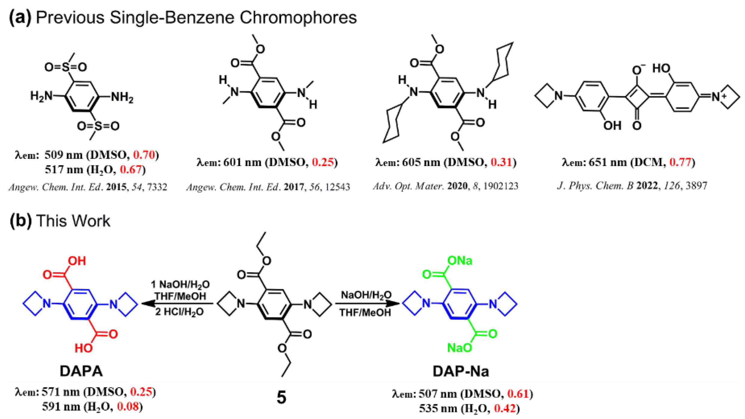
:1. Introduction
2. Results and Discussion
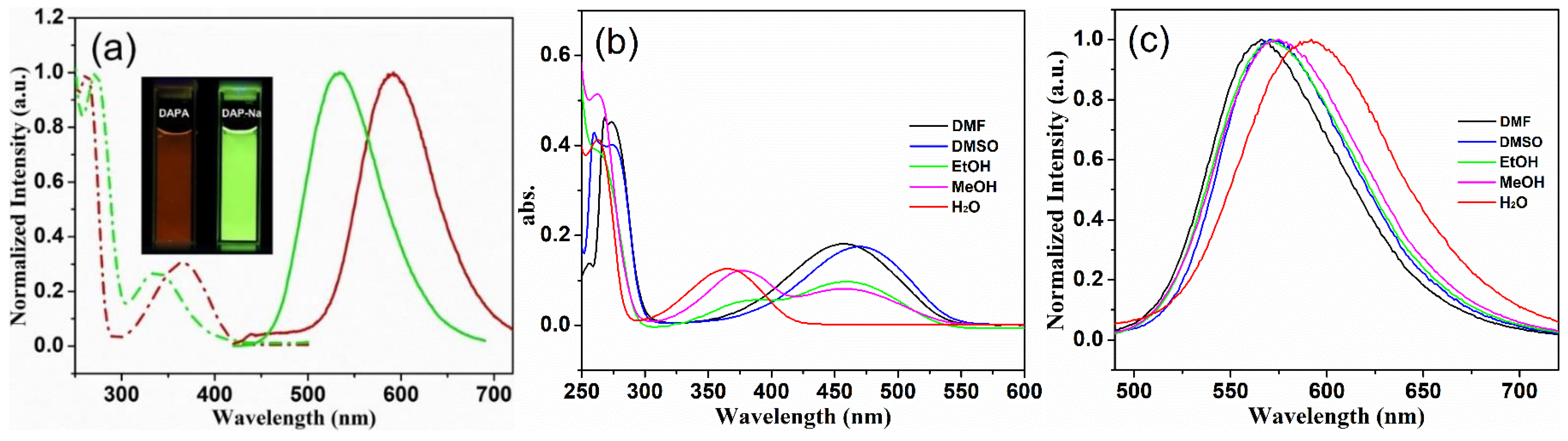
2.1. Photophysical Properties of DAPA and DAP-Na
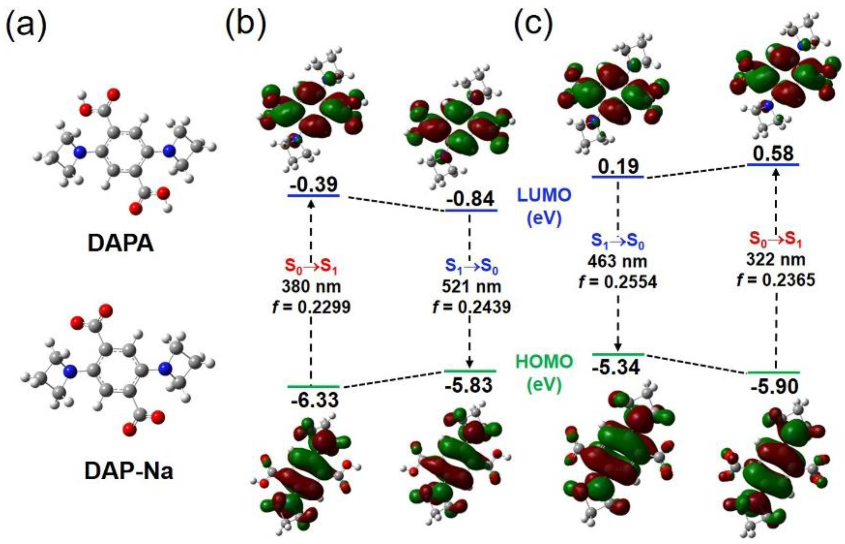
2.2. Theoretical Calculations
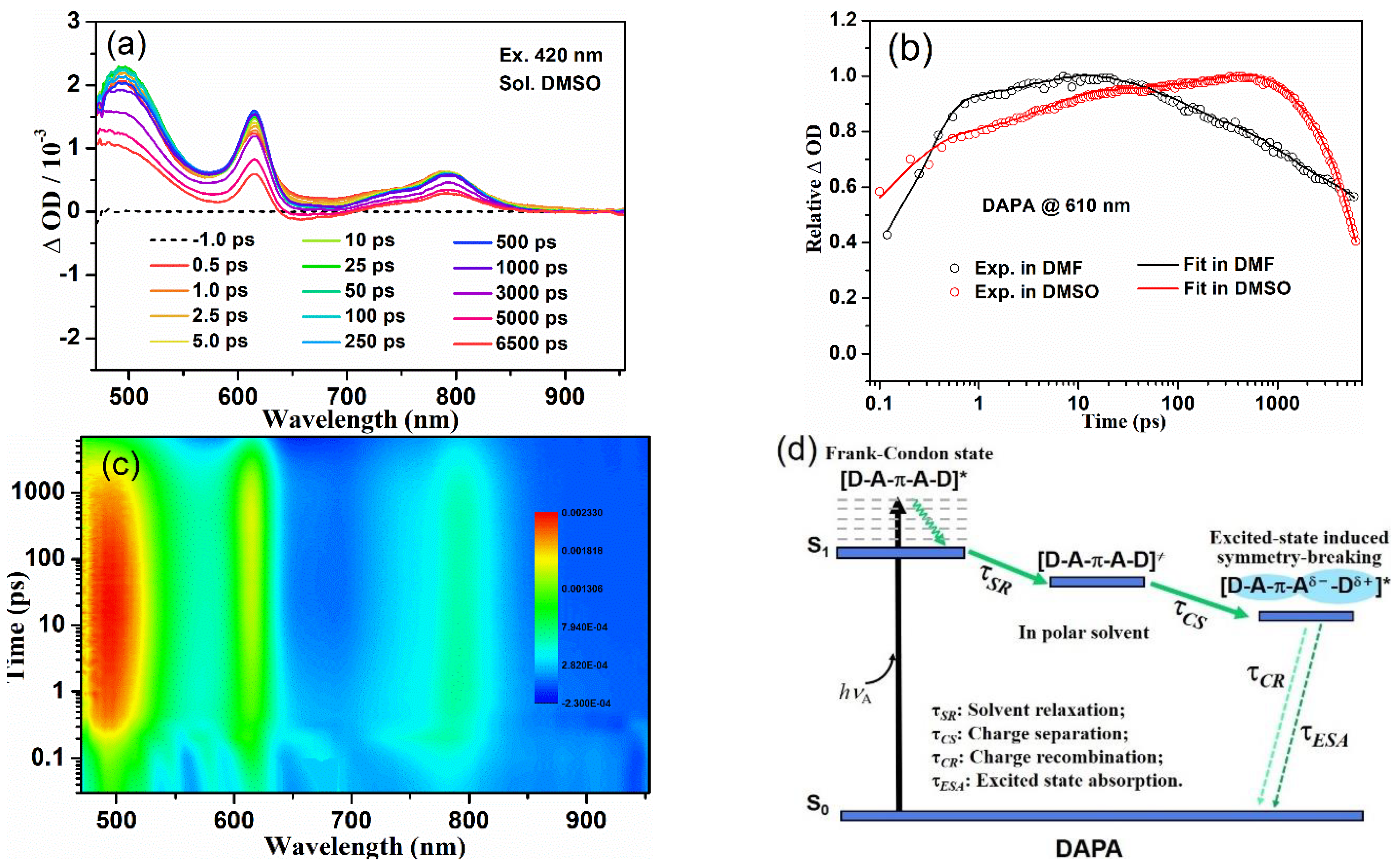
2.3. Transient Absorption Spectroscopy
2.4. pH-Dependent Fluorescence
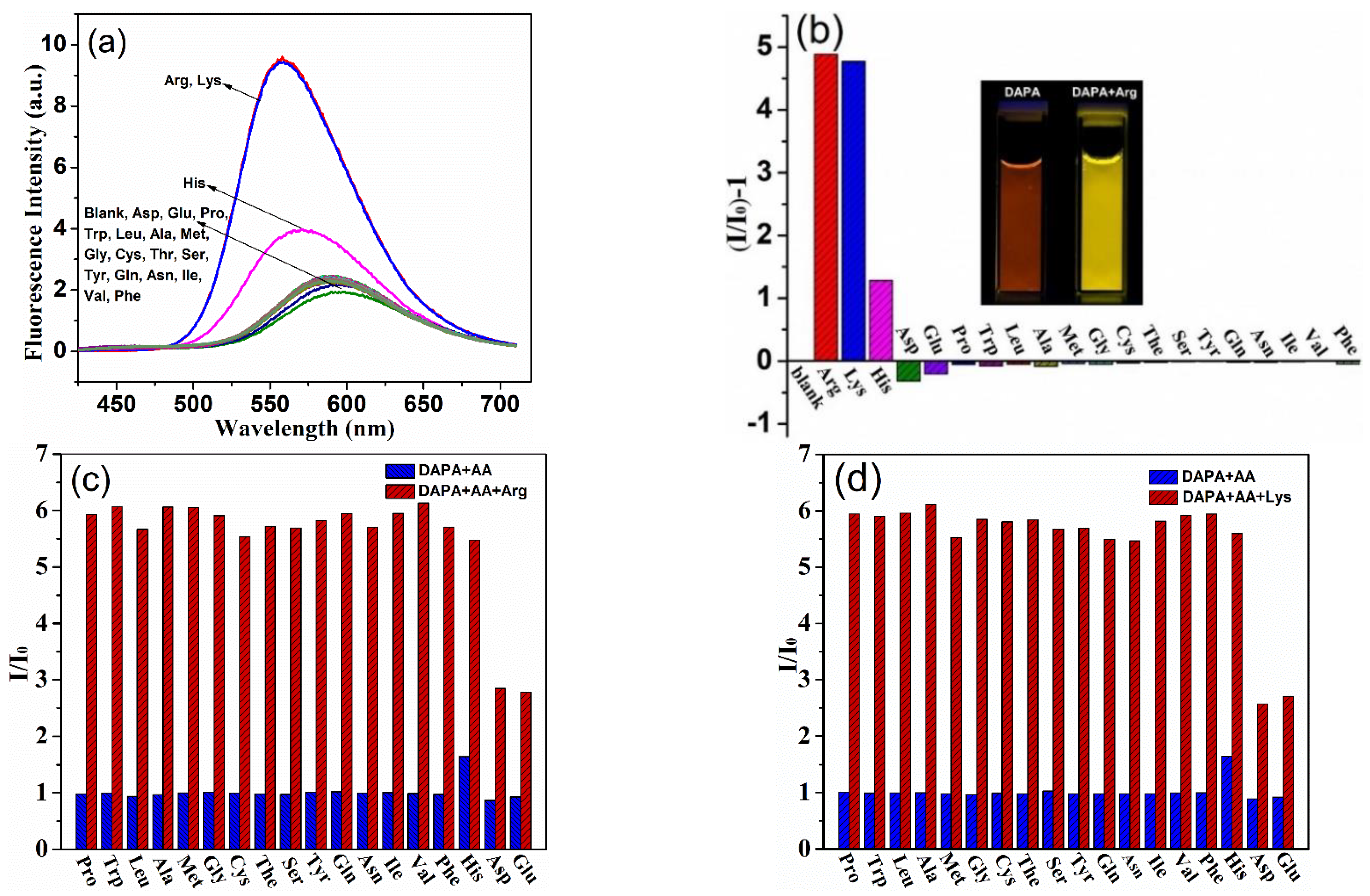
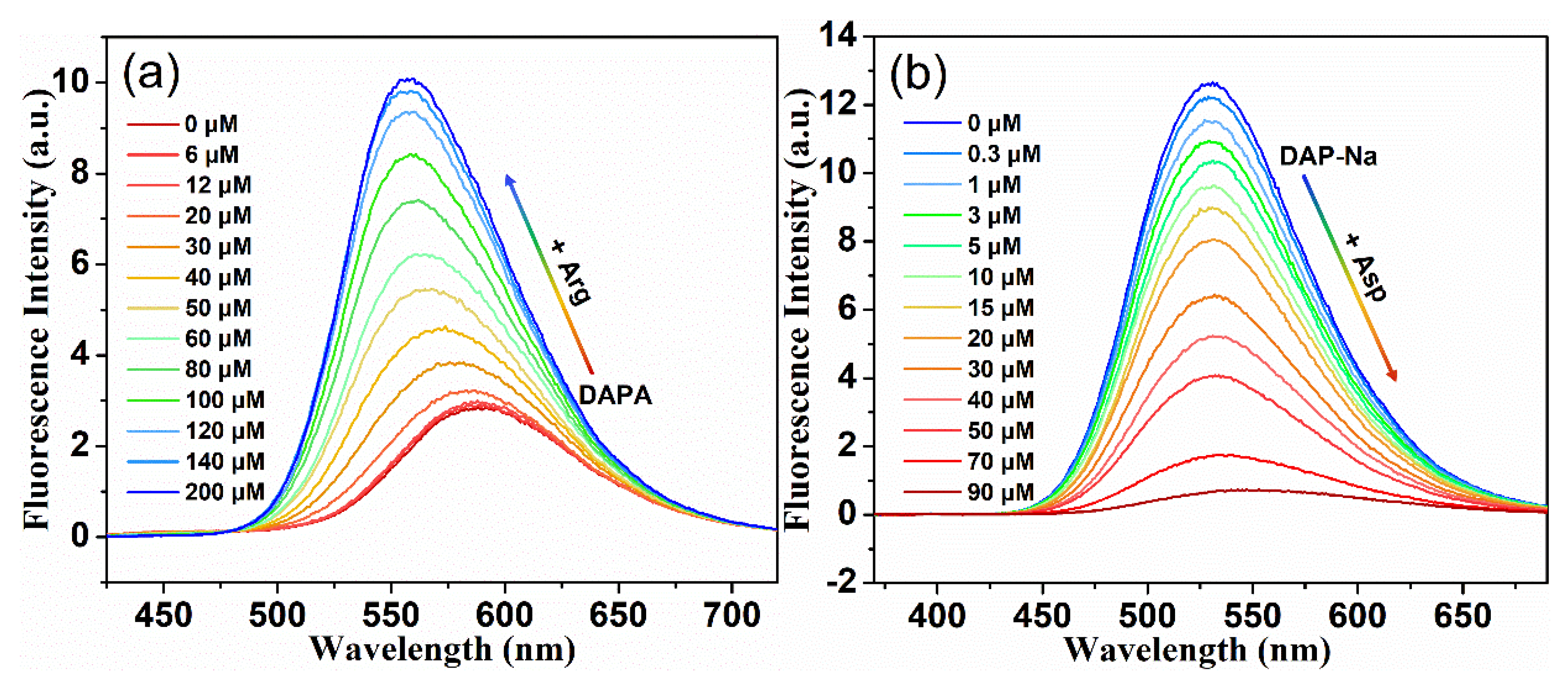
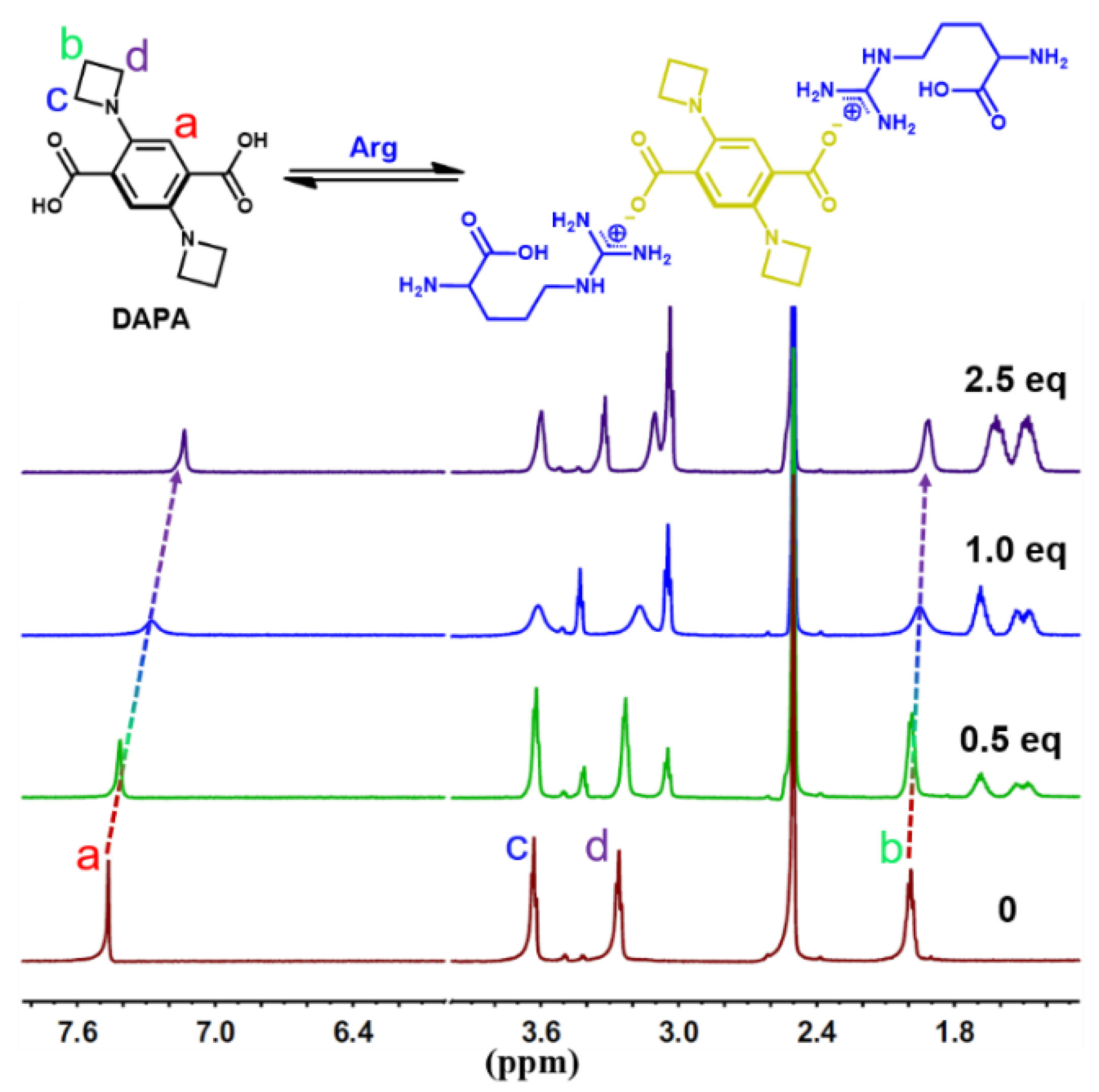
2.5. Sensitive and Discriminative Detection of Amino Acids
3. Materials and Methods
3.1. Reagents and Instruments
3.2. Synthesis of Compound DAPA
3.3. Synthesis of Compound DAP-Na
4. Conclusions
Supplementary Materials
Author Contributions
Funding
Institutional Review Board Statement
Informed Consent Statement
Data Availability Statement
Acknowledgments
Conflicts of Interest
Sample Availability
References
- Friedman, M. Chemistry, nutrition, and microbiology of d-amino acids. J. Agric. Food Chem. 1999, 47, 3457–3479. [Google Scholar] [CrossRef] [PubMed]
- Wu, G.; Wu, Z.; Dai, Z.; Yang, Y.; Wang, W.; Liu, C.; Wang, B.; Wang, J.; Yin, Y. Dietary requirements of “nutritionally non-essential amino acids” by animals and humans. Amino Acids 2013, 44, 1107–1113. [Google Scholar] [CrossRef] [PubMed]
- Wang, B.; Han, J.; Bojanowski, N.M.; Bender, M.; Ma, C.; Seehafer, K.; Herrmann, A.; Bunz, U.H.F. An optimized sensor array identifies all natural amino acids. ACS Sens. 2018, 3, 1562–1568. [Google Scholar] [CrossRef] [PubMed]
- Sonsalla, P.K.; Nicklas, W.J.; Heikkila, R.E. Role for excitatory amino acids in metamphetamine-induced nigrostriatal dopaminergic toxicity. Science 1989, 243, 398–400. [Google Scholar] [CrossRef] [PubMed]
- Fornai, F.; Vaglini, F.; Maggio, R.; Bonuccelli, U.; Corsini, G.U. Species differences in the role of excitatory amino acids in experimental parkinsonism. Neurosci. Biobehav. Rev. 1997, 21, 401–415. [Google Scholar] [CrossRef]
- Das, S.; Guha, S.; Banerjee, A.; Lohar, S.; Sahana, A.; Das, D. 2-(2-Pyridyl) benzimidazole based Co(II) complex as an efficient fluorescent probe for trace level determination of aspartic and glutamic acid in aqueous solution: A displacement approach. Org. Biomol. Chem. 2011, 9, 7097–7104. [Google Scholar] [CrossRef]
- Stechmiler, J.K.; Childress, B.; Cowan, L. Arginine supplementation and wound healing. Nutr. Clin. Pract. 2005, 20, 52–61. [Google Scholar] [CrossRef]
- Zhou, Y.; Won, J.; Lee, J.Y.; Yoon, J. Studies leading to the development of a highly selective colorimetric and fluorescent chemosensor for lysine. Chem. Commun. 2011, 47, 1997–1999. [Google Scholar] [CrossRef]
- Zhang, Z.; Wei, T.; Chen, Y.; Chen, T.; Chi, B.; Wang, F.; Chen, X. A polydiacetylenes-based colorimetric and fluorescent probe for L-arginine and L-lysine and its application for logic gate. Sens. Actuators B 2018, 255, 2211–2217. [Google Scholar] [CrossRef]
- Niebroj-Dobosz, I.; Janik, P. Amino acids acting as transmitters in Amyotrophic Lateral Sclerosis (ALS). Acta Neurol. Scand. 1999, 100, 6–11. [Google Scholar] [CrossRef]
- Felig, P. Amino acids metabolism in man. Annu. Rev. Biochem. 1975, 44, 933–955. [Google Scholar] [CrossRef] [PubMed]
- Hirayama, C.; Suyama, K.; Horie, Y.; Tanimoto, K.; Kato, S. Plasma amino acid patterns in Hepatocellular Carcinoma. Biochem. Med. Metab. Biol. 1987, 38, 127–133. [Google Scholar] [CrossRef]
- Song, Y.; Xu, C.; Kuroki, H.; Liao, Y.; Tsunoda, M. Recent trends in analytical methods for the determination of amino acids in biological samples. J. Pharm. Biomed. Anal. 2018, 147, 35–49. [Google Scholar] [CrossRef]
- Ferré, S.; González-Ruiz, V.; Guillarme, D.; Rudaz, S. Analytical strategies for the determination of amino acids: Past, present and future trends. J. Chromatogr. B 2019, 1132, 121819. [Google Scholar] [CrossRef] [PubMed]
- Lavgnini, M.G.; Moscone, D.; Campagnone, D.; Crimisini, C.; Palleschi, G. Amperometric lysine bioprobes analysis in feeds. Talanta 1993, 40, 1301–1306. [Google Scholar] [CrossRef]
- Inoue, T.; Kirchhoff, J.R. Determination of thiols by capillary electrophoresis with amperometric detection at a coenzyme pyrroloquinoline quinone modified electrode. Anal. Chem. 2002, 74, 1349–1354. [Google Scholar] [CrossRef] [PubMed]
- Zhou, Y.; Yoon, J. Recent progress in fluorescent and colorimetric chemosensors for detection of amino acids. Chem. Soc. Rev. 2012, 41, 52–67. [Google Scholar] [CrossRef]
- Lu, W.; Gao, Y.; Jiao, Y.; Shuang, S.; Li, C.; Dong, C. Carbon nano-dots as a fluorescent and colorimetric dual-readout probe for the detection of arginine and Cu2+ and its logic gate operation. Nanoscale 2017, 9, 11545–11552. [Google Scholar] [CrossRef]
- Hao, J.; Wang, M.; Wang, S.; Huang, Y.; Cao, D. Dissolution-enhanced emission of 1,3,6,8-tetrakis(p-benzoic acid)pyrene for detecting arginine and lysine amino acids. Dyes Pigment. 2020, 175, 108131. [Google Scholar] [CrossRef]
- Guan, W.; Zhou, W.; Lu, J.; Lu, C. Luminescent films for chemo- and biosensing. Chem. Soc. Rev. 2015, 44, 6981–7009. [Google Scholar] [CrossRef]
- Klymchenko, A.S. Solvatochromic and fluorogenic dyes as environment-sensitive probes: Design and biological applications. Acc. Chem. Res. 2017, 50, 366–375. [Google Scholar] [CrossRef] [PubMed]
- Liu, T.; Miao, R.; Peng, H.; Liu, J.; Ding, L.; Fang, Y. Adlayer chemistry on film-based fluorescent gas sensors. Acta Phys.-Chim. Sin. 2020, 36, 1908025. [Google Scholar]
- Qiao, M.; Fan, J.; Ding, L.; Fang, Y. Fluorescent ensemble sensors and arrays based on surfactant aggregates encapsulating pyrene-derived fluorophores for differentiation applications. ACS Appl. Mater. Interfaces 2021, 131, 8395–8412. [Google Scholar] [CrossRef] [PubMed]
- Chinta, J.P.; Acharya, A.; Kumar, A.; Rao, C.P. Spectroscopy and microscopy studies of the recognition of amino acids and aggregation of proteins by Zn(II) complex of lower rim naphthylidene conjugate of calix[4]arene. J. Phys. Chem. B 2009, 113, 12075–12083. [Google Scholar] [CrossRef] [PubMed]
- Wang, J.; Liu, H.B.; Tong, Z.; Ha, C.S. Fluorescent/luminescent detection of natural amino acids by organometallic systems. Coordin. Chem. Rev. 2015, 303, 139–184. [Google Scholar] [CrossRef]
- Singh, H.; Sidhu, J.S.; Mahajan, D.K.; Singh, N. A carbon quantum dot and rhodamine-based ratiometric fluorescent complex for the recognition of histidine in aqueous systems. Mater. Chem. Front. 2019, 3, 476–483. [Google Scholar] [CrossRef]
- Fu, H.; Hu, O.; Fan, Y.; Hu, Y.; Huang, J.; Wang, Z.; She, Y. Rational design of an “on-off-on” fluorescent assay for chiral amino acids based on quantum dots and nanoporphyrin. Sens. Actuators B 2019, 287, 1–8. [Google Scholar] [CrossRef]
- Rawat, K.A.; Kailasa, S.K. Visual detection of arginine, histidine and lysine using quercetin-functionalized gold nanoparticles. Microchim. Acta 2014, 181, 1917–1929. [Google Scholar] [CrossRef]
- Lohar, S.; Safin, D.A.; Sengupta, A.; Chattopadhyay, A.; Matalobos, J.S.; Babashkina, M.G.; Robeyns, K.; Mitoraj, M.P.; Kubisiak, P.; Garcia, Y.; et al. Ratiometric sensing of lysine through the formation of the pyrene excimer: Experimental and computational studies. Chem. Commun. 2015, 51, 8536–8539. [Google Scholar] [CrossRef]
- Mohan, N.; Sreejith, S.S.; Begum, P.M.S.; Kurup, M.R.P. Dual responsive salen-type Schiff bases for the effective detection of L-arginine via a static quenching mechanism. New J. Chem. 2018, 42, 13114–13121. [Google Scholar] [CrossRef]
- Li, J.; Yue, Y.; Huo, F.; Yin, C. Rational design of cysteine-specific ratiometric probe based on steric hindrance effect and its biological application. Dyes Pigment. 2019, 164, 335–340. [Google Scholar] [CrossRef]
- Jeon, S.; Kim, T.; Jin, H.; Lee, U.; Bae, J.; Bouffard, J.; Kim, Y. Amine-reactive activated esters of meso-carboxybodipy: Fluorogenic assays and labeling of amines, amino acids, and proteins. J. Am. Chem. Soc. 2020, 142, 9231–9239. [Google Scholar] [CrossRef] [PubMed]
- Yang, L.; Xie, Y.; Chen, Q.; Zhang, J.; Li, L.; Sun, H. Colorimetric and fluorescent dual-signal chemosensor for lysine and arginine and its application to detect amines in solid-phase peptide synthesis. ACS Appl. Bio Mater. 2021, 4, 6558–6564. [Google Scholar] [CrossRef]
- Gujar, V.; Roshni, V.; Suryawanshi, M.; Bobade, V.; Ottoor, D. Pattern recognition of amino acids based on highly fluorescent SDS modified pyridyl thiazole derivative. Sens. Actuators B 2020, 310, 127840. [Google Scholar] [CrossRef]
- Shi, Y.G.; Yao, J.H.; Duan, Y.L.; Mi, Q.L.; Chen, J.H.; Xu, Q.Q.; Gou, G.Z.; Zhou, Y.; Zhang, J.F. 1,8-Naphthalimide–Cu(II) ensemble based turn-on fluorescent probe for the detection of thiols in organic aqueous media. Bioorg. Med. Chem. Lett. 2013, 23, 2538–2542. [Google Scholar] [CrossRef] [PubMed]
- Tuccitto, N.; Fichera, L.; Ruffino, R.; Cantaro, V.; Sfuncia, G.; Nicotra, G.; Sfrazzetto, G.T.; Li-Destri, G.; Valenti, A.; Licciardello, A.; et al. Carbon quantum dots as fluorescence nanochemo-sensors for selective detection of amino acids. ACS Appl. Nano Mater. 2021, 4, 6250–6256. [Google Scholar] [CrossRef]
- Hou, P.; Sun, J.; Wang, H.; Liu, L.; Zou, L.; Chen, S. TCF-imidazo[1,5-α]pyridine: A potential robust ratiometric fluorescent probe for glutathione detection with high selectivity. Sens. Actuators B 2020, 304, 127244. [Google Scholar] [CrossRef]
- Cao, J.; Ding, L.; Hu, W.; Chen, X.; Chen, X.; Fang, Y. Ternary system based on fluorophore-surfactant assemblies-Cu2+ for highly sensitive and selective detection of arginine in aqueous solution. Langmuir 2014, 30, 15364–15372. [Google Scholar] [CrossRef] [PubMed]
- Cao, J.; Ding, L.; Zhang, Y.; Wang, S.; Fang, Y. A ternary sensor system based on pyrene derivative-SDS assemblies-Cu2+ displaying dual responsive signals for fast detection of arginine and lysine in aqueous solution. J. Photochem. Photobiol. A 2016, 314, 66–74. [Google Scholar] [CrossRef]
- Beppu, T.; Tomiguchi, K.; Masuhara, A.; Pu, Y.J.; Katagiri, H. Single benzene green fluorophore: Solid-state emissive, water soluble, and solvent- and pH-independent fluorescence with large stokes shifts. Angew. Chem. Int. Ed. 2015, 54, 7332–7335. [Google Scholar] [CrossRef]
- Tang, B.; Wang, C.; Wang, Y.; Zhang, H. Efficient red-emissive organic crystals with amplified spontaneous emissions based on a single benzene framework. Angew. Chem. Int. Ed. 2017, 56, 12543–12547. [Google Scholar] [CrossRef] [PubMed]
- Huang, R.; Wang, C.; Wang, Y.; Zhang, H. Elastic self-doping organic single crystals exhibiting flexible optical waveguide and amplified spontaneous emission. Adv. Mater. 2018, 30, 1800814. [Google Scholar] [CrossRef]
- Zhou, R.; Cui, Y.; Dai, J.; Wang, C.; Liang, X.; Yan, X.; Liu, F.; Liu, X.; Sun, P.; Zhang, H.; et al. A red-emissive fluorescent probe with a compact single-benzene-based skeleton for cell imaging of lipid droplets. Adv. Opt. Mater. 2020, 8, 1902123. [Google Scholar] [CrossRef]
- Liu, H.; Yan, S.; Huang, R.; Gao, Z.; Wang, G.; Ding, L.; Fang, Y. Single-benzene-based solvatochromic chromophores: Color tunable and bright fluorescence in the solid and solution states. Chem. Eur. J. 2019, 25, 16732–16739. [Google Scholar] [CrossRef]
- Liu, H.; Zhang, S.; Ding, L.; Fang, Y. Dual-state efficient chromophore with pH-responsive and solvatofluorochromic properties based on an asymmetric single benzene framework. Chem. Commun. 2021, 57, 4011–4014. [Google Scholar] [CrossRef] [PubMed]
- Shimizu, M.; Takeda, Y.; Higashi, M.; Hiyama, T. 1,4-Bis(alkenyl)-2,5-dipiperidinobenzenes: Minimal fluorophores exhibiting highly efficient emission in the solid state. Angew. Chem. Int. Ed. 2009, 48, 3653–3656. [Google Scholar] [CrossRef]
- Xiang, Z.; Wang, Z.; Ren, T.; Xu, W.; Liu, Y.; Zhang, X.; Wu, P.; Yuan, L.; Zhang, X. A general strategy for development of a single benzene fluorophore with full-color-tunable, environmentally insensitive, and two-photon solid-state emission. Chem. Commun. 2019, 55, 11462–11465. [Google Scholar] [CrossRef]
- Grimm, J.B.; English, B.P.; Chen, J.; Slaughter, J.P.; Zhang, Z.; Revyakin, A.; Patel, R.; Macklin, J.J.; Normanno, D.; Singer, R.H.; et al. A general method to improve fluorophores for live-cell and single-molecule microscopy. Nat. Methods 2015, 12, 244–250. [Google Scholar] [CrossRef]
- Liu, H.; Xu, X.; Shi, Z.; Liu, K.; Fang, Y. Solvatochromic probes displaying unprecedented organic liquids discriminating characteristics. Anal. Chem. 2016, 88, 10167–10175. [Google Scholar] [CrossRef]
- Bondar, M.V.; Faryadras, S.; Munera, N.; Chang, H.J.; Uddin, M.; Belfield, K.D.; Kachkovsky, O.D.; Stryland, E.W.V.; Hagan, D.J. New two-photon absorbing squaraine derivative with efficient near-infrared fluorescence, superluminescence, and high photostability. J. Phys. Chem. B 2022, 126, 3897–3907. [Google Scholar] [CrossRef]
- Strehmel, B.; Sarker, A.M.; Detert, H. The influence of δ and π-acceptors on two-photon absorption and solvatochromism of dipolar and quadrupolar unsaturated organic compounds. ChemPhysChem 2003, 4, 249–259. [Google Scholar] [CrossRef]
- Terenziani, F.; Painelli, A.; Katan, C.; Charlot, M.; Blanchard-Desce, M. Charge instability in quadrupolar chromophores: Symmetry breaking and solvatochromism. J. Am. Chem. Soc. 2006, 128, 15742–15755. [Google Scholar] [CrossRef] [PubMed]
- Ricci, F.; Elisei, F.; Foggi, P.; Marrocchi, A.; Spalletti, A.; Carlotti, B. Photobehavior and nonlinear optical properties of push-pull, symmetrical, and highly fluorescent benzothiadiazole derivatives. J. Phys. Chem. C 2016, 120, 23726–23739. [Google Scholar] [CrossRef]
- Dereka, B.; Rosspeintner, A.; Stężycki, R.; Ruckebusch, C.; Gryko, D.T.; Vauthey, E. Excited-state symmetry breaking in a quadrupolar molecule visualized in time and space. J. Phys. Chem. Lett. 2017, 8, 6029–6034. [Google Scholar] [CrossRef] [PubMed]
- Söderberg, M.; Dereka, B.; Marrocchi, A.; Carlotti, B.; Vauthey, E. Ground-state structural disorder and excited-state symmetry breaking in a quadrupolar molecule. J. Phys. Chem. Lett. 2019, 10, 2944–2948. [Google Scholar] [CrossRef] [PubMed]
- Guo, Y.; Ma, Z.; Niu, X.; Zhang, W.; Tao, M.; Guo, Q.; Wang, Z.; Xia, A. Bridge-mediated charge separation in isomeric N-annulated perylene diimide dimers. J. Am. Chem. Soc. 2019, 141, 12789–12796. [Google Scholar] [CrossRef]
- Zhang, S.; Ma, L.; Ma, W.; Chen, L.; Gao, K.; Yu, S.; Zhang, M.; Zhang, L.; He, G. Selenoviologen-appendant metallacycles with highly stable radical cations and long-lived charge separation states for electrochromism and photocatalysis. Angew. Chem. Int. Ed. 2022, e202209054. [Google Scholar]
- Pogliani, L. Molecular connectivity model for determination of isoelectric point of amino acids. J. Pharm. Sci. 1992, 81, 334–336. [Google Scholar] [CrossRef]
- Bhosalea, R.S.; Shitrea, G.V.; Kumara, R.; Biradarb, D.O. A 8-hydroxypyrene-1,3,6-trisulfonic acid trisodium salt (HPTS) based colorimetric and green turn-on fluorescent sensor for the detection of arginine and lysine in aqueous solution. Sens. Actuators B 2017, 241, 1270–1275. [Google Scholar] [CrossRef]
- Liu, H.; Huang, R.; Fang, Y. New fluorescent conjugates displaying solvatochromic properties. Chin. J. Chem. 2017, 35, 707–715. [Google Scholar] [CrossRef]
- Wang, T.; Pang, Q.; Tong, Z.; Xiang, H.; Xiao, N. A hydrazone-based spectroscopic off-on probe for sensing of basic arginine and lysine. Spectrochim. Acta A 2021, 258, 119824. [Google Scholar] [CrossRef] [PubMed]
- Wang, Z.; Sun, Y.; Lin, S.; Wang, G.; Chang, X.; Gou, X.; Liu, T.; Jin, S.; He, G.; Wei, Y.-C.; et al. Orthogonal carbazole-perylene bisimide pentad: A photoconversion-tunable photosensitizer with diversified excitation and excited-state relaxation pathways. Sci. China Chem. 2021, 64, 2193–2202. [Google Scholar] [CrossRef]
- Feng, W.; Jiang, Q.; Wang, Z.; Zang, J.; Wang, G.; Liu, K.; Peng, H.; Liu, T.; Ding, L.; Fang, Y. Rigid bay-conjugated perylene bisimide rotors: Solvent-induced excited-state symmetry breaking and resonance-enhanced two-photon absorption. J. Phys. Chem. B 2022, 126, 4939–4947. [Google Scholar] [CrossRef] [PubMed]
- Zhang, S.; Wu, D.; Jiang, X.; Xie, F.; Jia, X.; Song, X.; Yuan, Y. A novel fluorescent probe with one-excitation and dual-emission for selective and simultaneous detection of Glutathione and Arginine in NIR and blue regions. J. Phys. Chem. B 2022, 290, 691–697. [Google Scholar] [CrossRef]
- Li, S.; Sun, X.; Zheng, T.; Xu, Z.; Song, Y.; Gu, X. Coumarin-based multifunctional chemosensor for arginine/lysine and Cu2+/Al3+ ions and its Cu2+ complex as colorimetric and fluorescent sensor for biothiols. Sens. Actuators B Chem. 2019, 279, 400–409. [Google Scholar] [CrossRef]
- Guria, S.; Ghosh, A.; Manna, K.; Pal, A.; Adhikary, A.; Adhikari, S. Rapid detection of aspartic acid and glutamic acid in water by BODIPY-Based fluorescent probe: Live-cell imaging and DFT studies. Dye Pigment. 2019, 168, 111–122. [Google Scholar] [CrossRef]
- Ding, H.; Li, B.; Pu, S.; Liu, G.; Jia, D.; Zhou, Y. A fluorescent sensor based on a diarylethene-rhodamine derivative for sequentially detecting Cu2+ and arginine and its application in keypad lock. Sens. Actuators B 2017, 247, 26–35. [Google Scholar] [CrossRef]


| Solvent | ET(30) | λabs | λem | Φf [a] | τf | ||||
|---|---|---|---|---|---|---|---|---|---|
| DAPA | DAP-Na | DAPA | DAP-Na | DAPA | DAP-Na | DAPA | DAP-Na | ||
| DMF | 43.2 | 457 | 345 | 563 | 508 | 0.20 | 0.36 | 9.36 | 10.70 |
| DMSO | 45.1 | 470 | 347 | 571 | 507 | 0.25 | 0.61 | 7.65 | 13.62 |
| EtOH | 51.9 | 459/394 | 345 | 571 | 527 | 0.22 | 0.21 | 5.69 | 7.75 |
| MeOH | 55.4 | 456/377 | 344 | 574 | 533 | 0.20 | 0.23 | 5.61 | 7.56 |
| H2O | 63.1 | 365 | 338 | 591 | 535 | 0.08 | 0.42 | 2.61 | 17.60 |
Publisher’s Note: MDPI stays neutral with regard to jurisdictional claims in published maps and institutional affiliations. |
© 2022 by the authors. Licensee MDPI, Basel, Switzerland. This article is an open access article distributed under the terms and conditions of the Creative Commons Attribution (CC BY) license (https://creativecommons.org/licenses/by/4.0/).
Share and Cite
Fan, Y.; Ma, J.; Liu, H.; Liu, T. Water-Soluble Single-Benzene Chromophores: Excited State Dynamics and Fluorescence Detection. Molecules 2022, 27, 5522. https://doi.org/10.3390/molecules27175522
Fan Y, Ma J, Liu H, Liu T. Water-Soluble Single-Benzene Chromophores: Excited State Dynamics and Fluorescence Detection. Molecules. 2022; 27(17):5522. https://doi.org/10.3390/molecules27175522
Chicago/Turabian StyleFan, Yingge, Jin Ma, Huijing Liu, and Taihong Liu. 2022. "Water-Soluble Single-Benzene Chromophores: Excited State Dynamics and Fluorescence Detection" Molecules 27, no. 17: 5522. https://doi.org/10.3390/molecules27175522