
Pharmacological Justification for the Medicinal Use of Plumeria rubra Linn. in Cardiovascular Disorders

 , , , ,
Abstract
:1. Introduction
2. Results
2.1. Phytochemical Evaluation
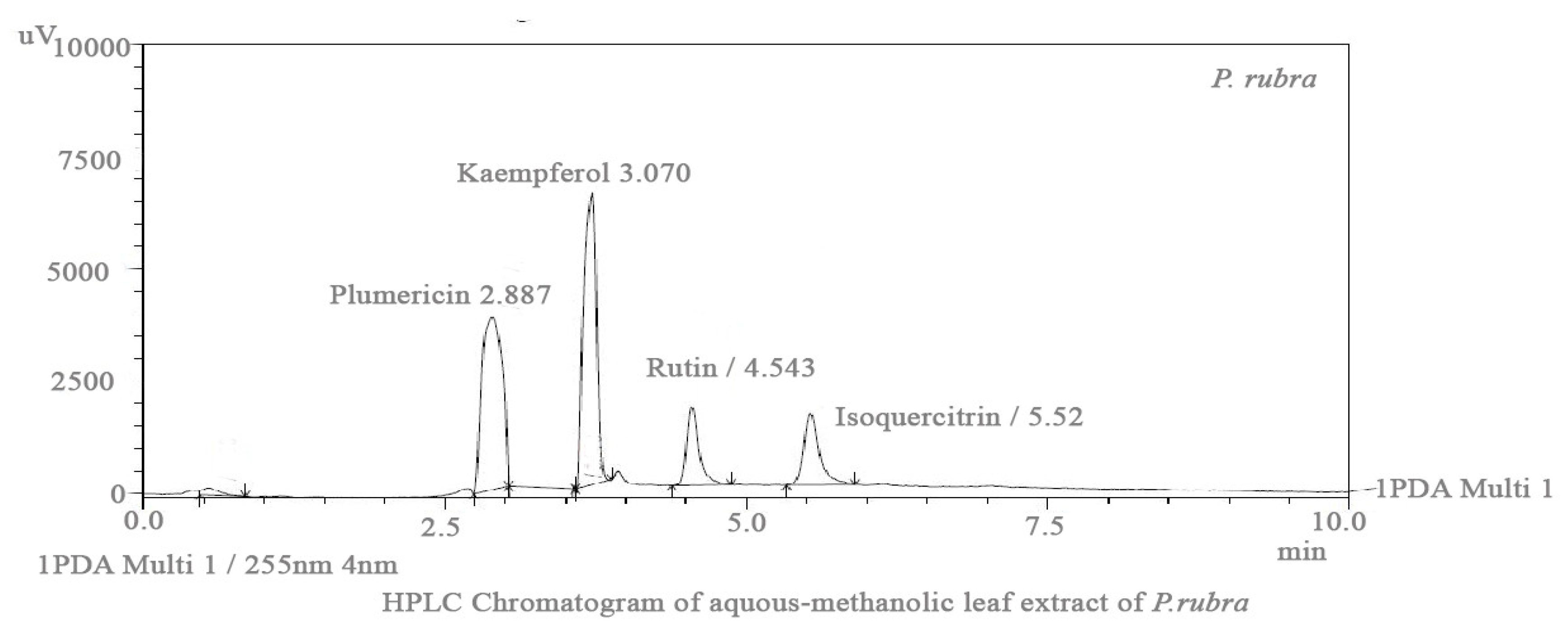
2.2. HPLC Analysis
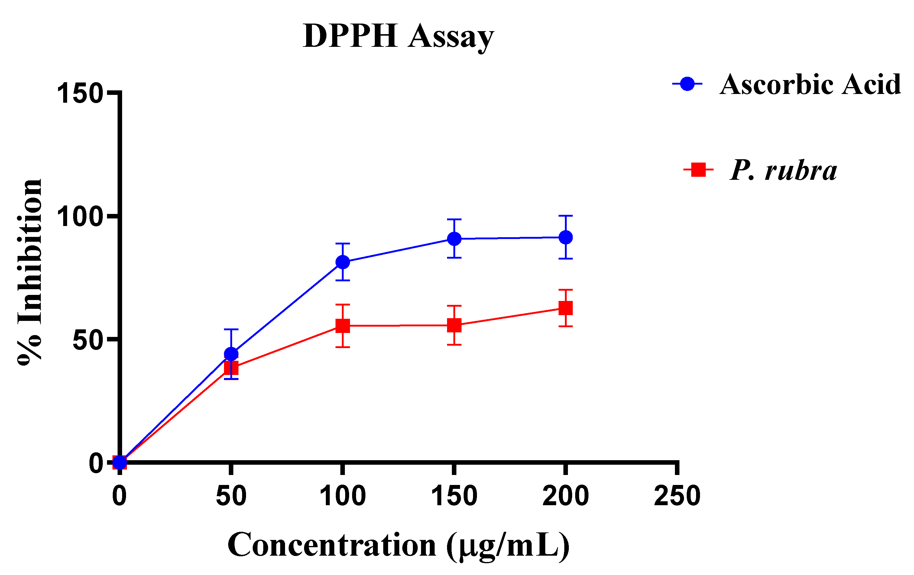
2.3. DPPH Assay
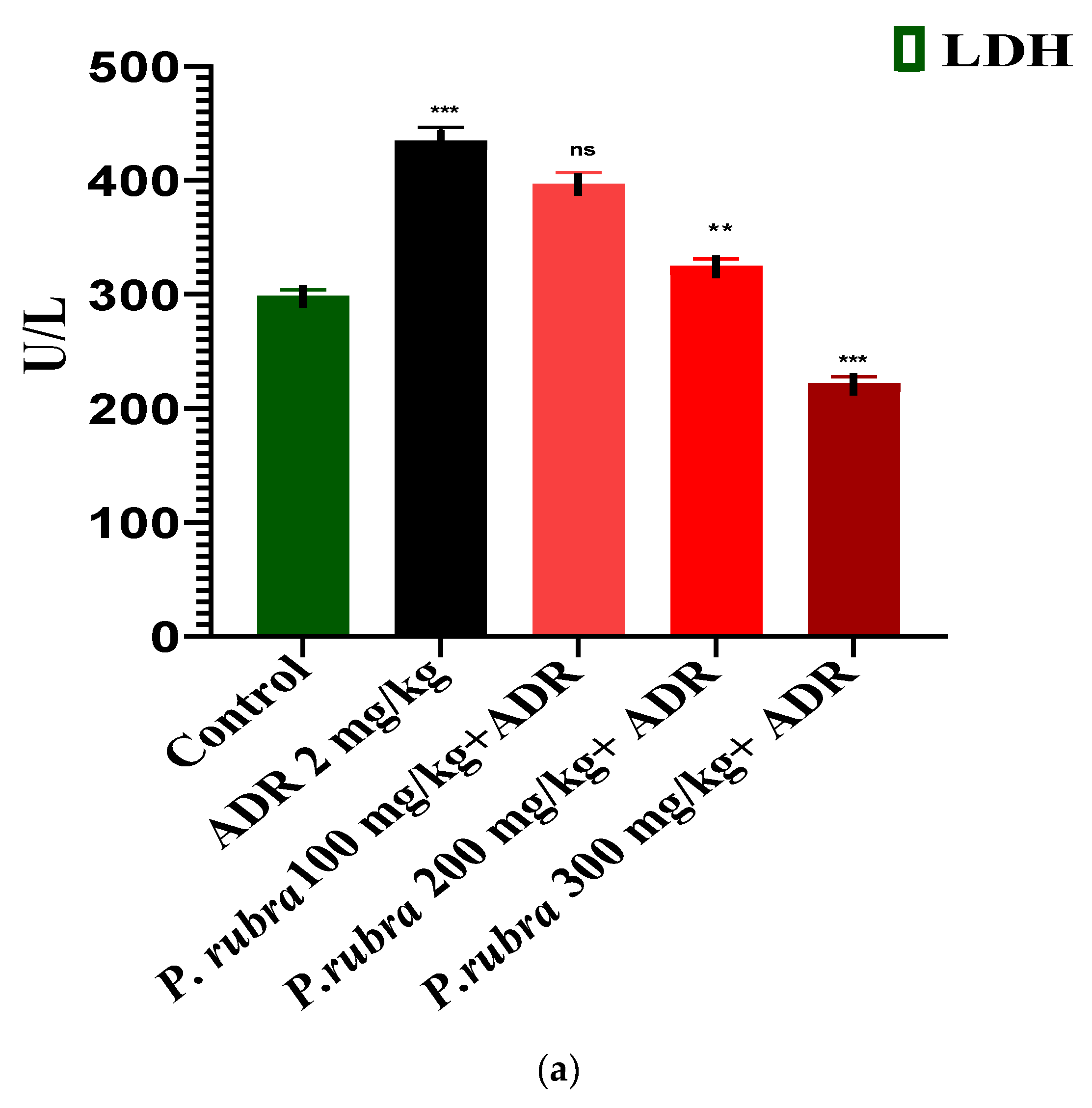
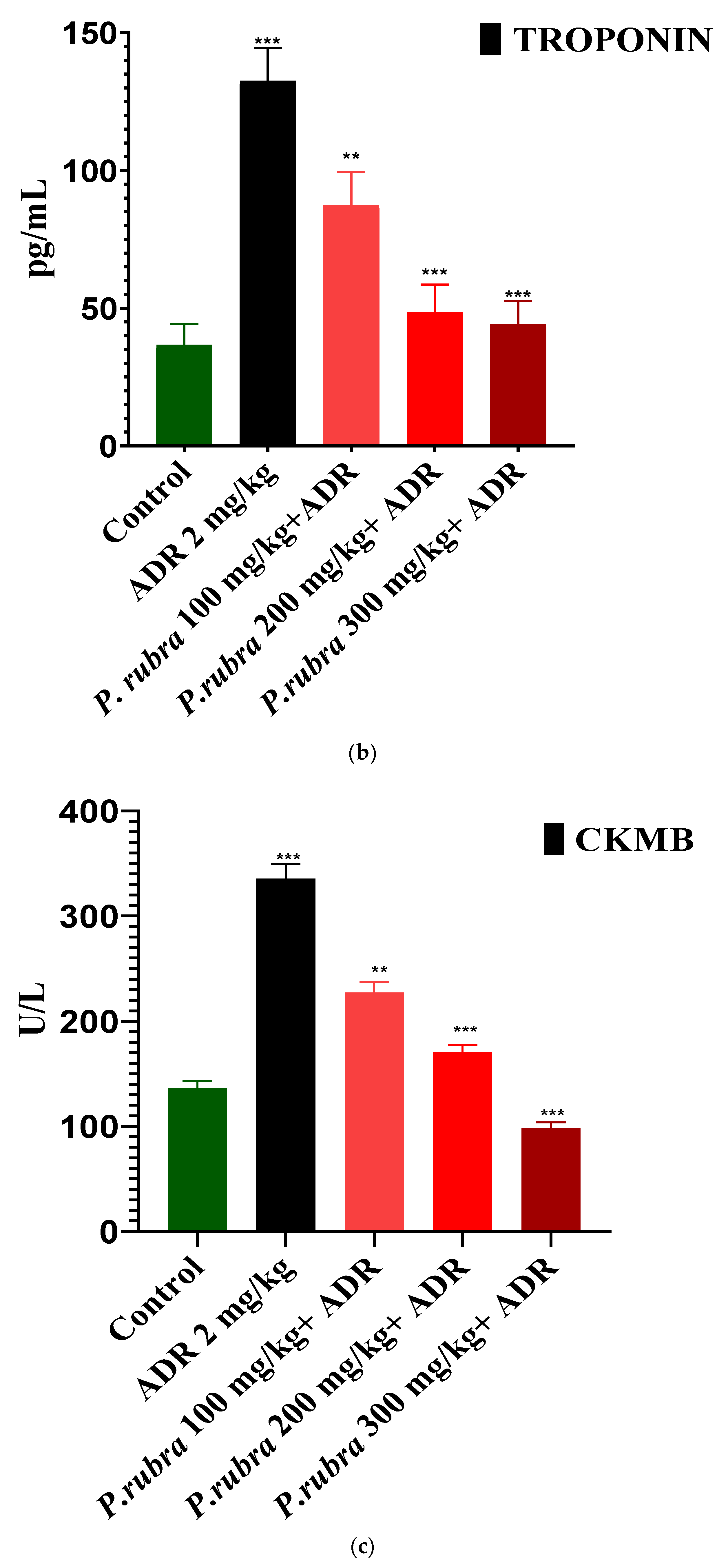
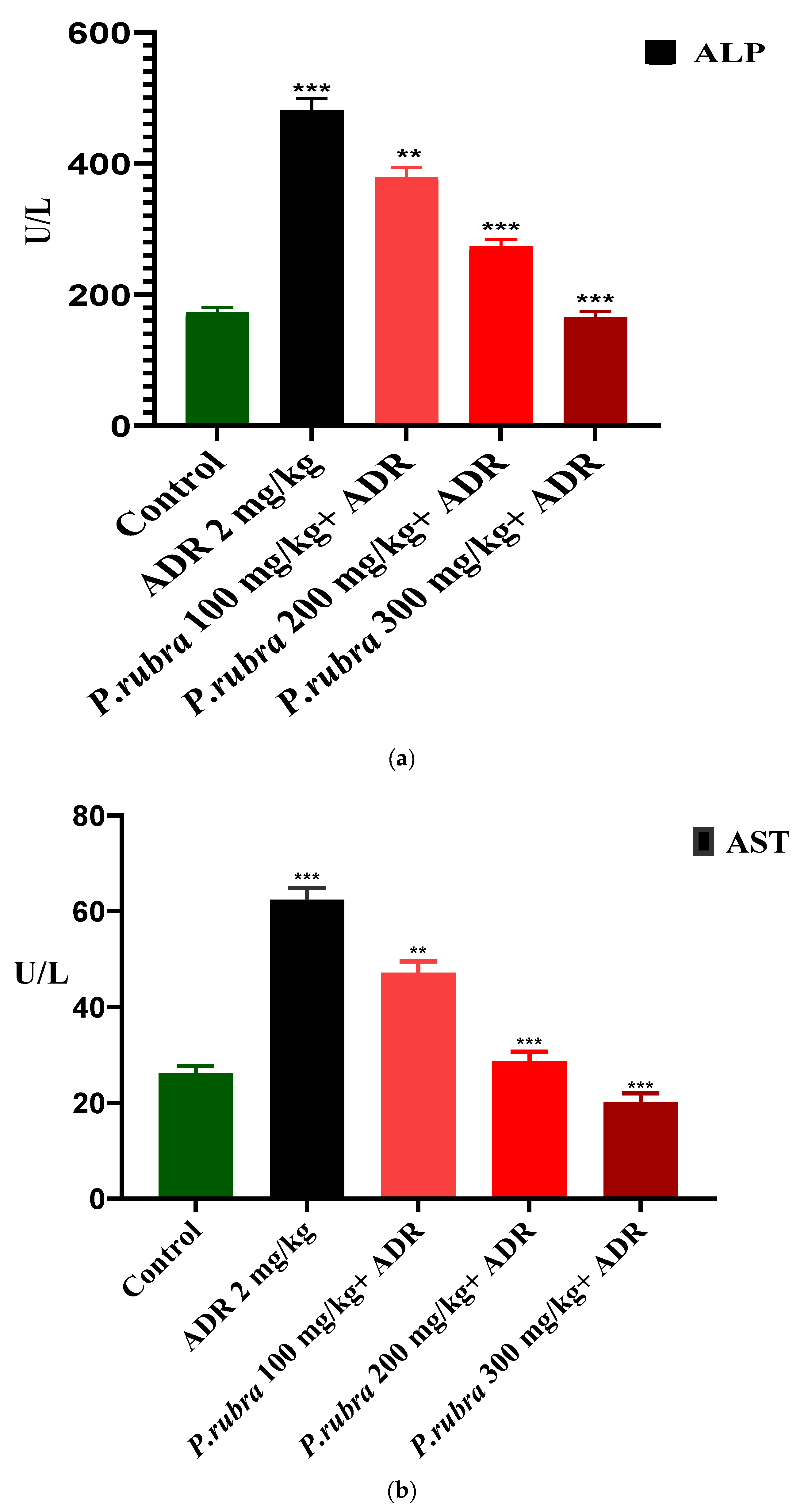
2.4. Evaluation of Myocardial Infarction
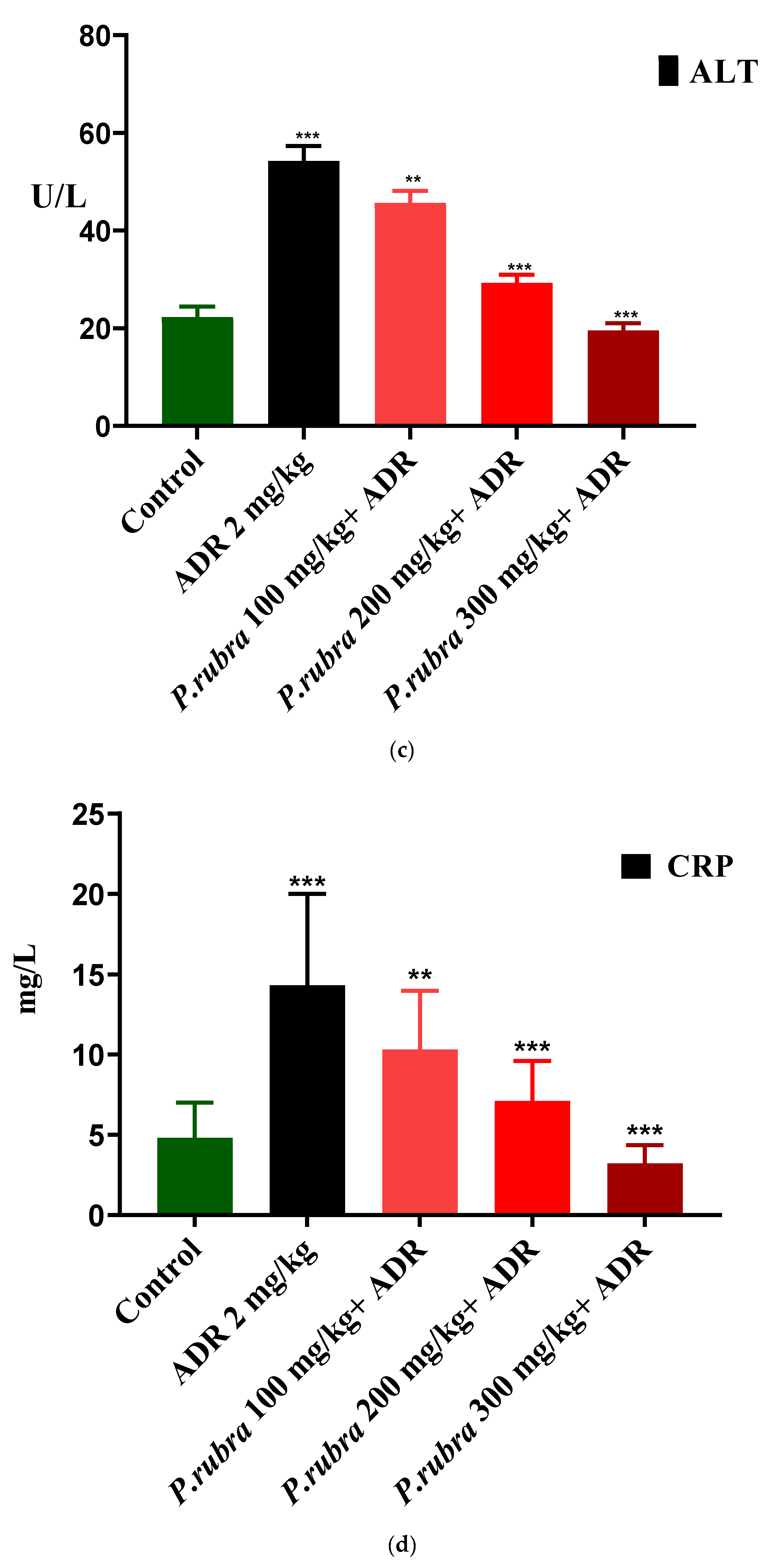
Effect on Heart to Bodyweight Ratio
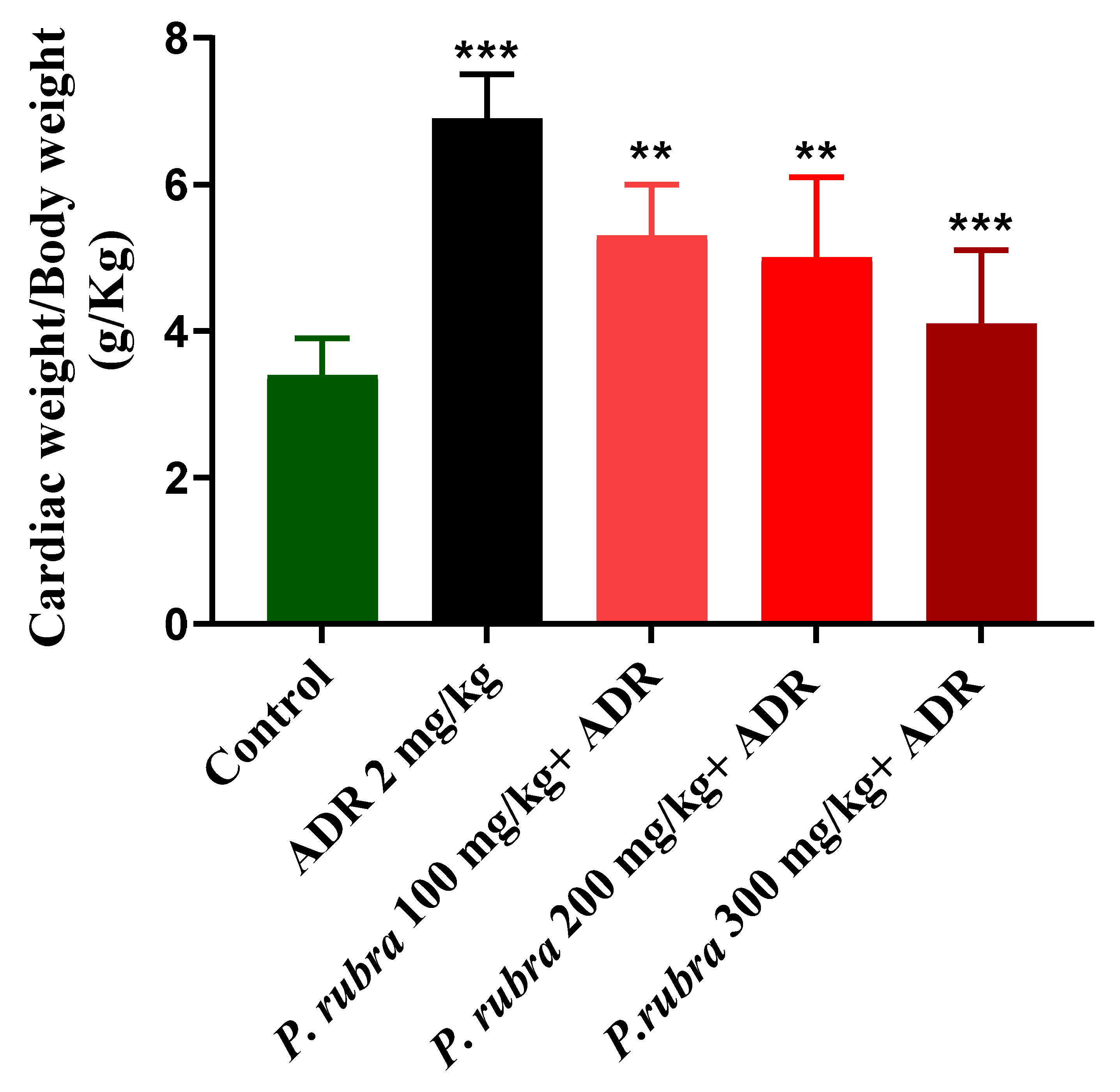
2.5. Histopathology
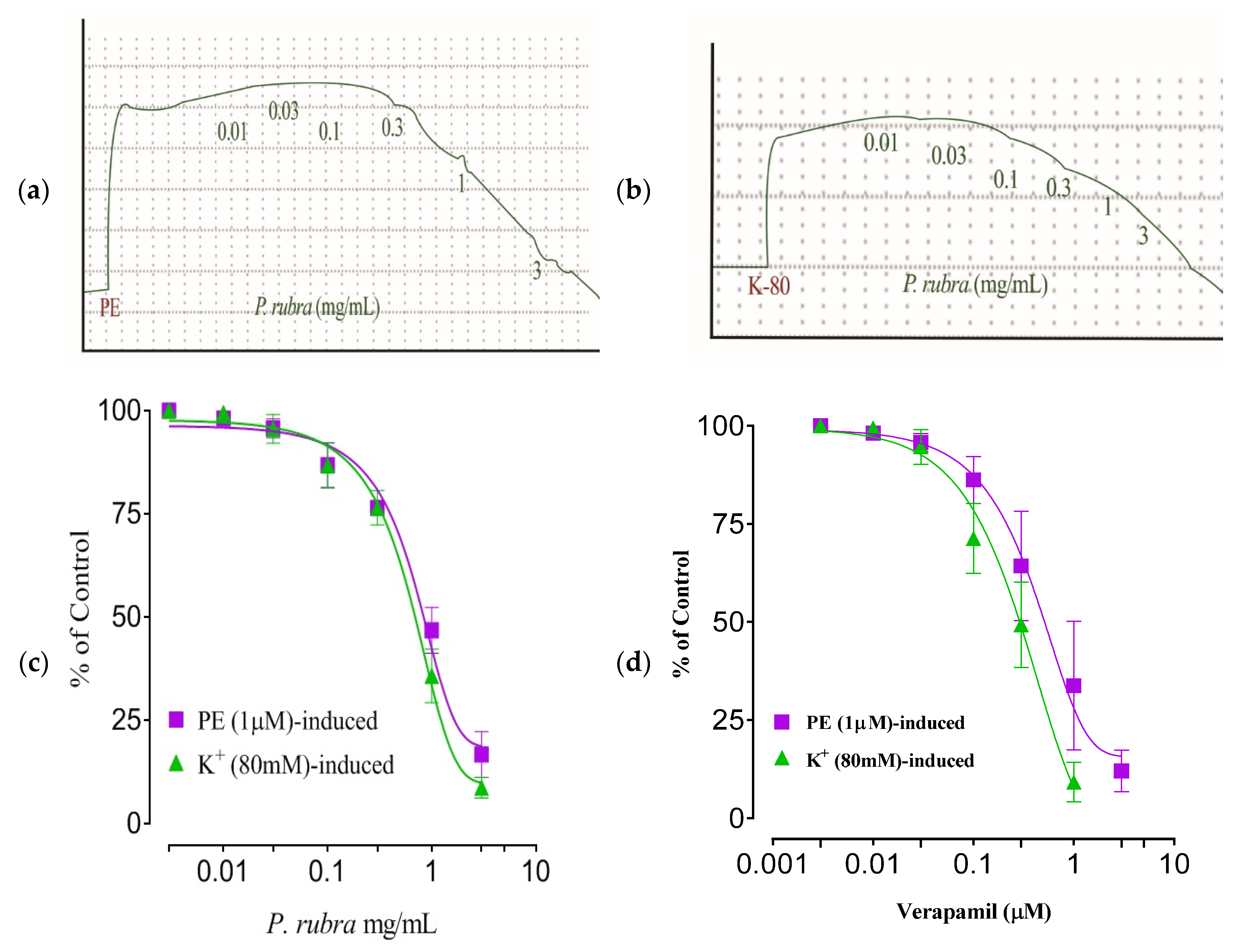
2.6. Aortic Tissue Preparation and Vasorelaxant Activity
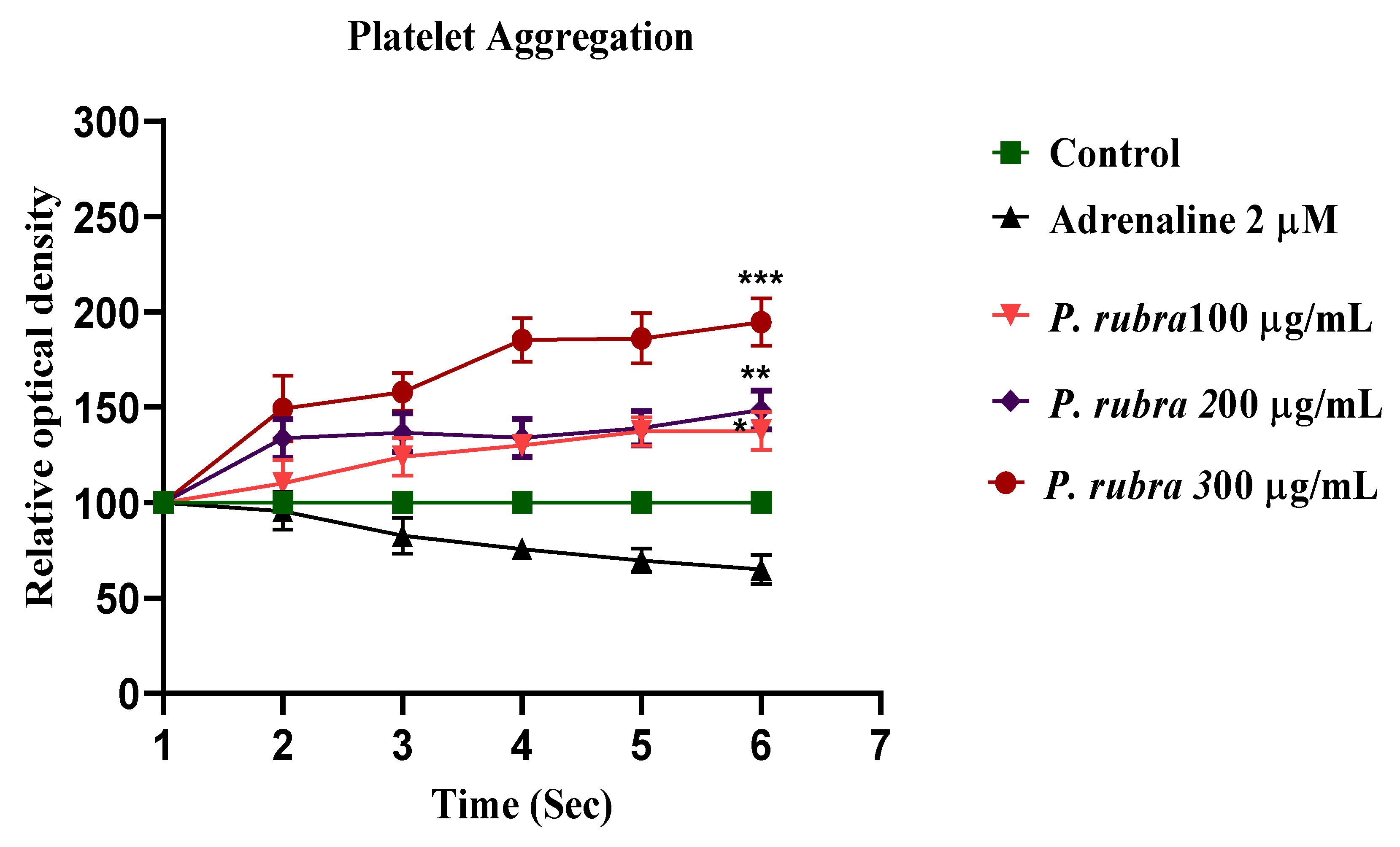
2.7. Antiplatelet Aggregatory Effect
2.8. Acute Oral Toxicity Dose Test
3. Discussion
4. Materials and Methods
4.1. Plant Materials
4.2. Extract Preparation
4.3. Animals
4.4. Chemicals
4.5. Preliminary Phytochemical Evaluation
4.6. HPLC Analysis
4.7. Acute Oral Toxicity Dose Test
4.8. Determination of DPPH Assay
4.9. Acute Myocardial Infarction Study
Screening of Cardiac Weight to Bodyweight Ratio
4.10. Histopathology
4.11. Isolated Aortic Tissue Preparation and Vasorelaxant Activity
4.12. Calcium Channel Blocking Activity
4.13. Adrenaline-Induced Platelet Activation and Aggregation
4.14. Statistical Analysis
5. Conclusions
Author Contributions
Funding
Institutional Review Board Statement
Informed Consent Statement
Data Availability Statement
Acknowledgments
Conflicts of Interest
Sample Availability
References
- Saqib, F.; Ali, A.; Ahmedah, H.A.; Irimie, C.A.; Toma, S.I.; Popovici, B.E.; Moga, M.; Irimie, M. Cardioprotective, hypotensive and toxicological studies of Populusciliata (Wall. ex Royle). Biomed. Pharmacother. 2021, 142, 1–16. [Google Scholar] [CrossRef] [PubMed]
- Khan, I.A. Pharmacotherapeutic modifications in cardiopulmonary patients during COVID-19 outbreak. J. Coll. Phys. Surg. Pak. 2020, 30, 15–17. [Google Scholar]
- Ashraf, N.; Rashid, A.; Naz, U.; Ashraf, M.M.; Khaliq, S.; Khan, I.A.; Nazir, A.; Sarwar, A.; Asif, A. Detection of antibiotics residues in protein containing diets (meat and eggs) of human through different methods. J. Univ. Med. Coll. 2018, 9, 1–11. [Google Scholar]
- Quiñones, M.; Miguel, M.; Aleixandre, G. Beneficial effects of polyphenols on cardiovascular disease. Pharmacol. Res. 2013, 68, 125–131. [Google Scholar] [CrossRef] [PubMed]
- Khan, I.A.; Hussain, M.; Munawar, S.H.; Iqbal, M.O.; Arshad, S.; Manzoor, A.; Shah, M.A.; Abbas, K.; Shakeel, W.; Syed, S.K. Jasminum sambac: A Potential candidate for drug development to cure cardiovascular ailments. Molecules 2021, 26, 5664. [Google Scholar] [CrossRef] [PubMed]
- Saqib, F.; Arif Aslam, M.; Mujahid, K.; Marceanu, L.; Moga, M.; Ahmedah, H.T.; Chicea, L. Studies to elucidate the mechanism of cardio protective and hypotensive activities of Anogeissusacuminata (Roxb. ex DC.) in rodents. Molecules 2020, 25, 3471. [Google Scholar] [CrossRef] [PubMed]
- Amin, M.M.; El-Gazayerly, O.N.; Gawad, N.A.; Halim, S.M. Effect of formulation variables on design, in vitro evaluation of valsartan SNEDDS and estimation of its antioxidant effect in adrenaline-induced acute myocardial infarction in rats. Pharm. Dev. Technol. 2016, 21, 909–920. [Google Scholar] [CrossRef]
- Fathiazad, F.; Matlobi, S.; Khorrami, N. Phytochemical screening and evaluation of cardioprotective activity of ethanolic extract of Ocimumbasilicum L. (basil) against isoproterenol induced myocardial infarction in rats. DARU J. Pharm. Sci. 2012, 20, 198–203. [Google Scholar] [CrossRef] [Green Version]
- Tsoupras, A.; Lordan, R.; Zabetakis, I. Inflammation, not cholesterol, is a cause of chronic disease. Nutrients 2018, 10, 604. [Google Scholar] [CrossRef] [Green Version]
- Shaito, A.; Thuan, D.T.B.; Nguyen, T.H.D.; Hasan, H.; Halabi, S.; Abdelhady, S.; Nasrallah, G.K.; Pintus, G. Herbal medicine for cardiovascular diseases: Efficacy, mechanisms, and safety. Front. Pharmacol. 2020, 11, 422–428. [Google Scholar] [CrossRef] [Green Version]
- Khan, I.A.; Aziz, A.; Raza, M.A.; Saleem, M.; Bashir, S.; Alvi, A. Study pertaining to the hypothermic activity of Plumeria rubra, Linn. in prostaglandin 1 and typhoid vaccine-induced pyrexia models in rabbits. West Indian Med. J. 2015, 12, 1. [Google Scholar] [CrossRef]
- Khan, I.A.; Lodhi, A.H.; Munawar, S.H.; Manzoor, A.; Manzoor, Z.; Raza, M.A. Dermatological evaluation of anti-Irritant and anti-Inflammatory effect of Plumerin-R isolated from the latex of Plumeria rubra Linn. Lat. Am. J. Pharm. 2018, 37, 317–320. [Google Scholar]
- Bihani, T. Plumeria rubra L.–A review on its ethnopharmacological, morphological, phytochemical, pharmacological and toxicological studies. J. Ethnopharmacol. 2021, 264, 113291. [Google Scholar] [CrossRef]
- Aziz, A.; Khan, I.A.; Munawar, S.H.; Shaheed, S. Antipyretic activity of methanolic bark extract of Plumeria rubra Linn. in various pyrexia induced models. Int. J. Res. Dev. Pharm. Life Sci. 2013, 2, 680–685. [Google Scholar]
- Khare, C.P. Encyclopedia of Indian Medicinal Plants, Rational Western Therapy. Ayurvedic and Other Traditional Usage, Botany; Springer: Berlin/Heidelberg, Germany, 2004; pp. 314–315. [Google Scholar]
- Omer, M.I.; Khan, I.A.; Manzoor, M.; Arshad, S.; Sial, A.S. Cardioprotective effect of hydroalcoholic leaf extract of Jatropha mollissima on isoproterenol-induced myocardial infarction in rats. Pharm. Mag. 2021, 17, 251–254. [Google Scholar]
- Omer, M.I.; Akhtar, I.; Rizvi, A.A.; Arshad, S.; Sial, A.S. The nephroprotective effects of Daucus carota and Eclipta prostrata against cisplatin-induced nephrotoxicity in rats. Metabolism 2021, 12, 12702–12721. [Google Scholar]
- Hawa, N.S.; Juriyati, J.A.; Yusof, A.; Yusof, K. Roles of rutin in cardiac remodeling. J. Funct. Foods 2020, 64, 606–619. [Google Scholar]
- Huang, S.H.; Xu, M.; Wu, H.M.; Wan, C.X.; Wang, H.B.; Wu, Q.Q.; Liao, H.H.; Deng, W.; Tang, Q.Z. Isoquercitrin attenuated cardiac dysfunction via AMPKα-dependent pathways in LPS-treated mice. Mol. Nutr. Food Res. 2018, 62, 303–321. [Google Scholar] [CrossRef]
- Feng, H.; Cao, J.; Zhang, G.; Wang, Y. Kaempferol attenuates cardiac hypertrophy via regulation of ASK1/MAPK signaling pathway and oxidative stress. Planta Med. 2017, 83, 837–845. [Google Scholar] [CrossRef] [PubMed] [Green Version]
- Bucki, R.; Pastore, J.J.; Giraud, F.; Sulpice, J.C.; Janmey, P.A. Flavonoid inhibition of platelet procoagulant activity and phosphoinositide synthesis. J. Thromb. Haemost. 2003, 1, 1820–1828. [Google Scholar] [CrossRef] [PubMed] [Green Version]
- Khan, I.A.; Aziz, A.; Saqib, F.; Munawar, S.H.; Manzoor, A.; Manzoor, Z.; Raza, M.A. Pharmacological evaluation of Rumex vesicarius Linn leaf extract and fractions in rabbit gastrointestinal ailment. Afr. J. Pharm. Pharmacol. 2014, 8, 333–341. [Google Scholar]
- Panchal, S.K.; Brown, L. Cholesterol versus Inflammation as Cause of Chronic Diseases. Nutrients 2019, 11, 2332. [Google Scholar] [CrossRef] [Green Version]
- Salma, A.; El-Marasy, S.A.; Awdan, E.; Hassan, H.; Heba, M.I. Cardioprotective effect of thymol against adrenaline-induced myocardial injury in rats. Heliyon 2020, 7, 35–39. [Google Scholar]
- He, Y.; Hu, Z.; Li, A.; Zhu, Z.; Yang, N.; Ying, Z. Recent advances in biotransformation of saponins. Molecules 2019, 24, 2365. [Google Scholar] [CrossRef] [Green Version]
- Dong, Y.; Duan, L.; Chen, H.W.; Liu, Y.M.; Zhang, Y. Network pharmacology-based prediction and verification of the targets and mechanism for Panax notoginseng saponins against coronary heart disease. Evid.-Based Complement. Altern. Med. 2019, 65, 37–52. [Google Scholar] [CrossRef] [PubMed]
- Mundugaru, R.; Udaykumar, P.; Senthilkumar, S.; Bhat, S. Cardioprotective activity of fruit of Garcinia pedunculata on isoprenaline induced myocardial infarction in rat. J. Pharmacol. 2018, 11, 231–235. [Google Scholar] [CrossRef] [Green Version]
- Prince, P.S.; Rajakumar, S.; Dhanasekar, K. Protective effects of vanillic acid on electrocardiogram, lipid peroxidation, antioxidants, proinflammatory markers and histopathology in isoproterenol induced cardiotoxic rats. Eur. J. Pharmacol. 2011, 668, 233–240. [Google Scholar] [CrossRef] [PubMed]
- Sun, J.; Yu, X.; Huangpu, H.; Yao, F. Ginsenoside Rb3 protects cardiomyocytes against hypoxia/reoxygenation injury via activating the antioxidation signaling pathway of PERK/Nrf2/HMOX1. Biomed. Pharmacother. 2019, 109, 254–261. [Google Scholar] [CrossRef] [PubMed]
- Satoh, S.; Nishida, S. Electropharmacological actions of Ginkgo biloba extract on vascular smooth and heart muscles. Clin. Chim. Acta. 2004, 342, 13–22. [Google Scholar] [CrossRef] [PubMed]
- Wang, X.L. Potential herb-drug interaction in the prevention of cardiovascular diseases during integrated traditional and western medicine treatment. Chin. J. Integr. Med. 2015, 21, 3–9. [Google Scholar] [CrossRef] [PubMed]
- Khan, I.A.; Lodhi, A.H.; Munawar, S.H.; Manzoor, A.; Manzoor, Z.; Raza, M.A. Formulation and evaluation of allicin and curcumin gel improves normal and diabetic ulcers in rabbits. Lat. Am. J. Pharm. 2018, 37, 1602–1607. [Google Scholar]
- National Institute of Health. Guide for the Care and Use of Laboratory Animals; No. 85–23; NIH Publication: Bethesda, MD, USA, 1985. [Google Scholar]
- Trease, G.E.; Evans, W.C. Trease and Evans’ Pharmacognosy, 16th ed.; Saunders/Elsevier: Edinburgh, Australia, 2009. [Google Scholar]
- Khan, I.A.; Lodhi, A.H.; Munawar, S.H.; Manzoor, A.; Manzoor, Z.; Raza, M.A.; Iqbal, O. Assessment of ameliorative effect ofAab-e-Shifa polyherbal formulation in experimentally-induced wound in rabbits. Trop. J. Pharm. Res. 2020, 19, 2357–2362. [Google Scholar]
- Chowdhur, A.; Dasgupta, B.; Kalita, G. Hepatoprotective activity of Plumeria alba extract against paracetamol induced-hepatotoxicity in rats. Int. J. Pharm. Pharm. Sci. 2012, 4, 618–620. [Google Scholar]
- Khan, I.A.; Lodhi, A.H.; Munawar, S.H.; Manzoor, A.; Manzoor, Z.; Raza, M.A. Formulation and evaluation of rutin-allicin gelagainst diabetic foot ulcer. Lat. Am. J. Pharm. 2020, 39, 725–729. [Google Scholar]
- Imtiaz, S.M.; Aleem, A.; Saqib, F.; Ormenisan, A.N.; Elena Neculau, A.; Anastasiu, C.V. The Potential involvement of an ATP-dependent potassium channel-opening mechanism in the smooth muscle relaxant properties of Tamarixdioica Roxb. Biomolecules 2019, 9, 722. [Google Scholar] [CrossRef] [PubMed] [Green Version]
- Isik, B.S.; Altay, F.; Capanoglu, E. The uniaxial and coaxial encapsulations of sour cherry (Prunus cerasus L.) concentrate byelectro spinning and their in vitro bioaccessibility. Food Chem. 2018, 265, 260–273. [Google Scholar] [CrossRef] [PubMed]
- Gilani, A.H.; Janbaz, K.H.; Zaman, H.; Lateef, A.; Suria, A. Possible presence of calcium channel blocker(s) in Rubiacordifolia: An indigenous medicinal plant. JPMA 1994, 44, 84–89. [Google Scholar]
- Singh, S.; Malm, C.J.; Ramström, S.; Hesse, C.; Jeppsson, A. Adrenaline enhances in vitro platelet activation and aggregation inblood samples from ticagrelor-treated patients. Res. Pract. Thromb. Haemost. 2018, 2, 718–725. [Google Scholar] [CrossRef] [Green Version]


| Tests | Observation | Results |
|---|---|---|
| Alkaloid | PPT | Present |
| Phenols | Light purple colour | Present |
| Tannins | Light purple colour | Present |
| Coumarins | Yellow fluorescence | Present |
| Saponins | 1 cm froth | Present |
| Anthraquinones | Pink colour | Present |
| Flavonoid | Light yellow colour | Present |
Publisher’s Note: MDPI stays neutral with regard to jurisdictional claims in published maps and institutional affiliations. |
© 2021 by the authors. Licensee MDPI, Basel, Switzerland. This article is an open access article distributed under the terms and conditions of the Creative Commons Attribution (CC BY) license (https://creativecommons.org/licenses/by/4.0/).
Share and Cite
Khan, I.A.; Hussain, M.; Syed, S.K.; Saadullah, M.; Alqahtani, A.M.; Alqahtani, T.; Aldahish, A.A.; Asiri, S.; Zeng, L.-H. Pharmacological Justification for the Medicinal Use of Plumeria rubra Linn. in Cardiovascular Disorders. Molecules 2022, 27, 251. https://doi.org/10.3390/molecules27010251
Khan IA, Hussain M, Syed SK, Saadullah M, Alqahtani AM, Alqahtani T, Aldahish AA, Asiri S, Zeng L-H. Pharmacological Justification for the Medicinal Use of Plumeria rubra Linn. in Cardiovascular Disorders. Molecules. 2022; 27(1):251. https://doi.org/10.3390/molecules27010251
Chicago/Turabian StyleKhan, Imran Ahmad, Musaddique Hussain, Shahzada Khurram Syed, Malik Saadullah, Ali M. Alqahtani, Taha Alqahtani, Afaf A. Aldahish, Saeed Asiri, and Ling-Hui Zeng. 2022. "Pharmacological Justification for the Medicinal Use of Plumeria rubra Linn. in Cardiovascular Disorders" Molecules 27, no. 1: 251. https://doi.org/10.3390/molecules27010251