Article

Mammalian Arginase Inhibitory Activity of Methanolic Extracts and Isolated Compounds from Cyperus Species

1
PEPITE EA 4267, FHU INCREASE, University Bourgogne Franche-Comté, 25000 Besançon, France
2
MIB-UR Oenologie, EA 4577, USC 1366 INRA, University of Bordeaux, ISVV, 33882 Villenave d’Ornon, France
*
Author to whom correspondence should be addressed.
Molecules 2021, 26(6), 1694; https://doi.org/10.3390/molecules26061694
Submission received: 16 February 2021 / Revised: 12 March 2021 / Accepted: 14 March 2021 / Published: 18 March 2021
(This article belongs to the Collection Bioactive Compounds)

Abstract

:
Polyphenolic enriched extracts from two species of Cyperus, Cyperus glomeratus and Cyperus thunbergii, possess mammalian arginase inhibitory capacities, with the percentage inhibition ranging from 80% to 95% at 100 µg/mL and 40% to 64% at 10 µg/mL. Phytochemical investigation of these species led to the isolation and identification of two new natural stilbene oligomers named thunbergin A-B (12), together with three other stilbenes, trans-resveratrol (3), trans-scirpusin A (4), trans-cyperusphenol A (6), and two flavonoids, aureusidin (5) and luteolin (7), which were isolated for the first time from C. thunbergii and C. glomeratus. Structures were established on the basis of the spectroscopic data from MS and NMR experiments. The arginase inhibitory activity of compounds 17 was evaluated through an in vitro arginase inhibitory assay using purified liver bovine arginase. As a result, five compounds (1, 47) showed significant inhibition of arginase, with IC50 values between 17.6 and 60.6 µM, in the range of those of the natural arginase inhibitor piceatannol (12.6 µM). In addition, methanolic extract from Cyperus thunbergii exhibited an endothelium and NO-dependent vasorelaxant effect on thoracic aortic rings from rats and improved endothelial dysfunction in an adjuvant-induced arthritis rat model.

Graphical Abstract

1. Introduction

Arginase is a trimeric metalloenzyme hydrolyzing l-arginine to l-ornithine and urea. Arginase plays an important role in ammonia detoxification in mammals [1,2], but also in the regulation of the production of many key biological intermediates, such as polyamines (via l-ornithine), which are responsible for cell proliferation and collagen production (wound healing), and nitric oxide (NO) (via l-arginine availability), a strong vasodilating agent [1,3,4]. However, it is well known that excessive arginase activity can produce l-ornithine oversupply, which is responsible for tissue stiffening, and L-arginine depletion, which is responsible for a decrease in NO availability, by substrate competition with NO synthase [5]. This contribute to the appearance of endothelial dysfunction, which can be observed in various diseases for which an arginase overactivity has been implicated (inflammatory, cardiovascular, or cancer diseases) [6,7]. Previous studies conducted on animal models or in humans showed that inhibition of arginase enhanced NO bioavailability thereby restoring normal vascular function [8]. Even though this represents a new therapeutic strategy [9], no drug has been developed. Among the few synthetic arginase inhibitors commercially available, boronic acid derivatives (S-(2-boronoethyl)-l-cysteine (BEC), 2-(S)-amino-6-boronohexanoic acid (ABH), and Nω-hydroxy-nor-L-arginine (nor-NOHA) are the most potent, but their toxicity and poor pharmacokinetic profile limits their potential therapeutic use in humans [10]. Finding new arginase inhibitors suitable for the treatment of endothelial dysfunction associated with several diseases in humans still poses a challenge. Therefore, natural substances constitute a promising source in this area [11]. Among naturally occurring metabolites, flavonoids and stilbenoids have been shown to be the most active on arginase [11,12]. Cyperus genus attracted our attention because it possesses a wealth of these kinds of compounds.
The genus Cyperus belongs to the Cyperaceae family and includes more than 900 species distributed worldwide. Cyperus species constitute one of the three most widely and traditionally used genera in China [13], and Cyperus rotundus L., is prevalent in several systems of medicine (Ayurveda, Traditional Chinese Medicine, and medicine from Japan and Iran), where it is commonly used for treating a large variety of diseases (infectious and parasitic diseases, cancers, inflammation). Numerous pharmacological studies provide scientific evidence of the biological activities of these plants [14,15] because they possess an abundance of different phytochemicals. In general, Cyperus are renowned sources of biologically active compounds, such as essential oils [16], terpenes [17,18], coumarins, flavonoids [19,20,21], and stilbenes [21,22,23]. We recently isolated stilbene oligomers scirpusin B and cyperusphenol B from Cyperus eragrostis and demonstrated their in vitro inhibitory activity against liver bovine arginase [23]. In order to extend the research initiated on Cyperus genus, based on a survey of traditional uses and literature, we chose to continue with the study of the aerial parts of Cyperus thunbergii and Cyperus glomeratus. Arginase inhibitory activities of polyphenolic enriched extracts were investigated by means of in vitro and ex vivo studies. The isolation, identification, and arginase inhibitory activity of seven polyphenolic compounds, including two new stilbene oligomers, are also reported here.

2. Results and Discussion

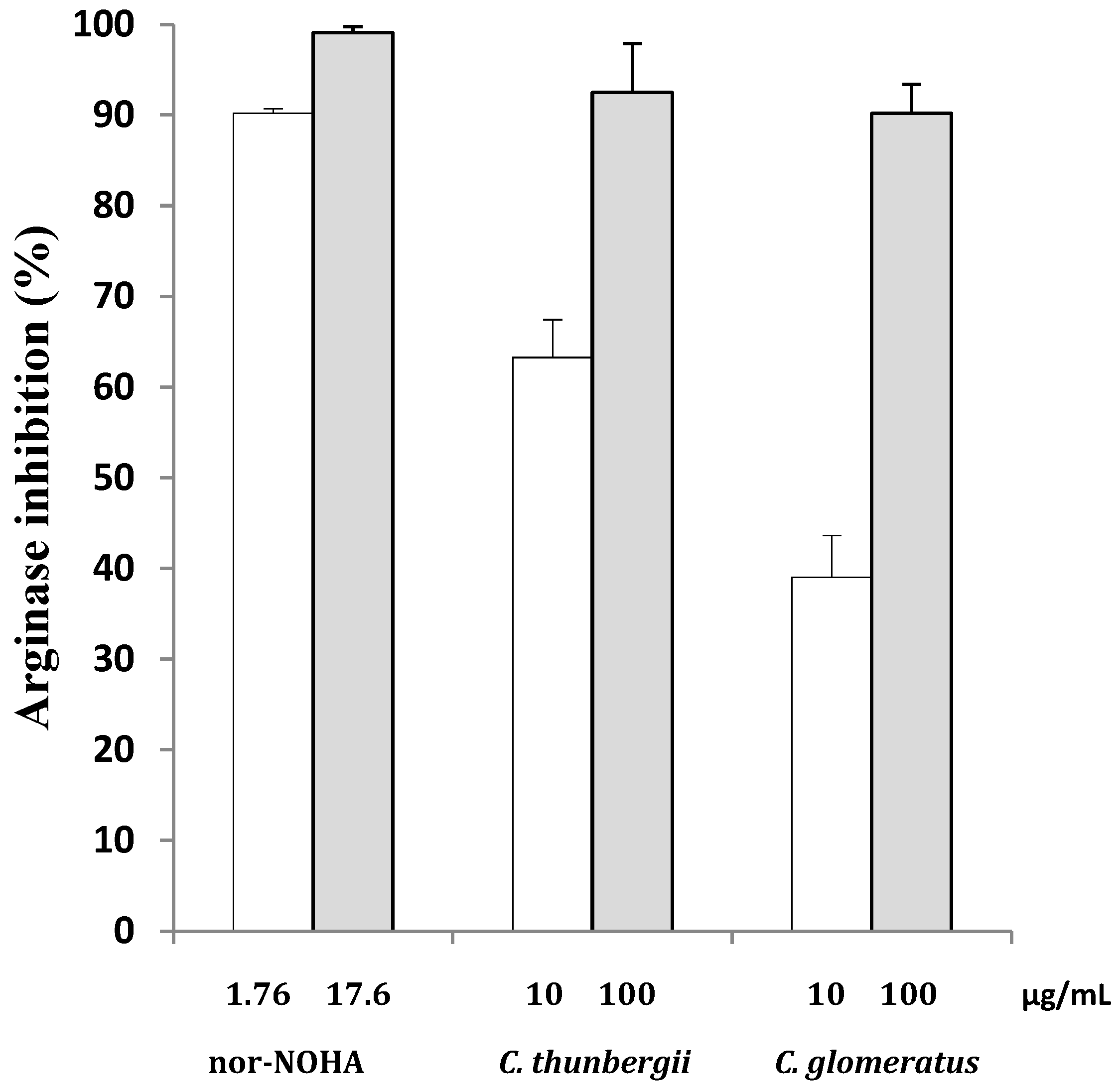
2.1. In Vitro Arginase Inhibitory Activity of Cyperus thunbergii and C. glomeratus

The aerial parts of two Cyperus species, Cyperus thunbergii and Cyperus glomeratus, were extracted with methanol (MeOH) using maceration at room temperature. The dried extracts were subsequently subjected to solid phase extraction (SPE) in order to recover polyphenolic compounds. The in vitro evaluation [12] of these polyphenolic enriched extracts revealed that they were able to inhibit arginase (more than 80% inhibition at 100 µg/mL) (Figure 1).

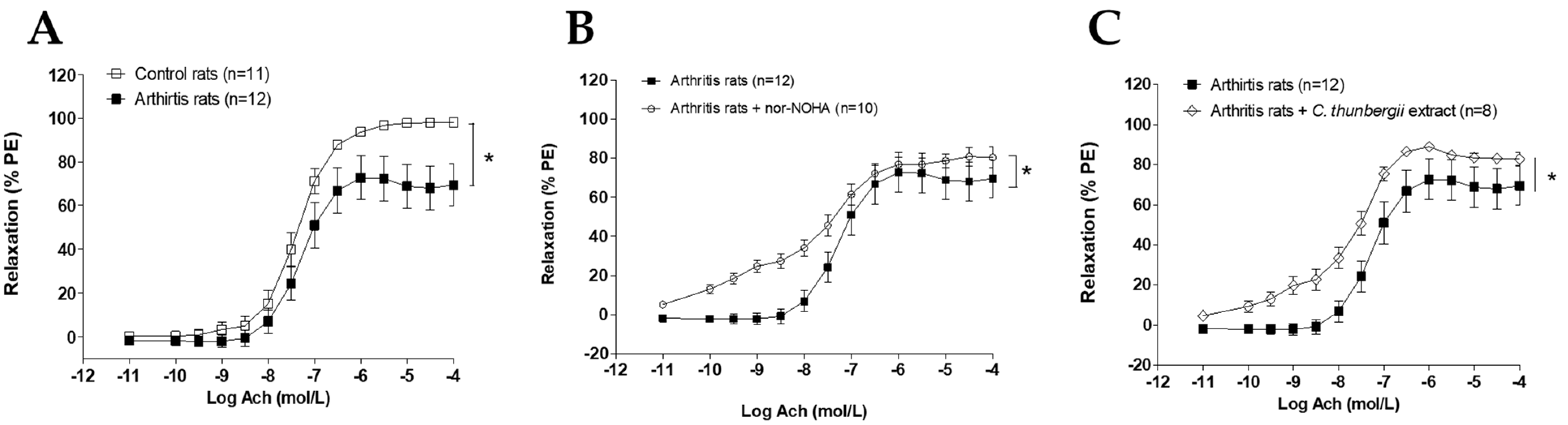
2.2. Ex Vivo C. thunbergii Improved Endothelial Dysfunction in Arthritic Rats

The most active polyphenolic enriched extract, obtained from C. thunbergii, was evaluated for its effect on arginase-related endothelial dysfunction in the rat model of arthritis. At a severe stage of the arthritis model, endothelial dysfunction on the aorta vascular bed was attested by the depressed endothelium-dependent vasorelaxation in comparison to controls (Figure 2A). As expected [6], acetylcholine (Ach)-induced relaxation was higher in the presence of arginase inhibitor nor-NOHA (Figure 2B). Interestingly, C. thunbergii extract was also able to significantly improve this depressed relaxation. These data confirmed the arginase inhibitor activity of the present extract in ex vivo conditions.

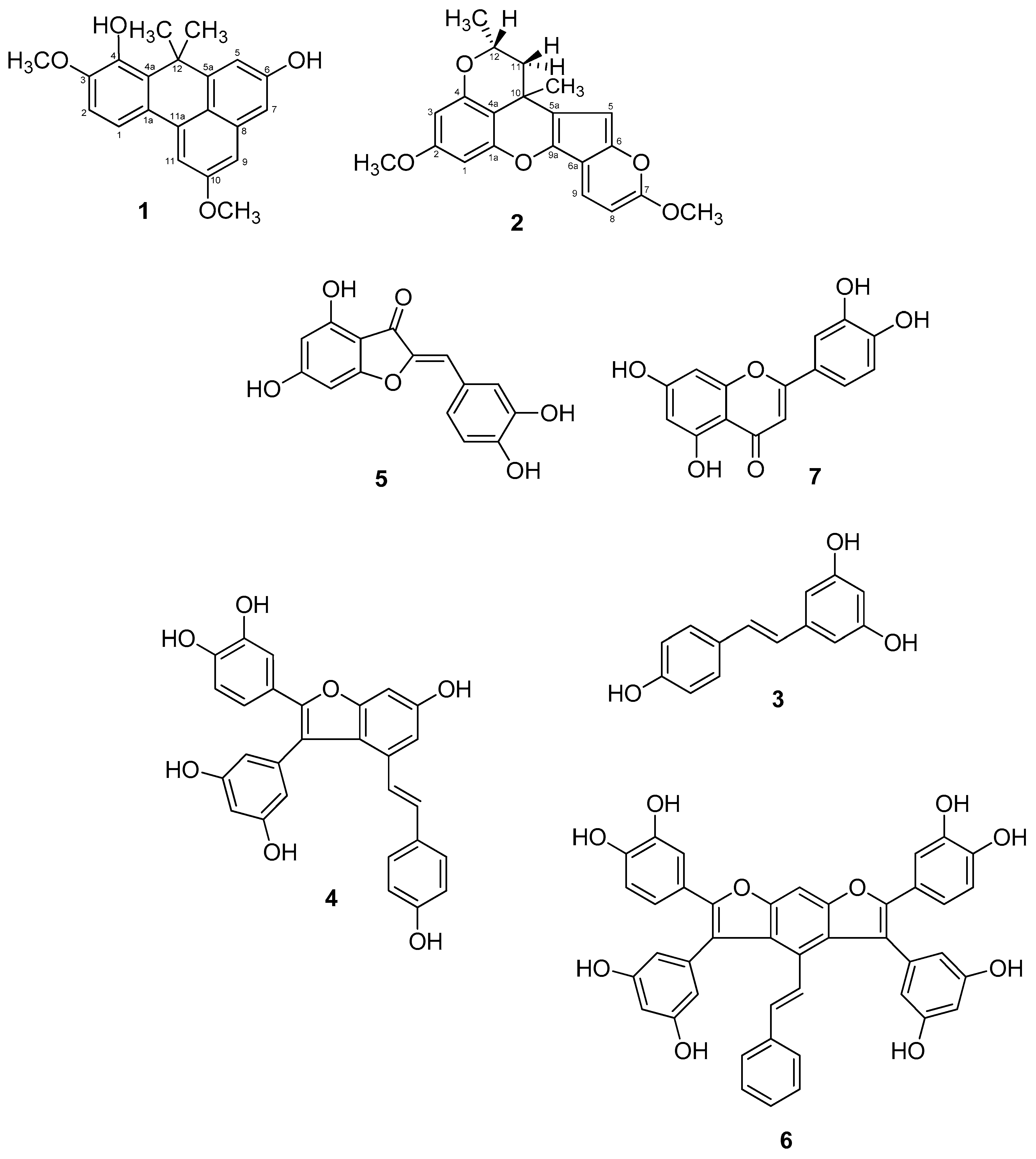
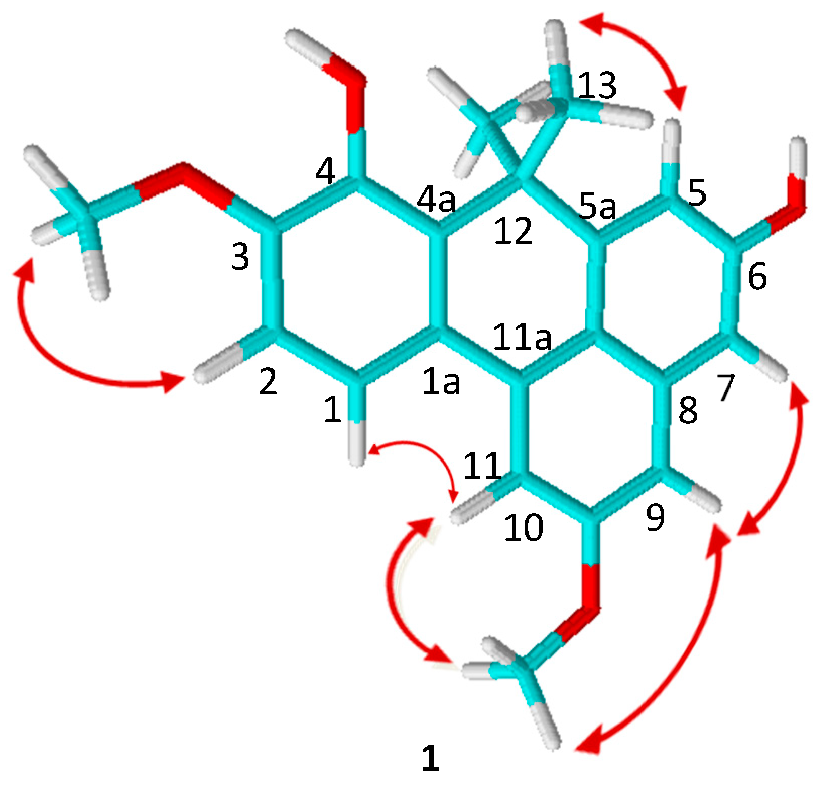
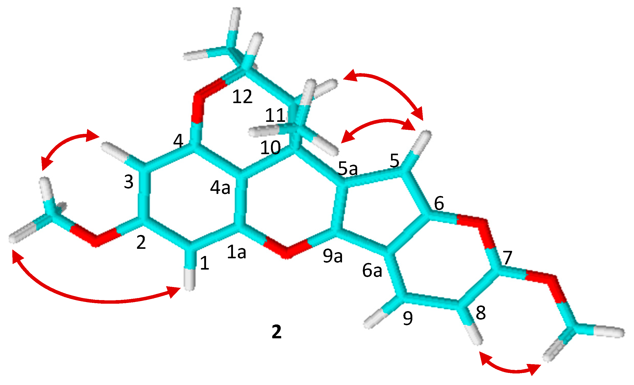
2.3. Isolation and Structural Elucidation of Compounds 17

In order to isolate active compounds, the phytochemical composition of both extracts was explored. This is the first phytochemical study of C. thunbergii and C. glomeratus. The reverse-phase preparative liquid chromatographic (PLC) investigation resulted in the isolation of two new stilbene compounds, thunbergin A (1) and thunbergin B (2), from C. thunbergii, together with five previously known compounds 37, from C. glomeratus (Figure 3).
The structures of 37 were established by comparing their observed data with those published in the literature, and identified as trans-resveratrol (3), trans-scirpusin A (4), aureusidin (5), trans-cyperusphenol A (6), and luteolin (7) (Table 1).
Compound 1 was obtained as a brownish powder amorphous solid. Its molecular formula, C21H20O4, was deducted from high resolution electrospray ionization mass spectroscopy (HRESIMS), through the presence of a peak at m/z 337.1432 [M + H]+. All 1H, 13C, and distortionless enhancement by polarization transfer (DEPT) nuclear magnetic resonance assignments for 1 were performed using 2D NMR spectroscopic data: heteronuclear simple quantum correlation (HSQC) (Figure S3), heteronuclear multiple bond correlation (HMBC) (Figure S4), correlation spectroscopy (COSY) (Figure S5), and nuclear Overhauser effect spectroscopy (NOESY) (Figure S6). Twenty-one carbon signals were observed in the 13C NMR spectrum (Figure S2), discriminated by the DEPT experiment into two methyl, two methoxyl (OCH3) groups, six methane, and eleven quaternary carbon signals. Analysis of the 1H NMR spectrum (Figure S1) in methanol-d6 immediately revealed the presence of two groups of ortho-coupled aromatic protons at δH 6.76 (1H, d, J = 8.4 Hz) and δH 6.84 (1H, d, J = 8.4 Hz), four groups of meta-coupled aromatic protons δH 6.17 (1H, d, J = 2.2 Hz), δH 6.80 (1H, d, J = 2.2 Hz), δH 7.15 (1H, d, J = 2.2 Hz), and δH 6.61 (1H, d, J = 2.2 Hz), two methyls at δH 1.59 (6H, s), and two signals corresponding to methoxyl groups at δH 3.69 (3H, s) and δH 3.81 (3H, s). (Table 2).
In the 1H–1H-COSY spectrum, correlations were observed between δH 6.76 d and δH 6.84 d (H-1/H-2), δH 6.61 d and δH 7.15 d (H-5/H-7), and δH 6.80 d and δH 6.17 d (H-9/H-11). The two methoxyl groups were placed to C-3 and C-10 as confirmed by HMBC correlations of the OCH3 (δH 3.81) with C-3 (δC 148.7) and the OCH3 (δH 3.69) with C-10 (δC 158.5) (Table 3). The location of the methyl groups was also concluded from the HMBC spectrum, as a proton signal at δH 1.59 (H6-13) showed correlations with δC 145.2 (C-4a), 130.2 (C-5a), and 36.9 (C-12). The hydroxyl groups linked to C-4 and C-6 were confirmed by the chemical shift of the quaternary carbons (δC-4 149.2 and δC-6 145.9).
The NOESY correlations further confirmed the structure of compound 1. Nuclear Overhauser effects were detected between H-2/H-1, H2/OCH3-3, H-5/H-13, H-9/H-7, H-9/OCH3-10, and H-11/OCH3-10 (Figure 4). All of the above confirmed the planar structure of compound 1 as 4,6-dihydroxy-12-dimethyl-3,10-dimethoxybenzophenanthrene, which was named thunbergin A (Figure 3).
Compound 2 was isolated as a brownish powder amorphous solid. It gave [M + H]+ at m/z 353.1386 in HRESIMS consistent with the molecular formula C21H20O5. All 1H (Figure S1), 13C (Figure S3), and J-modulated spin-echo (JMOD) NMR assignments for compound 2 were performed using 2D NMR spectroscopic data (HSQC (Figure S3), HMBC (Figure S4), COSY (Figure S5), NOESY (Figure S6)). Analysis of the 1H NMR spectrum in methanol-d6 (Table 4) displayed signals of two meta-coupled aromatic protons δH 6.63 (1H, d, J = 2.2 Hz, H-1) and δH 6.27 (1H, d, J = 2.2 Hz, H-3), two ortho-coupled aromatic protons δH 6.76 (1H, d, J = 8.4 Hz, H-8) and δH 6.65 (1H, d, J = 8.4 Hz, H-9), one aromatic proton as a singlet δH 6.66 (1H, s, H-5), two methyls, two methylenes, one sp3 methine, and signals of protons belonging to two methoxyls. In the JMOD spectrum, the presence of 21 carbon signals was detected (Table 3).
In the 1H–1H-COSY spectrum, correlations were observed between δH 6.63 d and δH 6.27 d (H-1/H-3), δH 6.76 d and δH 6.65 d (H-8/H-9), δH 2.73 dd and δH 3.08 m (H-11/H-12), and δH 2.96 dd and δH 3.08 m (H-11/H-12). The methine multiplet at δH 3.08, a methyl doublet at δ H 1.00, and two methylene protons at δH 2.73 dd and δH 2.96 dd provided evidence of the presence of a CH3CHCH2 structural unit (C-12, C-11) in the molecule. According to the 1H and 13C NMR signals at δ H 3.79 and δ C 54.4, and δ H 3.87 and δC 55.2, methoxyl groups could be identified and connected to C-2 and C-7, as confirmed by an HMBC correlation between OCH3 (δH 3.79) and C-2 (δC 160.5), and OCH3 (δH 3.87) and C-7 (δC 147.2) (Table 4). Moreover, on the basis of HMBC correlations between C-10/10-Me, C-11/10-Me, and C-5a/10-Me, the methyl groups (δH 1.79) were placed at C-10 (δC 38.0). On the basis of HMBC correlations between C-12/12-Me, C-11/12-Me, and C-10/12-Me, the methyl groups (δH 1.00) were placed at C-12 (δC 50.2). The NOESY correlations further confirmed the structure of compound 2. Nuclear Overhauser effects were detected between H-1/OCH3-2, H3/OCH3-2, H-5/H-11, H-5/C, H3-10, and H-8/OCH3-7 (Figure 4), which was named thunbergin B (Figure 3).
Table 1 shows the chromatographic data set and the m/z values of the isolated compounds. NMR and HRESIMS data were used to identify compounds 5 and 7 as new compounds from the Cyperus genus.
Aureusidin (5) belongs to the less studied subclass of flavonoids called aurones. This molecule, which rarely occurs in nature, was previously isolated from mosses marine brown algae and flowering plants [24,25]. Aureusidin possesses a pharmacological profile showing high antioxidant and lipoxygenase inhibitory activity [26], as well as anti-inflammatory effects [27,28]. Luteolin (7), a flavone, which is abundant in edible plants, displays a wide range of biological activities including antioxidant, anti-carcinogenic [29], cardioprotective [30], anti-inflammatory, and antipruritic [31] activities. Scirpusin A (4) and cyperusphenol A (6) are stilbene oligomers, most of which are potent antioxidants showing cardioprotective properties. Their structures result from the condensation of resveratrol and piceatannol. Scirpusin A (4), a hydroxystilbene dimer, acts as an effective singlet oxygen quencher and DNA damage protector [32].

2.4. Arginase Inhibitory Activity of Compounds 17

Compounds 17 were screened for their arginase inhibitory activity using purified bovine liver arginase [12]. Their IC50 values are indicated in Table 5.
The synthesized compound Nω-hydroxy-nor-l-arginine (nor-NOHA), a well-known reference inhibitor of arginase, was used as a positive control (IC50 = 1.7 ± 0.7 µM). Although all of the evaluated compounds remained less active than nor-NOHA, it should be noted that compound 4 (IC50 = 17.6 ± 2.2 µM), compound 6 (IC50 = 19.4 ± 1.3 µM), and compound 1 (IC50 = 28.8 ± 2.5 µM) all show an activity close to that of the natural inhibitor piceatannol (IC50 = 12.6 ± 0.6 µM), one of the most active natural compounds on mammalian arginase [12,23].

3. Materials and Methods

3.1. Reagents

All reagents were from Sigma-Aldrich (Saint-Quentin Fallavier, France). They were used without further purification, except for purified liver bovine arginase 1, which was purchased from MP Biomedicals (Illkirch-Graffenstaden, France) (one unit (1U) of bovine arginase corresponds to the amount of enzyme able to convert 1 µMol of L-arginine to urea and L-ornithine per minute at pH 9.5 and 37 °C). MeOH, acetonitrile (MeCN), and dimethylsulfoxide (DMSO) were obtained from two companies: Carlo Erba Reagents (Val de Reuil, France) and VWR Chemicals (Fontenay-sous-Bois, France). Deuterated solvents and trifluoroacetic acid (TFA) were purchased from Eurisotop (Tewksbury, MA, USA) and Fisher Scientific (Illkirch, France), respectively. Water was purified (resistivity > 18 mΏ/cm) using an water purification system (ELGA LabWater, UK).

3.2. Plant Materials

Cyperus thunbergii Vahl. (XX-0-TUEB-3630 ex JB Tubingen) and Cyperus glomeratus (FR-0-LYJB-005964W ex JB Lyon) aerial parts were collected in the Botanical Garden of Talence (Talence, France) between 2017 and 2018. Each plant was authenticated by one of the authors (A.B). The samples were thoroughly dried and kept free from moisture.

3.3. Extraction and Isolation

The aerial parts of C. thunbergii and C. glomeratus were ground into powder. A sample of each (80 g) was extracted, then macerated and stirred in methanol at room temperature (600 mL × 5 × 24 h). The methanolic solutions were recovered by filtration, then pooled and concentrated under reduced pressure to obtain dry extracts. These crude extracts (12 g) were dissolved in 30% MeOH (1 g of extract in 600 µL of MeOH and 1.4 mL of H2O) by vortexing and sonicating. Each extract was pre-purified using a solid phase extraction (SPE) mini column Strata® C18-E (55 µM, 70Å). Each sample (2 mL) was loaded onto the C18 mini column, washed with H2O 4 mL water, and then eluted with 90% MeOH. The recovered solution contained polyphenols. Each extract was evaporated until dry, using the same vacuum evaporator. Before HPLC analyses, the dried extract was redissolved in 50% MeOH HPLC grade by vortexing and sonicating, before filtration through an Acrodisc® (25 mm Syringe Filters) 0.2 μm nylon HPLC-certified membrane.

3.4. Identification of Pure Compounds

Identification and structural elucidation of the purified compounds were carried out on a mass spectrometer (high-resolution electrospray ionization mass spectra or HRESIMS) and an NMR spectrometer. HRESIMS data were acquired on an SCA Illkirch QToF instrument. 1H NMR at 400 MHz and 13C NMR data at 100 MHz were acquired using a Bruker AC300 spectrometer (Bruker BioSpin, Billerica, MA, USA). All compounds were dissolved in methanol-d4 and acetone-d6 for 1D NMR and 2D NMR measurements (including COSY, HSQC, NOESY, and HMBC). Chemical shifts (δ) were reported in parts per million (ppm) relative to the residual solvent signals. Coupling constants (J) were reported in Hz. Data were presented as follows: chemical shift (δ, ppm), multiplicity (s, singlet; br s, broad singlet; d, doublet; dd, doublet of doublets; t, triplet; q, quartet; m, multiplet), coupling constant (J, Hz), integration.
The purification was achieved through preparative liquid chromatography (PLC). Polyphenolic extracts were separated on a Gilson PLC 2020 Kinetex® EVO reverse-phase C18 column (250 × 21.2 mm, 5 µM). The solvent system used was ultrapure H2O acidified with 0.1% TFA (solvent A), and MeCN acidified with 0.1% TFA (solvent B). The elution program at 20 mL/min was 20% B (0–5 min), 20–60% B (5–35 min), 60% B (35−45 min), followed by a 5 min wash with 100% B. The injections were 500 µL with a concentration of 50 mg/mL. The chromatograms were registered at 286 and 306 nm. Preparative PLC performed on Cyperus thunberghii extract yielded two novel compounds: 1 (4.3 mg, tR 19.7 min) and 2 (12.7 mg, tR 21.5 min), whereas PLC performed on C. glomeratus yielded compounds 3 (4.6 mg, tR 13.8 min), 4 (15.1 mg, tR 18.5 min), 5 (3 mg, tR 29.2 min), 6 (6.3 mg, tR 22.5 min), and 7 (4.8 mg, tR 36.6 min). Compound purity was controlled by analytical HPLC. Compounds 37 were respectively identified as resveratrol [33], trans-scirpusin A [32], aureusidin [26], trans-cyperusphenol A [22], and luteolin [34] through a comparison with the data reported in the literature.

3.5. Measurement of Arginase Activity

3.5.1. In Vitro with Bovine Arginase

The amount of urea produced by the hydrolysis of L-arginine by arginase (purified liver bovine arginase (b-ARGI)) can be detected using a color reactant (α-isonitrosopropiophenone) followed by a colorimetric assay, as described below. In each well of a 96-well microplate, the solutions were added in the following order: (1) buffer containing Tris-HCl (50 mM, pH 7.5) and 0.1% of bovine serum albumin (TBSA buffer) (10 μL), with or without (control) arginase (0.025 U/μL); (2) Tris-HCl solution (50 mM, pH 7.5) containing 10 mM MnCl2 as a cofactor (30 μL); (3) a solution containing an inhibitor or its solvent (as a control) (10 μL); (4) a solution of L-arginine (pH 9.7, 0.05 M) (20 μL). The microplate was incubated for 60 min in a 37 °C water bath after covering with a plastic sealing film. The addition of 120 μL of H2SO4/H3PO4/H2O (1:3:7) quenched the reaction. The microplate was left on ice for 5 min. Thereafter, 10 μL of α-isonitrosopropiophenone (5% in absolute ethanol (EtOH)) was added, and the microplate was heated in an oven at 100 °C for 45 min, after covering with an aluminum sealing film. As the colored product is photosensitive, the microplate was kept in the dark until reading. After 5 min of centrifugation and cooling for another 10 min, the microplate was shaken for 2 min and the absorbance was read at 550 nm and 25 °C using a spectrophotometer (Synergy HT BioTeck). The level of arginase activity was expressed as relative to the “100% arginase activity”. The experiment was repeated three times with each microplate under similar experimental conditions (e.g., various inhibitor concentrations.)
The percentage of arginase inhibitory activity and IC50 values was evaluated as previously described [12]. A stock solution (70 mM) was prepared in DMSO and stored at −26 °C for each compound. These stock solutions were extemporaneously and successively diluted in ultrapure H2O to afford the following concentrations: 7000, 2100, 700, 210, 70, 21, 7, 2.1, and 0.7 μM, corresponding to final concentrations in the wells of 1000, 300, 100, 30, 10, 3, 1, 0.3, 0.1 μM, respectively. For a first screening, compounds were tested at final concentrations of 10 and 100 μM. Each solution was incubated with arginase for 1 h, as described above. The percentage of arginase inhibition was calculated by conversion of the resulting absorbance (relative to the absorbance of controls with only solvent (“100% arginase activity”)) and plotted on a semilogarithmic scale. The IC50 values were estimated by nonlinear sigmoidal curve-fitting by using Prism (GraphPad Software, version 5.0.3).

3.5.2. Ex Vivo, in Isolated Aortic Rings from Arthritic Rats

This part of the experiment was performed on 15 male Lewis rats (6 weeks old), purchased from JanvierLabs (Le Genest Saint Isle, France). The experimental procedures were approved by the local ethics committee for animal experimentation No. 2015/001-CD/5PR of Franche-Comté University (Besançon, France) and complied with the “Animal Research: Reporting In Vivo Experiments” (ARRIVE) guidelines.
Arthritis was induced by a single intradermal injection to the tail of 120 μL of 10 mg·ml−1 heat-killed Mycobacterium butyricum suspended in Freund’s incomplete adjuvant, as described previously [35]. Non-arthritic age-matched rats were used as controls and received saline at the base of the tail.
At 33 days post-immunization, corresponding to the acute phase of arthritis, the rats were anesthetized using sodium pentobarbital 60 mg/kg (Ceva Santé Animale, France). The descending thoracic aorta was excised and carefully cleaned for vascular study as previously described [23]. Acetylcholine (10−11−10−4 M) relaxation curves were achieved in aortic rings from adjuvant induced-arthritis and controls rats. To assess the effect of arginase inhibitors, experiments were repeated in the presence of nor-NOHA (10−4 M) and C. thunbergii extract (at EC50 obtained by in vitro test), respectively.

3.6. Data and Statistical Analysis

Values were presented as means ± SD. Data were analyzed with Prism (GraphPad Software, version 5.0.3). The comparison between two values was assessed by unpaired Student’s t test or Mann–Whitney U test when data were not normally distributed. Concentration-response curves were compared by two-way analysis of variance (ANOVA) for repeated measures. A p < 0.05 was considered significant.

4. Conclusions

In conclusion, studies were carried out on polyphenolic enriched methanolic extracts from aerial parts of Cyperus thunbergii and C. glomeratus, due to their interesting mammalian arginase inhibitory effect. Seven compounds were isolated for the first time from these two species, two of which are new stilbenes: thunbergin A (1) and B (2). Compounds 1, 47 showed arginase inhibitory activities close to those of the natural reference inhibitor piceatannol. Firstly, our results suggest that polyphenolic enriched extracts from Cyperus species constitute a valuable source from which to discover new natural arginase inhibitors. Notably, C. thunbergii extract improved endothelial dysfunction in arthritic rats. Secondly, these data highlight the potential benefits of polyphenolic-enriched extracts or stilbenes-type compounds isolated from Cyperus sp. for the vascular management of arthritis via an arginase inhibitory activity.

Supplementary Materials

The following are available online. Figure S1: 1H NMR spectrum of compound 1, Figure S2: 13C NMR spectrum of compound 1, Figure S3: HSQC spectrum of compound 1, Figure S4: HMBC spectrum of compound 1, Figure S5: 1H-1H COSY spectrum of compound 1, Figure S6: NOESY spectrum of compound 1, Figure S7: 1H NMR spectrum of compound 2, Figure S8: 13C NMR spectrum of compound 2, Figure S9: HSQC spectrum of compound 2, Figure S10: HMBC spectrum of compound 2, Figure S11: 1H-1H COSY spectrum of compound 2, Figure S12: NOESY spectrum of compound 2.

Author Contributions

Conceptualization, C.G. and C.D.; methodology, K.A. and P.T.; formal analysis, K.A. and P.T.; investigation, J.M., A.Z., K.A. and P.T.; resources, A.D. and A.B.; writing—original draft preparation, K.A and P.T. writing—review and editing, A.B., C.D. and C.G.; supervision, A.D., C.D. and C.G.; project administration, C.G.; funding acquisition, C.D. and C.G. All authors have read and agreed to the published version of the manuscript.

Funding

This research received no external funding.

Institutional Review Board Statement

The animal study was conducted according to the guidelines of the “Animal Research: Reporting In Vivo Experiments” (ARRIVE), and approved by the local ethics committee for animal experimentation of Franche-Comté University CEBEA#058 (Besançon, France): no. 2015/001-CD/5PR, on 4 January 2015.

Informed Consent Statement

Not applicable.

Data Availability Statement

The data presented in this study are available on request from the corresponding author.

Conflicts of Interest

We wish to confirm that there are no known conflicts of interest associated with this publication and there has been no significant financial support for this work that could have influenced its outcome.

Abbreviations

ABH(S)-amino-6-boronohexanoic acid
Achacétylcholine
BECS-(2-boronoethyl)-L-cysteine
COSYcorrelated spectroscopy
DEPTdistortionless enhancement by polarization transfer
DMSOdimethylsulfoxide
DNAdeoxyribonucleic acid
EthanolEtOH
HMBCheteronuclear multiple bond correlations
(HR)ESIMS(high resolution) electrospray ionization mass spectroscopy
HSQCheteronuclear simple quantum correlation
J-modulated spin-echoJMOD
MeCNacetonitrile
MeOHmethanol
NMRnuclear magnetic resonance
NONitric oxide
NOESYnuclear Overhauser effect spectroscopy
nor-NOHANω-hydroxy-nor-L-arginine
OCH3methoxyl
PLCpreparative liquid chromatography
SEMstandard error of mean
SDstandard deviation
SPEsolid phase extraction

References

  1. Jenkinson, C.P.; Grody, W.W.; Cederbaum, S.D. Comparative properties of arginases. Comp. Biochem. Physiol. B. Biochem. Mol. Biol. 1996, 114, 107–132. [Google Scholar] [CrossRef]
  2. Di Costanzo, L.; Sabio, G.; Mora, A.; Rodriguez, P.C.; Ochoa, A.C.; Centeno, F.; Christianson, D.W. Crystal structure of human arginase I at 1.29-Å resolution and exploration of inhibition in the immune response. Proc. Natl. Acad. Sci. USA 2005, 102, 13058–13063. [Google Scholar] [CrossRef] [Green Version]
  3. Caldwell, R.B.; Toque, H.A.; Narayanan, S.P.; Caldwell, R.W. Arginase: An old enzyme with new tricks. Trends Pharmacol. Sci. 2015, 36, 395–405. [Google Scholar] [CrossRef] [PubMed] [Green Version]
  4. Holowatz, L.A.; Kenney, W.L. Up-regulation of arginase activity contributes to attenuated reflex cutaneous vasodilatation in hypertensive humans. J. Physiol. 2007, 581, 863–872. [Google Scholar] [CrossRef]
  5. Ash, D.E. Structure and function of arginase. J. Nutr. 2004, 134, 2760S–2764S. [Google Scholar] [CrossRef] [PubMed]
  6. Prati, C.; Berthelot, A.; Kantelip, B.; Wendling, D.; Demougeot, C. Treatment with the arginase inhibitor Nw-hydroxy-nor-L-arginine restores endothelial function in rat adjuvant-induced arthritis. Arthritis Res. Ther. 2021, 14, R130. [Google Scholar] [CrossRef] [PubMed] [Green Version]
  7. Pham, T.N.; Liagre, B.; Girard-Thernier, C.; Demougeot, C. Research on novel anticancer agent targeting arginase inhibition. Drug Discov. Today 2018, 23, 871–878. [Google Scholar] [CrossRef] [PubMed]
  8. Pernow, J.; Jung, C. Arginase as a potential target in the treatment of cardiovascular disease: Reversal of arginine steal? Cardiovasc. Res. 2013, 98, 334–343. [Google Scholar] [CrossRef] [Green Version]
  9. Pudlo, M.; Demougeot, C.; Girard-Thernier, C. Arginase inhibitors: A rational approach over one century. Med. Res. Rev. 2017, 37, 475–513. [Google Scholar] [CrossRef] [PubMed]
  10. Ivanenkov, Y.A.; Chufarova, N.V. Small-molecule arginase inhibitors. Pharm. Pat. Anal. 2014, 3, 65–85. [Google Scholar] [CrossRef]
  11. Girard-Thernier, C.; Pham, T.N.; Demougeot, C. The Promise of Plant-Derived Substances as Inhibitors of Arginase. Mini-Rev. Med. Chem. 2015, 15, 798–808. [Google Scholar] [CrossRef] [PubMed]
  12. Bordage, S.; Pham, T.-N.; Zedet, A.; Gugglielmetti, A.S.; Nappey, M.; Demougeot, C.; Girard-Thernier, C. investigations of mammal arginase inhibitory properties of natural ubiquitous polyphenols by using an optimized colorimetric microplate assay. Planta Med. 2017, 83, 647–653. [Google Scholar] [CrossRef] [Green Version]
  13. Zhang, Y.; Xu, H.; Chen, H.; Wang, F.; Huai, H. Diversity of wetland plants used traditionally in China: A literature review. J. Ethnobiol. Ethnomed. 2014, 10, 72–89. [Google Scholar] [CrossRef] [Green Version]
  14. Pirzada, A.S.; Ali, H.H.; Naeem, M.; Latif, M.; Bukhari, A.H.; Tanveeer, A. Cyperus rotundus L.: Traditional uses, phytochemistry, and pharmacological activities. J. Ethnopharmacol. 2015, 174, 540–560. [Google Scholar] [CrossRef]
  15. Kamala, A.; Middha, S.K.; Karigar, C.S. Plants in traditional medicine with special reference to Cyperus rotundus L.: A review. Biotech 2018, 8, 309–319. [Google Scholar] [CrossRef]
  16. Feizbakhsh, A.; Naeemy, A. Chemical Composition of the Essential Oil of Cyperus conglomeratus Rottb. from Iran. J. Chem. 2011, 8, 293–296. [Google Scholar]
  17. Thebtaranonth, C.; Thebtaranonth, Y.; Wanauppathamkul, S.; Yuthavong, Y. Antimalarial sesquiterpenes from tubers of Cyperus rotundus: Structure of 10,12-peroxycalamenene, a sesquiterpene endoperoxide. Phytochemistry 1995, 40, 125–128. [Google Scholar] [CrossRef]
  18. Jin, J.H.; Lee, D.U.; Kim, Y.S.; Kim, H.P. Anti-allergic activity of sesquiterpenes from the rhizomes of Cyperus rotundus. Arch. Pharm. Res. 2011, 34, 223–228. [Google Scholar] [CrossRef] [PubMed]
  19. Nassar, M.; Abu-Mustafa, E.; Abdel-Razik, A.; Dawidar, A. A new flavanan isolated from Cyperus conglomeratus. Pharmazie 1998, 53, 806–807. [Google Scholar]
  20. Abdel-Mogib, M.; Bassaif, S.; Ezmirly, S. Two novel flavans from Cyperus conglomeratus. Pharmazie 2000, 55, 693–695. [Google Scholar]
  21. Zaki, A.A.; Ross, S.A.; El-Amier, Y.A.; Khan, I. New flavans and stilbenes from Cyperus conglomeratus. Phytochem. Lett. 2018, 26, 159–163. [Google Scholar]
  22. Ito, T.; Endo, H.; Shinohara, H.; Oyama, M.; Akao, Y.; Iinuma, M. Occurrence of stilbene oligomers in Cyperus rhizomes. Fitoterapia 2012, 83, 1420–1429. [Google Scholar] [CrossRef]
  23. Arraki, K.; Totoson, P.; Decendit, A.; Badoc, A.; Zedet, A.; Jolibois, J.; Pudlo, M.; Demougeot, C.; Girard-Thernier, C. Cyperaceae species are potential sources of natural mammalian arginase inhibitors with positive effects on vascular function. J. Nat. Prod. 2017, 80, 2432–2438. [Google Scholar] [CrossRef] [PubMed]
  24. Huang, H.Q.; Li, H.-L.; Tang, J.; Lv, Y.F.; Zhang, W.-D. A new aurone and other phenolic constituents from Veratrum schindleri Loes. f. Biochem. Syst. Ecol. 2008, 36, 590–592. [Google Scholar] [CrossRef]
  25. Mohan, P.; Joshi, T. Two anthochlor pigments from heartwood of Pterocarpus marsupium. Phytochemistry 1989, 28, 2529–2530. [Google Scholar] [CrossRef]
  26. Detsi, A.; Majdalani, M.; Kontogiorgis, C.A.; Hadjipavlou-Litina, D.; Kefalas, P. Natural and synthetic 2′-hydroxy-chalcones and aurones: Synthesis, characterization and evaluation of the antioxidant and soybean lipoxygenase inhibitory activity. Bioorg. Med. Chem. 2009, 17, 8073–8085. [Google Scholar] [CrossRef] [PubMed]
  27. Ren, J.; Su, D.; Li, L.; Cai, H.; Zhang, M.; Zhai, J.; Li, M.; Wu, X.; Hu, K. Anti-inflammatory effects of Aureusidin in LPS-stimulated RAW264.7 macrophages via suppressing NF-κB and activating ROS- and MAPKs-dependent Nrf2/HO-1 signaling pathways. Toxicol. Appl. Pharmacol. 2020, 387, 114846. [Google Scholar] [CrossRef] [PubMed]
  28. Yang, Y.; Han, C.; Sheng, Y.; Wang, J.; Zhou, X.; Li, W.; Guo, L.; Ruan, S. The Mechanism of Aureusidin in Suppressing Inflammatory Response in Acute Liver Injury by Regulating MD2. Front. Pharmacol. 2020, 11, 570776. [Google Scholar] [CrossRef]
  29. Seelinger, G.; Merfort, I.; Wölfle, U.; Schempp, C.M. Anti-carcinogenic effects of the flavonoid luteolin. Molecules 2008, 13, 2628–2651. [Google Scholar] [CrossRef]
  30. Luo, Y.; Schang, P.; Li, D. Luteolin: A Flavonoid that Has Multiple Cardio-Protective Effects and Its Molecular Mechanisms. Front. Pharmacol. 2017, 8, 692–702. [Google Scholar] [CrossRef] [Green Version]
  31. Jeon, I.H.; Kim, H.S.; Kang, H.J.; Lee, H.-S.; Jeong, S.I.; Kim, S.J.; Jang, S.I. Anti-inflammatory and antipruritic effects of luteolin from Perilla (P. frutescens L.) leaves. Molecules 2014, 19, 6941–6951. [Google Scholar] [CrossRef] [PubMed] [Green Version]
  32. Kong, Q.; Ren, X.; Jiang, L.; Pan, Y.; Sun, C. Scirpusin A, a hydroxystilbene dimer from Xinjiang wine grape, acts as an effective singlet oxygen quencher and DNA damage protector. J. Sci. Food Agric. 2010, 90, 823–828. [Google Scholar] [CrossRef] [PubMed]
  33. Mattivi, F.; Reniero, R.; Korhammer, S.J. Isolation, Characterization, and Evolution in Red Wine Vinification of Resveratrol Monomers. J. Agric. Food. Chem. 1995, 43, 1820–1830. [Google Scholar] [CrossRef]
  34. Kumar, S.; Singh, A.; Kumar, B. Identification and characterization of phenolics and terpenoids from ethanolic extracts of Phyllanthus species by HPLC-ESI-QTOF-MS/MS. J. Pharm. Anal. 2017, 7, 214–222. [Google Scholar] [CrossRef]
  35. Totoson, P.; Maguin-Gaté, K.; Prigent-Tessier, A.; Monnier, A.; Verhoeven, F.; Marie, C.; Wendling, D.; Demougeot, C. Etanercept improves endothelial function via pleiotropic effects in rat adjuvant-induced arthritis. Rheumatology 2016, 55, 1308–1317. [Google Scholar] [CrossRef] [PubMed] [Green Version]
Figure 1. Arginase inhibition (at 10 and 100 μg/mL) of the methanolic extracts of Cyperus thunbergii, Cyperus glomeratus, and Nω-hydroxy-nor-l-arginine (nor-NOHA) (positive control at 10 and 100 μM corresponding to 1.76 and 17.6 μg/mL, respectively). Results are expressed as means ± standard deviation (SD) obtained from three distinct experiments performed in duplicate.
Molecules 26 01694 g001
Figure 2. Acetylcholine (Ach)-induced relaxation in arthritic rats and the effect of Cyperus thunbergii extract. Experiments were performed on aortic rings from control and arthritis rats on day 33 post-immunization. Arteries were constricted with phenylephrine (10−6 mol/L) and relaxed with cumulative concentrations of Ach (A). The same experiments were performed in rings from arthritis rats in the presence of nor-NOHA (10−4 mol/L) (B) and Cyperus thunbergii extract (2.6 10−3 mg/mL) (C). Values are means ± standard error of mean (SEM) from n = number of aorta rings. * (p < 0.05).
Molecules 26 01694 g002
Figure 3. Structures of compounds 17.
Molecules 26 01694 g003
Figure 4. Key NOESY correlations for compounds 1 and 2.
Molecules 26 01694 g004aMolecules 26 01694 g004b
Table 1. Identification of compounds using retention times and electrospray ionization mass spectroscopy (ESIMS) data.
CompoundtR (min)[M + H]+Identification
Cyperus thunbergii
119.7337compound 1
221.5353compound 2
Cyperus glomeratus
313.8229resveratrol
418.5471trans-scirpusin A
529.2287aureusidin
622.5713trans-cyperusphenol A
736.6287luteolin
Table 2. NMR spectroscopic data for compound 1 (400 MHz, Methanol-d4).
PositionδH(m, J in Hz)δC, TypeCOSY (H→H)
16.84 (1 H, d, J = 8.4)121.6, CHH2
1a 126.5, C
26.76 (1 H, d, J = 8.4)108.3, CHH1
3 148.7, C
4 149.2, C
4a 145.2, C
56.61 (1 H, d, J = 2.2)117.9, CHH7
5a 130.2, C
6 145.9, C
77.15 (1 H, d, J = 2.2)125.3, CHH5
8 135.3, C
8a 137.0, C
96.80 (1 H, d, J = 2.2)98.3, CHH11
10 158.5, C
116.17 (1 H, d, J = 2.2)100.3, CHH9
11a 123.0, C
12 36.9, C
131.59 (6 H, s)28.5, CH3
3-OMe3.81 (3 H, s)55.1, CH3
10-OMe3.69 (3 H, s)54.6, CH3
The coupling constants (J) are given in parentheses and reported in Hz; chemical shifts (δ) are given in ppm.
Table 3. Major HMBC correlations for compounds 1 and 2.
12
PositionHMBC (H→C)PositionHMBC (H→C)
12, 3, 1a, 4a13, 4a, 2, 1a
21, 3, 1a31, 4a, 4, 2
58a, 6, 7, 12, 1355a, 10, 6
78a, 5, 6, 889, 6, 6a, 7
97, 8, 10, 1198, 7, 9a, 6a
1111a, 9, 101110, 12, 4a, 1a, 4
134a, 5a, 121212-Me, 11, 10
3-OMe310-Me10, 11, 5a
10-OMe1012-Me12, 11, 10
2-OMe2
7-OMe7
Table 4. NMR spectroscopic data for compound 2 (400 MHz, Methanol-d4).
PositionδH (m, J in Hz)δC, TypeCOSY (H→H)
16.63 (1 H, d, J = 2.2)95.6, CHH3
1a 142.2, C
2 160.5, C
36.27 (1 H, d, J = 2.2)101.2, CHH1
4 154.1, C
4a 124.6, C
56.66 (1 H, s)115.5, CH
5a 128.9, C
6 144.6, C
6a 129.2, C
7 147.2, C
86.76 (1 H, d, J = 8.4)108.1, CHH9
96.65 (1 H, d, J = 8.4)118.1, CHH8
9a 142.1, C
10 38.0, C
112.73 (1 H, dd, J = 16.2, 6.9)
2.96 (1 H, dd, J = 16.2, 9.1)
27.3, CHH12
123.08 (1 H, m)50.2, CHH11
10-Me1.79 (3 H, s)26.2, CH3
12-Me1.00 (3 H, s)17.9, CH3
2-OMe3.79 (3 H, s)54.4, CH3
7-OMe3.87 (3 H, s)55.2, CH3
The coupling constants (J) are given in parentheses and reported in Hz; chemical shifts (δ) are given in ppm.
Table 5. Arginase inhibitory activity of compounds 110 a.
CompoundArginase Inhibition, IC50 (µM)
nor-NOHA b1.7 ± 0.2
128.8 ± 2.5
274.1 ± 3.7
3105.2 ± 4.1
417.6 ± 2.2
557.1 ± 2.3
619.4 ± 1.3
760.6 ± 3.1
a Values are means ± SEM and were obtained from three distinct experiments performed in triplicate. b p < 0.05, significantly different from nor-NOHA (reference inhibitor).
Publisher’s Note: MDPI stays neutral with regard to jurisdictional claims in published maps and institutional affiliations.

