Article

Chemical Constituent Profiling of Phyllostachys heterocycla var. Pubescens with Selective Cytotoxic Polar Fraction through EGFR Inhibition in HepG2 Cells

1
Department of Pharmacognosy, Faculty of Pharmacy, Suez Canal University, Ismailia 41522, Egypt
2
Garden for Medicinal Plants, Graduate School of Biomedical Sciences, Nagasaki University, Bunkyo-machi 1-14, Nagasaki 852–8521, Japan
3
Department of Pharmacognosy, College of Pharmacy, Prince Sattam Bin Abdulaziz University 173, Al-Kharj 11942, Saudi Arabia
4
Department of Chemistry, Faculty of Science, Suez Canal University, Ismailia 41522, Egypt
*
Author to whom correspondence should be addressed.
These authors have equal contribution to this work.
Molecules 2021, 26(4), 940; https://doi.org/10.3390/molecules26040940
Submission received: 25 January 2021 / Revised: 6 February 2021 / Accepted: 7 February 2021 / Published: 10 February 2021
(This article belongs to the Collection Bioactive Compounds)

Abstract

:
Different extracts of the Bamboo shoot skin Phyllostachys heterocycla var. pubescens were screened against panel of cancer cell lines and normal one. The cell viability results exhibited that the ethyl acetate extract showed the least vitality percentage of 2.14% of HepG2 cells. Accordingly, it was subjected to chromatographic separation, which resulted in the isolation of a new natural product; 7-hydroxy, 5-methoxy, methyl cinnamate (1), together with four known compounds. The structures of the pure isolated compounds were deduced based on different spectroscopic data. The new compound (1) was screened against the HepG2 and MCF-7 cells and showed IC50 values of 7.43 and 10.65 µM, respectively. It induced apoptotic cell death in HepG2 with total apoptotic cell death of 58.6% (12.44-fold) compared to 4.71% in control by arresting cell cycle progression at the G1 phase. Finally, compound 1 was validated as EGFR tyrosine kinase inhibitor in both enzymatic levels (IC50 = 98.65 nM compared to Erlotinib (IC50 = 78.65 nM). Finally, in silico studies of compound 1 through the molecular docking indicated its high binding affinity towards EGFR protein and the ADME pharmacokinetics indicated it as a drug-like.

1. Introduction

Cancer is considered as a significant cause of the deaths around the world [1,2]. Treatment of cancer using plant-derived products is an emerging optimistic option that can defeat this aggressive killer. Today, many examples of phytochemicals and their derived analogs are identified as potential candidates for anticancer therapy. Among these examples are vinca alkaloids, epipodophyllotoxins, taxanes, and camptothecin derivatives [3]. Bamboo is a plant of wide occurrence around the world. It includes about 75 genera and 1250 species [4]. The young shoots of bamboo are estimated as one of the most useful health foods as they accumulate a tremendous number of vitamins, proteins, minerals, carbohydrates, and fibers [5,6]. In addition, many therapeutic uses of bamboo leaves were reported as treatment of different cardiovascular diseases and cancer [7]. Previous studies proved the potential of vigorous bamboo leaf extract as a tumor suppressive and cancer-preventive food supplement [8]. In addition, another species, Phyllostachys edulis, extract proved to induce apoptosis signaling in osteosarcoma cells, associated with AMPK activation [9]. The present study deals with Phyllostachys heterocycla var. pubescens, which is a plant considered as a useful food and its consumption is very common in Southeast Asian and East Asian countries. The methanol extract of the plant was fractionated, and the different extracts were examined for the cytotoxic activity. The ethyl acetate extract revealed the highest cytotoxic activity; accordingly, it was investigated for the main chemical constituents. The chemical structures of the isolated compounds were proved where one of the compounds was found to be new natural product, isolated for the first time from a natural source.

2. Results and Discussion

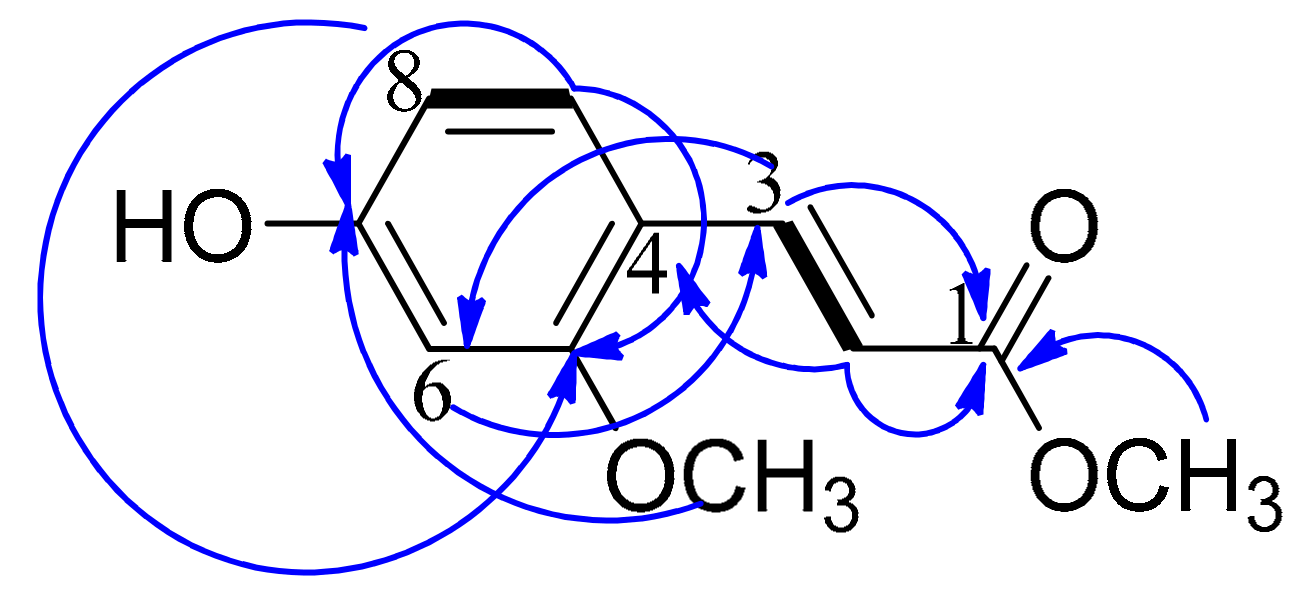
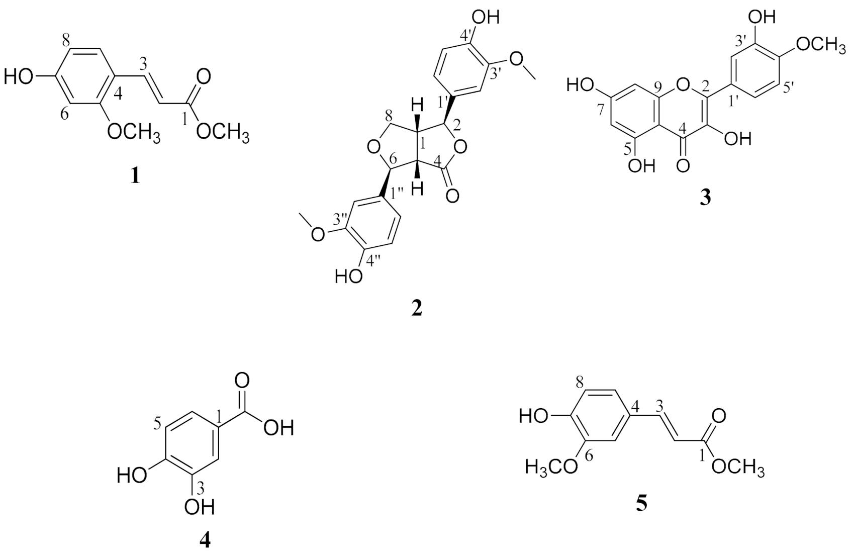
2.1. Structure Elucidation of the Isolated Compounds

Compound 1 was isolated as white amorphous powder. Combined spectral data including HRMS, HSQC, 1H NMR, and 13C NMR established its molecular formula as C11H12O4. Both 1H NMR and 13C NMR spectra confirmed the presence of a trisubstituted benzene ring. 1H NMR revealed three aromatic protons with ABX system resonating at δH 7.20 (d, J = 2.5), 6.70 (dd, J = 8.5, 2.5), and 7.80 (d, J = 8.5). Two of the aromatic carbons resonated at δC 160.9 and 168.0, thus confirmed to be oxygenated. The third substituted aromatic carbon was revealed to be substituted with a propenoate moiety. This was clear from the two sp2 carbons detected at δC 114.1 and 145.6 with their corresponding protons resonating at δH 6.16 and 7.52, respectively. The configuration at the double bond between C-2 and C-3 was determined to be E based on the large coupling constant between H-2 and H-3 (J = 15) [10], whereas in the vicinal coupling for Z geometry olefin, J = 11 Hz [11]. The 13C NMR spectrum revealed the presence of a carbonyl functionality resonating at δC 169.1 and assigned for C-1. The remaining two carbon signals were proved as methoxyl group from the signals detected at δC 51.9 and 52.1 with their corresponding protons resonating at δH 3.70 and 3.78, respectively. A final confirmation of the structure was illustrated by HMBC correlations (Figure 1 and Table 1). Accordingly, compound 1 was assigned as 7-hydroxy, 5-methoxy, methyl cinnamate. According to the available literature, compound 1 is a new natural product reported here for the first time from a natural source.
The chemical structures of compounds 25 were determined based on different spectroscopic data including 1D and 2D NMR, MS data, as well as comparison of the data with the previously reported in the literature. Compound 2 was elucidated as 4-keto-pinoresinol [12], compound 3 was identified as tamarixetin [13], compound 4 was found to be 3,4-dihydroxybenzoic acid [14], and finally, 5 was methyl ferulate [15,16]. The structures of all the isolated compounds (15) are declared in Figure 2.

2.2. Biological Evaluation of the Crude Extract and the Isolated Compounds

2.2.1. Cytotoxic Assay

Crude methanol extract of bamboo shoot skin as well as hexane, ethyl acetate, and butanol extracts were screened against a panel of cancer cell lines HepG2, Hela, A549, and MCF-7 and noncancerous liver cells (THP-1) cells. Percentages of cell viabilities are recorded in Table 2. Crude methanol extract exhibited the least vitality percentage of 6.6% of Hela cells among the tested cell lines at the highest concentrations (100 µg/mL). On the other hand, ethyl acetate extract exhibited the least vitality percentage of 2.14% of HepG2 cells. It is worthy to mention that the 4 known compounds that were isolated from ethyl acetate extract were previously tested for the cytotoxic activity. According to the literature, 4-keto-pinoresinol was reported to be active against SK-Mel-2, B16F1, ovarian cancer SKOV-3, CaoV-3, and cervical cancer HeLa cell lines cycle [17]. Tamarixetin was previously reported to possess cytotoxic action against A-549 and HCC-44 lung adenocarcinoma cells, gastric carcinoma (AGS), skin melanoma (B16F10), brain carcinoma (C6), epitheliod cervix carcinoma (HeLa) cells, MCF-7 and HCT-116 cells [18,19,20]. Additionally, 3,4-dihydrozybenzoic acid was also reported to cause apoptosis on human breast cancer MCF-7 cell, lung cancer A-549 cell, HepG2 cell, cervix HeLa cell, prostate cancer LNCaP cell, and A549 human lung cancer [21,22,23]. Moreover, methyl ferulate previously showed antiproliferative effect on colorectal cancer cells, MCF-7, PC-3, HL-60, MCF-7, and A-549 cells [24,25,26]. This can justify the potent cytotoxic effect of the ethyl acetate extract. In order to complete the profile of ethyl acetate extract activity, the new compound 1 isolated from this extract was screened against the HepG2 and MCF-7 cells and showed IC50 values of 7.43 and 10.65 µM, respectively. So, compound 1 was further tested for its apoptotic activity against HepG2 cells.

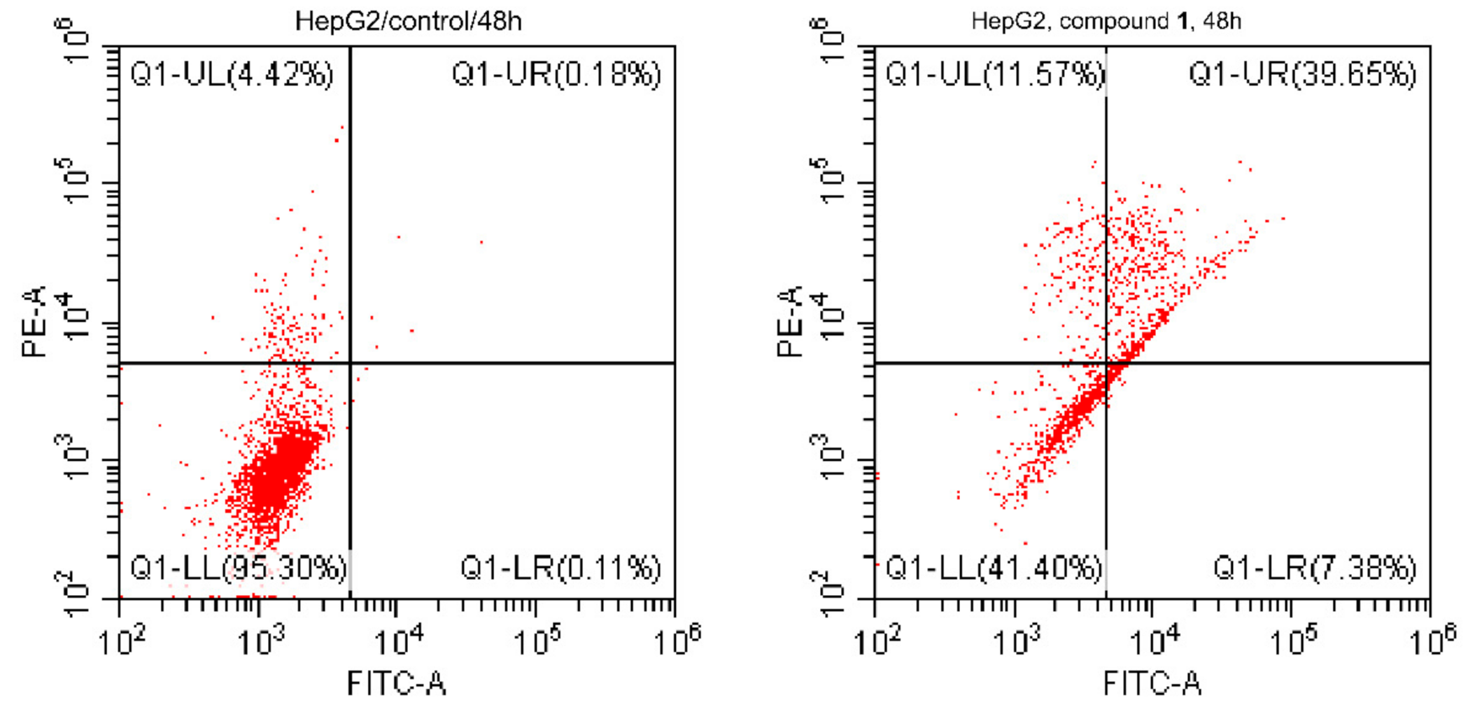
2.2.2. Annexin V/PI Staining

Staining with Annexin V/PI on HepG2 cells treated with compound 1 (IC50 = 7.43 µM, 48 h) and untreated cells for investigating the apoptotic activity of its treatment in the HepG2 cells was done. As shown in Figure 3, results indicated that the treatment caused apoptosis in HepG2 cells with total apoptotic cell death of 58.6% (12.44-fold) compared to 4.71% in control. It induced apoptosis by 7.38%, (early), 11.57% (intermediate), and 39.65% (late). These results are with good evidence for apoptosis induction in the HepG2-treated cells.

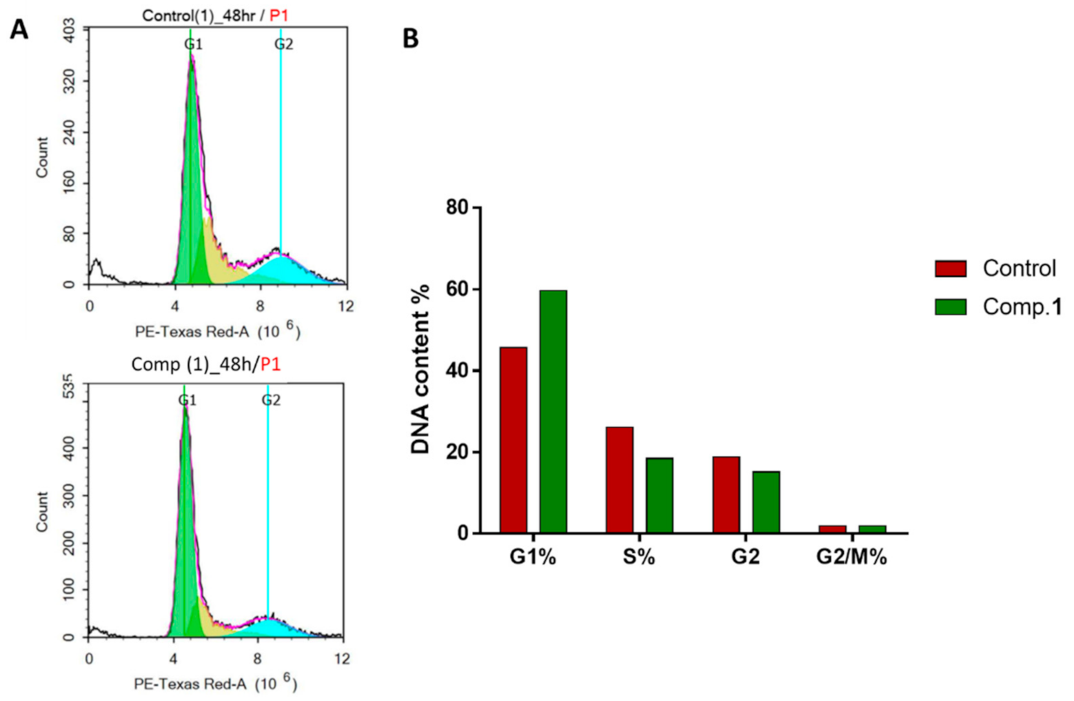
2.2.3. Cell Cycle Analysis

A significant test that shows the proportion of cell proliferation in each phase of cell development following treatment with cytotoxic compound is the cell cycle analysis. Therefore, to analyze the cell cycle kinetics of the HepG2 cells, the cells were treated with compound 1 (IC50 = 7.43 µM, 48 h). As shown in Figure 4, compound 1 treatment showed an increase at G1-phase cell-cycle arrest with 1.3-fold (59.63%, compared to 45.75% for control), while it decreased the cell population distribution at S (18.46 compared to 26.20%), G2 (15.22 compared to 18.77%), and G2/M (1.88 compared to 1.89%) phases; this may have resulted in compound 1 treatment-induced cytotoxic activity in HepG2 cells by arresting cell cycle progression at the G1 phase. These results agreed with our previous studies [27,28], which investigated the apoptotic activity of some solvent extracts using the flow cytometric analyses.

2.3. In Silico Studies

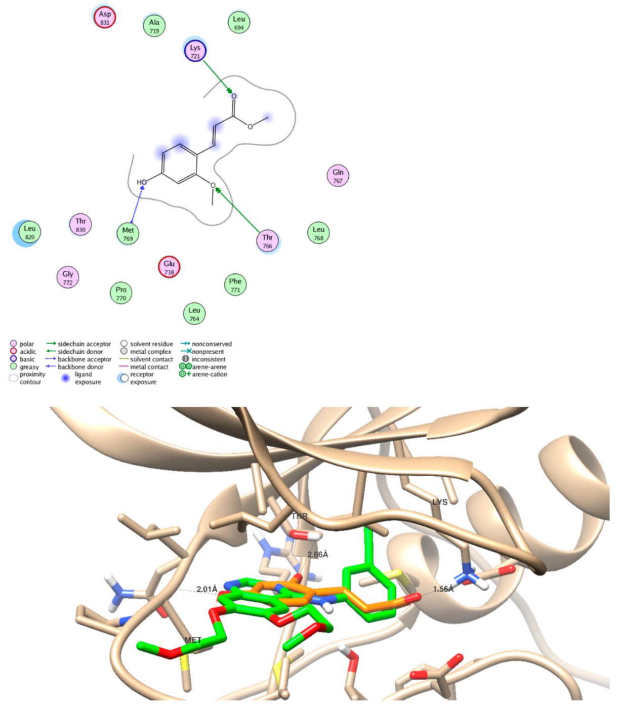
2.3.1. EGFR Inhibition Activity

The five identified compounds were screened for their binding activities through molecular docking studies, they were docked inside the EGFR binding site (PDB = 1M17) with binding energies (−9.74 to −18.82 Kcal/mol) and formed strong hydrogen bond interactions with the key amino acid Met 769. All interactions between moieties of ligand and receptor are summarized in Table 3 and Figure 5. The new compound 1 formed one hydrogen bond through its carboxylic group as HBA with binding energy of −18.82 Kcal/mol. Interestingly, compound 3 forms three hydrogen bonds with Met 769 as HBD and HBA through the hydroxylic and carbonyl groups, respectively. Since the molecular docking results exhibited good binding activity towards the EGFR protein, the new compound 1 was further tested against the EGFR enzymatic assay; it exhibited good inhibitory activity with IC50 value of 98.65 nM compared to erlotinib (IC50 = 78.65 nM). The agreement of the in silico and in situ results validate the EGFR inhibitor activity.

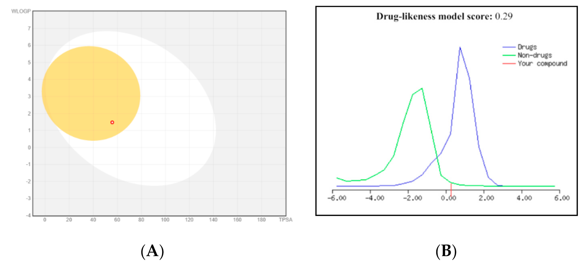
2.3.2. ADME Pharmacokinetics

The five identified compounds were screened for their ADME pharmacokinetics as previously described [29] (Table 4 and Figure 6). All tested compounds possess accepted values of H-bond donors (1–4) and H-bond acceptors (3–7), which are following the right criteria for hydrogen-bonding capacity for good drug permeability [30]. When the H-bond donors exceed 5, and the H-bond acceptors reach 10, drugs will be poorly absorbed. All compounds have log p values ≤ 5, so they had good membrane permeability. All of the compounds obeyed to Lipinski’s five rule, so, they could, therefore, be considered as drugs applicants for oral absorption. Furthermore, compounds 1, 3, and 4 showed positive values for drug-likeness scores, which indicated them as drug-like.

3. Experimental

3.1. General Experimental Procedures

1D and 2D NMR spectra (chemical shifts in ppm, coupling constants in Hz) were recorded on Bruker Avance DRX 500 MHz spectrometers (MA, USA). HRMS were determined by direct injection using Thermo Scientific UPLC RS Ultimate 3000-Q Exactive (Thermo Fisher Scientific, Waltham, MA, USA) hybrid quadrupole-Orbitrap mass spectrometer combined with high-performance quadrupole precursor selection with high-resolution, accurate-mass (HR/AM) Orbitrap™ detection. Detection was done in both positive and negative modes separately. Column chromatographic separations were carried out using Sephadex LH-20 (0.25–0.1 mm, Pharmacia, Sigma-Aldrich, St. Louis, Missouri, USA) and silica gel 60 (0.04–0.063 mm). TLC was accomplished using TLC plates precoated with silica gel 60 F254 (0.2 mm, Merck, NY, USA). Spots were visualized by UV absorption at λ of 255 and 366 nm followed by spraying with P-anisaldehyde/H2SO4.

3.2. Plant Material

Phyllostachys heterocycla var. pubescens was harvested in Isahaya, Nagasaki, Japan and collected on October 2011. The plant was stored at −24 °C until used. It was identified by Koji Yamada, Garden for Medicinal Plants, School of Pharmacy, Nagasaki University, Japan. A voucher specimen was kept under registration number KY-11 in the herbarium of Pharmacognosy Department, Faculty of Pharmacy, Suez Canal University, Ismailia, Egypt.

3.3. Extraction and Purification of Compounds 15

An amount of 12.7 kg was repeatedly extracted with methanol (20 L) followed by further extraction with CHCl3: MeOH (1:1) (20 L) at room temperature and the combined extracts were concentrated in vacuo to give a residue of 110 g. The residue was suspended in H2O (4 L) and extracted with n-hexane, EtOAc, n-BuOH, successively. The EtOAc extract was subjected to SiO2 column, eluted with CHCl3/MeOH gradient, and monitored by TLC to compile the resulted similar fractions. Based on TLC analysis, two fractions were subjected to further investigation.
First fraction was applied over SiO2 column with gradual elution using MeOH: CHCl3. Subfractions eluted were investigated and similar ones were combined to afford three main subfractions. Subfraction 1 was chromatographed on silica gel column packed in CHCl3 and eluted with a step gradient of MeOH: CHCl3 to afford semipure compound that was finally purified over Sephadex LH-20 column and eluted with CHCl3: MeOH (1:1) to obtain pure compound 2 (1.8 mg). Subfraction 2 was purified by dissolving it in CHCl3: MeOH (1:1) followed by decantation and crystallization to obtain the pure compound 3 (2.3 mg). Subfraction 3 was purified on preparative TLC using CHCl3: MeOH (20:1) and a drop of glacial acetic acid to yield pure compound 4 (3.3 mg). The second fraction was chromatographed over Sephadex LH-20 column and eluted with CHCl3: MeOH (1:1) followed by purification on preparative TLC using CHCl3: MeOH (20:1) as developing solvent mixture to obtain two pure compounds; compound 5 (11 mg) and compound 1 (23.6 mg).

3.4. Spectroscopic Data of the Isolated Compounds

7-Hydroxy, 5-methoxy, methyl cinnamate (1): White amorphous powder; HRMS analysis (negative mode) m/z: 207.0658 [M − H], molecular formula: C11H12O4, NMR data: see Table 1.
4-Keto-pinoresinol (2): White amorphous powder; 1H-NMR (500 MHz, CD3OD): δH = 6.79: 6.98 (6H, m, aromatic-H), 5.79 (1H, s, -OH), 5.78 (1H, s, -OH), 3.34 (1H, m, H-1), 5.24 (1H, d, J = 5, H-2), 3.68 (1H, dd, J = 5, 10, H-5), 5.40 (1H, d, J = 5, H-6), 4.29 (1H, dd, J = 5, 10, H-8a), 4.03 (1H, dd, J = 5, 10 H-8b), 3.88 (3H, s, OCH3), 3.87 (3H, s, OCH3). 13C-NMR (125 MHz, CD3OD): δC = 51.0 (C-1), 85.1 (C-2), 179.9 (C-4), 54.5 (C-5), 87.2 (C-6), 73.8 (C-8), 132.4 (C-1′), 110.6 (C-2′), 149.2 (C-3′), 147.5 (C-4′), 116.2 (C-5′), 119.5 (C-6′), 133.2 (C-1′′), 110.7 (C-2′′), 149.4 (C-3′′), 148.2 (C-4′′), 116.4 (C-5′′), 119.8 (C-6′′), 56.4 (3′-OCH3), 56.5 (3′′-OCH3).
Tamarixetin (3): Yellow powder; 1H-NMR (500 MHz, DMSO-d6): δH 6.29, brs (H-6), 6.64, brs (H -8), 7.39, brs, (H-2′), 7.06, brs, (H-5′), 7.41, brs, (H-6′), 3.88, s, (OCH3), 13.05 (OH). 13C-NMR (125 MHz, DMSO-d6): δC 148.9 (C-2), 136.0 (C-3), 177.0 (C-4), 162.2 (C-5), 99.5 (C-6), 165.1 (C-7), 94.8 (C-8), 153.9 (C-9), 104.8 (C-10), 123.0 (C-1′), 114.1 (C-2′), 146.3 (C-3′), 149.0 (C-4′), 119.5 (C-5′), 123.8 (C-6′), 56.9 (OCH3).
3,4-Dihydroxybenzoic acid (4): White amorphous powder; 1H-NMR (500 MHz, CD3OD): δH 7.44, brs (H-2), 6.81, d, J = 10.0 (H-5), 7.46, brd, J = 10.0 (H-6). 13C-NMR (125 MHz, CD3OD): δC 122.0 (C-1), 116.4 (C-2), 144.6 (C-3), 150.0 (C-4), 114.5 (C-5), 122.5 (C-6), 169.9 (CO).
Methyl ferulate (5): White amorphous powder; 1H-NMR (500 MHz, CDCI3): δH 6.18, d, J = 15.9 (H-2), 7.41, d, J = 15.9 (H-3), 6.80, brs (H-5), 6.89, d, J = 8.4 (H-8), 7.52, brd, J = 8.4 (H-9), 4.10, s (1-OCH3), 4.20, s, (6-OCH3). 13C-NMR (125 MHz, CDCI3): δC 167.9 (C-1), 114.8 (C-2), 145.0 (C-3), 126.8 (C-4), 109.5 (C-5), 148.1 (C-6), 146.9 (C-7), 114.8 (C-8), 123.1 (C-9), 51.9 (1-OCH3), 55.8 (6-OCH3).

3.5. Biological Evaluation of the Compounds

3.5.1. Cytotoxic Activity

Cell viability using the MTT assay was performed to investigate the effect of the crude extract, different extracts of hexane, ethyl acetate, and butanol and pure of compound 1 [31]. Full methodology is supported in the Supplementary File.

3.5.2. Apoptotic Investigation Using Flow Cytometric Analysis

Cell cycle analysis and apoptotic assays were performed to determine in which phase cells would be arrested and to calculate the percentage of apoptotic activity. Full methodologies of flow cytometric assays are provided in the Supplementary File [32]. After treatment with compound 1 (IC50 = 7.43 μM, 48 h), the harvested HepG2 cells were subjected to flow cytometric analyses including “FITC/Annexin-V-FITC/PI differential apoptosis/necrosis assessment and DNA content flow cytometry aided cell cycle analysis.”

3.5.3. EGFR Inhibition Activity

EGFR-TK assay was performed to evaluate the inhibitory potency of novel compound 1 against the EGFR [33]. For full methodology see the Supplementary File.

3.5.4. In Silico Studies

Molecular Docking Simulation

For the docking studies, the crystal structure of the epidermal growth factor receptor tyrosine kinase (EGFR) was obtained from the Protein data bank (PDB code: 1M17). According to routine work [34], preparation and optimization of ligand and receptor were carried out. All molecular modeling studies were conducted on a computational software basis using the “Molecular Operating Environment (MOE 2014-09 Chemical Computing Group, Canada)”. Each ligand–receptor complex was tested for interaction analysis, 2D images were made using the MOE visualizing tool, and 3D images were taken by Chimera as a visualizing software [35].

ADME Pharmacokinetics

ADME pharmacokinetics parameters of the most active compounds were calculated using a set of software including “MolSoft,” “Molinspiration”, and “SwissADME” websites as previously described by Youssef et al. 2020 [36].

3.6. Flow Cytometric Analysis

3.6.1. FITC/Annexin-V-FITC/PI Differential Apoptosis/Necrosis Assessment

Apoptosis and necrosis cell populations are determined using Annexin V-FITC apoptosis detection kit (Abcam Inc., Cambridge Science Park, Cambridge, UK) coupled with 2 fluorescent channels flowcytometry. After treatment with test compounds for 48 h, cells (105 cells) are collected by trypsinization and washed twice with ice-cold PBS (pH 7.4). Then, cells are incubated in dark with 0.5 mL of Annexin V-FITC/PI solution for 30 min in dark at room temperature according to manufacturer protocol. After staining, cells are injected via ACEA Novocyte™ flowcytometer (ACEA Biosciences Inc., San Diego, CA, USA) and analyzed for FITC and PI fluorescent signals using FL1 and FL2 signal detector, respectively (λex/em 488/530 nm for FITC and λex/em 535/617 nm for PI). For each sample, 12,000 events are acquired and positive FITC and/or PI cells are quantified by quadrant analysis and calculated using ACEA NovoExpress™ software (ACEA Biosciences Inc.,San Diego, CA, USA).

3.6.2. DNA Content-Flow Cytometry Aided Cell Cycle Analysis

After treatment with test compounds for 48 h, cells (105 cells) are collected by trypsinization and washed twice with ice-cold PBS (pH 7.4). Cells are re-suspended in two milliliters of 60% ice-cold ethanol and incubated at 4 °C for 1 h for fixation. Fixed cells are washed twice again with PBS (pH 7.4) and re-suspended in 1 mL of PBS containing 50 μg/mL RNAase A and 10 μg/mL propidium iodide (PI). After 20 min of incubation in dark at 37 C, cells are analyzed for DNA contents using flow cytometry analysis using FL2 (λex/em 535/617 nm) signal detector (ACEA Novocyte™ flowcytometer, ACEA Biosciences Inc., San Diego, CA, USA). For each sample, 12,000 events are acquired. Cell cycle distribution is calculated using ACEA NovoExpress™ software (ACEA Biosciences Inc., San Diego, CA, USA).

3.7. EGFR Inhibitory Assay

EGFR-TK assay was performed to evaluate the inhibitory potency of novel compound 1 against EGFR. Baculoviral expression vectors including pBlueBacHis2B and pFASTBacHTc were used separately to clone 1.6 kb cDNA coding for EGFR cytoplasmic domain (EGFR-CD, amino acids 645–1186). 5ʹ upstream to the EGFR sequence comprised a sequence that encoded (His)6. HepG2 cells were infected for 48h for protein expression. The pellets of HepG2 cells were solubilized in a buffer containing sodium vanadate (100 µM), aprotinin (10 µg/mL), triton (1%), HEPES buffer (50 mM), ammonium molybdate (10 µM), benzamidine HCl (16 µg/mL), NaCl (10 mM), leupeptin (10 µg/mL) and pepstatin (10 µg/mL) at 0 °C for 20 min at pH 7.4, followed by centrifugation for 20 min. To eliminate the nonspecifically bound material, a Ni-NTA superflow packed column was used to pass through and wash the crude extract supernatant first with 10 mM and then with 100 mM imidazole. Histidine-linked proteins were first eluted with 250 and then with 500 mM imidazole subsequent to dialysis against NaCl (50 mM), HEPES (20 mM), glycerol (10%) and 1 µg/mL each of aprotinin, leupeptin and pepstatin for 120 min. The purification was performed either at 4 °C or on ice. To record autophosphorylation level, EGFR kinase assay was carried out on the basis of DELFIA/Time-Resolved Fluorometry. The compound was first dissolved in DMSO absolute, subsequent to dilution to appropriate concentration using HEPES (25 mM) at pH 7.4. Each compound (10 µL) was incubated with recombinant enzyme (10 µL, 5 ng for EGFR, 1:80 dilution in 100 mM HEPES) for 10 min at 25 °C, subsequent to the addition of 5X buffer (10 µL, containing 2 mM MnCl2, 100 µM Na3VO4, 20 mM HEPES and 1 mM DTT) and ATP-MgCl2 (20 µL, containing 0.1 mM ATP and 50 mM MgCl2) and incubation for 1h. The negative and positive controls were included in each plate by the incubation of enzyme either with or without ATP-MgCl2. The liquid was removed after incubation and the plates were washed thrice using wash buffer. Europium-tagged antiphosphotyrosine antibody (75 µL, 400 ng) was added to each well followed by incubation of 1h and then washing of the plates using buffer. The enhancement solution was added to each well and the signal was recorded at excitation and emission wavelengths of 340 at 615 nm. The autophosphorylation percentage inhibition by compounds was calculated using the following equation:
100% − [(negative control)/(positive control) – (negative control)]
Using the curves of percentage inhibition of five concentrations of each compound, IC50 was calculated. Majority of signals detected by antiphosphotyrosine antibody were from EGFR because the enzyme preparation contained low impurities.

4. Conclusions

The ethyl acetate extract of Phyllostachys heterocycla var. pubescens exhibited potent cytotoxic activity and showed vitality percentage of 2.14% of HepG2 cells. This extract afforded five compounds of which one new compound was identified as 7-hydroxy, 5-methoxy, methyl cinnamate. The new compound was screened against the HepG2 and MCF-7 cells and exhibited IC50 values of 7.43 and 10.65 µM, respectively, with non-cytotoxic activity against the THLE2 cells. It induced total apoptotic cell death of 58.6% (12.44-fold) compared to 4.71% in control by arresting cell cycle progression at the G1 phase. Moreover, the EGFR activity was validated by the mechanism of action through both enzymatic and in silico levels. The study demonstrated the edible plant Phyllostachys heterocycla var. pubescens as an excellent promising anticancer agent.

Supplementary Materials

Figure S1. 1HNMR spectrum of compound (1); Figure S2. 13CNMR spectrum of compound (1); Figure S3. COSY spectrum of compound (1); Figure S4. HSQC spectrum of compound (1); Figure S5. HMBC spectrum of compound (1); Scheme 1. Mass spectrum of compound (1).

Author Contributions

R.F.A.A. and K.Y. conceived and designed the experiment; K.Y. collected and identified the plant material; R.F.A.A., A.K.I. (Ahmed K. Ibrahim), and E.S.H. performed the experiments, including isolation and structure elucidation of the pure compounds; M.S.N. performed cytotoxic activity using the MTT assay, Annexin-V/PI staining, cell cycle analysis, EGFR enzymatic assay, and molecular modeling; M.S.A.-K. conducted the spectral analysis of the pure compounds; S.A.A., R.F.A.A., and M.S.N. carried out writing—original draft; and J.M.B. and A.K.I. (Amany K. Ibrahim) carried out writing—review and editing. All authors have read and agreed to the published version of the manuscript.

Funding

This research received no external funding.

Institutional Review Board Statement

Not applicable.

Informed Consent Statement

Not applicable.

Data Availability Statement

No new data were created or analyzed in this study. Data sharing is not applicable to this article. The data presented in this study are available in Supplementary Materials.

Acknowledgments

This publication was supported by the Deanship of Scientific Research at Prince Sattam Bin Abdulaziz University, Alkharj, Saudi Arabia, as well as Suez Canal University, Ismailia, Egypt.

Conflicts of Interest

The authors declare no conflict of interest.

References

  1. Bray, F.; Ferlay, J.; Soerjomataram, I.; Siegel, R.L.; Torre, L.A.; Jemal, A. Global cancer statistics 2018: GLOBOCAN estimates of incidence and mortality worldwide for 36 cancers in 185 countries. CA Cancer J. Clin. 2018, 68, 394–424. [Google Scholar] [CrossRef] [Green Version]
  2. Zaorsky, N.G.; Churilla, T.M.; Egleston, B.L.; Fisher, S.G.; Ridge, J.A.; Horwitz, E.M.; Meyer, J.E. Causes of death among cancer patients. Ann. Oncol. 2017, 28, 400–407. [Google Scholar] [CrossRef] [PubMed]
  3. Iqbal, J.; Abbasi, B.A.; Mahmood, T.; Kanwal, S.; Ali, B.; Shah, S.A.; Khalil, A.T. Plant-derived anticancer agents: A green anticancer approach. Asian Pac. J. Trop. Biomed. 2017, 7, 1129–1150. [Google Scholar] [CrossRef]
  4. Yuming, Y.; Kanglin, W.; Shengji, P.; Jiming, H. Bamboo Diversity and Traditional Uses in Yunnan, China. Mt. Res. Dev. 2004, 24, 157–165. [Google Scholar] [CrossRef]
  5. Chongtham, N.; David, E.; Sharma, M.L. Changes in nutrient components during ageing of emerging juvenile bamboo shoots. Int. J. Food Sci. Nutr. 2007, 58, 612–618. [Google Scholar] [CrossRef]
  6. Chongtham, N.; Bisht, M.S.; Haorongbam, S. Nutritional Properties of Bamboo Shoots: Potential and Prospects for Utilization as a Health Food. Compr. Rev. Food Sci. F 2011, 10, 153–169. [Google Scholar] [CrossRef]
  7. Park, H.-S.; Lim, J.H.; Kim, H.J.; Choi, H.J.; Jung, D. Antioxidant flavone glycosides from the leaves of Sasa borealis. Arch. Pharm. Res. 2007, 30, 161–166. [Google Scholar] [CrossRef]
  8. Seki, T.; Maeda, H. Cancer preventive effect of Kumaizasa bamboo leaf extracts administered prior to carcinogenesis or cancer inoculation. Anticancer. Res. 2010, 30, 111–118. [Google Scholar]
  9. Chou, C.-W.; Cheng, Y.-W.; Tsai, C.-H. Phyllostachys edulis extract induces apoptosis signaling in osteosarcoma cells, associated with AMPK activation. Drug Des. Dev. Ther. 2014, 8, 1577–1584. [Google Scholar] [CrossRef] [PubMed] [Green Version]
  10. Youssef, D.T.; Badr, J.M.; Shaala, L.A.; Mohamed, G.A.; Bamanie, F.H. Ehrenasterol and biemnic acid; new bioactive compounds from the Red Sea sponge Biemna ehrenbergi. Phytochem. Lett. 2015, 12, 296–301. [Google Scholar] [CrossRef]
  11. Kamada, T.; Shean-Yeaw, N.G.; Phan, C.-S.; Vairappan, C.S. Chemical Composition and Antibacterial Activity of Bornean Medicinal Ginger Alpinia aquatic. Nat. Prod. Commun. 2018, 13, 741–742. [Google Scholar]
  12. Otsuka, H.; Takeuchi, M.; Inoshiri, S.; Sato, T.; Yamasaki, K. Phenolic compounds from Coix lachryma-jobi var. Ma-yuen. Phytochemistry 1989, 28, 883–886. [Google Scholar] [CrossRef]
  13. Benahmed, M.; Akkal, S.; Elomri, A.; Laouer, H.; Vérite, P.; Seguin, E. Constituents from Bupleurum montanum (Coss. & Dur.) (Apiaceae). Arab. J. Chem. 2014, 7, 1065–1069. [Google Scholar]
  14. Guria, M.; Mitra, P.; Ghosh, T.; Gupta, S.; Basu, B.; Mitra, P.K. 3, 4 diHydroxyBenzoic acid Isolated from the Leaves of Ageratum conyzoides L. Eur. J. Biotechnol. Biosci. 2013, 1, 25–28. [Google Scholar]
  15. Phuong, N.T.M.; Cuong, T.T.; Quang, D.N. Anti-inflammatory activity of methyl ferulate isolated from Stemona tuberosa Lour. Asian Pac. J. Trop. Med. 2014, 7, S327–S331. [Google Scholar] [CrossRef] [Green Version]
  16. Sajjadi, S.E.; Shokoohinia, Y.; Moayedi, N.S. Isolation and identification of ferulic acid from aerial parts of Kelussia odo-ratissima Mozaff. Jundishapur. J. Nat. Pharm. Prod. 2012, 7, 159–162. [Google Scholar]
  17. Moon, S.-S.; Rahman, A.A.; Kim, J.-Y.; Kee, S.-H. Hanultarin, a cytotoxic lignan as an inhibitor of actin cytoskeleton polymerization from the seeds of Trichosanthes kirilowii. Bioorg. Med. Chem. 2008, 16, 7264–7269. [Google Scholar] [CrossRef]
  18. Sak, K.; Lust, H.; Kase, M.; Jaal, J. Cytotoxic action of methylquercetins in human lung adenocarcinoma cells. Oncol. Lett. 2017, 15, 1973–1978. [Google Scholar] [CrossRef] [PubMed] [Green Version]
  19. Nicolini, F.; Burmistrova, O.; Marrero, M.T.; Torres, F.; Hernández, C.; Quintana, J.; Estévez, F. Induction of G 2 /M phase arrest and apoptosis by the flavonoid tamarixetin on human leukemia cells. Mol. Carcinog. 2013, 53, 939–950. [Google Scholar] [CrossRef] [PubMed]
  20. Abdelhady, M.I.S.; Kamal, A.M.; Othman, S.M.; Mubarak, M.S.; Ben Hadda, T. Total polyphenolic content, antioxidant, cytotoxic, antidiabetic activities, and polyphenolic compounds of Sophora japonica grown in Egypt. Med. Chem. Res. 2014, 24, 482–495. [Google Scholar] [CrossRef]
  21. Yin, M.-C.; Lin, C.-C.; Wu, H.-C.; Tsao, S.-M.; Hsu, C.-K. Apoptotic Effects of Protocatechuic Acid in Human Breast, Lung, Liver, Cervix, and Prostate Cancer Cells: Potential Mechanisms of Action. J. Agric. Food Chem. 2009, 57, 6468–6473. [Google Scholar] [CrossRef]
  22. Lin, H.-H.; Chen, J.-H.; Huang, C.-C.; Wang, C.-J. Apoptotic effect of 3,4-dihydroxybenzoic acid on human gastric carcinoma cells involving JNK/p38 MAPK signaling activation. Int. J. Cancer 2007, 120, 2306–2316. [Google Scholar] [CrossRef] [PubMed]
  23. Kuen, C.Y.; Galen, T.; Fakurazi, S.; Othman, S.S.; Masarudin, M.J. Increased Cytotoxic Efficacy of Protocatechuic Acid in A549 Human Lung Cancer Delivered via Hydrophobically Modified-Chitosan Nanoparticles as an Anticancer Modality. Polymers 2020, 12, 1951. [Google Scholar] [CrossRef] [PubMed]
  24. Abaza, M.S.I.; Afzal, M.; Rajáa, J.; Guleri, R. Methylferulate from Tamarix aucheriana inhibits growth and enhances chemo-sensitivity of human colorectal cancer cells: Possible mechanism of action. BMC Complement. Altern. Med. 2016, 16, 384. [Google Scholar] [CrossRef] [PubMed] [Green Version]
  25. Al Bujuq, N.; Arar, S.; Khalil, R. Synthesis and cytotoxic activity of 4-O-β-D-galactopyranosyl derivatives of phenolic acids esters. Nat. Prod. Res. 2018, 32, 2663–2669. [Google Scholar] [CrossRef]
  26. Cuong, T.D.; Lim, C.J.; Kim, S.W.; Park, J.E.; Hung, T.M.; Min, B.S. Isolation of compounds from Cimicifugae rhizoma and their cytotoxic activity. Nat. Prod. Sci. 2011, 17, 80–84. [Google Scholar]
  27. Abdelhameed, R.F.A.; Nafie, M.S.; Ibrahim, A.K.; Yamada, K.; Abdel-Kader, M.S.; Ibrahim, A.K.; Ahmed, S.A.; Badr, J.M.; Habib, E.S. Cytotoxic, Apoptosis-Inducing Activities, and Molecular Docking of a New Sterol from Bamboo Shoot Skin Phyllostachys heterocycla var. pubescens. Molecules 2020, 25, 5650. [Google Scholar] [CrossRef]
  28. Eltamany, E.E.; Elhady, S.S.; Ahmed, H.A.; Badr, J.M.; Noor, A.O.; Ahmed, S.A.; Nafie, M.S. Chemical Profiling, Antioxidant, Cytotoxic Activities and Molecular Docking Simulation of Carrichtera annua DC. (Cruciferae). Antioxidants 2020, 9, 1286. [Google Scholar] [CrossRef]
  29. Nafie, M.S.; Amer, A.M.; Mohamed, A.K.; Tantawy, E.S. Discovery of novel pyrazolo[3,4-b]pyridine scaffold-based derivatives as potential PIM-1 kinase inhibitors in breast cancer MCF-7 cells. Bioorg. Med. Chem. 2020, 28, 115828. [Google Scholar] [CrossRef]
  30. Lipinski, C.A.; Lombardo, F.; Dominy, B.W.; Feeney, P.J. Experimental and computational approaches to estimate solubility and permeability in drug discovery and development settings. Adv. Drug Deliv. Rev. 1997, 23, 3–25. [Google Scholar] [CrossRef]
  31. Sarhan, A.A.M.; Boraei, A.T.; Barakat, A.; Nafie, M.S. Discovery of hydrazide-based pyridazino[4,5-b]indole scaffold as a new phosphoinositide 3-kinase (PI3K) inhibitor for breast cancer therapy. RSC Adv. 2020, 10, 19534–19541. [Google Scholar] [CrossRef]
  32. Nafie, M.S.; Arafa, K.; Sedky, N.K.; Alakhdar, A.A.; Arafa, R.K. Triaryl dicationic DNA minor-groove binders with antiox-idant activity display cytotoxicity and induce apoptosis in breast cancer. Chem. Biol. Interact. 2020, 324, 109087. [Google Scholar] [CrossRef]
  33. Al-Wahaibi, L.H.; Gouda, A.M.; Abou-Ghadir, O.F.; Salem, O.I.A.; Ali, A.T.; Farghaly, H.S.; Abdelrahman, M.H.; Trembleau, L.; Abdu-Allah, H.H.M.; Youssif, B.G.M. Design and synthesis of novel 2,3-dihydropyrazino[1,2-a]indole-1,4-dione deriva-tives as antiproliferative EGFR and BRAFV600E dual inhibitors. Bioorg. Chem. 2020, 104, 104260. [Google Scholar] [CrossRef] [PubMed]
  34. Tantawy, E.S.; Amer, A.M.; Mohamed, E.K.; Abd Alla, M.M.; Nafie, M.S. Synthesis, characterization of some pyrazine deriv-atives as anti-cancer agents: In vitro and in Silico approaches. J. Mol. Struct. 2020, 1210, 128013. [Google Scholar] [CrossRef]
  35. Khodair, A.I.; Alsafi, M.A.; Nafie, M.S. Synthesis, molecular modeling and anti-cancer evaluation of a series of quinazoline derivatives. Carbohydr. Res. 2019, 486, 107832. [Google Scholar] [CrossRef]
  36. Youssef, E.; El-Moneim, M.A.; Fathalla, W.; Nafie, M.S. Design, synthesis and antiproliferative activity of new amine, amino acid and dipeptide-coupled benzamides as potential sigma-1 receptor. J. Iran. Chem. Soc. 2020, 17, 2515–2532. [Google Scholar] [CrossRef]
Figure 1. Selected COSY and HMBC correlations of compound 1.
Molecules 26 00940 g001
Figure 2. Chemical structures of the isolated compounds.
Molecules 26 00940 g002
Figure 3. Cytograms annexin-V/propidium iodide stained HepG2 cells showing induction of apoptosis in cancer cells by compound 1 (IC50 = 7.43 µM, 48 h) compared to untreated cells. “Quadrant charts show Q-UL (intermediate, AV–/PI+), Q-UR (late apoptotic cells, AV+/PI+), Q-LL (normal cells, AV–/PI–), and Q-LR (early apoptotic cells, AV+/PI–).”
Molecules 26 00940 g003
Figure 4. (A) Cytogram of the cell cycle distribution of untreated and treated HepG2 cells with. compound 1 (7.43 µM, 48 h); (B) bar chart representation of the percentage of cell population in different HepG2 cell cycle phases.
Molecules 26 00940 g004
Figure 5. Representative of superimposition and binding mode of compound 1 (Orange) and the cocrystalized ligand (Green) inside the epidermal growth factor receptor tyrosine kinase (EGFR)-binding site (PDB 1M17) in 2D and 3D.
Molecules 26 00940 g005
Figure 6. (A) BOILED-Egg model for compound 7 using SwissADME: “points located in the BOILED-Egg‘s yolk are molecules predicted to passively permeate through the blood–brain barrier (BBB), while points located in the BOILED-Egg‘s white are molecules predicted to be passively absorbed by gastrointestinal (GI) tract” and (B) drug likeness score of compound 1 using MolSoft: “The green color means nondrug-like behavior, and those fall under blue color area are considered as drug-like. Those compounds having negative or zero value should not be considered as drug like.”
Molecules 26 00940 g006
Table 1. NMR spectroscopic data of compound 1 (CDCl3, 500 and 125 MHz).
PositionδC (m) aδH (m, J in Hz)
1169.1 (C)
2114.1 (CH)6.16 (d, J = 15)
3145.6 (CH)7.52 (d, J = 15)
4126.3 (C)
5160.9 (C)
6130.1 (CH)7.20 (d, J = 2.5)
7168.0 (C)
8115.4 (CH)6.70 (dd, J = 8.5, 2.5)
9131.9 (CH)7.80 (d, J = 8.5)
1- OCH351.9 (CH3)3.70 (s)
5- OCH352.1 (CH3)3.78 (s)
a Multiplicities were deduced from multiplicity-edited HSQC.
Table 2. Percentages of cell viabilities of crude, solvent fractions, and identified compounds against four cancerous cell lines HepG2, Hela, A549, and MCF-7 and noncancerous cells.
Extracts% of Cell Viability at 100 µg/mL
HepG2HelaA549MCF-7THP-1
Crude methanol extract47.5 ± 0.766.6 ± 0.8933.18 ± 0.1936.6 ± 0.6486 ± 1.67
Hexane extract58.6 ± 1.2664.53 ± 1.2352.63 ± 1.1449.8 ± 0.8196 ± 1.98
Ethyl acetate extract2.14 ± 0.1524.23 ± 1.5215.05 ± 0.5712.14 ± 0.8186 ± 0.98
Butanol extract84.3 ± 1.9894.31 ± 0.0994.26 ± 0.2363.7 ± 0.3687 ± 1.04
IC50 ± SD *#
Compound 1
(µM)
7.43 ± 0.82NDND10.65 ± 1.01≥50
* Values are expressed as mean ± SD of 3 independent trials (n = 3). # IC50 were calculated using GraphPad Prism 7 software using nonlinear regression Dose-Inhibition curve fit.
Table 3. Ligand–receptor interactions of the docked compounds with the binding energy (Kcal/mol) inside the 1M17 as epidermal growth factor receptor tyrosine kinase (EGFR) inhibitors
CompoundsIn Silico Molecular Docking Simulation as EGFR InhibitorsIn Vitro EGFR Activity(nM) *
Binding Energy (Kcal/mol)HB Interactions with The Key Amino Acid (Met 769)
1−18.82HBA as Molecules 26 00940 i00198.65 ± 9.87
2−16.28HBA as Molecules 26 00940 i002
3−13.2HBD as Molecules 26 00940 i003
HBA as Molecules 26 00940 i004
4−9.74HBA as Molecules 26 00940 i005
5−16.77HBA as Molecules 26 00940 i006
Erlotinib78.65 ± 6.54
* Values are expressed as mean ± SD of 3 independent triplicates and are calculated by GraphPad Prism 7.
Table 4. Molecular properties of and drug-likeness.
WebsiteCompound
ADME
12345
Molinspiration 2018.10Mwt (D)208.21372.37316.26154.12208.21
MV (A3)189.55321.96257.61127.08189.55
PSA (A2)55.7794.46120.3677.7555.77
Log p1.852.461.990.881.86
nrotb44214
nviolations00000
MolSoftHBA37744
HBD12431
Solubility (mg/L)375.192726.41102.63378.71461.2
Drug-likeness score0.29−0.090.160.23−0.76
Mwt: molecular weight, MV: molecular volume, PSA: polar surface area, Log p: Log P: octanol-water partition coefficient, nrotb: number of rotatable bonds, nviolations: number of violations, HBA: hydrogen bond acceptor, HBD: hydrogen bond donor [29,30].
Publisher’s Note: MDPI stays neutral with regard to jurisdictional claims in published maps and institutional affiliations.

