Article

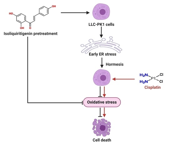
Isoliquiritigenin Pretreatment Induces Endoplasmic Reticulum Stress-Mediated Hormesis and Attenuates Cisplatin-Induced Oxidative Stress and Damage in LLC-PK1 Cells

by
Tania Gómez-Sierra
,
Omar Noel Medina-Campos
,
José D. Solano
,
María Elena Ibarra-Rubio
and
José Pedraza-Chaverri
*
Departamento de Biología, Facultad de Química, Universidad Nacional Autónoma de México, CDMX 04510, Mexico
*
Author to whom correspondence should be addressed.
Molecules 2020, 25(19), 4442; https://doi.org/10.3390/molecules25194442
Submission received: 25 August 2020 / Revised: 19 September 2020 / Accepted: 26 September 2020 / Published: 27 September 2020

Abstract

:
Isoliquiritigenin (IsoLQ) is a flavonoid with antioxidant properties and inducer of endoplasmic reticulum (ER) stress. In vitro and in vivo studies show that ER stress-mediated hormesis is cytoprotective; therefore, natural antioxidants and ER stress inducers have been used to prevent renal injury. Oxidative stress and ER stress are some of the mechanisms of damage involved in cisplatin (CP)-induced nephrotoxicity. This study aims to explore whether IsoLQ pretreatment induces ER stress and produces hormesis to protect against CP-induced nephrotoxicity in Lilly Laboratories Cell-Porcine Kidney 1 (LLC-PK1) cells. During the first stage of this study, both IsoLQ protective concentration and pretreatment time against CP-induced toxicity were determined by cell viability. At the second stage, the effect of IsoLQ pretreatment on cell viability, ER stress, and oxidative stress were evaluated. IsoLQ pretreatment in CP-treated cells induces expression of glucose-related proteins 78 and 94 kDa (GRP78 and GRP94, respectively), attenuates CP-induced cell death, decreases reactive oxygen species (ROS) production, and prevents the decrease in glutathione/glutathione disulfide (GSH/GSSG) ratio, free thiols levels, and glutathione reductase (GR) activity. These data suggest that IsoLQ pretreatment has a moderately protective effect on CP-induced toxicity in LLC-PK1 cells, through ER stress-mediated hormesis, as well as by the antioxidant properties of IsoLQ.

Graphical Abstract

1. Introduction

The flavonoid 4,2′,4′-trihydroxichalcone, also known as isoliquiritigenin (IsoLQ) (Figure 1), proceeds from the roots and rhizomes of licorice (Glycyrrizha glabra). It is used in Asia as an herbal medicine due to its anti-inflammatory, antimicrobial, antitumoral, and antioxidant properties [1,2,3,4,5,6,7,8]. Regarding its antioxidant activity, it has been reported that IsoLQ scavenges reactive oxygen species (ROS), such as the superoxide anion (O2●−), hydroxyl radical, hydrogen peroxide, and singlet oxygen [4,7], and induces phase II cytoprotective enzymes’ expression [9,10]. In addition, IsoLQ has been used as an endoplasmic reticulum (ER) stress inducer in cancer cell lines to induce apoptosis [8,10,11,12,13,14,15,16] and also has cytoprotective effects in non-cancer cells, such as liver and kidney cells [1,4,9,16,17,18].
On the other hand, cisplatin or cis-diaminodichloroplatin II (CP), is an antineoplastic drug approved by the United States Federal Drug Administration since 1978 used for the treatment of many cancers, such as head, neck, bladder, lung, stomach, uterus, and testicular. However, its application has been limited because 25–35% of treated patients develop kidney damage [19,20,21,22,23]. The mechanism of CP nephrotoxicity has been studied for the last forty years and binding to multiple targets such as deoxyribonucleic acid or glutathione (GSH) have been identified. Also, mechanisms such as oxidative stress, inflammation, mitochondrial dysfunction, and ER stress have been associated to kidney damage [19,20,21,22,24], and although the contribution of each pathway is not exactly known, oxidative stress plays a key role [19,24,25,26].
Under acute ER stress, glucose-related protein 78 kDa (GRP78) and glucose-related protein 94 kDa (GRP94) are released from the ER membrane and activate the unfolded protein response (UPR) to restore ER functions, such as protein synthesis and transport, protein folding, lipid and steroid synthesis, among others [27,28,29,30,31], in addition to the antioxidant response and autophagy [27,32,33,34], which maintain cellular homeostasis. However, if the ER stress is excessive or chronic and cannot be corrected, UPR signaling promotes apoptosis [29,31,35,36,37,38,39]. These different effects of UPR on cellular function have generated interest because of their similarity to those occurring in hormesis, a process usually referred to as a biological favorable response originated by low dose/concentration or short exposure to chemical/natural compounds or environmental factors [40,41,42,43,44]. However, a high dose/concentration or long exposure inhibits this favorable response [40,41,45,46,47,48,49]. It has been suggested that this biphasic dose-response could protect against more than one type of stress, and play a key role in the treatment of different pathologies [36,40,41,50,51,52,53].
Based on the knowledge of the main pathways involved in CP-induced nephrotoxicity, such as oxidative stress [19,21,22,25,54], apoptosis [20,21,22,25], mitochondrial dysfunction [20,21,22,25], kinases activation [21,22,54], and ER stress [21,24,55], treatments with natural compounds have been implemented to prevent CP-induced nephrotoxicity [20,21,24,25,56,57,58]. However, it is necessary to explore more compounds and other targets less studied, such as ER stress, due to the complexity of cisplatin toxicity. Therefore, the aim of this study was to explore whether IsoLQ-induced ER stress produces a hormetic effect that protects against CP-induced nephrotoxicity in Lilly Laboratories Cell-Porcine Kidney 1 (LLC-PK1) cells.

2. Results

2.1. First Stage

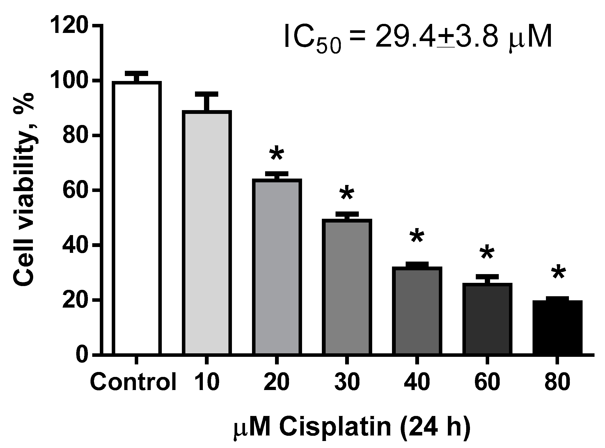
2.1.1. Estimation of Hormetic Zone of IsoLQ in CP-Induced Toxicity in LLC-PK1 Cells

LLC-PK1 cells were treated with CP (10–80 μM) for 24 h. Figure 2 shows that 10 μM CP was not cytotoxic, whereas concentrations higher than 20 μM induced cytotoxicity (p < 0.05) and the half-maximal (50%) inhibitory concentration (IC50) calculated was 29.4 ± 3.8 μM. Figure 3 presents the typical inverted J-shaped hormetic relationship, where IsoLQ concentrations between 5 and 30 μM attenuated CP-induced cell death (p < 0.05), whereas IsoLQ concentrations higher than 35 μM increased CP-induced cell death (p < 0.05).

2.1.2. Determination of IsoLQ Protective Concentration and Pretreatment Time against CP-Induced Toxicity in LLC-PK1 Cells

According to the data shown in Figure 2, 40 μM CP caused ~66% of cell death (p < 0.05), while Figure 3 shows that 15 and 25 μM IsoLQ attenuated CP-induced decrease on cell viability (p < 0.05). Therefore, these two concentrations were used to determine the IsoLQ pretreatment time that could have a protective effect against CP-induced toxicity in LLC-PK1 cells. Cell viability results indicate that 6 and 8 h pretreatment with 15 and 25 μM IsoLQ promoted cytoprotective effects against 40 μM CP (p < 0.05) (Figure 4).

2.1.3. Pretreatment of IsoLQ Induces ER Stress

According to the data shown in Figure 4, 15 and 25 μM IsoLQ pretreatment attenuated CP-induced cell death (p < 0.05), so cells were treated with the highest concentration of IsoLQ (25 μM) to evaluate ER stress. Figure 5 shows that GRP94 and GRP78 increased at 4 and 6 h (p < 0.05) respectively, after pretreatment with 25 μM IsoLQ, and remained increased at 24 h.

2.2. Second Stage

2.2.1. IsoLQ Pretreatment Enhanced ER Stress in CP-Induced Nephrotoxicity in LLC-PK1 Cells

Based on cell viability results as well as protein levels of GRP78 and GRP94 obtained from first-stage experiments, the experimental protocol was designed as follows: cells were pretreated with 25 μM IsoLQ for 8 h (the highest effects on cell viability and GRP78 and GRP94 expression were observed in these conditions) and subsequently with 40 μM CP (the highest CP concentration evaluated) for a time-course study. After pretreatment of LLC-PK1 cells with 25 μM IsoLQ for 8 h and exposure to 40 μM CP for 16 and 24 h, GRP78 and GRP94 expression increased (p < 0.05) (Figure 6A). This increase was higher than that under IsoLQ treatment or with CP alone (Figure 5 and Figure 6B, respectively). In addition, the expression of the eukaryotic initiation factor 2 alpha (p-eIF2α) did not change (data not shown).

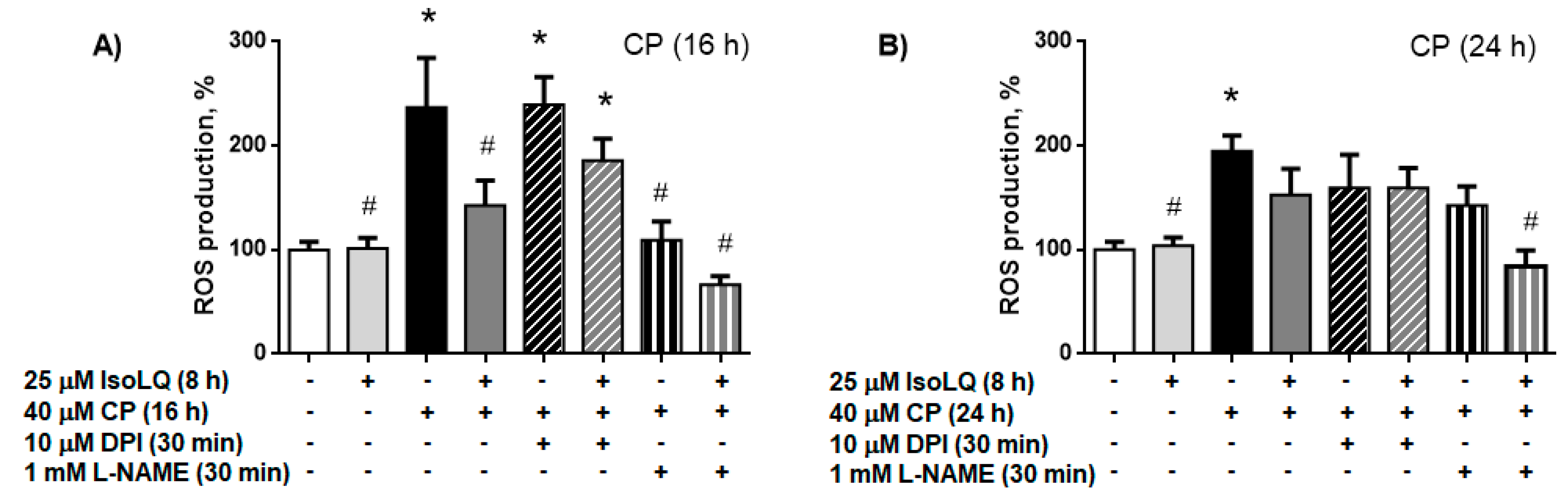
2.2.2. IsoLQ-Induced Hormesis Attenuates Oxidative Stress in CP-Treated LLC-PK1 Cells

LLC-PK1 cells were pretreated with 25 μM IsoLQ for 8 h and then with 40 μM CP for 16 and 24 h, as described for the second stage of the experimental design. 25 μM IsoLQ pretreatment decreased ROS production by ~37% and ~26% after exposure to 40 μM CP for 16 and 24 h (p < 0.05) respectively, and attenuated the decrease in GSH/glutathione disulfide (GSSG) ratio, free thiols, and glutathione reductase (GR) activity 24 h after CP treatment (p < 0.05) (Table 1).

Nitric Oxide Synthase (NOS) is the Source of ROS Production in LLC-PK1 Cells Pretreated with IsoLQ and Exposed to CP
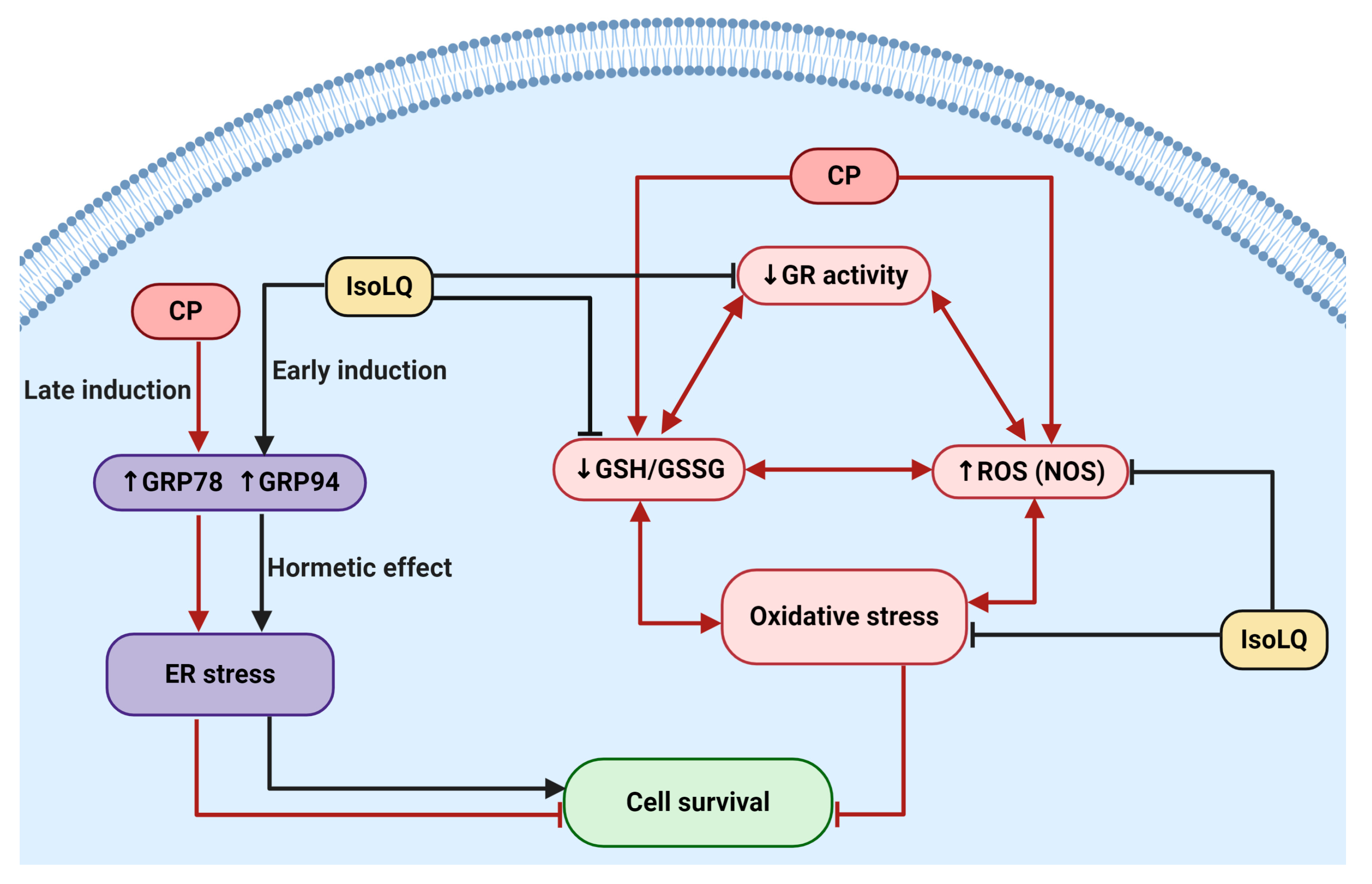
In order to determine intracellular ROS sources, we used diphenyleneiodonium chloride (DPI), an inhibitor of nicotinamide adenine dinucleotide phosphate hydrogen (NADPH) oxidase (NOX), or N-nitro-L-arginine methyl ester (L-NAME), an inhibitor of NOS. DPI did not decrease ROS production in any of the groups of treated cells, whereas L-NAME decreased ROS production at 16 h in IsoLQ + CP-treated cells to similar levels as the control group (p < 0.05). This effect remained until 24 h (p < 0.05) (Figure 7); whereas in CP-treated cells, ROS production decreased only at 16 h (p < 0.05).

3. Discussion

IsoLQ is a flavonoid with anti-inflammatory, antitumoral, and antioxidant properties [1,2,3,4,5,6,7,16,59] that exerts protective effects in liver and kidney cells [3,4,9,16,17,18]. In addition, it has been demonstrated that IsoLQ does not interfere with CP antineoplastic activity [16]. The effect of IsoLQ on cell viability in CP-induced nephrotoxicity was a biphasic dose-response: low IsoLQ concentrations attenuated the decrease on cell viability, but high IsoLQ concentrations inhibited it. This effect is known as hormesis, a term that refers to a biological favorable cellular response [40,47,48,49,50,60]. The IsoLQ + CP dose-response curve showed an inverted J-shaped hormetic effect and this biphasic curve has an interval called the hormetic zone where the beneficial effects are evident (Figure 3) [45,47,48,49]. The low-dose stimulation reflects the capacity of the cells to distribute biological resources that help to defend the organism from a wide range of stressor agents, such as CP, and this adaptive mechanism includes stress protein responses that reduce the damage more effectively than the response induced only by exposure to the stressor [40,41,46,47]. Therefore, hormesis is more than simply a dose-response relationship, it is a quantitative and temporal manifestation of reparative processes that is adaptive in nature, such as UPR elicited by ER stress [42,43,46,47,50,60,61].
Data obtained show that 25 μM IsoLQ pretreatment induced ER stress and also attenuated 40 μM CP-induced cell death in LLC-PK1 cells. The increase in the expression of the chaperones GRP78 and GRP94 indicates the occurrence of ER stress and suggests the activation of UPR [27,29,37,62,63,64,65]. The early induction of ER stress could play a key role in cell survival, because ER stress induced by IsoLQ pretreatment generated a hormetic effect to activate adaptive responses such as UPR, that protects against higher stress levels as occurs in CP-induced nephrotoxicity. In in vitro and in vivo models of kidney diseases, like renal ischemia-reperfusion or exposure to cytotoxic compounds, the a priori induction of ER stress with pharmacological or natural compounds generates tolerance against these insults (hypoxia, oxidative stress) in order to inhibit the apoptotic cell death [37,52,53,62,66,67,68,69].
On the other hand, oxidative stress is also characteristic in CP-induced nephrotoxicity and is an important factor in the development and progression of other associated damage mechanisms, such as mitochondrial dysfunction, inflammation, and ER stress [19,20,21,22,25,26,54,70]. ROS are generated naturally and regulate cell functions [71,72]. However, excessive quantities of ROS can result in damage to proteins, modifying their function and disrupting ER homeostasis [27,32,34,73]. ROS overproduction can be sensed by the ER, which is highly sensitive to changes in ROS levels through redox sensors, such as, for example, the thiol groups of cysteines [33,70,74,75,76,77], causing misfolded protein accumulation and consequently increasing ROS production, which leads to a vicious cycle able to activate UPR, generate chronic stress, and induce apoptosis [32,33,34,70,72]. Moreover, ROS overproduction causes a decrease in the GSH/GSSG ratio and in antioxidant enzyme levels, whilst oxidizing lipids, DNA, and proteins [19,20,21,22,25,26]. In our experimental model, ROS production could be associated to NOS activity and consequently to overproduction of O2●− that enhances oxidative stress. However, IsoLQ can attenuate this oxidative imbalance due to its ROS scavenging capacity [4,7]. In CP-induced nephrotoxicity, the decrease in the GSH/GSSG ratio is characteristic due to the formation of GSH-CP adducts. The positively charged species of CP (their active forms) have a great affinity for thiol groups [20,22,25,78,79] and consequently, there is an alteration in the activity of enzymes related to GSH metabolism [19,26,78]. In addition, the decrease in GSH could be associated with the formation of disulfide bonds among misfolded proteins in a coupled reaction with the protein disulfide isomerase and ER oxidoreductase 1 [73,80,81,82,83].
Interestingly, ER stress induction is also associated with an increase in GSH biosynthesis in order to form disulfide bonds between proteins and recycle unfolded proteins. This effect could explain the increase in free thiols, such as GSH, in cells pretreated with IsoLQ [33,82]. Moreover, it has been described that ER stress increases the activity of antioxidant enzymes such as GR, glutathione peroxidase, and glutathione S-transferase [33,63,82]. Probably, augmented GR activity in cells pretreated with IsoLQ was due to this effect, since IsoLQ is also a bifunctional antioxidant able to induce expression of phase II detoxifying enzymes through nuclear factor erythroid 2 (Nrf2) transcriptional regulation [9,10]. Under ER stress, protein kinase ribonucleic acid (RNA)-activated-like ER kinase (PERK), one the of the signaling pathways of UPR, directly phosphorylates Nrf2, causing Nrf2 to dissociates from its negative regulator (Kelch-like erythroid-derived cap-n-collar homology-(ECH-)associated protein 1); after which, Nrf2 translocates to the nucleus, leading the transcription of genes that encode phase II detoxifying enzymes that maintain redox homeostasis and may contribute to cell survival [84,85,86]. In addition, eIF2α is another target of the PERK signaling pathway [27,29,63,65]. Even though the data on p-eIF2α expression are inconclusive (data not shown), it is possible that the PERK pathway has become activated. Nevertheless, studies on the role of UPR proteins are required to describe and understand the mechanisms involved in the hormetic effect produced by IsoLQ in this experimental model. In summary, Figure 8 shows a possible mechanism of the protective effect of IsoLQ against CP-induced nephrotoxicity in LLC-PK1 cells. We derived our assumptions from the main observations found in this study.

4. Materials and Methods

4.1. Reagents

LLC-PK1 cells were obtained from the American Type Culture Collection (ATCC, Rockville, MD, USA). IsoLQ was purchased from AK Scientific (Union City, NJ, USA). Bovine serum albumin, CP, 1-chloro-2,4-dinitrobenzene, dimethyl sulfoxide, DPI, 5,5’-dithiobis-(2-nitrobenzoic acid), fluorescein diacetate (FDA), glycerophosphate, GSH, GSSG, GR, L-NAME, NADPH, 4-nonylphenyl-polyethylene glycol (NP-40), protease inhibitor cocktail, sodium deoxycholate, sodium dodecyl sulfate (SDS), sodium fluoride (NaF), sodium pyrophosphate (Na4P2O7), sodium orthovanadate (Na3VO4), sulfosalicylic acid, triethanolamine, Tris-hydrochloride (Tris-HCl), trypan blue, triton X-100, tween 20, 2-vinylpirydine, mouse anti-α-tubulin (cat. T9026) antibodies, and rabbit anti-GRP94 (cat. G4420) antibodies were from Sigma Aldrich Co. (St. Louis, MO, USA). Dipotassium hydrogen orthophosphate, ethylenediaminetetraacetic acid disodium salt (EDTA), potassium chloride, potassium dihydrogen orthophosphate, sodium chloride (NaCl), sodium bicarbonate (NaHCO3), and sodium phosphate dibasic were from J.T. Baker (Xalostoc, Edo. Mex, Mex). Bradford reagent was from Bio-Rad (Hercules, CA, USA). Dulbecco’s Modified Eagle’s Medium (DMEM), fetal bovine serum (FBS), and penicillin/streptomycin were from Biowest (Riverside, MO, USA). TrypLE Express was from Thermo Fisher Scientific (Waltham, MA, USA). Dichlorodihydrofluorescein diacetate (H2DCFDA) was from Molecular Probes (Eugene, OR, USA). Mouse anti-GRP78 (cat. SC-3768) antibodies were from Santa Cruz Biotechnology Inc. (Dallas, TX, USA). Fluorescent secondary antibodies anti-rabbit 800CW and anti-mouse 680RD were from LI-COR Biosciences (Lincoln, NE, USA). All the other chemicals and reagents used were of analytical grade and commercially available.

4.2. Cell Culture

LLC-PK1 cells are a proximal tubule cell line derived from porcine kidneys, widely used in experimental models [9,25,53,87]. LLC-PK1 cells were cultured in DMEM supplemented with 10% FBS, 0.33% NaHCO3, 100 U/mL penicillin, and 50 U/mL streptomycin under a humidified atmosphere of 5% CO2 at 37 °C. The cells were sub-cultured with TrypLE Express upon reaching 90% confluence. LLC-PK1 cells passage numbers 8 to 33 were used.

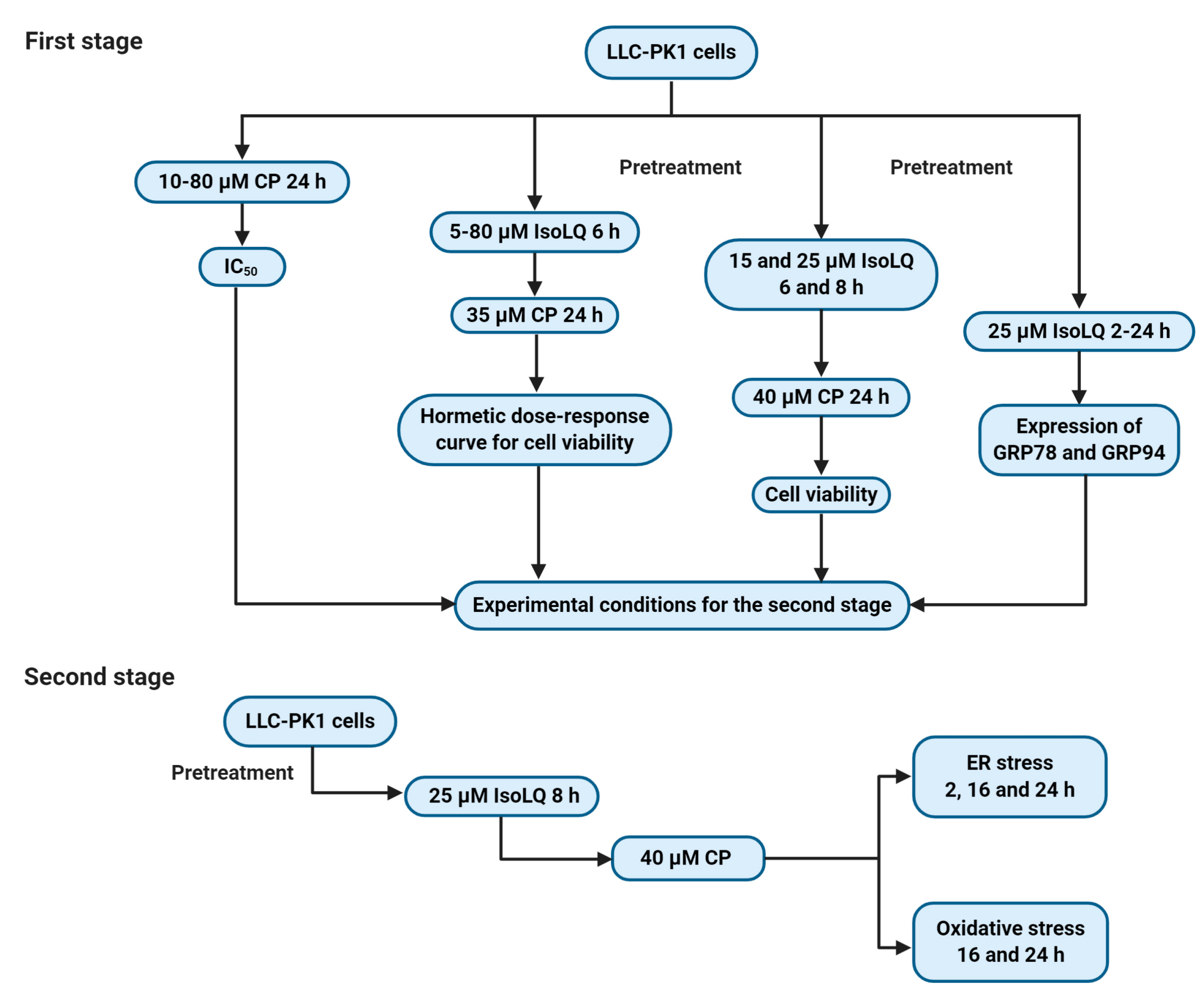
4.3. Experimental Design

4.3.1. First Stage

The aim of the first stage set of experiments was to determine the potential IsoLQ protective concentration and pretreatment time against CP-induced toxicity in LLC-PK1 cell culture. Cells were treated with CP (10–80 μM) for 24 h to calculate the IC50. Additionally, cells were pretreated with IsoLQ (5–80 μM) for 6 h and later incubated with one concentration higher than the IC50 of CP (35 μM) for 24 h to evaluate the potentially hormetic dose-response curve for cell viability. Finally, two IsoLQ concentrations within the hormetic zone (15 and 25 μM) and one concentration higher than the IC50 of CP (40 μM) were selected to test the putative protective effect by IsoLQ on cell viability.
On the other hand, cells were pretreated with 25 μM IsoLQ (highest concentration within the hormetic zone) in a time-course study (2–24 h) to evaluate the expression of GRP78 and GRP94.

4.3.2. Second Stage

Based on the first stage results, LLC-PK1 cells were pretreated with 25 μM IsoLQ for 8 h and subsequently incubated, in a time-course study, with 40 μM CP to evaluate ER stress (2, 16, and 24 h) and oxidative stress (16 and 24 h). The experimental design is summarized in Figure 9.

4.4. Cell Viability

Subsequently after treatments, DMEM was removed and cells were washed with phosphate-buffered saline (PBS) followed by the addition of 12 μM FDA and incubation for 5 min at 37 °C in darkness [88]. Fluorescence was determined by a microplate reader at excitation and emission wavelengths of 485 and 520 nm, respectively. The results are expressed as the percentage of the control (without treatments).

4.5. Extraction of Total Fractions of Proteins for Western Blot

LLC-PK1 cells were lysed with radioimmunoprecipitation assay (RIPA) buffer (50 mM Tris-HCl, pH 7.4, 150 mM NaCl, 1 mM EDTA, 0.5% sodium deoxycholate, 1% NP-40, 0.1% SDS, 25 mM NaF, 1 mM Na4P2O7, 1 mM Na3VO4, 0.5 mM glycerophosphate, and protease inhibitor cocktail) for one hour at 4 °C. Lysates were centrifuged at 12,000× g during 10 min at 4 °C and the supernatant was collected and stored at −70 °C until use. Total protein content was determined by the Bradford assay [89].

4.6. ER Stress Evaluation

Western Blot Analysis

Proteins were separated by SDS-polyacrylamide gel electrophoresis (PAGE) and transferred to polyvinylidene fluoride membranes, which were blocked for one hour with 5% skimmed milk or bovine serum albumin. Membranes were left incubating overnight at 4 °C with the appropriate primary antibodies against GRP78 (1:1000), GRP94 (1:1000), and α-tubulin (1:8000). Subsequently, membranes were washed and incubated with the appropriate secondary antibodies for two hours. The proteins of interest were detected with the LI-COR Odyssey Sa Infrared Imaging System (Lincoln, NE, USA). Image analysis was performed with Image Studio Lite Software version 5.2.

4.7. Oxidative Stress Evaluation

4.7.1. Measurement of ROS Production

After the second stage cell treatments, DMEM was removed and cells were washed with PBS followed by incubation with 10 µM H2DCFDA for 30 min at 37 °C in the darkness. Fluorescence intensity was visualized and determined in a Cytation 5 Cell Imaging Reader (Biotek Instruments, Inc., Winooski, VT, USA) at excitation and emission wavelengths of 485 and 520 nm, respectively. Relative fluorescence intensity of treated cells was expressed as a percentage of the control (without treatments).
In order to determine the intracellular ROS sources, LLC-PK1 cells were pretreated with IsoLQ for 8 h, then with CP for 16 or 24 h. At 30 min before the end of the CP treatments, the NOX inhibitor 10 μM DPI and the NOS inhibitor 1 mM L-NAME, were added separately to the cell cultures. Finally, the growth medium was removed, and the measurement of ROS was carried out as described above.

4.7.2. Measurement of GSH/GSSG Ratio

The redox status, expressed as the GSH/GSSG ratio, was determined following a method previously described [90].

4.7.3. Free Thiols Levels

Free thiols were determined following a method previously described [91]. Total protein content was determined by the Bradford assay [89]. Results are expressed as nmol/mg protein.

4.7.4. GR Activity

Activity of GR was determined by measuring the disappearance of NADPH at 340 nm. Total protein content was determined by the Bradford assay [89]. Results are expressed as U/mg protein, where U equals one μmol of oxidized NADPH per minute [92].

4.8. Statistical Analysis

The results are expressed as mean ± standard error of the mean (SEM). Statistical analysis was performed for each determination using the software Graph-Pad Prism 6.0 (San Diego, CA, USA) comparing the mean values through a one-way analysis of variance, followed by the Tukey’s multiple comparison test. For the time-course studies of Western blot, a Student’s t-test was carried out with respect to the control. A p-value < 0.05 was considered significant.

5. Conclusions

The mechanism of cisplatin toxicity is complex and involves many cellular targets, some of which are less studied, such as ER stress, which could have cytoprotective or cytotoxic effects. However, if ER stress is induced early with natural compounds such as IsoLQ, the effects could be beneficial because the cell adapts to stress, distributing biological resources to help defend the organism from a wide range of stressor agents, such as CP. However, to describe the mechanisms involved in this specific hormetic effect, it is necessary to study and explore each UPR pathway activated in this particular experimental model.
In conclusion, our results suggest that pretreatment with IsoLQ induces the expression of GRP78 and GRP94, consequently inducing ER stress-mediated hormesis and ameliorating oxidative stress, both of which have a moderately protective effect against CP-induced nephrotoxicity in LLC-PK1 cells. This study provides another strategy or perspective to prevent cisplatin-induced toxicity, which can be applied to the study of other reticulum stress-inducing compounds.

Author Contributions

Conceptualization, T.G.-S., O.N.M.-C. and J.P.-C.; methodology, formal analysis, investigation, and data curation, T.G.-S.; writing—original draft preparation, T.G.-S., O.N.M.-C. and J.P.-C.; writing—review and editing, T.G.-S., O.N.M.-C., J.D.S., M.E.I.-R. and J.P.-C.; supervision and funding acquisition, J.P.-C. All authors have read and agreed to the published version of the manuscript.

Funding

This research was funded by (1) Consejo Nacional de Ciencia y Tecnología (CONACYT, A1-S-7495), (2) Programa de Apoyo a Proyectos de Investigación e Innovación Tecnológica (PAPIIT, IN202219), Dirección General de Asuntos del Personal Académico, Universidad Nacional Autónoma de México (UNAM), and (3) Programa de Apoyo a la Investigación y al Posgrado (PAIP, 5000-9105).

Acknowledgments

T.G.-S. is a PhD student who received a scholarship from CONACYT-México and would like to thank Posgrado en Ciencias Biológicas, UNAM. This article is a requirement for obtaining the Doctor of Science degree of the Programa de Posgrado de Ciencias Biológicas, UNAM. We thank Elena Martínez-Klimova for English and style corrections to the manuscript.

Conflicts of Interest

The authors declare no conflict of interest.

References

  1. Chen, C.; Huang, S.; Chen, C.L.; Su, S.B.; Fang, D.D. Isoliquiritigenin Inhibits Ovarian Cancer Metastasis by Reversing Epithelial-to-Mesenchymal Transition. Molecules 2019, 24, 3725. [Google Scholar] [CrossRef] [PubMed] [Green Version]
  2. Kim, J.Y.; Park, S.J.; Yun, K.J.; Cho, Y.W.; Park, H.J.; Lee, K.T. Isoliquiritigenin isolated from the roots of Glycyrrhiza uralensis inhibits LPS-induced iNOS and COX-2 expression via the attenuation of NF-κB in RAW 264.7 macrophages. Eur. J. Pharmacol. 2008, 584, 175–184. [Google Scholar] [CrossRef] [PubMed]
  3. Lee, Y.; Chin, Y.W.; Bae, J.K.; Seo, J.; Choi, Y. Pharmacokinetics of Isoliquiritigenin and Its Metabolites in Rats: Low Bioavailability Is Primarily Due to the Hepatic and Intestinal Metabolism. Planta Med. 2013, 79, 1656–1665. [Google Scholar] [CrossRef] [PubMed] [Green Version]
  4. Ramalingam, M.; Kim, H.; Lee, Y.; Lee, Y.I. Phytochemical and Pharmacological Role of Liquiritigenin and Isoliquiritigenin From Radix Glycyrrhizae in Human Health and Disease Models. Front. Aging Neurosci. 2018, 10, 348. [Google Scholar] [CrossRef] [Green Version]
  5. Takahashi, T.; Takasuka, N.; Iigo, M.; Baba, M.; Nishino, H.; Tsuda, H.; Okuyama, T. Isoliquiritigenin, a flavonoid from licorice, reduces prostaglandin E2 and nitric oxide, causes apoptosis, and suppresses aberrant crypt foci development. Cancer Sci. 2004, 95, 448–453. [Google Scholar] [CrossRef] [Green Version]
  6. Tanemoto, R.; Okuyama, T.; Matsuo, H.; Okumura, T.; Ikeya, Y.; Nishizawa, M. The constituents of licorice (Glycyrrhiza uralensis) differentially suppress nitric oxide production in interleukin-1β-treated hepatocytes. Biochem. Biophys. Rep. 2015, 2, 153–159. [Google Scholar] [CrossRef] [Green Version]
  7. Wang, A.; Lu, Y.; Shi, P.; Zhang, H. Hydroxyl and hydroperoxyl radicals scavenging by isoliquiritigenin and liquiritigenin: A quantum chemical study. Struct. Chem. 2017, 28, 1181–1186. [Google Scholar] [CrossRef]
  8. Zhao, H.; Yuan, X.; Li, D.; Chen, H.; Jiang, J.; Wang, Z.; Sun, X.; Zheng, Q. Isoliquiritigen Enhances the Antitumour Activity and Decreases the Genotoxic Effect of Cyclophosphamide. Molecules 2013, 18, 8786–8798. [Google Scholar] [CrossRef] [Green Version]
  9. Moreno-Londoño, A.P.; Bello-Alvarez, C.; Pedraza-Chaverri, J. Isoliquiritigenin pretreatment attenuates cisplatin induced proximal tubular cells (LLC-PK1) death and enhances the toxicity induced by this drug in bladder cancer T24 cell line. Food Chem. Toxicol. 2017, 109, 143–154. [Google Scholar] [CrossRef]
  10. Orlikova, B.; Tasdemir, D.; Golais, F.; Dicato, M.; Diederich, M. Dietary chalcones with chemopreventive and chemotherapeutic potential. Genes Nutr. 2011, 6, 125–147. [Google Scholar] [CrossRef] [Green Version]
  11. Chen, H.Y.; Huang, T.C.; Shieh, T.M.; Wu, C.H.; Lin, L.C.; Hsia, S.M. Isoliquiritigenin Induces Autophagy and Inhibits Ovarian Cancer Cell Growth. IJMS 2017, 18, 2025. [Google Scholar] [CrossRef] [PubMed] [Green Version]
  12. Hu, F.W.; Yu, C.C.; Hsieh, P.L.; Liao, Y.W.; Lu, M.Y.; Chu, P.M. Targeting oral cancer stemness and chemoresistance by isoliquiritigenin-mediated GRP78 regulation. Oncotarget 2017, 8, 93912–93923. [Google Scholar] [CrossRef] [PubMed] [Green Version]
  13. Kanazawa, M.; Satomi, Y.; Mizutani, Y.; Ukimura, O.; Kawauchi, A.; Sakai, T.; Baba, M.; Okuyama, T.; Nishino, H.; Miki, T. Isoliquiritigenin Inhibits the Growth of Prostate Cancer. Eur. Urol. 2003, 43, 580–586. [Google Scholar] [CrossRef]
  14. Si, L.; Yang, X.; Yan, X.; Wang, Y.; Zheng, Q. Isoliquiritigenin induces apoptosis of human bladder cancer T24 cells via a cyclin-dependent kinase-independent mechanism. Oncol. Lett. 2017, 14, 241–249. [Google Scholar] [CrossRef]
  15. Zhou, G.S.; Song, L.J.; Yang, B. Isoliquiritigenin inhibits proliferation and induces apoptosis of U87 human glioma cells in vitro. Mol. Med. Rep. 2013, 7, 531–536. [Google Scholar] [CrossRef]
  16. Lee, C.K.; Son, S.H.; Park, K.K.; Park, J.H.Y.; Lim, S.S.; Chung, W.Y. Isoliquiritigenin Inhibits Tumor Growth and Protects the Kidney and Liver Against Chemotherapy-Induced Toxicity in a Mouse Xenograft Model of Colon Carcinoma. J. Pharm. Sci 2008, 106, 444–451. [Google Scholar] [CrossRef] [Green Version]
  17. Peng, F.; Du, Q.; Peng, C.; Wang, N.; Tang, H.; Xie, X.; Shen, J.; Chen, J. A Review: The Pharmacology of Isoliquiritigenin: A Review: The Pharmacology of ISL. Phytother. Res. 2015, 29, 969–977. [Google Scholar] [CrossRef]
  18. Wang, Z.F.; Liu, J.; Yang, Y.A.; Zhu, H.L. A Review: The Anti-inflammatory, Anticancer and Antibacterial Properties of Four Kinds of Licorice Flavonoids Isolated from Licorice. CMC 2020, 27, 1997–2011. [Google Scholar] [CrossRef]
  19. Chirino, Y.I.; Pedraza-Chaverri, J. Role of oxidative and nitrosative stress in cisplatin-induced nephrotoxicity. Exp. Toxicol. Pathol. 2009, 61, 223–242. [Google Scholar] [CrossRef]
  20. dos Santos, N.A.G.; Carvalho Rodrigues, M.A.; Martins, N.M.; dos Santos, A.C. Cisplatin-induced nephrotoxicity and targets of nephroprotection: An update. Arch. Toxicol. 2012, 86, 1233–1250. [Google Scholar] [CrossRef]
  21. Manohar, S.; Leung, N. Cisplatin nephrotoxicity: A review of the literature. J. Nephrol. 2018, 31, 15–25. [Google Scholar] [CrossRef] [PubMed]
  22. Miller, R.P.; Tadagavadi, R.K.; Ramesh, G.; Reeves, W.B. Mechanisms of Cisplatin Nephrotoxicity. Toxins 2010, 2, 2490–2518. [Google Scholar] [CrossRef] [PubMed] [Green Version]
  23. Sancho-Martínez, S.M.; Prieto-García, L.; Prieto, M.; López-Novoa, J.M.; López-Hernández, F.J. Subcellular targets of cisplatin cytotoxicity: An integrated view. Pharmacol. Ther. 2012, 136, 35–55. [Google Scholar] [CrossRef] [PubMed]
  24. Gómez-Sierra, T.; Eugenio-Pérez, D.; Sánchez-Chinchillas, A.; Pedraza-Chaverri, J. Role of food-derived antioxidants against cisplatin induced-nephrotoxicity. Food Chem. Toxicol. 2018, 120, 230–242. [Google Scholar] [CrossRef] [PubMed]
  25. Pabla, N.; Dong, Z. Cisplatin nephrotoxicity: Mechanisms and renoprotective strategies. Kidney Int. 2008, 73, 994–1007. [Google Scholar] [CrossRef] [PubMed] [Green Version]
  26. Peres, L.A.B.; da Cunha Júnior, A.D. Acute nephrotoxicity of cisplatin: Molecular mechanisms. J. Bras. Nefrol. 2013, 35, 332–340. [Google Scholar] [CrossRef]
  27. Almanza, A.; Carlesso, A.; Chintha, C.; Creedican, S.; Doultsinos, D.; Leuzzi, B.; Luís, A.; McCarthy, N.; Montibeller, L.; More, S.; et al. Endoplasmic reticulum stress signaling-from basic mechanisms to clinical applications. FEBS J. 2019, 286, 241–278. [Google Scholar] [CrossRef]
  28. Braakman, I.; Hebert, D.N. Protein Folding in the Endoplasmic Reticulum. Cold Spring Harb. Perspect. Biol. 2013, 5, a013201. [Google Scholar] [CrossRef] [PubMed] [Green Version]
  29. Schönthal, A.H. Endoplasmic Reticulum Stress: Its Role in Disease and Novel Prospects for Therapy. Scientifica 2012, 2012, 857516. [Google Scholar] [CrossRef] [Green Version]
  30. Schwarz, D.S.; Blower, M.D. The endoplasmic reticulum: Structure, function and response to cellular signaling. Cell. Mol. Life Sci. 2016, 73, 79–94. [Google Scholar] [CrossRef] [PubMed] [Green Version]
  31. Wang, Q.; Groenendyk, J.; Michalak, M. Glycoprotein Quality Control and Endoplasmic Reticulum Stress. Molecules 2015, 20, 13689–13704. [Google Scholar] [CrossRef] [PubMed] [Green Version]
  32. Malhotra, J.D.; Kaufman, R.J. Endoplasmic Reticulum Stress and Oxidative Stress: A Vicious Cycle or a Double-Edged Sword? Antioxid. Redox Signal. 2007, 9, 2277–2294. [Google Scholar] [CrossRef] [Green Version]
  33. Ozgur, R.; Uzilday, B.; Iwata, Y.; Koizumi, N.; Turkan, I. Interplay between the unfolded protein response and reactive oxygen species: A dynamic duo. J. Exp. Bot. 2018, 69, 3333–3345. [Google Scholar] [CrossRef] [PubMed] [Green Version]
  34. Reyes-Fermín, L.M.; Aparicio-Trejo, O.E.; Avila-Rojas, S.H.; Gómez-Sierra, T.; Martínez-Klimova, E.; Pedraza-Chaverri, J. Natural antioxidants’ effects on endoplasmic reticulum stress-related diseases. Food Chem. Toxicol. 2020, 138, 111229. [Google Scholar] [CrossRef] [PubMed]
  35. Boyce, M.; Yuan, J. Cellular response to endoplasmic reticulum stress: A matter of life or death. Cell Death Differ. 2006, 13, 363–373. [Google Scholar] [CrossRef]
  36. Inagi, R.; Kumagai, T.; Nishi, H.; Kawakami, T.; Miyata, T.; Fujita, T.; Nangaku, M. Preconditioning with Endoplasmic Reticulum Stress Ameliorates Mesangioproliferative Glomerulonephritis. JASN 2008, 19, 915–922. [Google Scholar] [CrossRef] [Green Version]
  37. Kitamura, M. Endoplasmic reticulum stress and unfolded protein response in renal pathophysiology: Janus faces. Am. J. Physiol.-Ren. Physiol. 2008, 295, F323–F334. [Google Scholar] [CrossRef] [Green Version]
  38. Shi, S.; Tan, P.; Yan, B.; Gao, R.; Zhao, J.; Wang, J.; Guo, J.; Li, N.; Ma, Z. ER stress and autophagy are involved in the apoptosis induced by cisplatin in human lung cancer cells. Oncol. Rep. 2016, 35, 2606–2614. [Google Scholar] [CrossRef] [Green Version]
  39. Song, S.; Tan, J.; Miao, Y.; Li, M.; Zhang, Q. Crosstalk of autophagy and apoptosis: Involvement of the dual role of autophagy under ER stress. J. Cell Physiol. 2017, 232, 2977–2984. [Google Scholar] [CrossRef]
  40. Mattson, M.P. Hormesis defined. Ageing Res. Rev. 2008, 7, 1–7. [Google Scholar] [CrossRef]
  41. Zimmermann, A.; Bauer, M.; Kroemer, G.; Madeo, F.; Carmona-Gutierrez, D. When less is more: Hormesis against stress and disease. MIC 2014, 1, 150–153. [Google Scholar] [CrossRef] [PubMed] [Green Version]
  42. Mollereau, B. Establishing Links between Endoplasmic Reticulum-Mediated Hormesis and Cancer. Mol. Cell. Biol. 2013, 33, 2372–2374. [Google Scholar] [CrossRef] [PubMed] [Green Version]
  43. Mollereau, B.; Manié, S.; Napoletano, F. Getting the better of ER stress. J. Cell Commun. Signal. 2014, 8, 311–321. [Google Scholar] [CrossRef] [PubMed] [Green Version]
  44. Marques, F.Z.; Markus, M.A.; Morris, B.J. Hormesis as a pro-healthy aging intervention in human beings? Dose Response 2009, 8, 28–33. [Google Scholar] [CrossRef] [PubMed]
  45. Breithaupt, H. Fierce creatures: Zoonoses, diseases that jump from animals to humans, are a growing health problem around the world. Understanding their causes and their effects on humans have therefore become an important topic for global public health. EMBO Rep. 2003, 4, 921–924. [Google Scholar] [CrossRef] [PubMed] [Green Version]
  46. Calabrese, E.J. Hormesis: Why it is important to toxicology and toxicologists. Environ. Toxicol. Chem. 2008, 27, 1451. [Google Scholar] [CrossRef]
  47. Calabrese, E.J.; Baldwin, L.A. The Dose Determines the Stimulation (and Poison): Development of a Chemical Hormesis Database. Int. J. Toxicol. 1997, 16, 545–559. [Google Scholar] [CrossRef]
  48. Calabrese, E.J.; Baldwin, L.A. U-Shaped Dose-Responses in Biology, Toxicology, and Public Health. Annu. Rev. Public Health 2001, 22, 15–33. [Google Scholar] [CrossRef]
  49. Lushchak, V.I. Dissection of the Hormetic Curve: Analysis of Components and Mechanisms. Dose-Response 2014, 12. [Google Scholar] [CrossRef]
  50. Calabrese, E.J.; Mattson, M.P. How does hormesis impact biology, toxicology, and medicine? NPJ Aging Mech. Dis. 2017, 3, 13. [Google Scholar] [CrossRef] [Green Version]
  51. Foufelle, F.; Fromenty, B. Role of endoplasmic reticulum stress in drug-induced toxicity. Pharm. Res. Perspect 2016, 4, e00211. [Google Scholar] [CrossRef] [PubMed]
  52. Inagi, R. Endoplasmic reticulum stress as a progression factor for kidney injury. Curr. Opin. Pharmacol. 2010, 10, 156–165. [Google Scholar] [CrossRef] [PubMed]
  53. Peyrou, M.; Cribb, A.E. Effect of endoplasmic reticulum stress preconditioning on cytotoxicity of clinically relevant nephrotoxins in renal cell lines. Toxicol. In Vitro 2007, 21, 878–886. [Google Scholar] [CrossRef] [PubMed]
  54. Dasari, S.; Bernard Tchounwou, P. Cisplatin in cancer therapy: Molecular mechanisms of action. Eur. J. Pharmacol. 2014, 740, 364–378. [Google Scholar] [CrossRef] [PubMed] [Green Version]
  55. Tian, J.; Liu, R.; Qu, Q. Role of endoplasmic reticulum stress on cisplatin resistance in ovarian carcinoma. Oncol. Lett. 2017, 13, 1437–1443. [Google Scholar] [CrossRef] [Green Version]
  56. Mohan, I.K.; Khan, M.; Shobha, J.C.; Naidu, M.U.R.; Prayag, A.; Kuppusamy, P.; Kutala, V.K. Protection against cisplatin-induced nephrotoxicity by Spirulina in rats. Cancer Chemother. Pharm. 2006, 58, 802–808. [Google Scholar] [CrossRef]
  57. Zou, P.; Song, J.; Jiang, B.; Pei, F.; Chen, B.; Yang, X.; Liu, G.; Hu, Z. Epigallocatechin-3-gallate protects against cisplatin nephrotoxicity by inhibiting the apoptosis in mouse. Int. J. Clin. Exp. Pathol. 2014, 7, 4607–4616. [Google Scholar]
  58. Khan, S.A.; Priyamvada, S.; Khan, W.; Khan, S.; Farooq, N.; Yusufi, A.N.K. Studies on the protective effect of green tea against cisplatin induced nephrotoxicity. Pharmacol. Res. 2009, 60, 382–391. [Google Scholar] [CrossRef]
  59. Sun, C.; Zhang, H.; Ma, X.; Zhou, X.; Gan, L.; Liu, Y.; Wang, Z. Isoliquiritigenin Enhances Radiosensitivity of HepG2 Cells via Disturbance of Redox Status. Cell Biochem. Biophys. 2013, 65, 433–444. [Google Scholar] [CrossRef]
  60. Bhakta-Guha, D.; Efferth, T. Hormesis: Decoding Two Sides of the Same Coin. Pharmaceuticals 2015, 8, 865–883. [Google Scholar] [CrossRef]
  61. Cox, L.A. (Tony) Hormesis for Fine Particulate Matter (PM 2.5). Dose-Response 2012, 10. [Google Scholar] [CrossRef]
  62. Cybulsky, A.V. Endoplasmic reticulum stress, the unfolded protein response and autophagy in kidney diseases. Nat. Rev. Nephrol. 2017, 13, 681–696. [Google Scholar] [CrossRef]
  63. Dufey, E.; Sepúlveda, D.; Rojas-Rivera, D.; Hetz, C. Cellular Mechanisms of Endoplasmic Reticulum Stress Signaling in Health and Disease. 1. An overview. Am. J. Physiol. Cell Physiol. 2014, 307, C582–C594. [Google Scholar] [CrossRef]
  64. Hiramatsu, N.; Chiang, W.-C.; Kurt, T.D.; Sigurdson, C.J.; Lin, J.H. Multiple Mechanisms of Unfolded Protein Response–Induced Cell Death. Am. J. Pathol. 2015, 185, 1800–1808. [Google Scholar] [CrossRef] [PubMed]
  65. Urra, H.; Dufey, E.; Lisbona, F.; Rojas-Rivera, D.; Hetz, C. When ER stress reaches a dead end. Biochim. Biophys. Acta (BBA) Mol. Cell Res. 2013, 1833, 3507–3517. [Google Scholar] [CrossRef] [Green Version]
  66. Bedard, K.; MacDonald, N.; Collins, J.; Cribb, A. Cytoprotection Following Endoplasmic Reticulum Stress Protein Induction in Continuous Cell Lines. Basic Clin. Pharmacol. Toxicol. 2004, 94, 124–131. [Google Scholar] [CrossRef] [PubMed] [Green Version]
  67. Prachasilchai, W.; Sonoda, H.; Yokota-Ikeda, N.; Oshikawa, S.; Aikawa, C.; Uchida, K.; Ito, K.; Kudo, T.; Imaizumi, K.; Ikeda, M. A protective role of unfolded protein response in mouse ischemic acute kidney injury. Eur. J. Pharmacol. 2008, 592, 138–145. [Google Scholar] [CrossRef] [PubMed]
  68. Xu, Y.; Guo, M.; Jiang, W.; Dong, H.; Han, Y.; An, X.F.; Zhang, J. Endoplasmic reticulum stress and its effects on renal tubular cells apoptosis in ischemic acute kidney injury. Ren. Fail. 2016, 38, 831–837. [Google Scholar] [CrossRef] [PubMed] [Green Version]
  69. Yan, M.; Shu, S.; Guo, C.; Tang, C.; Dong, Z. Endoplasmic reticulum stress in ischemic and nephrotoxic acute kidney injury. Ann. Med. 2018, 50, 381–390. [Google Scholar] [CrossRef]
  70. Cao, S.S.; Kaufman, R.J. Targeting endoplasmic reticulum stress in metabolic disease. Expert Opin. Ther. Targets 2013, 17, 437–448. [Google Scholar] [CrossRef]
  71. Halliwell, B. Biochemistry of oxidative stress. Biochem. Soc. Trans. 2007, 35, 1147–1150. [Google Scholar] [CrossRef] [PubMed]
  72. Small, D.M.; Coombes, J.S.; Bennett, N.; Johnson, D.W.; Gobe, G.C. Oxidative stress, anti-oxidant therapies and chronic kidney disease: Oxidative stress and CKD. Nephrology 2012, 17, 311–321. [Google Scholar] [CrossRef] [PubMed]
  73. Bhandary, B.; Marahatta, A.; Kim, H.-R.; Chae, H.-J. An Involvement of Oxidative Stress in Endoplasmic Reticulum Stress and Its Associated Diseases. IJMS 2012, 14, 434–456. [Google Scholar] [CrossRef] [PubMed]
  74. Goligorsky, M.S. Oxidative Stress and the Kidney: Riding on the Curve of Hormesis. Antioxid. Redox Signal. 2016, 25, 117–118. [Google Scholar] [CrossRef] [PubMed]
  75. Mailloux, R.J.; Jin, X.; Willmore, W.G. Redox regulation of mitochondrial function with emphasis on cysteine oxidation reactions. Redox Biol. 2014, 2, 123–139. [Google Scholar] [CrossRef] [Green Version]
  76. Ursini, F.; Maiorino, M.; Forman, H.J. Redox homeostasis: The Golden Mean of healthy living. Redox Biol. 2016, 8, 205–215. [Google Scholar] [CrossRef]
  77. Xiong, Y.; Uys, J.D.; Tew, K.D.; Townsend, D.M. S-Glutathionylation: From Molecular Mechanisms to Health Outcomes. Antioxid. Redox Signal. 2011, 15, 233–270. [Google Scholar] [CrossRef] [Green Version]
  78. Haynes, C.M.; Titus, E.A.; Cooper, A.A. Degradation of Misfolded Proteins Prevents ER-Derived Oxidative Stress and Cell Death. Mol. Cell 2004, 15, 767–776. [Google Scholar] [CrossRef]
  79. Townsend, D.M. Metabolism of Cisplatin to a Nephrotoxin in Proximal Tubule Cells. J. Am. Soc. Nephrol. 2003, 14, 1–10. [Google Scholar] [CrossRef] [Green Version]
  80. Birk, J.; Meyer, M.; Aller, I.; Hansen, H.G.; Odermatt, A.; Dick, T.P.; Meyer, A.J.; Appenzeller-Herzog, C. Endoplasmic reticulum: Reduced and oxidized glutathione revisited. J. Cell Sci. 2013, 126, 1604–1617. [Google Scholar] [CrossRef] [Green Version]
  81. Ognjanović, B.I.; Djordjević, N.Z.; Matić, M.M.; Obradović, J.M.; Mladenović, J.M.; Štajn, A.Š.; Saičić, Z.S. Lipid Peroxidative Damage on Cisplatin Exposure and Alterations in Antioxidant Defense System in Rat Kidneys: A Possible Protective Effect of Selenium. IJMS 2012, 13, 1790–1803. [Google Scholar] [CrossRef] [PubMed] [Green Version]
  82. Uzilday, B.; Ozgur, R.; Sekmen, A.H.; Turkan, I. Endoplasmic reticulum stress regulates glutathione metabolism and activities of glutathione related enzymes in Arabidopsis. Funct. Plant. Biol. 2018, 45, 284. [Google Scholar] [CrossRef] [PubMed]
  83. Zaazaa, A.M.; Motelp, B.A.A.E.; Aniss, N.N.D. Potential Protective Role of Rutin and Alpha-lipoic Acid Against Cisplatin-induced Nephrotoxicity in Rats. Pak. J. Biol. Sci. 2019, 22, 361–371. [Google Scholar] [CrossRef]
  84. Cullinan, S.B.; Zhang, D.; Hannink, M.; Arvisais, E.; Kaufman, R.J.; Diehl, J.A. Nrf2 Is a Direct PERK Substrate and Effector of PERK-Dependent Cell Survival. MCB 2003, 23, 7198–7209. [Google Scholar] [CrossRef] [Green Version]
  85. Huang, Y.; Li, W.; Su, Z.; Kong, A.N.T. The complexity of the Nrf2 pathway: Beyond the antioxidant response. J. Nutr. Biochem. 2015, 26, 1401–1413. [Google Scholar] [CrossRef] [PubMed]
  86. Zhu, Y.; Zheng, Z.; Hu, S.; Ru, X.; Fan, Z.; Qiu, L.; Zhang, Y. Unification of Opposites between Two Antioxidant Transcription Factors Nrf1 and Nrf2 in Mediating Distinct Cellular Responses to the Endoplasmic Reticulum Stressor Tunicamycin. Antioxidants 2019, 9, 4. [Google Scholar] [CrossRef] [PubMed] [Green Version]
  87. Thongnuanjan, P.; Soodvilai, S.; Chatsudthipong, V.; Soodvilai, S. Fenofibrate reduces cisplatin-induced apoptosis of renal proximal tubular cells via inhibition of JNK and p38 pathways. J. Toxicol. Sci. 2016, 41, 339–349. [Google Scholar] [CrossRef] [Green Version]
  88. Clarke, J.M.; Gillings, M.R.; Altavilla, N.; Beattie, A.J. Potential problems with fluorescein diacetate assays of cell viability when testing natural products for antimicrobial activity. J. Microbiol. Methods 2001, 46, 261–267. [Google Scholar] [CrossRef]
  89. Bradford, M.M. A rapid and sensitive method for the quantitation of microgram quantities of protein utilizing the principle of protein-dye binding. Anal. Biochem. 1976, 72, 248–254. [Google Scholar] [CrossRef]
  90. Rahman, I.; Kode, A.; Biswas, S.K. Assay for quantitative determination of glutathione and glutathione disulfide levels using enzymatic recycling method. Nat. Protoc. 2006, 1, 3159–3165. [Google Scholar] [CrossRef]
  91. Ellman, G.L. Tissue sulfhydryl groups. Arch. Biochem. Biophys. 1959, 82, 70–77. [Google Scholar] [CrossRef]
  92. Carlberg, I.; Mannervik, B. Purification and characterization of the flavoenzyme glutathione reductase from rat liver. J. Biol. Chem. 1975, 250, 5475–5480. [Google Scholar] [PubMed]
Sample Availability: Samples of the compounds are not available from the authors.
Figure 1. Chemical structure of isoliquiritigenin (IsoLQ).
Molecules 25 04442 g001
Figure 2. Cisplatin half-maximal (50%) inhibitory concentration (IC50) in 24 h treated Lilly Laboratories Cell-Porcine Kidney 1 (LLC-PK1) cells. Data are mean ± standard error of the mean (SEM), n = 3. * p < 0.05 vs. control (without cisplatin).
Molecules 25 04442 g002
Figure 3. Hormetic dose-response curve of isoliquiritigenin (IsoLQ) in cisplatin (CP)-induced LLC-PK1 cell death. Data are mean ± SEM, n = 3. * p < 0.05 vs. CP 24 h.
Molecules 25 04442 g003
Figure 4. Isoliquiritigenin (IsoLQ) pretreatment attenuated cell viability decrease in cisplatin (CP)-induced toxicity. (A) 6 h IsoLQ pretreatment, (B) 8 h IsoLQ pretreatment. Data are mean ± SEM, n = 3. * p < 0.05 vs. control (without IsoLQ and CP), # p < 0.05 vs. CP.
Molecules 25 04442 g004
Figure 5. 25 μM isoliquiritigenin (IsoLQ) induced endoplasmic reticulum (ER) stress in LLC-PK1 cells. Glucose-related protein 78 kDa (GRP78), glucose-related protein 94 kDa (GRP94). Data are mean ± SEM, n = 3–4. * p < 0.05 vs. control (without IsoLQ).
Molecules 25 04442 g005
Figure 6. Endoplasmic reticulum (ER) stress in LLC-PK1 cells. (A) 25 μM isoliquiritigenin (IsoLQ, 8 h) pretreatment + 40 μM cisplatin (CP) treatment. (B) 40 μM CP treatment. Glucose-related protein 78 kDa (GRP78), glucose-related protein 94 kDa (GRP94). Data are mean ± SEM, n = 3. * p < 0.05 vs. control (without IsoLQ and CP).
Molecules 25 04442 g006
Figure 7. Nitric oxide synthase (NOS), but not nicotinamide adenine dinucleotide phosphate hydrogen (NADPH) oxidase (NOX), is the source of reactive oxygen species (ROS) production in LLC-PK1 cells pretreated with 25 μM isoliquiritigenin (IsoLQ, 8 h) and subsequently with 40 μM cisplatin (CP). Diphenyleneiodonium chloride (DPI), N-nitro-L-arginine methyl ester (L-NAME). (A) 16 h CP treatment. (B) 24 h CP treatment. Data are mean ± SEM, n = 3. * p < 0.05 vs. control (without IsoLQ and CP), # p < 0.05 vs. CP.
Molecules 25 04442 g007
Figure 8. Mechanisms involved in the protective effect of isoliquiritigenin (IsoLQ) against cisplatin (CP)-induced damage in LLC-PK1 cells. Pretreatment with IsoLQ increased the expression of glucose-regulated protein 78 (GRP78) and glucose-regulated protein 94 (GRP94), markers of endoplasmic reticulum (ER)-stress. The generated hormetic effect induces a decrease in reactive oxygen species (ROS) production, associated to nitric oxide synthase (NOS) activity, as well as the attenuation in the reduction of glutathione/glutathione disulfide (GSH/GSSG) ratio and glutathione reductase (GR) activity. These combined effects, in addition to the antioxidant properties of IsoLQ, attenuated oxidative stress. Figure was created with Biorender.com.
Molecules 25 04442 g008
Figure 9. Scheme of the experimental design. CP, cisplatin; IsoLQ, isoliquiritigenin; GRP78, glucose-regulated protein 78 kDa; GRP94, glucose-regulated protein 94 kDa; ER, endoplasmic reticulum. Figure was created with Biorender.com.
Molecules 25 04442 g009
Table 1. Effect of isoliquiritigenin (IsoLQ)-induced hormesis on oxidative stress in LLC-PK1 cells after 16 and 24 h of cisplatin (CP) treatment.
GroupsControl25 μM IsoLQ
(8 h)
40 μM CP25 μM IsoLQ (8 h) + 40 μΜ CP
Time CP Treatment0 h0 h16 h24 h16 h24 h
ROS production, %100 ± 4.5100.1 ± 6.7 **,#236.5 ± 36.2 *206.8 ± 24.5 *147.9 ± 18.2 *,**152.3 ± 24.5 *
GSH/GSSG5.92 ± 0.35ND1.67 ± 0.19 *1.49 ± 0.16 *2.14 ± 0.13 *3.22 ± 0.44 *,#
Free thiols (nmol/mg protein)143.9 ± 2.9138.1 ± 13.6 #115.1 ± 3.685.1 ± 4.6 *148.9 ± 27.1204.9 ± 28.7 #
GR activity (U/mg protein)0.015 ± 0.0010.014 ± 0.001 #0.010 ± 0.001 *0.006 ± 0.001 *0.011 ± 0.001 *0.014 ± 0.001 #
ROS, reactive oxygen species; GSH/GSSG, glutathione/glutathione disulfide ratio; GR, glutathione reductase. Data are mean ± SEM, n = 3. * p < 0.05 vs. control (without IsoLQ and CP), ** p < 0.05 vs. CP 16 h, # p < 0.05 vs. CP 24 h. ND, not determined.

