Article

Anion Inhibition Studies of the Beta-Carbonic Anhydrase from Escherichia coli

by
Sonia Del Prete
1,
Viviana De Luca
1,2,
Alessio Nocentini
3,
Andrea Scaloni
2,
Margaret D. Mastrolorenzo
3,4,
Claudiu T. Supuran
3,* and
Clemente Capasso
1,*
1
Institute of Biosciences and Bioresources, CNR, Via Pietro Castellino 111, 80131 Napoli, Italy
2
Proteomics &
3
Department of Neurofarba, Section of Pharmaceutical and Nutraceutical Sciences, University of Florence, Via U. Schiff 6, Sesto Fiorentino, 50019 Florence, Italy
4
University of California, San Diego (UCSD), 3425 Lebon Drive, Unit 918, San Diego, CA 92122, USA
*
Authors to whom correspondence should be addressed.
Molecules 2020, 25(11), 2564; https://doi.org/10.3390/molecules25112564
Submission received: 2 May 2020 / Revised: 25 May 2020 / Accepted: 28 May 2020 / Published: 31 May 2020

Abstract

:
The interconversion of CO2 and HCO3 is catalyzed by a superfamily of metalloenzymes, known as carbonic anhydrases (CAs, EC 4.2.1.1), which maintain the equilibrium between dissolved inorganic CO2 and HCO3. In the genome of Escherichia coli, a Gram-negative bacterium typically colonizing the lower intestine of warm-blooded organisms, the cyn operon gene includes the CynT gene, encoding for a β-CA, and CynS gene, encoding for the cyanase. CynT (β-CA) prevents the depletion of the cellular bicarbonate, which is further used in the reaction catalyzed by cyanase. A second β-CA (CynT2 or Can or yadF), as well as a γ and ι-CAs were also identified in the E. coli genome. CynT2 is essential for bacterial growth at atmospheric CO2 concentration. Here, we characterized the kinetic properties and the anion inhibition profiles of recombinant CynT2. The enzyme showed a good activity for the physiological CO2 hydratase reaction with the following parameters: kcat = 5.3 × 10⁵ s−1 and kcat/KM = of 4.1 × 10⁷ M−1 s−1. Sulfamide, sulfamate, phenylboronic acid, phenylarsonic acid, and diethyldithiocarbamate were the most effective CynT2 inhibitors (KI = 2.5 to 84 µM). The anions allowed for a detailed understanding of the interaction of inhibitors with the amino acid residues surrounding the catalytic pocket of the enzyme and may be used as leads for the design of more efficient and specific inhibitors.

1. Introduction

Escherichia coli is a bacterium discovered in 1885 by the German bacteriologist Theodor Escherich who isolated it from the feces of a newborn [1]. This microorganism was initially named Bacterium coli, and lately, the term was modified to Escherichia coli to honor Escherich [1]. Escherichia coli is a harmless microbe, which typically colonizes the infant gastrointestinal tract within the first hours of life, establishing a mutual benefit with its host [2,3,4]. However, when the gastrointestinal mucosa is damaged by various factors affecting its integrity, the harmless microbe disseminates and provokes infection in the body, becoming a pathogen, which provokes a wide spectrum of diseases [2,5]. Although its discovery dates back to the previous century, only in 1935 E. coli was identified as the etiological agent responsible for the outbreak of diarrhea among infants [1]. The common genus Escherichia coli contains a broad variety of different forms: (i) pathogenic microorganisms, which can lead to death, or triggering severe disease outbreaks worldwide as well as serious infections, such as watery diarrhea, bloody diarrhea, urinary tract infection, meningitis, and sepsis [6,7,8]; (ii) opportunistic pathogens, which can cause disease if the host defenses are weakened [9]; and (iii) commensal microorganisms that innocuously colonize the healthy intestine of warm-blooded animals, including humans, with mutual benefits [10,11,12].
During their growth, bacteria need carbon dioxide (CO2) and bicarbonate (HCO3), which are necessary for supporting the central metabolism [13,14]. The interconversion of inorganic CO2 and HCO3 is naturally and correctly balanced to maintain the equilibrium between dissolved CO2 and HCO3 [15,16,17,18]. The naturally occurring reaction of interconversion of CO2 and H2O into bicarbonate and protons (CO2 + H2O ⇌ HCO3 + H+) cannot provide enough CO2/HCO3 to the bacterium, being the reaction rate (kcat, catalytic constant) too low at physiological pH (kcat hydration = 0.15 s−1 and kcat dehydration = 50.0 s−1) [19,20]. Intriguing, the catalyzed CO2 hydration/dehydration reaction (CO2 + H2O ⇌ HCO3 + H+) is the only known response of the bacterial metabolic pathway for rapidly obtaining and balancing the endogenous levels of CO2, H2CO3, HCO3, and CO32− [13,14,21]. The catalyzed reaction has a kcat ranging from 104 to 106 s−1 [22,23] and is carried out by a superfamily of ubiquitous metalloenzymes known as carbonic anhydrases (CAs, EC 4.2.1.1) [24,25,26,27,28]. The CA superfamily includes eight genetically distinct families (or classes), named with the Greek letters, α, β, γ, δ, ζ, η, θ, and ι [13,20,29]. The last three classes were only recently discovered [30,31]. Up to now, the exploration of the bacterial genome revealed only four of the eight CA-classes: α, β, γ, and ι [20,28,29,32,33,34,35,36], showing an intricate gene pattern distribution since the genome of some bacteria encodes for one, two, or even three different CA-families [13,20]. A fourth class, ι-CAs, recently discovered, was identified by our groups in the genome of Burkholderia territorii, which is a Gram-negative bacterium found in soil and water, which often shows resistance to common antibiotics [29,37]. In the genome of Escherichia coli, the cyn operon gene includes the CynT gene, encoding for a β-CA, and CynS gene, encoding for the cyanase, which catalyzes the reaction of cyanate with bicarbonate to give ammonia and carbon dioxide [38,39,40]. It has been hypothesized that the β-CA (CynT), catalyzing the CO2 hydration, prevents the depletion of the cellular bicarbonate, which thereafter will be used in the reaction catalyzed by the enzyme cyanase. Exploring the E. coli genome, a second β-CA (CynT2 or Can or yadF) was identified [41], whereas a γ-CA and a ι-CA (annotated as SgcJ/EcaC family oxidoreductase) were discovered by our groups (unpublished data from our laboratory and manuscript in preparation). CynT2 was characterized for its three-dimensional structure and for its essential role in allowing bacterial growth at atmospheric pCO2 [21,41]; no such information is available on the γ-and ι-CAs. In the latter context, it seems that the activity of E. coli CAs can promote bacterial growth and adaptation in the host. This observation is corroborated by the in vivo results demonstrating that CAs are crucial macromolecules for survival, pathogenicity, and virulence of several species of human pathogens, such as Helicobacter pylori [42,43,44], Vibrio cholerae [45], Brucella suis [46,47,48,49], Salmonella enterica [50], and Pseudomonas aeruginosa [51].
In this context, here, using a stopped-flow technique, we investigated the kinetic constants of the recombinant CynT2, a β-CA identified in the genome of Escherichia coli, for which the kinetic characterization has not yet been reported. Furthermore, since CynT2 is essential for bacterial growth at atmospheric CO2 concentration, its inhibition profile has been explored with a broad range of inorganic metal-complexing anions. These inhibitors are among the classical CA inhibitors (CAIs) and are very attractive because they are small molecules/ions, which can be efficiently transported in the body; in addition, they can exploit oxidation and ligand substitution reactions [52,53]. The aim of this work was to identify in vitro efficient inhibitors of CynT2, whose inactivation could impair the microbe diffusion in the host. Moreover, since E. coli is a microorganism that can be handled without risk in the laboratory, it might represent a bacterial study model to be used safely in vitro for cell-based tests, diversely from other human and animal pathogens, which require the use of particular levels of protection for reducing risks of contaminations. Moreover, we believe that the results of this study will be useful in further exploring novel approaches for the inhibition of bacterial CAs, which may lead to alternatives in the use of standard antibiotics for contrasting the growth and virulence of both human and animal pathogens.

2. Results and Discussion

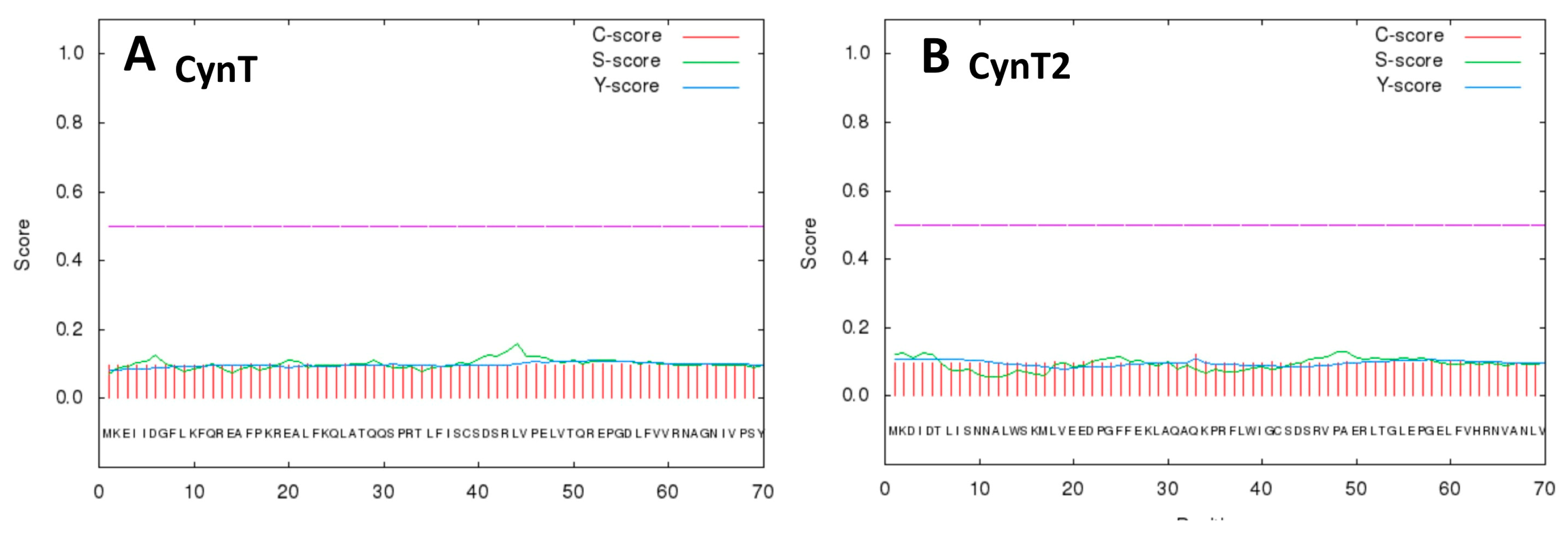
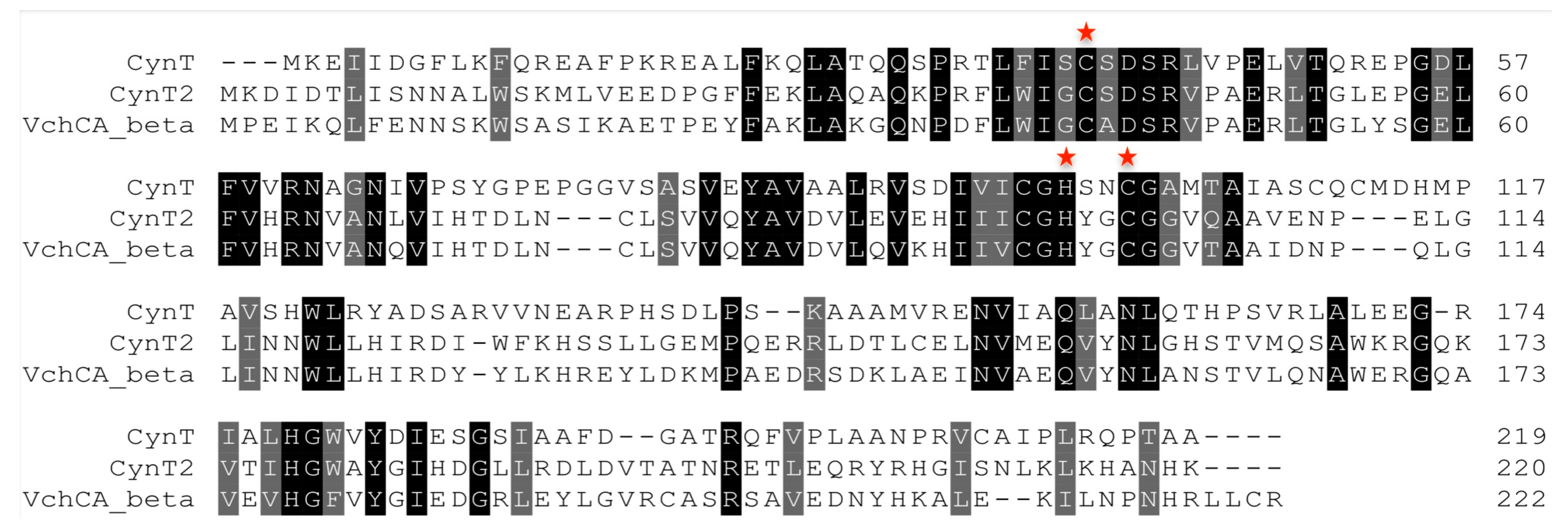
2.1. Primary Structure Analysis

The genome of Escherichia coli was inspected with BLAST (Basic Local Alignment Search Tool) to identify the CA-classes encoded by the bacterial genome, using individually the amino acid sequences belonging to the eight CA-classes (α, β, γ, δ, ζ, η, θ, and ι) as query sequences. Table 1 summarizes the results obtained with the BLAST analysis. Three CA classes were identified in the E. coli genome: β, γ- and ι-CAs; furthermore, different isoforms were detected for each class of CAs.
Table 1 shows that no representative of the α-class was detected in the E. coli genome. In our previous works, it has been documented that the bacterial α-CAs identified in the Gram-negative bacteria are characterized by the presence of a short secretory or signal peptide at the N-terminal end of the polypeptide chain [13,19,29]. This short peptide allows the translocation of the neo-synthesized protein into the bacterial periplasmic space, which is a typical feature of the Gram-negative bacteria. Lately, this secretory signal was also identified in the amino terminus of some polypeptide chains of β-, γ-, and ι-CAs from Gram-negative bacteria [13,29]. These findings prompted us to investigate the N-terminal portion of the two E. coli β-CA sequences reported in Table 1 (CynT and CynT2, with the GenBank IDs WP_033547590.1 and EEW0221051.1, respectively) for the presence of a putative signal peptide. Figure 1 shows the results obtained using the bioinformatics tool “SignalP 4.1” (http://www.cbs.dtu.dk/services/SignalP/), which is a software optimized for the prediction of a signal peptide in Bacteria, Archaea, and Eukarya.
From Figure 1, it is readily apparent that all the scores represented in the SignalP output were very low, close to the value of 0.1. Thus, E. coli β-CAs seemed characterized by the absence of a secretory signal at the amino terminus of their sequence. The translocation of CAs in the space between the two bacterial membranes (outer and inner layers) of the Gram-negative bacteria guarantees the rapid conversion of the periplasmic CO2 to bicarbonate, avoiding its depletion. Furthermore, the bicarbonate transported in the cytoplasm will be used by the cytoplasmic CA-classes (β and γ) to produce the CO2/HCO3 for the central bacterial metabolism [13,29]. From this consideration, it can be assumed that in E. coli, the lack of the α-CA is compensated by the presence of the ι-CA, whose polypeptide chain is typified by a secretory signal at the N-amino terminal [29]. This assumption is also corroborated by the existence of β- or γ-CAs with a signal peptide in Gram-negative bacteria, whose genome doesn’t encode for any α-CA. Figure 2 shows the multialignment of the two β-CAs amino acid sequences reported in Table 1 with a representative bacterial CA sequence belonging to the same family.
The isoform CynT showed an amino acid sequence identity of 28%, when compared with the isoform CynT2. The identity of CynT or CynT2 with the VchCA_beta from Vibrio cholerae was of 28% and 61%, respectively. This means that the two isoforms are characterized by multiple amino acid substitutions, even if the catalytic triad (two Cys and on His) is perfectly conserved (Figure 2). All the catalytically active CAs contain, independently of the genetic groups, a metal ion cofactor, which is necessary for enzyme catalysis [13,20,28,34]. The β-CAs use as catalytic metal a Zn2+ ion, which is coordinated by three amino acid residues; the fourth ligand is a water molecule/hydroxide ion acting as the nucleophile in the catalytic enzyme cycle, or an Asp residue in Type II β-CAs as CynT2 [41].

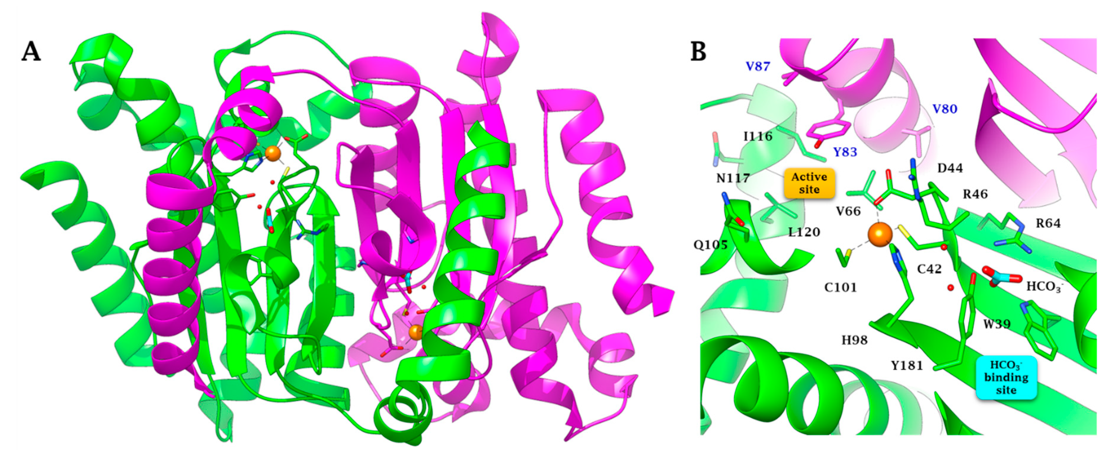
2.2. Three-Dimensional Structure Analysis

Figure 3 shows the X-ray crystal structure of the CynT2 from E. coli, which has been solved at 2.0 Å resolution; it reveals a dimeric arrangement of the protein, which is a tetramer, as two dimers interact with each other in the crystal packing [41]. The enzyme structure was determined as the closed conformation (type II β-CA) in which the zinc is tetrahedrally coordinated to Cys42, Asp44, His98, and Cys101 [41] (Figure 3A,B). The Asp44 side chain replacing the fourth ligand (water molecule) forms a non-canonical CA active site, which does not allow the CO2 hydration activity. This inactive form (closed active site) is present at pH values of less than 8.0 [41]. At pH values of ≥8.3, an incoming water molecule replaces the carboxylate moiety of the Asp residue, generating the nucleophile used in the typical catalytic cycle of the CAs [22,23]. Thus, the closed active site is converted to the open and active form (type I β-CA). As shown in Figure 3B, the closed structure is stabilized by HCO3, which occurs in a non-catalytic binding pocket close to the zinc ion, as reported for other few β-CAs, such as those from and Haemophilus influenzae and Vibrio cholerae [54,55].

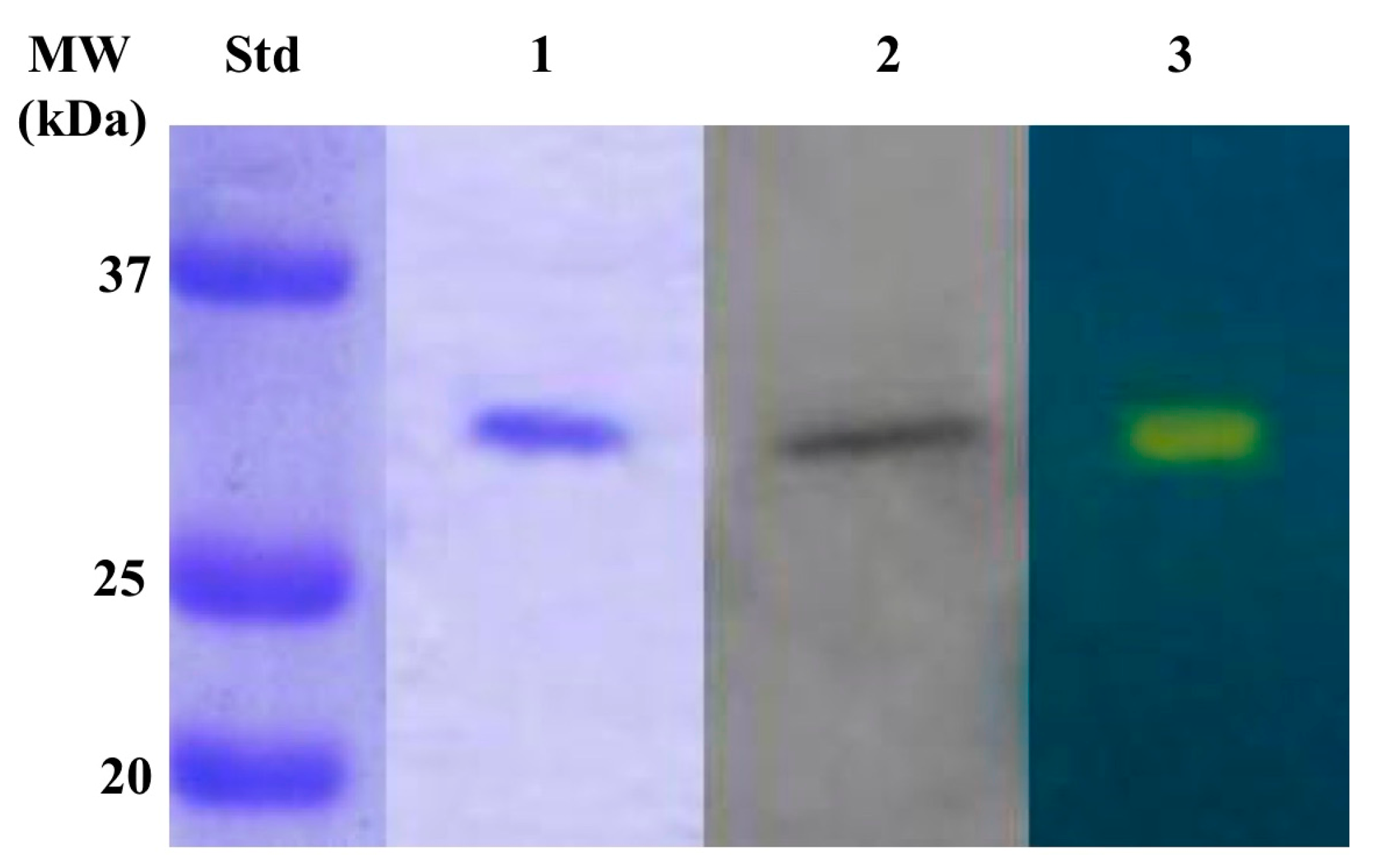
2.3. Production of Recombinat β-CA (CynT2)

We produced the bacterial CynT2 encoded in the E. coli genome, since a detailed investigation of its kinetic parameters is lacking to date, although, as mentioned above, the CynT2 three-dimensional structure has been solved [41]. Furthermore, this enzyme seems to be essential for bacterial growth at atmospheric pCO2 [21]. The purified enzyme, throughout the purification steps, was monitored following the Wilbur-Anderson Units (WAU) as described previously by Capasso’s group [56]. Figure 4 shows the purity of the recombinant CynT2 after the affinity column. Three biochemical techniques were used to verify the heterologous overexpression and purification of the bacterial enzyme, namely SDS-PAGE, Western Blot (WB), and protonography. SDS-PAGE and WB reported in Figure 4, (lane 1 and 2) indicated that the CynT2 fusion protein was purified to the homogeneity as a subunit with an apparent molecular weight of about 29.0 kDa. Moreover, the developed protonogram obtained by the protonography analysis evidenced that the recombinant enzyme was catalytically active (lane 3). It evidenced a yellow band due to the production of ions (H+) during the enzymatic CO2 hydration reaction at the molecular weight of 29.0 kDa, which corresponds to the mass of the recombinant CynT2 (Figure 4, lane 3).

2.4. Determination of the Kinetic Parameters Using the Stopped-Flow Technique

Using CO2 as a substrate, the recombinant CynT2 was subject to a stopped-flow analysis for the determination of the kinetic constants for the CO2 hydratase activity. As shown in Table 2, CynT2 showed a good biocatalyst activity for the physiological CO2 hydratase reaction to bicarbonate and protons, with kcat of 5.3 × 10⁵ s−1 and catalytic efficiency (kcat/KM) of 4.1 × 10⁷ M−1 s−1. In fact, the CynT2 kinetic constants were similar to those obtained for other bacterial CAs, as well as for hCA I. CynT2 was also inhibited by the sulfonamide acetazolamide (KI = 227 nM), which is a well-known pharmacological CA inhibitor (Table 2).
The results of Table 2 show that CynT2 was sensitive to 5-acetamido-1,3,4-thiadiazole-2-sulfonamide (acetazolamide, AZA) inhibition, similarly to the human isoform I (KI = 250 nM), but it was 19 times less inhibited than the human isoform hCA II (KI = 12 nM). Again, CynT2 with respect to VchCA_alpha from V. cholerae was 33 times less sensitive to AZA inhibition and 3.5 times less compared to the ι-CA from B. territorii (KI = 65 nM). The comparison with the β-CA from V. cholerae (KI = 451 nM) showed that the E. coli enzyme with a KI of 227 nM is two times more susceptible to AZA inhibition. These results are of extreme importance in the field of the inhibition of bacterial CAs because they prove that, even if these enzyme catalyze the same reaction, they can show a different inhibition pattern, whose investigation can lead to the discovery of novel inhibitors, which may impair the microbial growth as well as their virulence [57].

2.5. Inhibition Profile of Inorganic Metal-Complexing Anions

Since CAs are crucial for bacterial growth as well as for their virulence, we decided to investigate the inhibition profile of CynT2 with a broad range of inorganic metal-complexing anions (Table 1).
Anions and small molecules, such as diethyldithiocarbamate, iminodisulfonate, sulfamide, sulfamate, phenylboronic and phenlylarsonic acids, can complex the metal ion (orange sphere of the Figure 3) of the enzyme catalytic pocket, hindering the hydration of the CO2 to bicarbonate and protons. They bind the Zn2+ ion of the enzyme either in a tetrahedral geometry or as trigonal-bipyramidal adducts of the metal ion [59]. Generally, these inhibitors show an inhibition constant (KI) in the millimolar range. Even if the anion inhibitors are usually less effective than sulfonamides (KI is nM range), their investigation is essential for two fundamental aspects: (i) to design more efficient and selective inhibitors for the various CA-classes and their isoforms; (ii) potential for clinical applications for the treatment of diseases caused by pathogens, including bacteria. Here, we report the inhibition profile of CynT2 comparing these data with those obtained for the two human α-CAs, the isoforms hCA I and hCAII, and the bacterial enzyme (VchCA_beta), which have been previously investigated [58].
Table 3 lists the obtained results, which prompted us to elaborate the following observations:
  • A group of anions, such as iodide (I), cyanide (CN), azide (N3), perchlorate (ClO4) perosmate (OsO52), pyrophosphate (P2O72), divanadate (V2O72) perrhenate (ReO4), hexafluorophosphate (PF6), and trifluoromethanesulfonate (CF3SO3) were very weak inhibitors of the E.coli β-CA, with a KI > 10 mM (see Table 3). Within this group, VchCA_beta was inhibited with a KI ranging from 5.7 to 9.0 mM (Table 3), except for OsO52, PF6, and CF3SO3, whose KIs were even not measurable (Table 3). Moreover, N3 and P2O72 showed a KI > 10 mM for the CynT2, while ClO4 with a KI > 200 mM was an ineffective inhibitor of CynT2 as well as the V. cholerae enzyme. Interesting to note that some of these anions resulted to be quite effective inhibitors, when tested on the two human CAs. For example, cyanide and azide inhibited the human isoform h CA I with KI of 0.5 and 12 µM, respectively (Table 3). Thus, low concentrations of N3 and CN poison the two human enzymes as well as humans, whereas the bacterial CAs may better tolerate such toxic anions.
  • Another group of anion inhibitors, which weakly inhibited CynT2, showed a KI in the range between 1.5 and 9.4 mM. This is the case of fluoride (F), chloride (Cl), bromide (Br), thiocyanate (SCN), nitrite (NO2), nitrate (NO3), bisulfite (HSO3), sulfate (SO42), hydrogensulfide (HS), selenate (SeO42), perruthenate (RuO4), peroxydisulfate (S2O82), selenocyanate (SeCN), iminodisulfonate (NH(SO3)22), and trithiocarbonate (CS32). The inhibitory behavior of these anions was very similar to that exerted on the enzyme from V. cholerae (VchCA_beta); exceptions were HSO3, SO42, and NH(SO3)22, which were ineffective inhibitors for VchCA_beta with a KI > 200 mM. At the same time, HS inhibited the Vibrio enzyme with a KI > 20 mM. This last anion inhibited the human isoenzymes, hCA I and hCA II, with a KI = 0.6 µM and KI = 40 µM, respectively.
  • The submillimolar inhibitors of CynT2 showing a KI in the range 0.25–0.89 mM were cyanate (CNO), bicarbonate (HCO3), carbonate (CO32−), stannate (SnO32−), tetraborate (B4O72−), and fluorosulfonate (FSO3). These anions were millimolar inhibitors for the VchCA_beta (Table 1). The differences in the values of the inhibition constants can be due to the amino acid residues surrounding the catalytic pocket, which influence the interaction of the anion with the enzyme, even if the three-dimensional structure of the β-CAs from E. coli and V. cholerae are very similar. Intriguing is the result obtained using bicarbonate and carbonate as inhibitors. These two anions are not effective inhibitors (KI = 12–85 mM) for the two human isoforms (hCA I and hCA II), as well as for the β-CA from V. cholerae (KI = 5.9–6.7 mM). These results may reflect an evolutionary adaptation of the human and Vibrio CAs due to their continuous exposition to the high concentration of these two anions. The human enzymes are adapted to the high concentration of carbonate and bicarbonate present in the plasma, while V. cholerae colonizes the upper part of the small intestine characterized by high concentrations of bicarbonate, which is also a potent inducer of the expression of the genes involved in the virulence of the pathogen [45].
  • The best inhibitors of CynT2 resulted to be the small molecules, such as sulfamide (NH2SO2NH2), sulfamate (NH2SO3H), phenylboronic acid (PhB(OH)2), phenylarsonic acid (PhAsO3H2) as well as diethyldithiocarbamate (EtNCS2). As shown in Table 3, they showed KI values from 2.5 to 84 µM. These inhibitors were also effective against VchCA_beta (KI from 54 to 86 µM), except for EtNCS2, which showed a KI = 730 µM. Interesting to note, that dithiocarbamates were recently reported as a potent new class of CAIs targeting both the α- and β-classes of such enzymes [60,61,62].
From the mechanistic viewpoint, anions and small molecules as the ones investigated were shown to bind directly to the metal ion from the enzyme active site both for α- and β-CAs, as discussed extensively on the review by De Simone and Supuran [66] and by Whitesides’ group [67]. We estimate that anions investigated here show the same mechanism for the inhibition of CynT2 as the anions crystallized with various other α- and β-CAs.

3. Materials and Methods

3.1. Chemicals and Instruments

All the chemicals used in this study were of reagent grade and purchased from Sigma (Milano, Italy). The Affinity column (His-Trap FF) and the AKTA-Prime purification system were bought from GE Healthcare (Chicago, IL, USA). The SX20 Stopped-Flow was obtained by the AppliedPhotophysics. SDS-PAGE and Western-Blot apparatus were procured by BioRAD (Hercules, CA, USA).

3.2. Cloning, Expression, and Purification

The synthetic Escherichia coli gene encoding for the CynT2 was synthesized by the Invitrogen GeneArt (ThermoFisher Scientific, Waltham, MA, USA), a company specialized in gene synthesis, and cloned into the expression vector pET100D-Topo/CynT2. Briefly, the gene was designed to produce the recombinant CynT2 as fusion proteins with a tag containing nucleotides encoding for six histidines (His-Tag) at the amino terminus of neosynthesized recombinant protein. Competent E. coli BL21 (DE3) codon plus cells (Agilent) were transformed as described by Del Prete et al. [68]. Isopropyl β-D-1-thiogalactopyranoside (IPTG) at the concentration of 1 mM was added to the cellular culture to overexpress the recombinant CynT2. After growth, the cells were harvested and disrupted by sonication. Cellular extract was purified using a nickel affinity column (His-Trap FF), which allows the interaction between the matrix functionalized with Ni2+ ion and the His-Tag at the N-terminus of the protein. The HisTrap column (1 mL) was equilibrated with a 20-mL equilibration buffer (50 mM Tris, 20 mM imidazole and 150 mM sodium chloride, pH 7.5) at 1 mL/min. The supernatant from the cellular lysate was loaded onto the column at 1 mL/min, connected with AKTA Prime. The recombinant CynT2 was eluted from the column by fluxing a linear gradient of imidazole (0–300 mM) at a flow of 0.5 mL/min in a buffer composed of 50 mM Tris and 300 mM sodium chloride, pH 7.5. The recovered CynT2 was 90% pure. The protein quantification was carried out by Bradford method (BioRAD) [69].

3.3. Carbonic Anhydrase Assay for Monitoring the Recombinant Enzyme during the Purification Steps

CA activity assay was performed as described by Capasso et al. [56]. Briefly, the assay was based on the monitoring of pH variation due to the catalyzed conversion of CO2 to bicarbonate. Bromothymol blue was used as the indicator of pH variation. The assay was performed at 0 °C and a CO2-satured solution was used as substrate. The enzyme activity was calculated by measuring the time required for Bromothymol blue to change from blue to yellow. This time is inversely related to the quantity of enzyme present in the sample and allows the calculation of the Wilbur-Anderson units as described previously [56].

3.4. SDS-PAGE

A 12% Sodium Dodecyl Sulfate-polyacrylamide gel electrophoresis (SDS-PAGE) prepared as described by Laemmli [70] was used, loading on the gel the recovered CynT2 from the affinity column. The gel was stained with Coomassie Brilliant Blue-R.

3.5. Western Blot

CynT2 was subjected to a 12% (w/v) SDS-PAGE, and then was transferred to a PVDF (polyvinylidene fluoride) membrane with transfer buffer (25 mM Tris, 192 mM glycine, 20% methanol) using Trans-Plot SD Cell (Bio-Rad, Hercules, CA, USA). His-Tag Western blot was carried out using the Pierce Fast Western Blot Kit (Thermo Scientific, Waltham, MA, USA). Blotted membrane was placed in the wash blot solution Fast Western 1 Wash Buffer to remove transfer buffer. Primary Antibody Working Dilution was added to the blot and incubated for 30 min at room temperature (RT) with shaking. Invitrogen anti-His antibody (1:10,000) was used. Afterwards, the blot was removed from the primary antibody solution and incubated for 10 min with the FastWestern Optimised HRP ReagentWorking Dilution. Subsequently, the membrane was washed two times in about 20 mL of FastWestern 1 Wash Buffer. Finally, the membrane was incubated with the detection reagent working solution and incubated for 1 min, at room temperature, and then developed with X-ray film.

3.6. Protonography

To perform the protonography, wells of 12% SDS-PAGE gel were loaded with samples mixed with loading buffer not containing 2-mercaptoethanol and not subjected to boiling, in order to avoid protein denaturation. The gel was run at 150 V until the dye front ran off the gel. Following the electrophoresis, the 12% SDS-PAGE gel was subject to protonography to detect the yellows bands due to the hydratase activity on the gel as described previously [71,72,73,74].

3.7. Kinetic Parameters and Inhibition Constants Determined by the Stopped-Flow Technique

The CO2 hydration activity performed by the BteCAι was monitored using an Applied Photophysics stopped-flow instrument [59]. Phenol red (at a concentration of 0.2 mM) was used as indicator, working at the absorbance maximum of 557 nm, with 20 mM TRIS (pH 8.3) as buffer, and 20 mM NaClO4 (for maintaining constant the ionic strength), following the initial rates of the CA-catalyzed CO2 hydration reaction for a period of 10–100 s. To determine the kinetic parameters by Lineweaver-Burk plots and the inhibition constants, a concentration of CO2 between 1.7 to 17 mM was used. At least six measurements of the original 5–10% reaction were used to assess the initial velocity for each inhibitor. The uncatalyzed rates were identically determined and detracted from the total observed rates. Stock inhibitor solutions (10–100 mM) were prepared in distilled-deionized water and dilutions up to 0.01 mM were done with the buffer test. Inhibitor and enzyme solutions were preincubated together for 15 min at room temperature prior to assay, in order to allow for the formation of the E–I complex or for the eventual active site mediated hydrolysis of the inhibitor. The inhibition constants were obtained by non-linear least-squares methods using PRISM 6 and the Cheng-Prusoff equation, as reported earlier [58,60,61], and represent the mean from at least three different determinations. All CA isoforms were recombinant ones obtained in-house.

4. Conclusions

The E. coli genome encodes only for three of the eight CA-classes reported in the literature: β-, γ-, and ι-CAs. In the present paper, we produced the recombinant isoform CynT2, a CA belonging to the β-class. The enzyme kinetic parameters and its anion inhibition profile were determined, using the stopped-flow technique. CynT2 resulted to be a good biocatalyst for the CO2 hydration reaction showing a kcat = 5.3 × 10⁵ and a kcat/KM = 4.1 × 10⁷. These values are similar to those obtained for other enzymes belonging to the β-CA class. Moreover, we also investigated the inhibition profile of CynT2 with a broad range of inorganic metal-complexing anions, a well-known group of CA inhibitors capable of blocking the enzyme activity. Small molecules such as sulfamide, sulfamate, phenylboronic acid, phenylarsonic acid, and diethyldithiocarbamate resulted to be the most effective CynT2 inhibitors (KI = 2.5 to 84 µM). Despite these metal-complexing anions showed inhibition constants in the millimolar range, this anion investigation study is relevant, because it allowed a better understanding of the interaction of the CA inhibitors with the amino acid residues surrounding the enzyme catalytic pocket as well as the design of more efficient and specific inhibitors. Besides, it is important to keep in mind that the physiological role of CAs is to balance pH, CO2, and bicarbonate inside the bacterial cell, ensuring the right amounts of these molecules/ions to the bacterial metabolism. Thus, inhibition of such enzymes can impair bacterial growth as well as their virulence through a metabolic pathway, which is different from those used by the common pharmacological antibiotics. These could open a new solution to antibiotic resistance, which is occurring worldwide. Finally, E. coli is a microorganism that can easily be manipulated in the laboratory, avoiding the risk connected to the handling of pathogenic bacteria. Considering this aspect, E. coli could be considered as a model study organism for testing CA inhibitors in vitro, in cell-based assays, evaluating the effect of the inhibition on bacterial cell growth in a safe and facile way. To address the issues mentioned above, it is necessary to analyze in vitro the effect of CA inhibitors on all CA-classes encoded by the E. coli genome. That’s what we are doing.

Author Contributions

Investigation, S.D.P., V.D.L., A.N. and C.C.; data curation, A.S., M.D.M., and C.C.; supervision, C.T.S. and C.C; writing—original draft, C.C.; writing—review and editing, C.T.S. and C.C. All authors have read and agreed to the published version of the manuscript.

Funding

This research received no external funding.

Acknowledgments

We are grateful to Giovanni Del Monaco for technical assistance.

Conflicts of Interest

The authors declare no conflicts of interest.

References

  1. Mainil, J. Escherichia coli virulence factors. Vet. Immunol. Immunopathol. 2013, 152, 2–12. [Google Scholar] [CrossRef] [PubMed] [Green Version]
  2. Allocati, N.; Masulli, M.; Alexeyev, M.F.; Di Ilio, C. Escherichia coli in Europe: An overview. Int. J. Environ. Res. Public Health 2013, 10, 6235–6254. [Google Scholar] [CrossRef] [PubMed]
  3. Conway, T.; Cohen, P.S. Commensal and Pathogenic Escherichia coli Metabolism in the Gut. In Metabolism and Bacterial Pathogenesis; Conway, T., Cohen, P., Eds.; ASM Press: Washington, WA, USA, 2015. [Google Scholar]
  4. Robins-Browne, R.M.; Holt, K.E.; Ingle, D.J.; Hocking, D.M.; Yang, J.; Tauschek, M. Are Escherichia coli Pathotypes Still Relevant in the Era of Whole-Genome Sequencing? Front. Cell Infect. Microbiol. 2016, 6, 141. [Google Scholar] [CrossRef] [PubMed] [Green Version]
  5. Stromberg, Z.R.; Van Goor, A.; Redweik, G.A.J.; Wymore Brand, M.J.; Wannemuehler, M.J.; Mellata, M. Pathogenic and non-pathogenic Escherichia coli colonization and host inflammatory response in a defined microbiota mouse model. Dis. Model Mech. 2018, 11, dmm035063. [Google Scholar] [CrossRef] [Green Version]
  6. Pitout, J.D. Extraintestinal Pathogenic Escherichia coli: A Combination of Virulence with Antibiotic Resistance. Front. Microbiol. 2012, 3, 9. [Google Scholar] [CrossRef] [PubMed] [Green Version]
  7. Nataro, J.P.; Kaper, J.B. Diarrheagenic Escherichia coli. Clin. Microbiol. Rev. 1998, 11, 142–201. [Google Scholar] [CrossRef] [PubMed] [Green Version]
  8. Moriel, D.G.; Bertoldi, I.; Spagnuolo, A.; Marchi, S.; Rosini, R.; Nesta, B.; Pastorello, I.; Corea, V.A.; Torricelli, G.; Cartocci, E.; et al. Identification of protective and broadly conserved vaccine antigens from the genome of extraintestinal pathogenic Escherichia coli. Proc. Natl. Acad. Sci. USA 2010, 107, 9072–9077. [Google Scholar] [CrossRef] [Green Version]
  9. Agus, A.; Massier, S.; Darfeuille-Michaud, A.; Billard, E.; Barnich, N. Understanding host-adherent-invasive Escherichia coli interaction in Crohn’s disease: Opening up new therapeutic strategies. Biomed. Res. Int. 2014, 2014, 567929. [Google Scholar] [CrossRef] [Green Version]
  10. Clermont, O.; Olier, M.; Hoede, C.; Diancourt, L.; Brisse, S.; Keroudean, M.; Glodt, J.; Picard, B.; Oswald, E.; Denamur, E. Animal and human pathogenic Escherichia coli strains share common genetic backgrounds. Infect. Genet. Evol. 2011, 11, 654–662. [Google Scholar] [CrossRef]
  11. Reid, S.D.; Herbelin, C.J.; Bumbaugh, A.C.; Selander, R.K.; Whittam, T.S. Parallel evolution of virulence in pathogenic Escherichia coli. Nature 2000, 406, 64–67. [Google Scholar] [CrossRef]
  12. Szych, J.; Wolkowicz, T.; La Ragione, R.; Madajczak, G. Impact of antibiotics on the intestinal microbiota and on the treatment of Shiga-toxin-producing Escherichia coli and Salmonella infections. Curr. Pharm. Des. 2014, 20, 4535–4548. [Google Scholar] [CrossRef] [PubMed]
  13. Supuran, C.; Capasso, C. An Overview of the Bacterial Carbonic Anhydrases. Metabolites 2017, 7, 56. [Google Scholar] [CrossRef] [PubMed] [Green Version]
  14. Vullo, D.; Del Prete, S.; Di Fonzo, P.; Carginale, V.; Donald, W.A.; Supuran, C.T.; Capasso, C. Comparison of the Sulfonamide Inhibition Profiles of the beta- and gamma-Carbonic Anhydrases from the Pathogenic Bacterium Burkholderia pseudomallei. Molecules 2017, 22, 421. [Google Scholar] [CrossRef] [PubMed] [Green Version]
  15. Smith, K.S.; Ferry, J.G. Prokaryotic carbonic anhydrases. FEMS Microbiol. Rev. 2000, 24, 335–366. [Google Scholar] [CrossRef]
  16. Maeda, S.; Price, G.D.; Badger, M.R.; Enomoto, C.; Omata, T. Bicarbonate binding activity of the CmpA protein of the cyanobacterium Synechococcus sp. strain PCC 7942 involved in active transport of bicarbonate. J. Biol. Chem. 2000, 275, 20551–20555. [Google Scholar] [CrossRef] [PubMed] [Green Version]
  17. Joseph, P.; Ouahrani-Bettache, S.; Montero, J.L.; Nishimori, I.; Minakuchi, T.; Vullo, D.; Scozzafava, A.; Winum, J.Y.; Kohler, S.; Supuran, C.T. A new beta-carbonic anhydrase from Brucella suis, its cloning, characterization, and inhibition with sulfonamides and sulfamates, leading to impaired pathogen growth. Bioorg. Med. Chem. 2011, 19, 1172–1178. [Google Scholar] [CrossRef]
  18. Joseph, P.; Turtaut, F.; Ouahrani-Bettache, S.; Montero, J.L.; Nishimori, I.; Minakuchi, T.; Vullo, D.; Scozzafava, A.; Kohler, S.; Winum, J.Y.; et al. Cloning, characterization, and inhibition studies of a beta-carbonic anhydrase from Brucella suis. J. Med. Chem. 2010, 53, 2277–2285. [Google Scholar] [CrossRef]
  19. Supuran, C.T.; Capasso, C. New light on bacterial carbonic anhydrases phylogeny based on the analysis of signal peptide sequences. J. Enzym. Inhib. Med. Chem. 2016, 31, 1254–1260. [Google Scholar] [CrossRef] [Green Version]
  20. Capasso, C.; Supuran, C.T. An overview of the alpha-, beta- and gamma-carbonic anhydrases from Bacteria: Can bacterial carbonic anhydrases shed new light on evolution of bacteria? J. Enzym. Inhib. Med. Chem. 2015, 30, 325–332. [Google Scholar] [CrossRef] [Green Version]
  21. Merlin, C.; Masters, M.; McAteer, S.; Coulson, A. Why is carbonic anhydrase essential to Escherichia Coli. J. Bacteriol. 2003, 185, 6415–6424. [Google Scholar] [CrossRef] [Green Version]
  22. Supuran, C.T. Advances in structure-based drug discovery of carbonic anhydrase inhibitors. Expert Opin. Drug Discov. 2017, 12, 61–88. [Google Scholar] [CrossRef] [PubMed]
  23. Supuran, C.T. Structure and function of carbonic anhydrases. Biochem. J. 2016, 473, 2023–2032. [Google Scholar] [CrossRef] [PubMed]
  24. Annunziato, G.; Angeli, A.; D’Alba, F.; Bruno, A.; Pieroni, M.; Vullo, D.; De Luca, V.; Capasso, C.; Supuran, C.T.; Costantino, G. Discovery of New Potential Anti-Infective Compounds Based on Carbonic Anhydrase Inhibitors by Rational Target-Focused Repurposing Approaches. ChemMedChem 2016, 11, 1904–1914. [Google Scholar] [CrossRef] [PubMed]
  25. Ozensoy Guler, O.; Capasso, C.; Supuran, C.T. A magnificent enzyme superfamily: Carbonic anhydrases, their purification and characterization. J. Enzym. Inhib. Med. Chem. 2016, 31, 689–694. [Google Scholar] [CrossRef]
  26. Del Prete, S.; Vullo, D.; De Luca, V.; Carginale, V.; Ferraroni, M.; Osman, S.M.; AlOthman, Z.; Supuran, C.T.; Capasso, C. Sulfonamide inhibition studies of the beta-carbonic anhydrase from the pathogenic bacterium Vibrio cholerae. Bioorg. Med. Chem. 2016, 24, 1115–1120. [Google Scholar] [CrossRef]
  27. Del Prete, S.; De Luca, V.; De Simone, G.; Supuran, C.T.; Capasso, C. Cloning, expression and purification of the complete domain of the eta-carbonic anhydrase from Plasmodium falciparum. J. Enzym. Inhib. Med. Chem. 2016, 31, 54–59. [Google Scholar] [CrossRef] [Green Version]
  28. Capasso, C.; Supuran, C.T. An Overview of the Carbonic Anhydrases from Two Pathogens of the Oral Cavity: Streptococcus mutans and Porphyromonas gingivalis. Curr. Top. Med. Chem. 2016, 16, 2359–2368. [Google Scholar] [CrossRef]
  29. Del Prete, S.; Nocentini, A.; Supuran, C.T.; Capasso, C. Bacterial iota-carbonic anhydrase: A new active class of carbonic anhydrase identified in the genome of the Gram-negative bacterium Burkholderia territorii. J. Enzym. Inhib. Med. Chem. 2020, 35, 1060–1068. [Google Scholar] [CrossRef] [Green Version]
  30. Jensen, E.L.; Clement, R.; Kosta, A.; Maberly, S.C.; Gontero, B. A new widespread subclass of carbonic anhydrase in marine phytoplankton. Isme. J. 2019, 13, 2094–2106. [Google Scholar] [CrossRef] [Green Version]
  31. Kikutani, S.; Nakajima, K.; Nagasato, C.; Tsuji, Y.; Miyatake, A.; Matsuda, Y. Thylakoid luminal theta-carbonic anhydrase critical for growth and photosynthesis in the marine diatom Phaeodactylum tricornutum. Proc. Natl. Acad. Sci. USA 2016, 113, 9828–9833. [Google Scholar] [CrossRef] [Green Version]
  32. Capasso, C.; Supuran, C.T. Bacterial, fungal and protozoan carbonic anhydrases as drug targets. Expert Opin. Ther. Targets 2015, 19, 1689–1704. [Google Scholar] [CrossRef] [PubMed]
  33. Supuran, C.T.; Capasso, C. The eta-class carbonic anhydrases as drug targets for antimalarial agents. Expert Opin. Ther. Targets 2015, 19, 551–563. [Google Scholar] [CrossRef] [PubMed]
  34. Capasso, C.; Supuran, C.T. An Overview of the Selectivity and Efficiency of the Bacterial Carbonic Anhydrase Inhibitors. Curr. Med. Chem. 2015, 22, 2130–2139. [Google Scholar] [CrossRef]
  35. Capasso, C.; Supuran, C.T. Sulfa and trimethoprim-like drugs-antimetabolites acting as carbonic anhydrase, dihydropteroate synthase and dihydrofolate reductase inhibitors. J. Enzym. Inhib. Med. Chem. 2014, 29, 379–387. [Google Scholar] [CrossRef]
  36. Capasso, C.; Supuran, C.T. Anti-infective carbonic anhydrase inhibitors: A patent and literature review. Expert Opin. Ther. Pat. 2013, 23, 693–704. [Google Scholar] [CrossRef] [PubMed]
  37. De Smet, B.; Mayo, M.; Peeters, C.; Zlosnik, J.E.; Spilker, T.; Hird, T.J.; LiPuma, J.J.; Kidd, T.J.; Kaestli, M.; Ginther, J.L.; et al. Burkholderia stagnalis sp. nov. and Burkholderia territorii sp. nov., two novel Burkholderia cepacia complex species from environmental and human sources. Int. J. Syst. Evol. Microbiol. 2015, 65, 2265–2271. [Google Scholar] [CrossRef]
  38. Kozliak, E.I.; Guilloton, M.B.; Gerami-Nejad, M.; Fuchs, J.A.; Anderson, P.M. Expression of proteins encoded by the Escherichia coli cyn operon: Carbon dioxide-enhanced degradation of carbonic anhydrase. J. Bacteriol. 1994, 176, 5711–5717. [Google Scholar] [CrossRef] [Green Version]
  39. Guilloton, M.B.; Lamblin, A.F.; Kozliak, E.I.; Gerami-Nejad, M.; Tu, C.; Silverman, D.; Anderson, P.M.; Fuchs, J.A. A physiological role for cyanate-induced carbonic anhydrase in Escherichia coli. J. Bacteriol. 1993, 175, 1443–1451. [Google Scholar] [CrossRef] [Green Version]
  40. Guilloton, M.B.; Korte, J.J.; Lamblin, A.F.; Fuchs, J.A.; Anderson, P.M. Carbonic anhydrase in Escherichia coli. A product of the cyn operon. J. Biol. Chem. 1992, 267, 3731–3734. [Google Scholar]
  41. Cronk, J.D.; Endrizzi, J.A.; Cronk, M.R.; O’Neill, J.W.; Zhang, K.Y. Crystal structure of E. coli beta-carbonic anhydrase, an enzyme with an unusual pH-dependent activity. Protein Sci. 2001, 10, 911–922. [Google Scholar] [CrossRef]
  42. Modak, J.K.; Tikhomirova, A.; Gorrell, R.J.; Rahman, M.M.; Kotsanas, D.; Korman, T.M.; Garcia-Bustos, J.; Kwok, T.; Ferrero, R.L.; Supuran, C.T.; et al. Anti-Helicobacter pylori activity of ethoxzolamide. J. Enzym. Inhib. Med. Chem. 2019, 34, 1660–1667. [Google Scholar] [CrossRef] [PubMed] [Green Version]
  43. Ronci, M.; Del Prete, S.; Puca, V.; Carradori, S.; Carginale, V.; Muraro, R.; Mincione, G.; Aceto, A.; Sisto, F.; Supuran, C.T.; et al. Identification and characterization of the alpha-CA in the outer membrane vesicles produced by Helicobacter pylori. J. Enzym. Inhib. Med. Chem. 2019, 34, 189–195. [Google Scholar] [CrossRef] [PubMed] [Green Version]
  44. Buzas, G.M. Helicobacter pylori—2010. Orv. Hetil. 2010, 151, 2003–2010. [Google Scholar] [CrossRef] [PubMed]
  45. Abuaita, B.H.; Withey, J.H. Bicarbonate Induces Vibrio cholerae virulence gene expression by enhancing ToxT activity. Infect. Immun. 2009, 77, 4111–4120. [Google Scholar] [CrossRef] [PubMed] [Green Version]
  46. Kohler, S.; Ouahrani-Bettache, S.; Winum, J.Y. Brucella suis carbonic anhydrases and their inhibitors: Towards alternative antibiotics. J. Enzym. Inhib. Med. Chem. 2017, 32, 683–687. [Google Scholar] [CrossRef] [PubMed] [Green Version]
  47. Singh, S.; Supuran, C.T. 3D-QSAR CoMFA studies on sulfonamide inhibitors of the Rv3588c beta-carbonic anhydrase from Mycobacterium tuberculosis and design of not yet synthesized new molecules. J. Enzym. Inhb. Med. Chem. 2014, 29, 449–455. [Google Scholar] [CrossRef] [Green Version]
  48. Ceruso, M.; Vullo, D.; Scozzafava, A.; Supuran, C.T. Sulfonamides incorporating fluorine and 1,3,5-triazine moieties are effective inhibitors of three beta-class carbonic anhydrases from Mycobacterium tuberculosis. J. Enzym. Inhib. Med. Chem. 2014, 29, 686–689. [Google Scholar] [CrossRef]
  49. Carta, F.; Maresca, A.; Covarrubias, A.S.; Mowbray, S.L.; Jones, T.A.; Supuran, C.T. Carbonic anhydrase inhibitors. Characterization and inhibition studies of the most active beta-carbonic anhydrase from Mycobacterium tuberculosis, Rv3588c. Bioorg. Med. Chem. Lett. 2009, 19, 6649–6654. [Google Scholar] [CrossRef]
  50. Rollenhagen, C.; Bumann, D. Salmonella enterica highly expressed genes are disease specific. Infect. Immun. 2006, 74, 1649–1660. [Google Scholar] [CrossRef] [Green Version]
  51. Lotlikar, S.R.; Kayastha, B.B.; Vullo, D.; Khanam, S.S.; Braga Reygan, E.; Murray, A.B.; McKenna, R.; Supuran, C.T.; Patrauchan, M.A. Pseudomonas aeruginosa β-carbonic anhydrase, psCA1, is required for calcium deposition and contributes to virulence. Cell Calcium 2019, 84, 102080. [Google Scholar] [CrossRef]
  52. Alonso-de Castro, S.; Terenzi, A.; Gurruchaga-Pereda, J.; Salassa, L. Catalysis Concepts in Medicinal Inorganic Chemistry. Chemistry 2019, 25, 6651–6660. [Google Scholar] [CrossRef] [PubMed]
  53. Mjos, K.D.; Orvig, C. Metallodrugs in medicinal inorganic chemistry. Chem. Rev. 2014, 114, 4540–4563. [Google Scholar] [CrossRef] [PubMed]
  54. Ferraroni, M.; Del Prete, S.; Vullo, D.; Capasso, C.; Supuran, C.T. Crystal structure and kinetic studies of a tetrameric type II beta-carbonic anhydrase from the pathogenic bacterium Vibrio cholerae. Acta Cryst. D Biol. Cryst. 2015, 71, 2449–2456. [Google Scholar] [CrossRef] [PubMed]
  55. Cronk, J.D.; Rowlett, R.S.; Zhang, K.Y.; Tu, C.; Endrizzi, J.A.; Lee, J.; Gareiss, P.C.; Preiss, J.R. Identification of a novel noncatalytic bicarbonate binding site in eubacterial beta-carbonic anhydrase. Biochemistry 2006, 45, 4351–4361. [Google Scholar] [CrossRef] [PubMed]
  56. Capasso, C.; De Luca, V.; Carginale, V.; Cannio, R.; Rossi, M. Biochemical properties of a novel and highly thermostable bacterial alpha-carbonic anhydrase from Sulfurihydrogenibium yellowstonense YO3AOP1. J. Enzym. Inhib. Med. Chem. 2012, 27, 892–897. [Google Scholar] [CrossRef] [PubMed] [Green Version]
  57. Rahman, M.M.; Tikhomirova, A.; Modak, J.K.; Hutton, M.L.; Supuran, C.T.; Roujeinikova, A. Antibacterial activity of ethoxzolamide against Helicobacter pylori strains SS1 and 26695. Gut Pathog. 2020, 12, 20. [Google Scholar] [CrossRef] [Green Version]
  58. Del Prete, S.; Vullo, D.; De Luca, V.; Carginale, V.; di Fonzo, P.; Osman, S.M.; AlOthman, Z.; Supuran, C.T.; Capasso, C. Anion inhibition profiles of alpha-, beta- and gamma-carbonic anhydrases from the pathogenic bacterium Vibrio cholerae. Bioorg. Med. Chem. 2016, 24, 3413–3417. [Google Scholar] [CrossRef]
  59. Supuran, C.T. How many carbonic anhydrase inhibition mechanisms exist. J. Enzym. Inhib. Med. Chem. 2016, 31, 345–360. [Google Scholar] [CrossRef]
  60. Aspatwar, A.; Hammaren, M.; Parikka, M.; Parkkila, S.; Carta, F.; Bozdag, M.; Vullo, D.; Supuran, C.T. In vitro inhibition of Mycobacterium tuberculosis beta-carbonic anhydrase 3 with Mono- and dithiocarbamates and evaluation of their toxicity using zebrafish developing embryos. J. Enzym. Inhib. Med. Chem. 2020, 35, 65–71. [Google Scholar] [CrossRef] [Green Version]
  61. Carta, F.; Aggarwal, M.; Maresca, A.; Scozzafava, A.; McKenna, R.; Supuran, C.T. Dithiocarbamates: A new class of carbonic anhydrase inhibitors. Crystallographic and kinetic investigations. Chem. Commun. 2012, 48, 1868–1870. [Google Scholar] [CrossRef] [Green Version]
  62. Maresca, A.; Carta, F.; Vullo, D.; Supuran, C.T. Dithiocarbamates strongly inhibit the beta-class carbonic anhydrases from Mycobacterium tuberculosis. J. Enzym. Inhib. Med. Chem. 2013, 28, 407–411. [Google Scholar] [CrossRef] [PubMed]
  63. Khalifah, R.G. The carbon dioxide hydration activity of carbonic anhydrase. I. Stop-flow kinetic studies on the native human isoenzymes B and C. J. Biol. Chem. 1971, 246, 2561–2573. [Google Scholar]
  64. Del Prete, S.; Vullo, D.; De Luca, V.; Carginale, V.; di Fonzo, P.; Osman, S.M.; AlOthman, Z.; Supuran, C.T.; Capasso, C. Anion inhibition profiles of the complete domain of the eta-carbonic anhydrase from Plasmodium falciparum. Bioorg. Anicmed. Chem. 2016, 24, 4410–4414. [Google Scholar] [CrossRef] [PubMed]
  65. De Luca, V.; Vullo, D.; Del Prete, S.; Carginale, V.; Osman, S.M.; AlOthman, Z.; Supuran, C.T.; Capasso, C. Cloning, characterization and anion inhibition studies of a gamma-carbonic anhydrase from the Antarctic bacterium Colwellia psychrerythraea. Bioorg. Med. Chem. 2016, 24, 835–840. [Google Scholar] [CrossRef] [PubMed]
  66. De Simone, G.; Supuran, C.T. (In)organic anions as carbonic anhydrase inhibitors. J. Inorg. Biochem. 2012, 111, 117–129. [Google Scholar] [CrossRef] [PubMed]
  67. Fox, J.M.; Kang, K.; Sherman, W.; Héroux, A.; Sastry, G.M.; Baghbanzadeh, M.; Lockett, M.R.; Whitesides, G.M. Interactions between Hofmeister anions and the binding pocket of a protein. J. Am. Chem. Soc. 2015, 137, 3859–3866. [Google Scholar] [CrossRef] [PubMed]
  68. Del Prete, S.; Vullo, D.; Ghobril, C.; Hitce, J.; Clavaud, C.; Marat, X.; Capasso, C.; Supuran, C.T. Cloning, Purification, and Characterization of a beta-Carbonic Anhydrase from Malassezia restricta, an Opportunistic Pathogen Involved in Dandruff and Seborrheic Dermatitis. Int. J. Mol. Sci. 2019, 20, 2447. [Google Scholar] [CrossRef] [Green Version]
  69. Bradford, M.M. A rapid and sensitive method for the quantitation of microgram quantities of protein utilizing the principle of protein-dye binding. Anal. Biochem. 1976, 72, 248–254. [Google Scholar] [CrossRef]
  70. Laemmli, U.K. Cleavage of structural proteins during the assembly of the head of bacteriophage T4. Nature 1970, 227, 680–685. [Google Scholar] [CrossRef]
  71. De Luca, V.; Del Prete, S.; Supuran, C.T.; Capasso, C. Protonography, a new technique for the analysis of carbonic anhydrase activity. J. Enzym. Inhib. Med. Chem. 2015, 30, 277–282. [Google Scholar] [CrossRef]
  72. Del Prete, S.; De Luca, V.; Iandolo, E.; Supuran, C.T.; Capasso, C. Protonography, a powerful tool for analyzing the activity and the oligomeric state of the gamma-carbonic anhydrase identified in the genome of Porphyromonas gingivalis. Bioorg. Med. Chem. 2015, 23, 3747–3750. [Google Scholar] [CrossRef] [PubMed]
  73. Del Prete, S.; De Luca, V.; Supuran, C.T.; Capasso, C. Protonography, a technique applicable for the analysis of eta-carbonic anhydrase activity. J. Enzym. Inhib. Med. Chem. 2015, 30, 920–924. [Google Scholar] [CrossRef] [PubMed]
  74. Del Prete, S.; Vullo, D.; Caminiti-Segonds, N.; Zoccola, D.; Tambutte, S.; Supuran, C.T.; Capasso, C. Protonography and anion inhibition profile of the alpha-carbonic anhydrase (CruCA4) identified in the Mediterranean red coral Corallium rubrum. Bioorg. Chem. 2018, 76, 281–287. [Google Scholar] [CrossRef] [PubMed]
Sample Availability: All the samples of the compounds are available from the authors.
Figure 1. SignalP 4.1 graphical results obtained using the two β-CA polypeptide chains encoded by the Escherichia coli genome. Legend: Panel (A), CynT; Panel (B), CynT2; X-axis, the first 70 amino acid residues at the amino terminus of the polypeptide chain; Y-axis: C-score (red line), raw cleavage site score; S-score (green line); signal peptide score; Y-score (blue line), the combined cleavage site score. The violet line indicates the threshold value of 0.5.
Molecules 25 02564 g001
Figure 2. Multialignment of the two β-CAs (CynT and CynT2) from E. coli and the β-CA (VchCA_beta) from Vibrio cholerae. Legend: red stars indicate the metal ion coordinating residues. The alignment was formatted highlighting in black the identical residues and in gray the conservative substitutions. Multiple amino acid sequence alignment was performed with the program MUSCLE, version 3.7.
Molecules 25 02564 g002
Figure 3. β-CA (CynT2, PDB 2ESF) from E. coli. (A) Ribbon representation of the dimer. (B) Zoomed view of the active site and bicarbonate binding pocket. The Zn(II) ion is represented as an orange sphere. Chain A and chain B are colored green and magenta, respectively. The HCO3 ion is colored cyan. Amino acids composing the active site are labeled with one letter symbols (blue for chain A and black for chain B): A, Ala; C, Cys; D, Asp; H, His; I, Ile; L, Leu; N, Asn; Q, Gln; R, Arg; V, Val; W, Trp; Y, Tyr. The cyan box is the HCO3 pocket, while the yellow box represents the catalytic pocket.
Molecules 25 02564 g003
Figure 4. Combined results obtained with SDS-PAGE, Western blot and protonography, which were used to evaluate the recombinant CynT2 product purified by affinity column. The yellow band evidenced in the protonogram corresponds to the enzyme activity responsible for the drop of pH from 8.2 to the transition point of the dye in the control buffer. Lane STD, molecular markers (form bottom to the top: 20, 25, and 37 kDa); Lane 1, SDS-PAGE; Lane 2, Western Blot; Lane 3, Protonogram; Molecular markers (Lane STD).
Molecules 25 02564 g004
Table 1. CA-classes encoded by the Escherichia coli genome. This table reports only some representatives of the identified enzymes, such as two β, one γ, and ι-CAs.
CA-ClassPresence in GenomeAcronymNumber of Amino Acid Residues
αAbsent--
βPresentCynT219
CynT2220
γPresentEcoli_gamma256
ιPresentEcoli_iota150
- not applicable.
Table 2. CynT2 kinetic parameters for the CO2 hydration reaction. The CynT2 calculated kinetic constants were compared with those determined for the two human isoforms hCA I and II (α-class), the α-, β-, γ-CAs from Vibrio cholerae and ι-CAs from Burkholderia territorii. The reaction was performed at 25 °C, in 20 mM Tris buffer and 20 mM NaClO4, pH 8.3. Inhibition data with the clinically used acetazolamide are also provided.
OrganismsCA-ClassAcronymkcat 1
(s−1)
kcat/KM 1
(M−1 × s−1)
KI (Acetazolamide) 1
(nM)
Homo sapiensαhCA I 22.0 × 1055.0 × 107250
αhCA II 21.4 × 1061.5 × 10812
Vibrio choleraeαVchCA_alpha 28.2 × 1057.0 × 1076.8
βVchCA_beta 23.3 × 1054.1 × 107451
γVchCA_gamma 27.3 × 1056.4 × 107473
Burkholderia territoriiιBteCAι 33.0 × 1059.7 × 10765
Escherichia coliβCynT25.3 × 1054.1 × 107227
1 Mean from 3 different assays by a stopped flow technique (errors were in the range of ±5–10% of the reported values); 2 From reference [58]; 3 From reference [29].
Table 3. Anions inhibition constants for the human α-CAs isoforms I and II, CynT2 (E. coli β-CA) and the β-CA (VchCA_beta) identified in the genome of V. cholerae. The analysis was carried out by a stopped-flow assay [63,64,65].
AnionKI (mM) 1
hCA I 2hCA II 2CynT2VchCA_beta 2
F>300>3009.48.7
Cl62006.78.1
Br4633.87.4
I0.326>109.0
CNO0.00070.030.587.1
SCN0.21.65.79.5
CN0.00050.02>105.7
N30.00121.51>1020.5
NO28.4634.99.1
NO37352.48.4
HCO312850.815.9
CO32−15730.896.7
HSO318893.7>200
SO42−63>2001.7>200
HS0.00060.042.721.3
NH2SO2NH20.311.130.0110.054
NH2SO3H0.0210.390.00250.086
PhAsO3H231.7490.00610.079
PhB(OH)258.6230.00280.085
ClO4>200>200>100>200
SnO32−0.570.830.523.1
SeO42−1181123.13.4
TeO42−0.660.920.512.3
OsO52−0.920.95>10nt
P2O72−25.848>1015.1
V2O72−0.540.57>107.9
B4O72−0.640.950.253.4
ReO40.110.75>106.3
RuO40.1010.699.58.4
S2O82−0.1070.0846.43.4
SeCN0.0850.0863.15.3
NH(SO3)22−0.310.761.5>200
FSO30.790.460.838.9
CS32−0.00870.00883.17.0
EtNCS20.000790.00310.0840.73
PF6-->10-
CF3SO3-->10-
1 Mean from 3 different assays by a stopped flow technique (errors were in the range of ±5–10% of the reported values); 2 From reference [58]; -: not detected.

