Article

Crystal Structures and Cytotoxicity of ent-Kaurane-Type Diterpenoids from Two Aspilia Species

1
Department of Chemistry, University of Nairobi, P. O. Box 30197, 00100 Nairobi, Kenya
2
Department of Chemistry, University of Jyvaskyla, P.O. Box 35, 40014 Jyvaskyla, Finland
3
State Key Laboratory of Quality Research in Chinese Medicine/Macau Institute for Applied Research in Medicine and Health, Macau University of Science and Technology, Macau 999078, China
4
Department of Pharmacology and Pharmacognosy, School of Pharmacy, University of Nairobi, P. O. Box 19676, 00202 Nairobi, Kenya
5
Department of Chemistry–BMC, Uppsala University, Husargatan 3, 75237 Uppsala, Sweden
6
The Swedish NMR Centre, Medicinaregatan 5, 40530 Gothenburg, Sweden
7
Department of Chemistry and Molecular Biology, University of Gothenburg, 40530 Gothenburg, Sweden
*
Authors to whom correspondence should be addressed.
Molecules 2018, 23(12), 3199; https://doi.org/10.3390/molecules23123199
Submission received: 11 November 2018 / Revised: 26 November 2018 / Accepted: 30 November 2018 / Published: 4 December 2018
(This article belongs to the Collection Bioactive Compounds)

Abstract

:
A phytochemical investigation of the roots of Aspilia pluriseta led to the isolation of ent-kaurane-type diterpenoids and additional phytochemicals (123). The structures of the isolated compounds were elucidated based on Nuclear Magnetic Resonance (NMR) spectroscopic and mass spectrometric analyses. The absolute configurations of seven of the ent-kaurane-type diterpenoids (36, 6b, 7 and 8) were determined by single crystal X-ray diffraction studies. Eleven of the compounds were also isolated from the roots and the aerial parts of Aspilia mossambicensis. The literature NMR assignments for compounds 1 and 5 were revised. In a cytotoxicity assay, 12α-methoxy-ent-kaur-9(11),16-dien-19-oic acid (1) (IC50 = 27.3 ± 1.9 µM) and 9β-hydroxy-15α-angeloyloxy-ent-kaur-16-en-19-oic acid (3) (IC50 = 24.7 ± 2.8 µM) were the most cytotoxic against the hepatocellular carcinoma (Hep-G2) cell line, while 15α-angeloyloxy-16β,17-epoxy-ent-kauran-19-oic acid (5) (IC50 = 30.7 ± 1.7 µM) was the most cytotoxic against adenocarcinomic human alveolar basal epithelial (A549) cells.

1. Introduction

The genus Aspilia belongs to the family Asteraceae. The majority of plants in this family are herbaceous, while trees and shrubs are rare [1]. Plants belonging to the Asteraceae family are found worldwide, except Antarctica [2]. They are found in cooler montane habitats or temperate areas in tropical regions, and are not common dwellers of hot lowland tropical rain forests [1,2]. The family of Asteraceae is one of the largest plant families and the richest in vascular plants in the world. The family has about 1,600–1,700 genera and 24,000–30,000 species [1,3,4]. Plants from the genus Aspilia (Asteraceae) occur widely in South, South-West, and West Kenya, from the coast to Lake Victoria. The genus Aspilia exhibits biological activities, including antibacterial and antifungal effects, mainly attributed to the presence of kaurane-type diterpenoids [3,5] and sesquiterpene lactones [5,6].
Aspilia pluriseta Schweinf has been used in traditional medicine to treat lacerations, bruises and burns, and it is reputed to aid in the healing of cutaneous lesions [7]. The plant is found in Kenya and is commonly known as ‘Dwarf Aspilia’ [7]. The presence of diterpenoids from aerial parts of A. pluriseta has been reported previously [8], and four of these diterpenoids exhibited moderate activity against chloroquine-sensitive (D6) and chloroquine-resistant (W2) strains of Plasmodium falciparum. The aqueous extract of the plant was also reported by the same authors to exhibit hypoglycemic properties in alloxanized mice. A. pluriseta is locally known in Kenya as Muuti (Kikuyu), Wuti (Kamba), Ol-oiyabase (Maasai), and Shilambila (Luhya). Many communities in Kenya, as well as some in the rest of Eastern and Southern Africa, use the plant ethnomedically to treat wounds [7].
Aspilia mossambicensis (Oliv.) Wild is a shrub native to central and Eastern tropical Africa. The plant is found in the Democratic Republic of Congo, Ethiopia, Kenya, Malawi, Mozambique, Tanzania, Uganda, Zambia, and Zimbabwe [9]. In Eastern Africa, the plant is well known for the treatment of cystitis, gonorrhea, abdominal pain, intestinal worms, and skin infections [9,10,11,12]. The thiophene derivatives, thiarubrines A and B, have previously been isolated from Aspilia mossambicensis [9]. The roots of this plant exhibited antibacterial activity, which was suggested to explain its use by wild chimpanzees [9,11]. Herein, we report the phytochemical investigation and the cytotoxicity study of the constituents of Aspilia pluriseta Schweinf and Aspilia mossambicensis (Oliv.) Wild (Asteraceae).

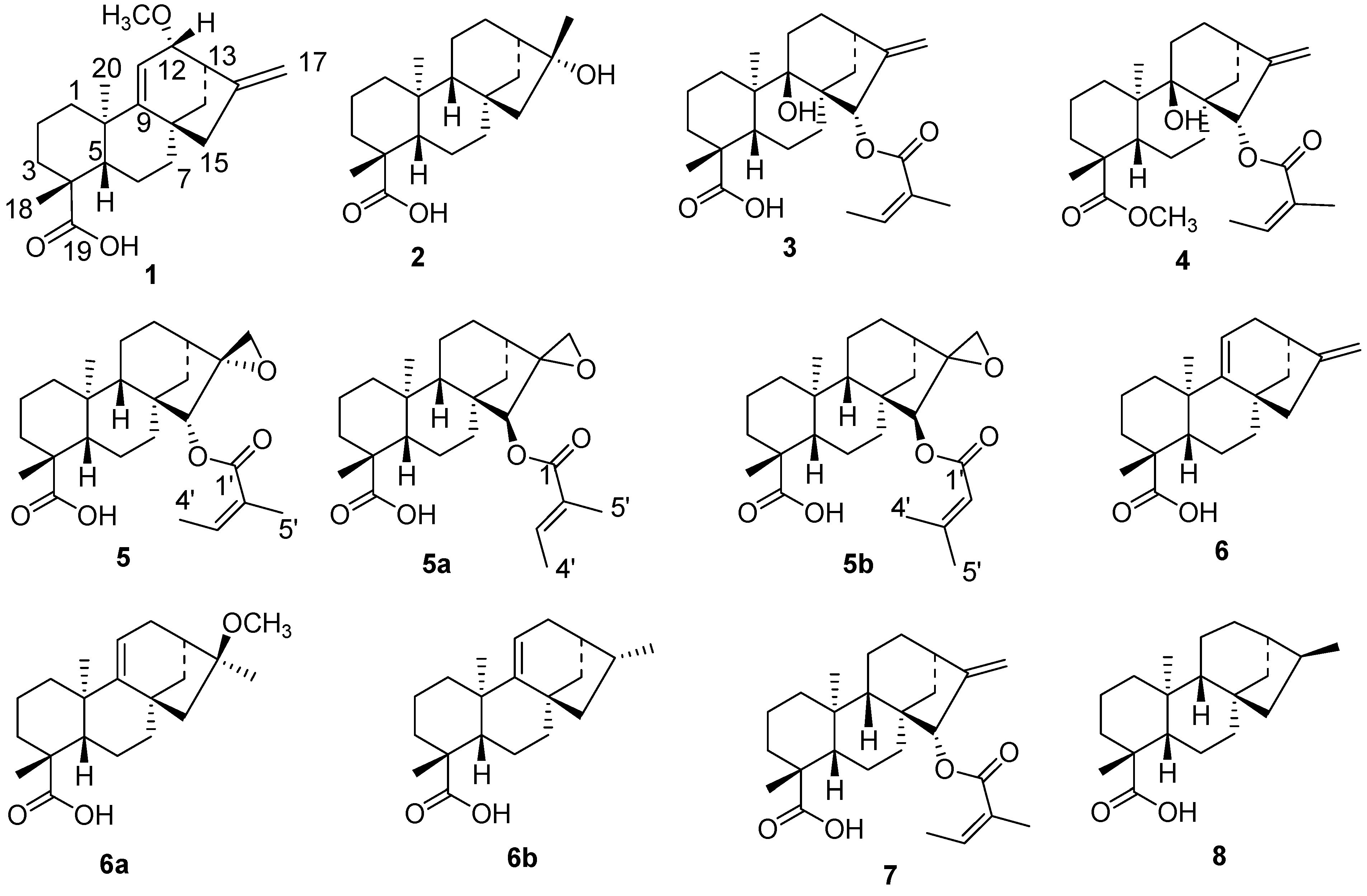
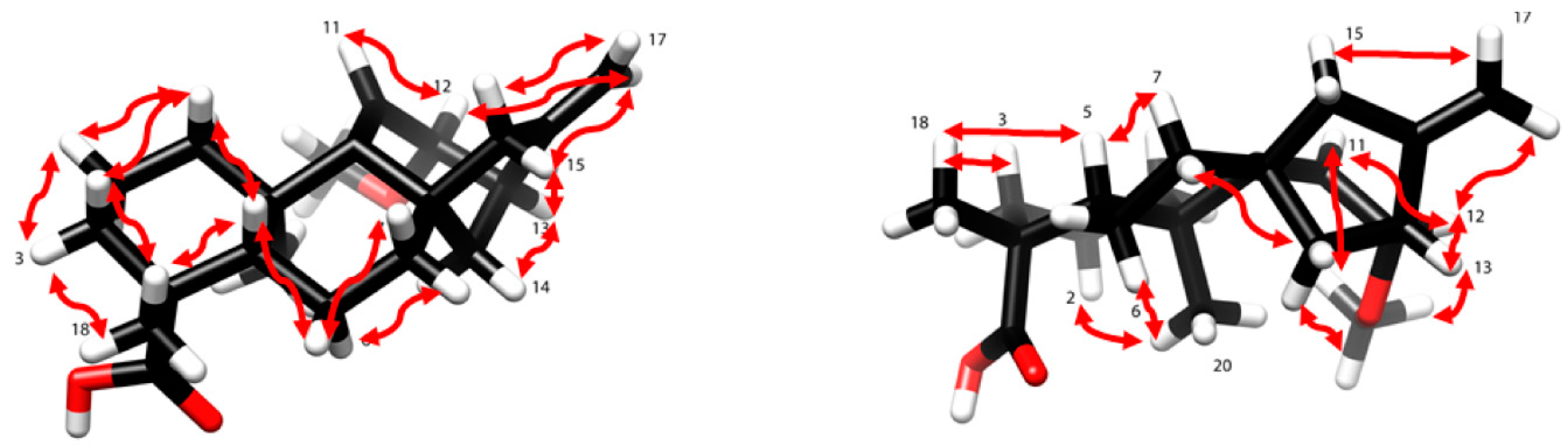
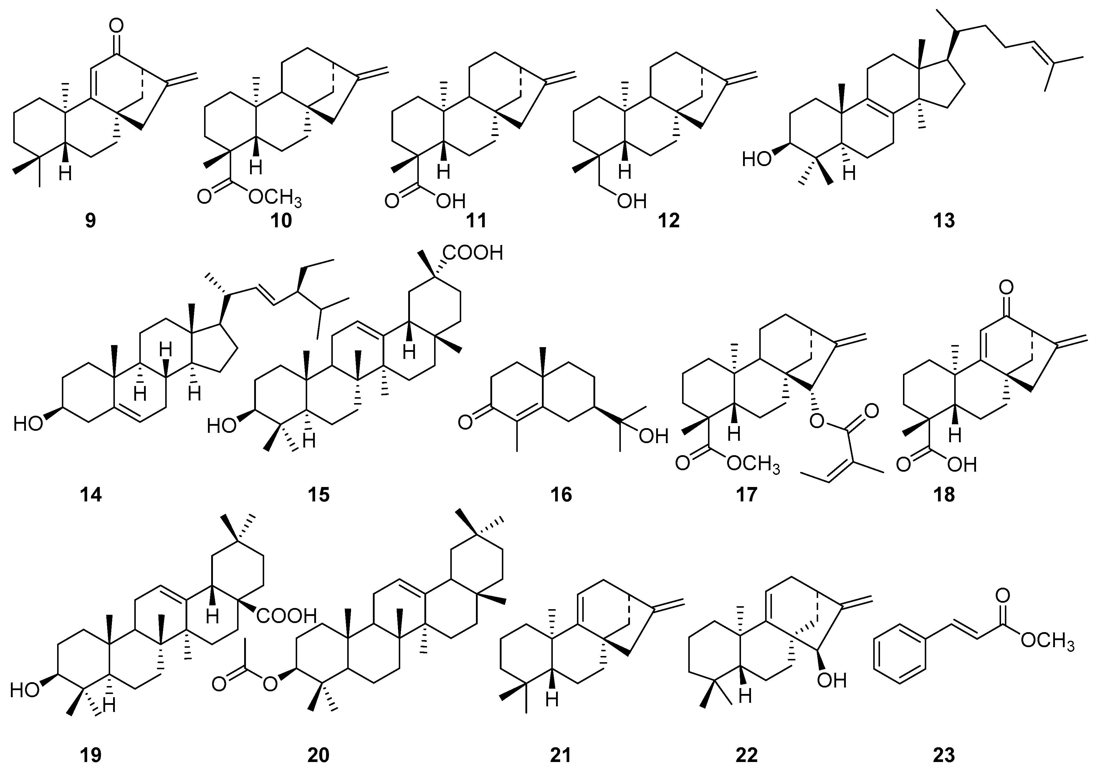
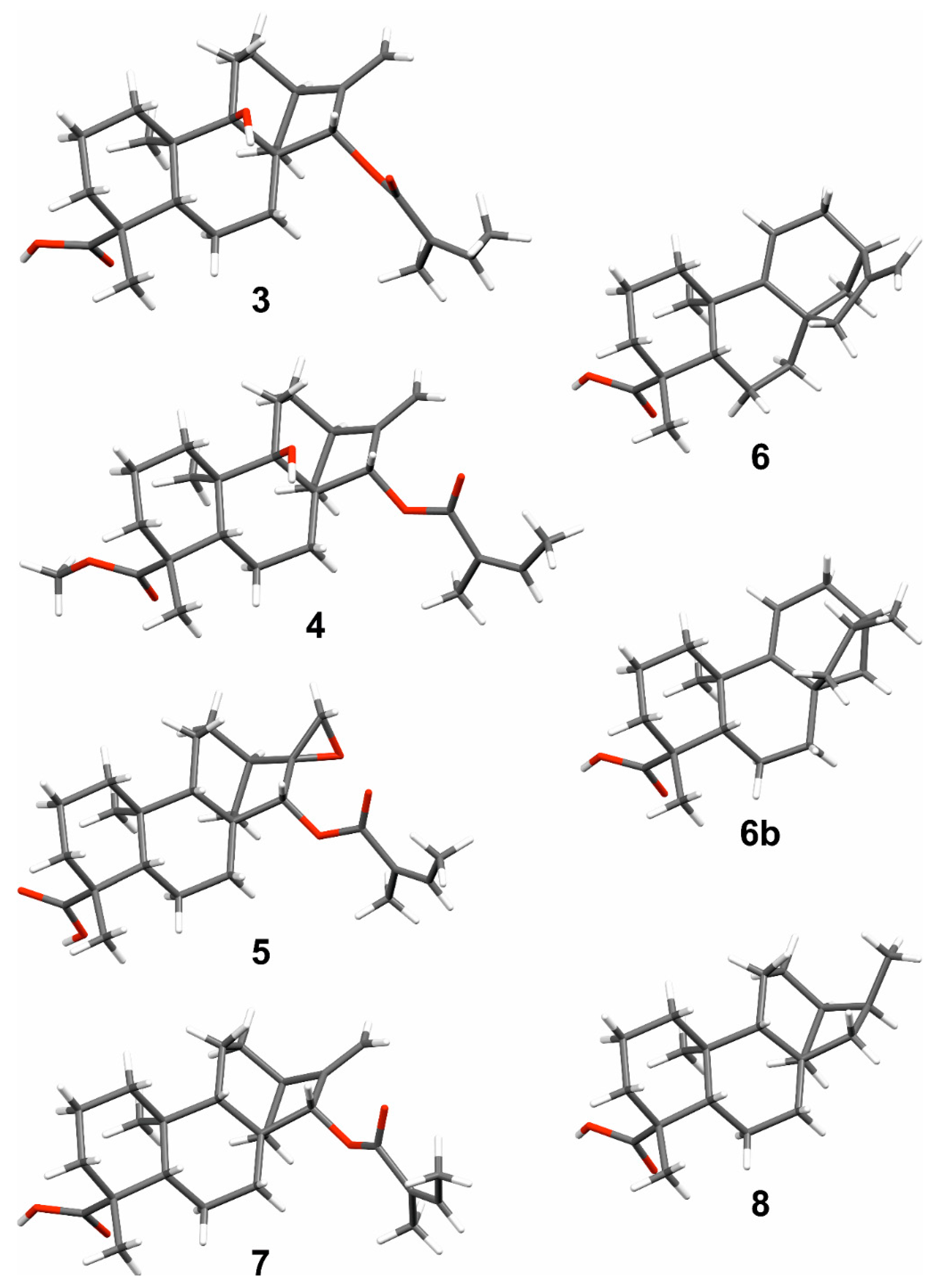
2. Results and Discussion

Compound 1, [α ] D 20 −88°, was isolated as colorless crystals (m.p. 184–186 °C) from the CH2Cl2/MeOH (1:1) extract of the roots of Aspilia pluriseta. HRMS (Figure S7, Supplementary Material) showed a [M–H] ion peak at m/z = 329.2191, which is in agreement with the molecular formula C21H30O3. The NMR spectra (Table 1) indicated that this compound is a kaurene diterpenoid (Figure 1). The 1H–NMR spectrum further revealed the presence of three olefinic protons, namely H-11 (δH 5.30), H-17a (δH 4.84) and H-17b (δH 4.94), suggesting two double bonds. The 13C–NMR chemical shifts of C-16 (δC 152.9) and C-17 (δC 108.1) are typical of a terminal double bond in an ent-kaurene skeleton.
The second double bond was placed between C-9 (δC 160.2) and C-11 (δH 5.30; δC 115.3) by comparison of the NMR data with that found in the literature [13,14,15]. Signals indicating the presence of a methoxy (δH 3.34, δC 56.5) and a carboxylic acid (δC 183.2) substituent were observed. The Heteronuclear Multiple Bond Correlations (HMBCs) of CH3-18 (δH 1.17), H-3 (δH 0.93), and H-5 (δH 1.56) with the carboxy resonance C-19 (δC 183.2) suggested the location of the carboxy group (C-19) at C-4. Out of the three methyl groups expected in kaurene diterpenoid, only two, i.e., CH3-18 (δH 1.17, δC 28.2) and CH3-20 (δH 1.01, δC 23.4), were observed. This corroborated the suggestion of the third methyl group being oxidized to a carboxylic acid (C-19, δC 183.2). The methoxy group OCH3-12 (δH 3.34) showed HMBC correlation with C-12 (δC 81.7), whereas H-12 (δH 3.38) showed HMBC correlation with C-9 (δC 160.2), C-11 (δC 115.3), C-13 (δC 43.7), C-16 (δC 152.9), and OCH3-12 (δC 56.5). Furthermore, CH3-20 (δH 1.01) showed HMBC correlation with C-1 (δC 40.6), C-5 (δC 46.1), the olefinic carbon C-9 (δC 160.2), and C-10 (δC 38.9). This confirmed that the second double bond in the molecule is located at C-9. Moreover, the HMBC correlation of CH2-14 (δH 1.31, 1.58) with a deshielded carbon C-12 (δC 81.7) is in agreement with OCH3 being connected to C-12. The above findings confirmed the identity of compound 1 as a C-12-methoxy substituted ent-kaur-9(11),16-dienoic acid derivative. The relative configuration at C-12 was deduced from the Nuclear Overhauser Effect (NOE) of OCH3-12 (δH 3.34) to H-13 (δH 2.89) (Figure 2), indicating them to be syn-oriented, and hence OCH3-12 to be α-oriented. It should be noted that H-12 (δH 3.38) also showed a weak NOE to H-13 (δH 2.89), which is expected in a strained ring system. The proposed configuration at C-12 is further corroborated by the NOE of H-12 (δH 3.38) with H-14b (δH 1.58). The NOE of H-12 (δH 3.38 ppm) with H-17b (δH 4.94 ppm) supported H-12 to be β-oriented, and hence OCH3-12 to be α-oriented. Based on the above spectroscopic evidence, compound 1, 12α-methoxy-ent-kaur-9(11),16-dien-19-oic acid, (Figure 1) was identified as (4R,4aS,6aS,9R,10S,11bR)-10-methoxy-4,11b-dimethyl-8-methylene-1,2,3,4,4a,5,6,7,8,9,10,11b-dodecahydro-6a,9-methanocyclohepta[a]naphthalene-4-carboxylic acid. This compound has previously been reported both as synthetic derivative [13] and as a natural product [14,15]. However, our NMR data assignment differs from that reported in the literature [13] for its C-3 and C-7. The accuracy of the corrected assignment, given in Table 1, is corroborated by the HMBC correlations of H-3 and H-7 (Table 1), by the HSQC crosspeaks of CH2-3 (δH 0.93/2.08) to C-3 (δC 38.1) along with the TOCSY(Total Correlation Spectroscopy) correlations of CH2-3 to CH2-1 (δH 1.14/1.90) and CH2-2 (δH 1.43/1.79), and by the HSQC (Heteronuclear Single Quantum Correlation) crosspeaks of CH2-7 (δH 1.42/1.95) to C-7 (δC 28.9) along with the TOCSY correlations of CH2-7 to CH2-6 (δH 1.82/2.43) and H-5 (δH 1.56) (Figures S4–S6, Supplementary Material). Besides the compound having been reported earlier, it is unlikely to be an extraction artifact as the extraction (with CH2Cl2/MeOH, 1:1) has been performed at low temperature at neutral pH that does not promote formation of methyl ethers. The compound has been detected in the crude extract indicating that the compound is a natural product and has not formed during the chromatographic isolation.
Additional compounds (Figure 1 and Figure 3) isolated from the roots of Aspilia pluriseta include (16R)-hydroxy-ent-kauran-19-oic acid (2) [16,17], 9β-hydroxy-15α-angeloyloxy-ent-kaur-16-en-19-oic acid (3) [18], methyl-9β-hydroxy-15α-angeloyloxy-ent-kaur-16-en-19-oate (4) [19], 15α-angeloyloxy-16β,17-epoxy-ent-kauran-19-oic acid (5) [20], ent-kaur-9(11),16-dien-19-oic acid (6) [21], 15α-angeloyloxy-ent-kaur-16-en-19-oic acid (7) [22], ent-kaur-9(11),16-dien-12-one (9) [23] and methyl-ent-kaur-16-en-19-oate (10) [24]. The aerial part of Aspilia pluriseta contained ent-kaur-16-en-19-oic acid (11) [21,25], ent-kaur-16-en-19-ol (12) [26], lanosterol (13) [27], stigmasta-5,22(E)-dien-3β-ol (14) [28], 3β-hydroxy-olean-12-en-29-oic acid (15) [29], and carissone (16) [30].
Similar phytochemical investigation of the roots of Aspilia mossambicensis resulted in the isolation of methyl-15α-angeloyloxy-ent-kaur-16-en-19-oate (17) [24,31], 12-oxo-ent-kaur-9(11),16-dien-19-oic acid (18) [32], (16S)-ent-kauran-19-oic acid (8) [33], oleanolic acid (19) [34] (Figure 3), and compounds 35. The aerial part of A. mossambicensis afforded compound 6 [21,26], 3β-acetyloxy-olean-12-ene (20) [35,36] ent-kaur-9(11),16-diene (21) [37], 15a-hydroxy-kaur-9(11),16-diene (22) [38], and methyl cinnamate (23) [39].
The crystal structures for compounds 38 (Figure 4) are also reported here, where the identities of the compounds were confirmed, and the absolute configurations established. In the crystal state, compound 3 exhibited a continuous network, involving intermolecular O(9)-H···O(19a) and O(19b)-H···O(21) hydrogen bonds. Similar O(9)-H···O(19a) hydrogen bonding motif was found in compound 4, which also showed static disorder exhibiting two different spatial orientations of (Z)-2-methylbut-2-enoyl group in an approximately 1:1 ratio. Compounds 58 showed common double hydrogen bonding motifs for carboxylic acids leading to the formation of hydrogen-bonded pairs.
Crystal structure of compound 6 was determined at 120 K, which corresponds to the previously reported structure at room temperature [40]. Crystal structure analysis of a synthetic derivative of compound 6 led to the identification of ent-kaur-9(11)-en-19-oic acid (6b). In the structure 6b, there is a slight (~10%) disorder in the main ring system, where carbons C7–C17 have different positions giving a shade of different conformations for the corresponding aliphatic rings. The data quality for compound 7 is slightly deficient and the absolute structure of it could not be justified on the basis of data. The X-ray diffraction data parameters, thermal ellipsoid diagrams, and hydrogen bonding geometries are presented in the Supplementary Information section of this article.
In most of the ent-kaurane-type diterpenoids (except for compounds 9, 21, and 22) isolated from the two Aspilia species, A. pluriseta and A. mossambicensis, the α-methyl group at C-4 (C-19) is oxidized into carboxylic acid or methyl ester groups, which could be a characteristic feature of the genus Aspilia [3,8]. There are also examples where oxidation has occurred at C-12 (compounds 1, 9 and 18), C-16 (compound 2), C-9 (compounds 3 and 4), and C-15 (compounds 4, 5, 6, and 18). In agreement with the literature [36], oxidation has not been observed at other carbon atoms in ent-kaurane-type diterpenoids of Aspilia species.
Some ent-kaurane-type diterpenoids, including 16,17-epoxy-15β-tigloyloxy-ent-kauran-18-oic acid (5a) and 16,17-epoxy-15β-senecioyloxy-ent-kauran-18-oic acid (5b), were reported earlier from Aspilia pluriseta [8]. These compounds were reported to have the 15β-tigloyloxy and 15β-senecioyloxy groups, respectively, occupying the less favorable orientation [8]; however, the authors have not provided evidence for these proposals. Our single crystal X-ray analyses have shown that the C-15 substituent of compounds 3, 4, 5, and 7 is an angeloyloxy group occupying the more favourable-15α-position (Figure 4). In fact, the proposed stereochemical assignment of ent-kaurane-type diterpenoids reported from this genus, particularly in highly functionalized compounds, lacks evidence. We have filled this knowledge gap by determining the absolute configuration of seven ent-kaurane-type diterpenoids, as shown in Figure 4, using single crystal X-ray analyses.
The 13C-NMR data of compound 5 (Table 2) is in close agreement to that previously reported in the literature [20], except for the chemical shift assignment of C-2 and C-12 (Table 2). Related structures, 5a and 5b, have been proposed for two compounds earlier reported [8] from Aspilia pluriseta. The 13C-NMR assignment (Table 2) for these compounds differs from our assignment, which is based on 2D NMR correlations, despite the common 16,17-epoxy-15-oxy-ent-kauran-18-oic acid skeleton (Figure 1). The 13C-NMR chemical shifts of C-4’ (δC 27.4) and C-5’ (δC 20.8) in compound 5a do not support a tigloyloxy group at C-15, as proposed in Reference [8]; methyl carbon atoms in such group are expected to resonate at ~14 ppm (for C-4’) and at ~11 ppm (for C-5’), based on chemical shift prediction [41] and previous literature [14]. The NMR spectra of compound 5b that are given in the supporting information of [8] are of low quality and do not allow confirmation of the proposed assignment. It should be noted that the numbering used in this paper does not follow the literature convention [3]. Hence, the carboxylic group of 5b and of its structural analogues should not be assigned as C-18, but rather as C-19, following reference [3]. Overall, several details reported [8] for these compounds appear debatable, and consequently so are the proposed structures. To avoid such uncertainties, the NMR assignments of all compounds discussed in this paper are presented in Tables S1 and S2 in the Supplementary Material.
Most of the compounds isolated in this study were assayed against two normal (BEAS-2B and LO2) and two cancer (A549 and Hep-G2, Table 3) cell lines. Of the fifteen compounds tested, 1, 3, 5, 9, and 18 showed cytotoxicity towards some of the cell lines. Compound 1 showed selective activity against the human hepatoma (HepG2) cancer cell line without significant toxicity to other cell lines (IC50 above 100 μM). Compound 3 was moderately cytotoxic against the Hep-G2 cancer cell line (IC50 = 24.7 ± 2.8), but also showed weak cytotoxicity towards the normal cell lines LO2 (IC50 = 57.2 ± 1.2) and BEAS-2B (IC50 = 89.9 ± 2.0 µM), indicating low selectivity. Compound 5 (IC50 = 30.7 ± 1.7 µM) was the most active against the A549 cell line, followed by compound 18 (IC50 = 80.5 ± 1.8 µM). The other compounds tested, i.e., 2, 4, 6, 7, 10, 11, 14, or 17, did not exhibit significant cytotoxicity (IC50 above 100 μM). The lack of cytotoxicity against the two normal cell lines of these diterpenoids is valuable information, considering that some kaurane-type diterpenoids from Aspilia species have showed antibacterial and antifungal effects [5].

3. Materials and Methods

3.1. General Experimental Procedures

NMR spectra were acquired on a Bruker Avance II 600 MHz, a Bruker Avance III HD 800 MHz (Bruker BioSpin AG, Fällanden, Switzerland) or a Varian Unity 500 MHz (Varian Inc, Palo Alto, CA, USA) NMR spectrometer, using the residual solvent peaks as a reference. The spectra were processed using the software MestReNova (version 10.0, Mestrelab Research S.L., Santiago de Compostela, Spain) Coupling constants (J) are given in Hz. EI-MS and LC-MS were carried out using 70 eV ionization electron voltage on a Micromass GC-TOF spectrometer (Micromass, Wythenshawe, Waters Inc., UK). TLC (Thin Layer Chromatography) was carried out on Merck pre-coated silica gel 60 F254 plates (Merck, Darmstadt, Germany). Preparative TLC was performed on 20 × 20 cm glass plates, pre-coated with silica gel 60F254 with thicknesses of 0.25 to 1 mm. Column chromatography was run on silica gel 60 Å (70–230 mesh). Gel filtration was performed on Sephadex LH-20(Merck, Darmstadt, Germany).

3.2. X-ray Diffraction Analyses

The single crystal X-ray diffraction data were collected using Agilent Super-Nova (Agilent Technologies, Wrocław, Poland) dual wavelength diffractometer with a micro-focus X-ray source and multilayer optics monochromatized Cu-Kα (λ = 1.54184 Å) radiation. Program CrysAlisPro [42] was used for the data collection and reduction. The intensities were corrected for absorption using analytical face index absorption correction method. The structures were solved with intrinsic phasing method (SHELXT [43]) and refined by full-matrix least squares on F2 with SHELXL-2018/3 [44]. Anisotropic displacement parameters were assigned to non-H atoms. All C-H hydrogen atoms were refined using riding models. Hydroxy hydrogens were found from electron density maps and restrained to the proper distance from oxygen atom (0.84 Å). All hydrogen atoms were refined with Ueq(H) of 1.5 × Ueq(C,O) for hydroxy and terminal methyl groups or 1.2 × Ueq(C) for other C-H groups. Further geometric least-squares restraints (s = 0.02) were applied to structures 4, 6b, and 7 to obtain more chemically reasonable bond distances between disordered atoms. Anisotropic displacement parameters of few disordered or terminal atoms were restrained (s = 0.01, st = 0.02) to be more equal in structures 3, 4, 6b, and 7. CCDC 1868318-1868324 contains the supplementary crystallographic data for this paper. These data can be obtained free of charge via http://www.ccdc.cam.ac.uk/conts/retrieving.html (or from the CCDC, 12 Union Road, Cambridge CB2 1EZ, UK; Fax: +44 1223 336033; E-mail: deposit@ccdc.cam.ac.uk).

3.3. Plant Materials

The roots and aerial parts of Aspilia pluriseta and Aspilia mossambicensis (Asteraceae) were collected from Ngon’g Forest, Kenya. The plants were identified by Mr. Patrick B. Chalo Mutiso, of the Herbarium, School of Biological Sciences, University of Nairobi, Kenya, where voucher specimens (SY2015/04 for Aspilia pluriseta, and SY2015/05 for Aspilia mossambicensis) were deposited.

3.4. Extraction, Isolation and Derivatization

The air-dried and ground roots of A. pluriseta (0.8 kg) were extracted with CH2Cl2/MeOH (1:1) for 24 h affording 47 g of extract. A portion of the extract (35 g) was subjected to column chromatography on silica gel (700 g) and eluted with hexane containing increasing amounts of EtOAc. The fraction eluted with 10% EtOAc in hexane was purified by crystallization from acetone affording compound 1 (36 mg) [9]. Crystallization (from acetone) of the combined fractions eluted with 5–10% EtOAc in hexane afforded compound 2 (47 mg) [12,13]. Preparative TLC separation of the fraction eluted with 15% EtOAc in hexane yielded 22 mg of compound 3 [14] and 31 mg of compound 4 [15]. Compound 5 (97 mg) [16] was obtained from the fraction eluted with 30% EtOAc in hexane, after purification over Sephadex LH-20 (CH2Cl2/MeOH; 1:1). Fractional crystallization (from acetone) of the combined fractions eluted with 35–40% EtOAc in hexane afforded 218 mg of compound 6 [17] and 89 mg of compound 7 [18]. Purification of the fraction obtained with 60% EtOAc in hexane on Sephadex LH-20 (eluted with CH2Cl2/MeOH; 1:1) led to the isolation of ent-kaur-9(11),16-dien-12-one (9, 27 mg) [19] and methyl-ent-kaur-16-en-19-oate (10, 36 mg) [20]. The isolated compounds have been observed in the crude extract, ahead of chromatographic separation, by TLC. This confirmed their presence in the crude extracts, and thus, these are natural products and were not formed during the chromatographic separation.
The air-dried and ground aerial part of A. pluriseta (0.8 kg) was extracted, as described above, giving 53 g of crude extract. A portion of the extract (40 g) was subjected to column chromatography on silica gel (800 g) and eluted with hexane containing increasing amounts of EtOAc. The fractions eluted with 5–15% EtOAc in hexane were combined and purified on Sephadex LH-20 (eluted with CH2Cl2/MeOH; 1:1) affording lanosterol (13, 58 mg) [27] and stigmasta-5,22(E)-dien-3β-ol (14, 71 mg) [28]. Elution of the main silica gel column with 20% EtOAc in hexane afforded 42 mg of 3β-hydroxy-olean-12-en-29-oic acid (15) [29]. The fraction eluted with 30% EtOAc in hexane led to the isolation of ent-kaur-16-en-19-oic acid (11, 367 mg) [17,21] and ent-kaur-16-en-19-ol (12, 32 mg) [8,26]. Purification of the fraction eluted with 40% EtOAc in hexane over Sephadex LH-20 (CH2Cl2/MeOH; 1:1) yielded carissone (16, 26 mg) [30].
The air-dried and ground roots (0.7 kg) of Aspilia mossambicensis were extracted with CH2Cl2/MeOH (1:1) by cold percolation (4 × 24 h) to give gummy brown extract (47 g). A portion of the extract (40 g) was subjected to column chromatography on silica gel (800 g) and eluted with hexane containing increasing amounts of EtOAc. Fractional crystallization of the eluent with 25% EtOAc in hexane led to the isolation of 64 mg of methyl-15α-angeloyloxy-ent-kaur-16-en-19-oate (17) [24,31] and 23 mg of 12-oxo-ent-kaur-9(11), and 16-dien-19-oic acid (18) [32]. Purification of the mother liquor on Sephadex LH-20 resulted in the separation of (16S)-ent-kauran-19-oic acid (8, 17 mg) 33] and oleanolic acid (19, 28 mg) [34]. Elution of the column with 40% EtOAc in hexane led to the isolation of additional amounts of compounds 3 (14 mg), 4 (7 mg), and 5 (10 mg).
The aerial part (1.0 kg) of A. mossambicensis was extracted as above, yielding 82 g of crude extract. A portion of this extract (40 g) was subjected to column chromatography over silica gel (800 g) and eluted with hexane containing increasing amounts of EtOAc. Elution of the column with 20% EtOAc in hexane led to the isolation of 24 mg of 3β-acetyloxy-olean-12-ene acetate (20) [35,36]; elution with 25% EtOAc in hexane afforded 73 mg of ent-kaur-9(11),16-diene (21) [39]. Elution with 30–40% EtOAc in hexane resulted in a mixture of two compounds. These were separated by column chromatography on Sephadex LH-20 (eluted with CH2Cl2/MeOH; 1:1), yielding 21 mg of 15α-hydroxy-kaur-9(11),16-diene (22) [38], and 15 mg of methyl cinnamate (23) [40].
Derivatization of compound 6. A concentrated H2SO4 (2 drops) was added to MeOH (2 mL) solution of compound 6 (0.05 g, 0.165 mmol). The reaction mixture was stirred at 25 °C for 18 h, after which the mixture was poured onto water (20 mL) and extracted with ethyl acetate (3 × 20 mL). The combined organic layer was dried over MgSO4, and was then filtered and concentrated in a rotary evaporator, giving 37 mg of 6a [45].
To hydrogenate ent-kaur-9(11),16-dien-19-oic acid (6), a solution was obtained by dissolving 100 mg in 20 mL ethanol, in a 50 mL round-bottomed flask, and 5% palladium on charcoal (30 mg) was then added to the solution. The flask was sealed with an airtight rubber stopper. Nitrogen gas was bubbled through the solution to eliminate traces of air using a small syringe inserted through the rubber stopper. Hydrogen gas was then bubbled into the system for 2 days at room temperature. The reaction mixture was filtered and the solvent evaporated yielding compound 6b (87 mg).
12α-Methoxy-ent-kaur-9(11),16-dien-19-oic Acid (1) colorless crystals, m.p. 158%–160 °C. [α ] D 20 −88 (c 0.25, acetone), 1H and 13C-NMR (CD2Cl2) data (Table 1). ESIMS, m/z (rel. int.) 329 (12, [M–H]), 315 (11), 299 (100), 253 (71), 281 (10), 171 (18), 182 (3). HRMS [M–H] m/z 329.2191 C21H29O3 (Calculated: 329.2117).
15α-Angeloyloxy-ent-kaur-16α,17-epoxy-ent-kauran-19-oic Acid (5). Colorless crystals, m.p. 242–243 °C. 1H and 13C-NMR (CDCl3) data, see Table 2. ESIMS, m/z (rel. int): 417 (100, [M+H]+), 317 (80), 299 (71), 271 (64), 253 (23).

3.5. Cell Culture

Reagent and cells. Adenocarcinomic human alveolar basal epithelial (A549) and human hepatoma (HepG2) cancer cell lines, immortalized normal human liver (LO2), human bronchial epithelial (BEAS-2B), and fibroblast-like CCD19Lu cells were purchased from ATCC (ATCC, Manassas, VA, USA). The cells were cultured in RPMI-1640 medium supplemented with 10% fetal bovine serum and antibiotics: Penicillin (50 U/Ml) and streptomycin (50 μg/mL; Invitrogen, Paisley, Scotland, UK). All cells were incubated at 37 °C in a 5% humidified CO2 incubator). All test compounds were dissolved in DMSO at a final concentration of 50 mM and stored at −20 °C before use.

3.6. Cytotoxicity Assay

Cytotoxicity was assessed using the 3-[4,5-dimethylthiazole-2-yl]-2,5-diphenyltetrazolium bromide (MTT) (5 mg/mL) assay, as described previously [46]. Briefly, 5 × 10³ cells per well were seeded in 96-well plates before drug treatments. After overnight cell culture, the cells were exposed to different concentrations of selected compounds (0.19–100 μM) for 72 h. Cells without drug treatment were used as controls. Subsequently, 10 μL of 5 mg/mL MTT solution was added to each well and incubated at 37 °C for 4 h, followed by the addition of 100 μL solubilization buffer (12 mM HCl in a solution of 10% SDS) and overnight incubation. The absorbance, A570 nm, was then determined in each well on the next day. The percentage cell viability was calculated using the expression: % Viability = Atreated/ Acontrol × 100, and was given as cytotoxicity in Table 3.

4. Conclusions

Twenty-three compounds, mostly ent-kaurane-type diterpenoids, were isolated from Aspilia pluriseta and A. mossambicensis. Besides giving a full NMR assignment, the absolute configuration of seven of the isolated compounds was established by single crystal X-ray diffraction analyses. The isolated compounds were tested for their cytotoxicity against four cell lines. Compounds 1, 3, 5, 9, and 18 showed moderate to weak cytotoxicity against the cell lines. Compound 3 was the most cytotoxic (IC50=24.7 ± 2.8 µM) against the human hepatoma (Hep-G2) cancer cell line without toxicity against the tested normal cell lines.

Supplementary Materials

The following are available online: NMR and MS spectra for the new compound 1, spectral data for the known compounds, single crystal X-ray diffraction data and refinement parameters and hydrogen bonding geometries. The original FIDs and spectra (mnova) of compounds 123. The NMReDATA file of 1 [47].

Author Contributions

The authors contributed to this work as follows. Extraction and isolation of compounds was performed by S.Y. under the supervision of A.Y. and S.D.; X-ray analyses was carried out by A.V.; NMR analyses was performed with the help of M.E.; Spectroscopic characterization of the compounds was carried out by S.Y., A.Y. and M.E.; cytotoxicity assays were performed by P.C., J.G. and V.K.W.W.; E.M.G. assisted in the interpretation and data analysis of the cytotoxicity assay. All authors contributed to the preparation of the manuscript.

Funding

This research was funded by the Swedish Research Council (2016-05857), the Academy of Finland (grant no. 314343), the International Science Program (KEN-02), the Macao Science and Technology Development Fund for financial support (0022/2018/A1) and an AFIMEGQ intra-ACP fellowship.

Acknowledgments

S.Y. acknowledges the support of the AFIMEGQ intra-ACP project for a fellowship. A.V. kindly acknowledges the Academy of Finland (grant no. 314343) for financial support. Funding was also provided by the Swedish Research Council (2016-05857) and the International Science Program (ISP Sweden, grant KEN-02). Patrick B. Chalo Mutiso, School of Biological Sciences, University of Nairobi, Kenya, is acknowledged for the identification of the plant material. V.K.W.W. acknowledges the Macao Science and Technology Development Fund for financial support (FDTC grant Project code: 0022/2018/A1).

Conflicts of Interest

The authors declare no conflict of interest.

References

  1. Faleye, F.J.; Ogundaini, O.A. Evaluation of antioxidant and antimicrobial activities of two isolates from Aspilia africana (Pers) C.D. Adams. Int. Res. J. Pharm. 2012, 655, 135–138. [Google Scholar]
  2. Bohm, B.A.; Stuessy, T.F. Flavonoids of the Sunflower Family (Asteraceae); Springer Science & Business Media: Wien, Austria, 2001; pp. 116–119. ISBN 3-211-83479-6. [Google Scholar]
  3. Alvarenga, S.A.; Ferreira, M.J.; Rodrigues, G.V.; Emerenciano, V.P. A general survey and some taxonomic implications of diterpenes in the Asteraceae. Bot. J. Linnean. Soc. 2005, 147, 291–308. [Google Scholar] [CrossRef] [Green Version]
  4. Moreira-Muñoz, A.; Muñoz-Schick, M. Classification, diversity, and distribution of Chilean Asteraceae: Implications for biogeography and conservation. Divers. Distrib. 2007, 13, 818–828. [Google Scholar] [CrossRef]
  5. Souza, J.M.; Chang, M.R.; Brito, D.Z.; Farias, K.S.; Damasceno-Junior, G.A.; Turatti, I.C.; Lopes, N.P.; Santos, E.A.; Carollo, C.A. Antimicrobial activity of Aspilia latissima (Asteraceae). Braz. J. Microbiol. 2015, 46, 1103–1110. [Google Scholar] [CrossRef] [PubMed]
  6. Seaman, F.; Bohlmann, F.; Zdero, C.; Mabry, T.J. Diterpenes of Flowering Plants—Compositae (Asteraceae); Springer: New York, NY, USA, 2012. [Google Scholar]
  7. Kuria, J.M. Efficacy of Aspilia pluriseta Schweinf in Cutaneous Wound Healing in a Mouse Model. Ph.D. Thesis, University of Nairobi, Nairobi, Kenya, 2014. [Google Scholar]
  8. Sebisubi, F.M.; Odyek, O.; Anokbonggo, W.W.; Ogwal-Okeng, J.; Carcache-Blanco, E.J.; Ma, C.; Orjala, J.; Tan, G.T. Antimalarial activity of Aspilia pluriseta, a medicinal plant from Uganda. Planta Med. 2010, 76, 1870–1873. [Google Scholar] [CrossRef] [PubMed]
  9. Norton, R.A.; Huang, D.Q.; Rodriguez, E. Aspilia mossambicensis: In Vitro Propagation and Production of Antibiotic Polyacetylenes by Root Cultures. In Medicinal and Aromatic Plants V; Bajaj, Y.P.S., Ed.; Springer: Berlin/Heidelberg, Germany, 1993; pp. 54–63. [Google Scholar]
  10. Musyimi, D.; Ogur, J.; Muema, P. Phytochemical compounds and antimicrobial activity of extracts of Aspilia plant (Aspilia mossambicensis) Oliv. wild. Int. J. Botany 2008, 4, 56–61. [Google Scholar] [CrossRef]
  11. Page, J.E.; Balza, F.; Nishida, T.; Towers, G.N. Biologically active diterpenes from Aspilia mossambicensis, a Chimpanzee medicinal plant. Phytochemistry 1992, 31, 3437–3439. [Google Scholar] [CrossRef]
  12. Page, J.E.; Huffman, M.; Smith, V.; Towers, G. Chemical basis for Aspilia leaf-swallowing by Chimpanzees: A reanalysis. J. Chem. Ecol. 1997, 23, 2211–2226. [Google Scholar] [CrossRef]
  13. Ahmed, M.; Jakupovic, J.; Castro, V. Kaurene derivatives from Lasianthea fruticosa, revision of stereochemistry of related compounds. Phytochemistry 1991, 30, 1712–1714. [Google Scholar] [CrossRef]
  14. Cai, C.; Zhang, Y.; Yang, D.; Hao, X.; Li, S. Two new kaurane-type diterpenoids from Wedelia chinensis (Osbeck.) Merr. Nat. Prod. Res. 2017, 31, 2531–2536. [Google Scholar] [CrossRef]
  15. Li, S.F.; Ding, J.Y.; Li, Y.T.; Hao, X.J.; Li, S.L. Antimicrobial Diterpenoids of Wedelia trilobata (L.) Hitchc. Molecules 2016, 21, 457. [Google Scholar] [CrossRef] [PubMed]
  16. Duan, H.; Takaishi, Y.; Momota, H.; Ohmoto, Y.; Taki, T.; Jia, Y.; Li, D. Immunodepressive diterpenoids from Tripterygium wilfordii. J. Nat. Prod. 1999, 62, 1522–1525. [Google Scholar] [CrossRef] [PubMed]
  17. Lee, I.S.; Jin, W.Y.; Zhang, X.; Hung, T.M.; Song, K.S.; Seong, Y.H.; Bae, K. Cytotoxic and COX-2 inhibitory constituents from the aerial parts of Aralia cordata. Arch. Pharm. Res. 2006, 29, 548–555. [Google Scholar] [CrossRef] [PubMed]
  18. Müller, S.; Tirapelli, C.R.; de Oliveira, A.M.; Murillo, R.; Castro, V.; Merfort, I. Studies of ent-kaurane diterpenes from Oyedaea verbesinoides for their inhibitory activity on vascular smooth muscle contraction. Phytochemistry 2003, 63, 391–396. [Google Scholar] [CrossRef]
  19. Delgado, G.; Vivar, A.R.D. Ent-kaurenoid methyl esters from Viguiera stenoloba, structural revision of stenolobin and its biomimetic conversion to zoapatlin. Chem. Lett. 1984, 13, 1237–1240. [Google Scholar] [CrossRef]
  20. Wafo, P.; Kamdem, R.S.; Ali, Z.; Anjum, S.; Begum, A.; Oluyemisi, O.O.; Khan, S.N.; Ngadjui, B.T.; Etoa, X.F.; Choudhary, M.I. Kaurane-type diterpenoids from Chromoleana odorata, their X-ray diffraction studies and potent α-glucosidase inhibition of 16-kauren-19-oic acid. Fitoterapia 2011, 82, 642–646. [Google Scholar] [CrossRef] [PubMed]
  21. Arciniegas, A.; Pérez-Castorena, A.L.; Meléndez-Aguirre, M.; Ávila, J.G.; García-Bores, A.M.; Villaseñor, J.L.; Romod, V.A. Chemical composition and antimicrobial activity of Ageratina deltoidea. Chem. Biodivers. 2018, 15, e1700529. [Google Scholar] [CrossRef]
  22. El Marsni, Z.; Torres, A.; Varela, R.M.; Molinillo, J.M.; Casas, L.; Mantell, C.; Martinez, E.J.D.L.O.; Macias, F.A. Isolation of bioactive compounds from sunflower leaves (Helianthus annuus L.) extracted with supercritical carbon dioxide. J. Agric. Food Chem. 2015, 63, 6410–6421. [Google Scholar] [CrossRef]
  23. Pinto, A.C.; Prado, S.K.D.; Pinchin, R. Two kaurenes from Vellozia caput-ardeae. Phytochemistry 1981, 20, 520–521. [Google Scholar] [CrossRef]
  24. Aráoz, M.V.C.; Mercado, M.I.; Grau, A.; Catalán, C.A. Ent-kaurane derivatives from the root cortex of Yacon and other three Smallanthus species (Heliantheae, Asteraceae). Biochem. Syst. Ecol. 2010, 38, 1042–1048. [Google Scholar] [CrossRef]
  25. Chen, Q.; Lin, H.; Wu, X.; Song, H.; Zhu, X. Preparative separation of six terpenoids from Wedelia prostrata Hemsl. By two-step high-speed counter-current chromatography. J. Liq. Chromatogr. Relat. Technol. 2018, 41, 408–414. [Google Scholar] [CrossRef]
  26. Peña, A.; Alarcón, L.; Baptista, J.G.; Aparicio, R.; Villasmil, T.; Usubillaga, A. A phytochemical analysis of Espeletia nana Cuatrec. A midget Espeletiinae from Paramo Ortiz, Venezuela. Av. Quim. 2012, 7, 187–192. [Google Scholar]
  27. Dias, J.R.; Gao, H. 13C nuclear magnetic resonance data of lanosterol derivatives—profiling the steric topology of the steroid skeleton via substituent effects on its 13C-NMR. Spectrochim. Acta Part A: Mol. Biomol. Spectrosc. 2009, 74, 1064–1071. [Google Scholar] [CrossRef] [PubMed]
  28. Chaturvedula, V.S.P.; Prakash, I. Isolation of stigmasterol and β-sitosterol from the dichloromethane extract of Rubus suavissimus. Int. Curr. Pharm. J. 2012, 1, 239–242. [Google Scholar] [CrossRef]
  29. Zhou, Y.; Yang, B.; Liu, Z.; Jiang, Y.; Liu, Y.; Fu, L.; Wang, X.; Kuang, H. Cytotoxicity of triterpenes from green walnut husks of Juglans mandshurica maxim in HepG-2 cancer cells. Molecules 2015, 20, 19252–19262. [Google Scholar] [CrossRef] [PubMed]
  30. Reisch, J.; Hussain, R.A.; Krebs, B.; Dartmann, M. Natural product chemistry, part 100: The structure of carissone, C15H24O2. Monatsh. Chem. 1990, 121, 941–944. [Google Scholar] [CrossRef]
  31. Ohno, N.; Mabry, T.J.; Zabelt, V.; Watson, W.H. Tetrachyrin, a new rearranged kaurenoid lactone, and diterpene acids from Tetrachyron orizabaensis and Helianthus debilis. Phytochemistry 1979, 18, 1687–1689. [Google Scholar] [CrossRef]
  32. Huang, W.; Liang, Y.; Wang, J.; Li, G.; Wang, G.; Li, Y.; Chung, H.Y. Anti-angiogenic activity and mechanism of kaurane diterpenoids from Wedelia chinensis. Phytomedicine 2016, 23, 283–292. [Google Scholar] [CrossRef]
  33. Tincusi, B.M.; Jiménez, I.A.; Bazzocchi, I.L.; Moujir, L.M.; Mamani, Z.A.; Barroso, J.P.; Ravelo, A.G.; Hernandez, B.V. Antimicrobial terpenoids from the oleoresin of the peruvian medicinal plant Copaifera paupera. Planta Med. 2002, 68, 808–812. [Google Scholar] [CrossRef]
  34. Seebacher, W.; Simic, N.; Weis, R.; Saf, R.; Kunert, O. Complete assignments of 1H and 13C-NMR resonances of oleanolic acid, 18α-oleanolic acid, ursolic acid and their 11-oxo derivatives. Magn. Reson. Chem. 2003, 41, 636–638. [Google Scholar] [CrossRef]
  35. Faleye, F.J. Terpenoid constituents of Aspilia africana [Pers.] C.D. Adams leaves. Int. J. Pharm. Sci. Rev. Res. 2012, 13, 138–142. [Google Scholar]
  36. Ogihara, K.; Iraha, R.; Higa, M.; Yogi, S. Studies on constituents from the twigs of Messerschmidia argentea II. Bull. Coll. Sci., Univ. Ryukyus 1997, 64, 53–59. [Google Scholar]
  37. Zhang, H.; Wynne, G.; Mander, L.N. Synthesis of ent-9α, 15α-cyclokaurene from grandiflorenic acid. ARKIVOC 2001, 8, 40–58. [Google Scholar]
  38. Nagashima, F.; Kondoh, M.; Fujii, M.; Takaoka, S.; Watanabe, Y.; Asakawa, Y. Novel cytotoxic kaurane-type diterpenoids from the New Zealand liverwort Jungermannia species. Tetrahedron 2005, 61, 4531–4544. [Google Scholar] [CrossRef]
  39. Gao, L.; Xu, X.; Nan, H.; Yang, J.; Sun, G.; Wu, H.; Zhong, M. Isolation of cinnamic acid derivatives from the root of Rheum tanguticum Maxim. Ex balf. And its significance. J. Med. Plants Res. 2012, 6, 929–931. [Google Scholar]
  40. Reynolds, W.F.; Lough, A.J.; Sawyer, J.F.; Enriquez, R.G.; Ortiz, B.; Walls, F. Structure of (4α)-kaura-9(11),16-dien-18-oic acid (grandiflorenic acid), an active ingredient of the Mexican medicinal plant zoapatle. Acta Crystallogr. Sect. C: Cryst. Struct. Commun. 1991, 47, 973–977. [Google Scholar] [CrossRef] [Green Version]
  41. Banfi, D.; Patiny, L. www.nmrdb.org: Resurrecting and processing NMR spectra on-line. Chimia 2008, 62, 280–281. [Google Scholar] [CrossRef]
  42. CrysAlisPro, version 1.171.38.41q; Rigaku Oxford Diffraction: Yarnton, UK, 2015; Available online: https://www.rigaku.com/en/products/smc/crysalis (accessed on 11 November 2018).
  43. Sheldrick, G.M. SHELXT—Integrated space-group and crystal-structure determination. Acta Crystallogr. Sect. A 2015, 71, 3–8. [Google Scholar] [CrossRef]
  44. Sheldrick, G.M. Crystal structure refinement with SHELXL. Acta Crystallogr. Sect. C 2015, 71, 3–8. [Google Scholar] [CrossRef]
  45. Boeck, P.; Sá, M.M.; Souza, B.S.D.; Cercená, R.; Escalante, A.M.; Zachino, S.A.; Cechinel, F.V.; Yunes, R.A. A simple synthesis of kaurenoic esters and other derivatives and evaluation of their antifungal activity. J. Brazil. Chem. Soc. 2005, 16, 1360–1366. [Google Scholar] [CrossRef] [Green Version]
  46. Wong, V.K.W.; Zhang, M.M.; Zhou, H.; Lam, K.Y.C.; Chan, P.L.; Law, C.K.M.; Yue, P.Y.K.; Liu, L. Saikosaponin-D enhances the anticancer potency of TNF-via overcoming its undesirable response of activating NF-kappa B signalling in cancer cells. Evid. Based Complement. Altern. Med. 2013, 2013, 1–14. [Google Scholar] [CrossRef] [PubMed]
  47. Pupier, M.; Nuzillard, J.M.; Wist, J.; Schlorer, N.E.; Kuhn, S.; Erdelyi, M.; Steinbeck, C.; Williams, A.J.; Butts, C.; Claridge, T.D.W.; et al. NMReDATA, a standard to report the NMR assignment and parameters of organic compounds. Magn. Reson. Chem. 2018, 56, 703–715. [Google Scholar] [CrossRef] [PubMed] [Green Version]
Sample Availability: Samples of the compounds are available from the authors.
Figure 1. Structures of compounds 18.
Molecules 23 03199 g001
Figure 2. Some of the key NOE correlations observed for compound 1. The NOESY (Nuclear Overhauser Effect Spectroscopy) spectrum (800 MHz, CDCl3, 25 °C, 700 ms mixing time) is shown in the Supplementary Materials.
Molecules 23 03199 g002
Figure 3. Structures of compounds 923.
Molecules 23 03199 g003
Figure 4. Crystal structure representations of compounds 38. The structures were deposited with the following CCDC (Cambridge Crystallographic Data Centre) codes: 3 (1868318), 4 (1868319), 5 (1868321), 6 (1868320), 6b (1868324), 7 (1868323), and 8 (1868322).
Molecules 23 03199 g004
Table 1. The 1H (800 MHz) and 13C-NMR (200 MHz) data for compound 1 acquired in CDCl3.
PositionδC Lit. [13] *δCδH, mult. (J in Hz)HMBC (2J, 3J)
138.1740.61.14 ddd (13.5, 9.5, 4.2)C-2, C-3, C-10, C-20
1.90 ddd (13.5, 3.5, 1.4)C-2, C-3, C-10, C-20
218.3520.01.43 dddd (14.2, 9.5, 3.9, 3.5)C-1, C-3, C-4, C-5, C-10
1.79 ddddd (14.2, 11.1, 4.2, 3.5, 1.4)C-1, C-4, C-5
329.0338.10.93 ddd (13.4, 11.1, 3.9)C-1, C-2, C-4, C-18, C-19
2.08 ddd (13.4, 3.5, 3.5)C-1, C-4, C-5, C-7
443.4344.6
543.8146.11.56 dd (11.1, 8.5)C-4, C-7, C-9, C-10, C-18, C-19, C-20
620.0718.31.82 dddd (14.2, 10.0, 8.5, 2.5)C-3, C-4, C-5, C-7, C-10
2.43 dddd (14.2, 11.1, 9.5, 3.5)C-4, C-5, C-8
740.6028.91.42 ddd (13.8, 3.5, 2.5)C-5, C-6, C-8, C-9, C-15
1.95 ddd (13.8, 10.0, 9.5)C-6, C-8, C-9, C-14, C-15
844.6643.4
9160.28160.2
1038.9438.9
11115.42115.35.30 dd (4.3, 1.4)C-8, C-9, C-10, C-12, C-13, C-15, C-20
1281.7981.73.38 dd (4.3, 2.9)C-9, C-11, C-13, C-16, C-20, OCH3-12
1346.1743.72.89 dd (2.9, 1.4)C-10, C-11, C-12, C-15, C-16
1440.6040.51.31 dd (10.8, 4.3)C-7, C-8, C-9, C-12, C-13, C-15
1.58 dd (10.8, 2.5)C-9, C-12, C-13, C-15, C-16
1547.1747.12.08 dd (15.4, 4.3)C-7, C-8, C-9, C-16, C-17
2.35 dd (15.4, 2.5)C-7, C-9, C-13, C-14, C-16, C-17
16153.00152.9
17108.12108.14.84 dd (3.0, 1.6)C-12, C-13, C-15, C-16
4.94 dd (3.0, 1.6)C-12, C-13, C-15, C-16
1828.2228.21.17 sC-3, C-4, C-5, C-8, C-19
19182.98183.2
2023.4123.41.01 sC-1, C-5, C-9, C-10
OCH3-1256.5356.53.34 sC-12
* CDCl3 at 100 MHz [13].
Table 2. The literature reported NMR data for 5, 5a, and 5b and the 1H (800 MHz) and 13C-NMR (200 MHz) data for compound 5 acquired in CDCl3.
Position5 [20]5a [8]5b [8]55
δCδCδCδCδH, mult. (J in Hz)
141.240.640.640.60.80 ddd (7.2, 7.1, 1.3)
1.86* dd (2.9, 1.4)
228.919.819.019.71.55 ddd (7.3, 3.6, 2.4)
1.75 dd (3.7, 3.6)
337.736.736.437.60.96 ddd (13.7, 13.6, 4.3)
2.11 dd (13.7, 3.1)
443.646.947.843.5
556.720.356.656.51.16 dd (9.1, 7.1)
619.041.220.320.81.76 ddd (5.7, 3.4, 2.1)
1.86* ddd (3.4, 3.4, 2.7)
735.447.841.235.31.25 ddd (14.4, 13.9, 4.4)
1.79 ddd (13.8, 13.2, 4.3)
847.952.943.647.8
952.943.653.052.81.28 dd (13.8, 3.8)
1039.856.639.839.7
1119.820.819.818.91.40 ddd (13.8, 3.4, 3.4, 3.1)
1.81 dd (13.8, 4.3)
1220.828.928.928.81.50 ddd (13.5, 7.8, 7.2)
1341.236.435.141.11.82 dd (13.8, 4.4)
1436.537.737.736.41.68 dd (14.5, 3.3)
1.97 dd (13.1, 3.4)
1581.981.281.281.94.73 br s
1666.366.466.466.3
1749.649.649.649.62.78 dd (5.6, 1.3)
3.09 dd (5.8, 1.3)
1828.828.928.928.71.28 s
19182.3182.6182.6182.7
2015.715.816.015.91.03 s
1’167.9166.5166.5167.8
2’128.1129.0115.9128.0
3’137.3137.1156.8137.35.96 q (7.1)
4’15.927.420.815.71.96 d (1.9)
5’20.620.827.420.6s
Table 3. Cytotoxicity (IC50, μM) of compounds isolated from Aspilia species against various cell lines.
CompoundNormal Cell LinesCancer Cell Lines
BEAS-2BLO2A549Hep-G2
1>100>100>10027.3 ± 1.9
2>100>100>100>100
389.9 ± 2.057.2 ± 1.2>10024.7 ± 2.8
4>100>100>100>100
5>100>10030.7 ± 1.7>100
6>100>100>100>100
6a>100>100>100>100
6b>100>100>100>100
7>100>100>100>100
9>10075.3 ± 2.8>100>100
10>100>100>100>100
11>100>100>100>100
14>100>100>100>100
17>100>100>100>100
1838.6 ± 2.530.0 ± 1.780.5 ± 1.881.3 ± 0.3
Paclitaxel<0.1<0.10.00330.19

