
Synthesis and Biological Evaluation of Benzochromenopyrimidinones as Cholinesterase Inhibitors and Potent Antioxidant, Non-Hepatotoxic Agents for Alzheimer’s Disease

 , ,
, ,
Abstract
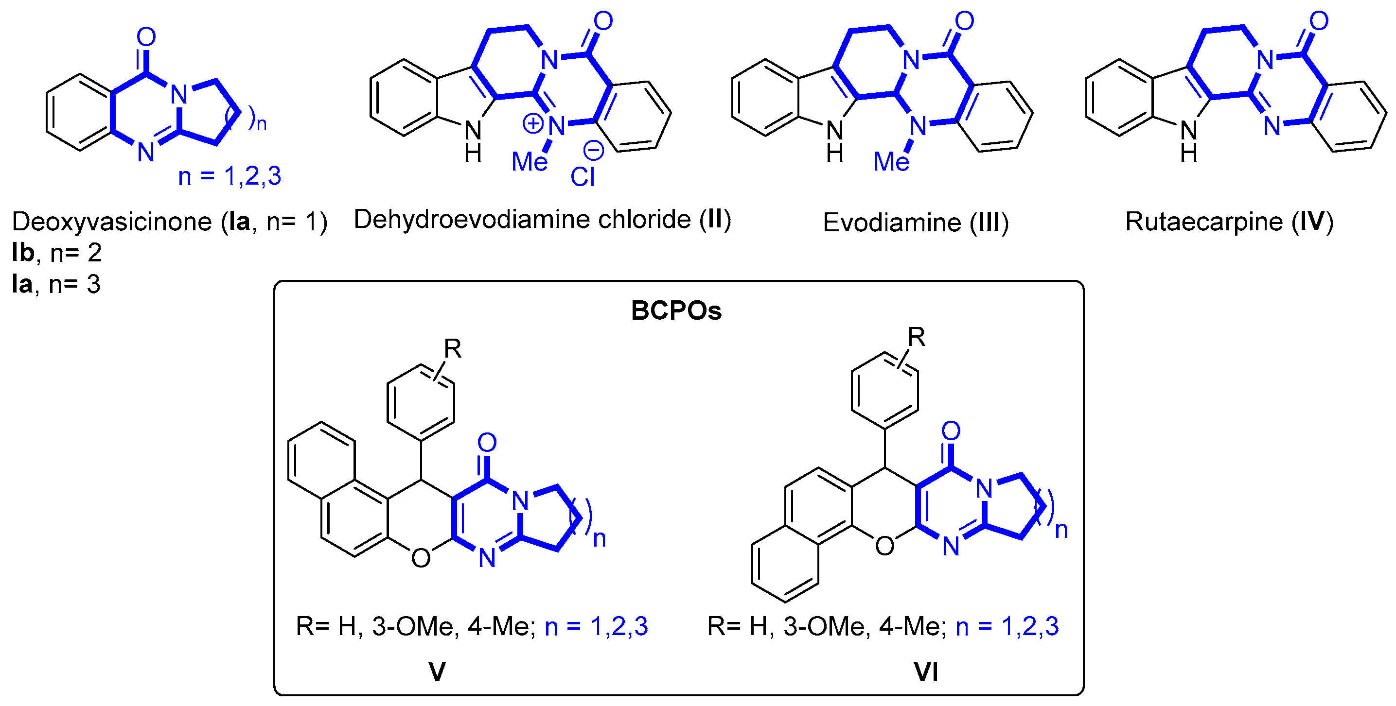
:1. Introduction
2. Results and Discussion
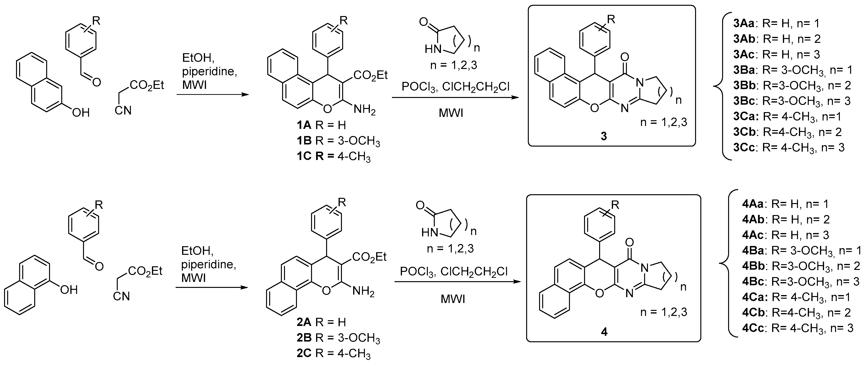
2.1. Synthesis
2.2. Evaluation of the Antioxidant Power
2.3. Evaluation of AChE and BuChE Inhibition
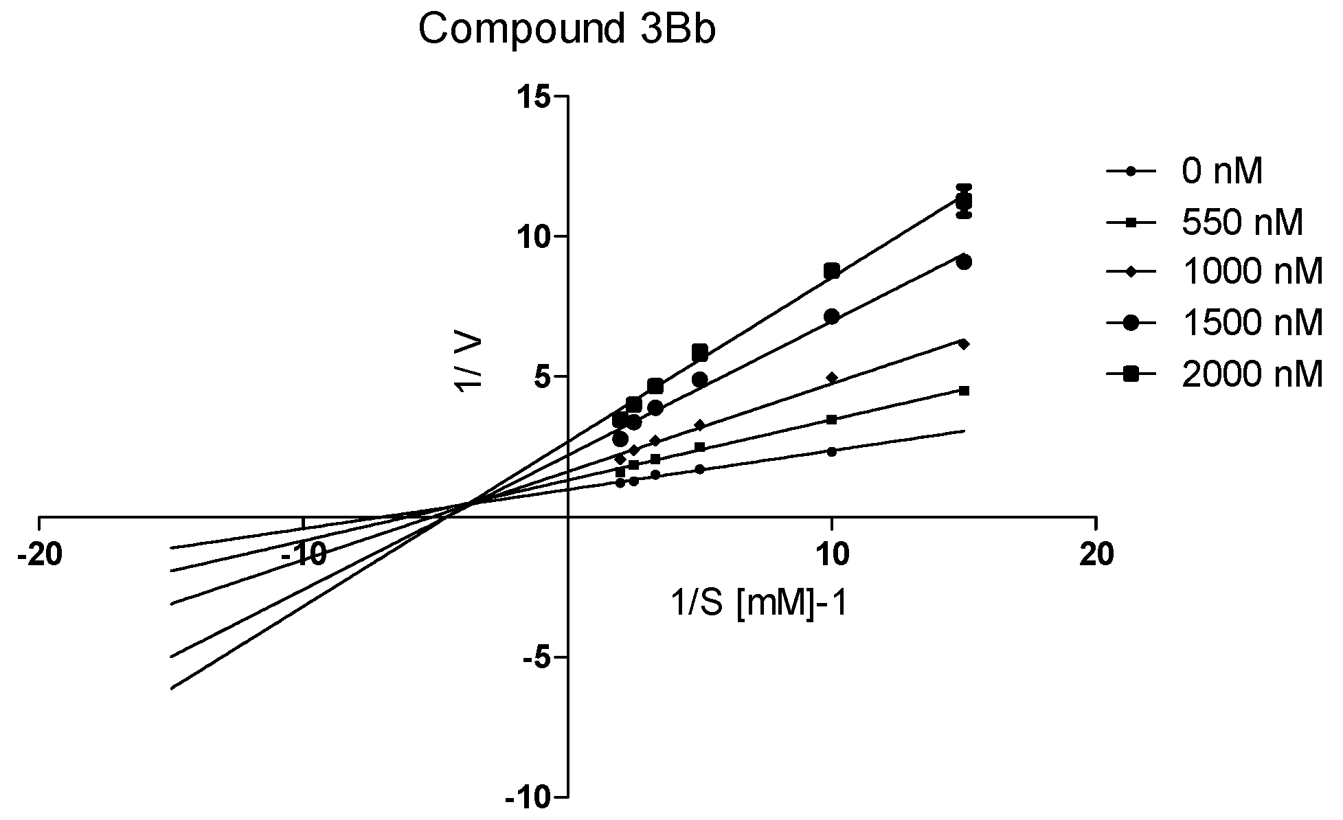
2.4. Kinetic Study of the hAChE Inhibition by Compound 3Bb
2.5. In Vitro Toxicity of Compounds 3Ab, 3Bb, 3Cb and 3Ba in HepG2 Cells
2.6. Blood Brain Barrier Penetration PAMPA Assay
3. Materials and Methods
3.1. Chemistry Methods
3.1.1. General Procedure for the Compounds 1A–C and 2A–C
3.1.2. General Procedure for the Synthesis of Benzochromenopyrimidinones (BCPOs)
3.2. Oxygen Radical Absorbance Capacity Assay
3.3. Inhibition of EeAChE and EqBuChE
3.4. Inhibition of hAChE
3.5. Kinetic Characterization of hAChE Inhibition
3.6. In vitro Toxicity of Compounds 3Ab, 3Bb, 3Cb and 3Ba in HepG2 Cells
3.7. PAMPA Assay
4. Conclusions
Supplementary Materials
Acknowledgments
Author Contributions
Conflicts of Interest
References
- World Alzheimer Report 2015. Available online: http://www.worldalzreport2015.org/ (accessed on 3 February 2016).
- Liu, L.; Luo, S.; Zeng, L.; Wang, W.; Yuan, L.; Jian, X. Degenerative alterations in noradrenergic neurons of the locus coeruleus in Alzheimer’s disease. Neural Regen. Res. 2013, 8, 2249–2255. [Google Scholar] [PubMed]
- Mesulam, M.-M. Cholinergic circuitry of the human nucleus basalis and its fate in Alzheimer’s disease. J. Comp. Neurol. 2013, 521, 4124–4144. [Google Scholar] [CrossRef] [PubMed]
- Zarow, C.; Lyness, S.A.; Mortimer, J.A.; Chui, H.C. Neuronal loss is greater in the locus coeruleus than nucleus basalis and substantia nigra in alzheimer and parkinson diseases. Arch. Neurol. 2003, 60, 337–341. [Google Scholar]
- Praticò, D.; Sung, S. Lipid peroxidation and oxidative imbalance: Early functional events in Alzheimer’s disease. J. Alzheimers Dis. JAD 2004, 6, 171–175. [Google Scholar] [PubMed]
- Yan, M.H.; Wang, X.; Zhu, X. Mitochondrial defects and oxidative stress in Alzheimer disease and Parkinson disease. Free Radic. Biol. Med. 2013, 62, 90–101. [Google Scholar] [CrossRef] [PubMed]
- Greenough, M.A.; Camakaris, J.; Bush, A.I. Metal dyshomeostasis and oxidative stress in Alzheimer’s disease. Neurochem. Int. 2013, 62, 540–555. [Google Scholar] [CrossRef] [PubMed]
- Candore, G.; Bulati, M.; Caruso, C.; Castiglia, L.; Colonna-Romano, G.; di Bona, D.; Duro, G.; Lio, D.; Matranga, D.; Pellicanò, M.; et al. Inflammation, cytokines, immune response, apolipoprotein E, cholesterol, and oxidative stress in Alzheimer disease: Therapeutic implications. Rejuvenation Res. 2010, 13, 301–313. [Google Scholar] [CrossRef] [PubMed] [Green Version]
- Bond, M.; Rogers, G.; Peters, J.; Anderson, R.; Hoyle, M.; Miners, A.; Moxham, T.; Davis, S.; Thokala, P.; Wailoo, A.; et al. The effectiveness and cost-effectiveness of donepezil, galantamine, rivastigmine and memantine for the treatment of Alzheimer’s disease (review of Technology Appraisal No. 111): A systematic review and economic model. Health Technol. Assess. 2012, 16. [Google Scholar] [CrossRef] [PubMed]
- Wilkinson, D.; Wirth, Y.; Goebel, C. Memantine in Patients with Moderate to Severe Alzheimer’s Disease: Meta-Analyses Using Realistic Definitions of Response. Dement. Geriatr. Cogn. Disord. 2014, 37, 71–85. [Google Scholar] [CrossRef] [PubMed]
- Watkins, P.B.; Zimmerman, H.J.; Knapp, M.J.; Gracon, S.I.; Lewis, K.W. Hepatotoxic effects of tacrine administration in patients with Alzheimer’s disease. JAMA 1994, 271, 992–998. [Google Scholar] [CrossRef] [PubMed]
- Bolea, I.; Juárez-Jiménez, J.; de los Rı́os, C.; Chioua, M.; Pouplana, R.; Luque, F.J.; Unzeta, M.; Marco-Contelles, J.; Samadi, A. Synthesis, Biological Evaluation, and Molecular Modeling of Donepezil and N-[(5-(Benzyloxy)-1-methyl-1H-indol-2-yl)methyl]-N-methylprop-2-yn-1-amine Hybrids as New Multipotent Cholinesterase/Monoamine Oxidase Inhibitors for the Treatment of Alzheimer’s Disease. J. Med. Chem. 2011, 54, 8251–8270. [Google Scholar] [PubMed]
- Samadi, A.; Chioua, M.; Bolea, I.; de los Ríos, C.; Iriepa, I.; Moraleda, I.; Bastida, A.; Esteban, G.; Unzeta, M.; Gálvez, E.; et al. Synthesis, biological assessment and molecular modeling of new multipotent MAO and cholinesterase inhibitors as potential drugs for the treatment of Alzheimer’s disease. Eur. J. Med. Chem. 2011, 46, 4665–4668. [Google Scholar] [CrossRef] [PubMed]
- Ismaili, L.; Refouvelet, B.; Benchekroun, M.; Brogi, S.; Brindisi, M.; Gemma, S.; Campiani, G.; Filipic, S.; Agbaba, D.; Esteban, G.; et al. Multitarget compounds bearing tacrine- and donepezil-like structural and functional motifs for the potential treatment of Alzheimer’s disease. Prog. Neurobiol. 2016. [Google Scholar] [CrossRef] [PubMed]
- Decker, M. Novel inhibitors of acetyl- and butyrylcholinesterase derived from the alkaloids dehydroevodiamine and rutaecarpine. Eur. J. Med. Chem. 2005, 40, 305–313. [Google Scholar] [CrossRef] [PubMed]
- Huang, G.; Kling, B.; Darras, F.H.; Heilmann, J.; Decker, M. Identification of a neuroprotective and selective butyrylcholinesterase inhibitor derived from the natural alkaloid evodiamine. Eur. J. Med. Chem. 2014, 81, 15–21. [Google Scholar] [CrossRef] [PubMed]
- Darras, F.H.; Wehle, S.; Huang, G.; Sotriffer, C.A.; Decker, M. Amine substitution of quinazolinones leads to selective nanomolar AChE inhibitors with “inverted” binding mode. Bioorg. Med. Chem. 2014, 22, 4867–4881. [Google Scholar] [CrossRef] [PubMed]
- Benchekroun, M.; Ismaili, L.; Pudlo, M.; Luzet, V.; Gharbi, T.; Refouvelet, B.; Marco-Contelles, J. Donepezil–ferulic acid hybrids as anti-Alzheimer drugs. Future Med. Chem. 2015, 7, 15–21. [Google Scholar] [CrossRef] [PubMed]
- Benchekroun, M.; Bartolini, M.; Egea, J.; Romero, A.; Soriano, E.; Pudlo, M.; Luzet, V.; Andrisano, V.; Jimeno, M.-L.; López, M.G.; et al. Novel Tacrine-Grafted Ugi Adducts as Multipotent Anti-Alzheimer Drugs: A Synthetic Renewal in Tacrine-Ferulic Acid Hybrids. ChemMedChem 2015, 10, 523–539. [Google Scholar] [CrossRef] [PubMed]
- Ou, B.; Hampsch-Woodill, M.; Prior, R.L. Development and validation of an improved oxygen radical absorbance capacity assay using fluorescein as the fluorescent probe. J. Agric. Food Chem. 2001, 49, 4619–4626. [Google Scholar] [CrossRef] [PubMed]
- Dávalos, A.; Gómez-Cordovés, C.; Bartolomé, B. Extending Applicability of the Oxygen Radical Absorbance Capacity (ORAC−Fluorescein) Assay. J. Agric. Food Chem. 2004, 52, 48–54. [Google Scholar] [CrossRef] [PubMed]
- Fang, L.; Kraus, B.; Lehmann, J.; Heilmann, J.; Zhang, Y.; Decker, M. Design and synthesis of tacrine–ferulic acid hybrids as multi-potent anti-Alzheimer drug candidates. Bioorg. Med. Chem. Lett. 2008, 18, 2905–2909. [Google Scholar] [CrossRef] [PubMed]
- Ellman, G.L.; Courtney, K.D.; Andres, V.; Feather-Stone, R.M. A new and rapid colorimetric determination of acetylcholinesterase activity. Biochem. Pharmacol. 1961, 7, 88–95. [Google Scholar] [CrossRef]
- Decker, M.; Krauth, F.; Lehmann, J. Novel tricyclic quinazolinimines and related tetracyclic nitrogen bridgehead compounds as cholinesterase inhibitors with selectivity towards butyrylcholinesterase. Bioorg. Med. Chem. 2006, 14, 1966–1977. [Google Scholar] [CrossRef] [PubMed]
- Esquivias-Pérez, M.; Maalej, E.; Romero, A.; Chabchoub, F.; Samadi, A.; Marco-Contelles, J.; Oset-Gasque, M.J. Nontoxic and neuroprotective β-naphthotacrines for Alzheimer’s disease. Chem. Res. Toxicol. 2013, 26, 986–992. [Google Scholar] [CrossRef] [PubMed]
- Di, L.; Kerns, E.H.; Fan, K.; McConnell, O.J.; Carter, G.T. High throughput artificial membrane permeability assay for blood–brain barrier. Eur. J. Med. Chem. 2003, 38, 223–232. [Google Scholar] [CrossRef]
- Lemes, L.F.N.; de Andrade Ramos, G.; de Oliveira, A.S.; da Silva, F.M.R.; de Castro Couto, G.; da Silva Boni, M.; Guimarães, M.J.R.; Souza, I.N.O.; Bartolini, M.; et al. Cardanol-derived AChE inhibitors: Towards the development of dual binding derivatives for Alzheimer’s disease. Eur. J. Med. Chem. 2016, 108, 687–700. [Google Scholar] [CrossRef] [PubMed]
- Cornish-Bowden, A. A Simple graphical method for determining the inhibition constants of mixed, uncompetitive and non-competitive inhibitors (Short Communication). Biochem. J. 1974, 137, 143–144. [Google Scholar] [CrossRef] [PubMed]
- Silverman, R.B. The Organic Chemistry of Enzyme-catalyzed Reactions; Academic Press: San Diego, CA, USA, 2000. [Google Scholar]
- Sugano, K.; Hamada, H.; Machida, M.; Ushio, H. High Throughput Prediction of Oral Absorption: Improvement of the Composition of the Lipid Solution Used in Parallel Artificial Membrane Permeation Assay. J. Biomol. Screen. 2001, 6, 189–196. [Google Scholar] [CrossRef] [PubMed]
- Wohnsland, F.; Faller, B. High-Throughput Permeability pH Profile and High-Throughput Alkane/Water log P with Artificial Membranes. J. Med. Chem. 2001, 44, 923–930. [Google Scholar] [CrossRef] [PubMed]
- Sample Availability: Samples of the compounds 3A–C and 4A–C are available from the authors.


| BCPO | EeAChE 1 (IC50, nM) | eqBuChE (% Inhibition at 10 µM) | hAChE (IC50, nM) | ORAC 2 |
|---|---|---|---|---|
| 3Aa | 518.4 ± 87.9 | 12.1 ± 1.4 | n.d. | 2.6 ± 0.1 |
| 3Ab | 55.5 ± 7.1 | 31.2 ± 1.8 | 3657 ± 59 | 2.3 ± 0.3 |
| 3Ac | 300.8 ± 6.5 | 28.4 ± 7.3 | n.d. | 2.5 ± 0.2 |
| 3Ba | 60.7 ± 4.5 | 51.0 ± 4.0 | 1527 ± 25 | 3.4 ± 0.2 |
| 3Bb | 30.5 ± 2.8 | 43.0 ± 1.9 | 1279 ± 32 | 4.7 ± 0.2 |
| 3Bc | 107.5 ± 7.2 | n.a. | n.d. | 3.5 ± 0.3 |
| 3Ca | 111.9 ± 21.7 | 17.4 ± 1.8 | n.d. | 2.7 ± 0.3 |
| 3Cb | 55.9 ± 12.7 | 19.0 ± 2.0 | 1591 ± 24 | 3.9 ± 0.3 |
| 3Cc | 166.6 ± 7.8 | n.a. | n.d. | 3.1 ± 0.2 |
| 4Aa | 317.8 ± 26.0 | 23.1 ± 3.1 | n.d. | 2.1 ± 0.1 |
| 4Ab | 383.5 ± 19.4 | 54.6 ± 1.8 | n.d. | 2.3 ± 0.2 |
| 4Ac | 290.5 ± 8.3 | 24.2 ± 3.1 | n.d. | 2.5 ± 0.1 |
| 4Ba | 326.7 ± 38.9 | 38.0 ± 1.6 | n.d. | 3.8 ± 0.1 |
| 4Bb | 153.2 ± 3.1 | n.a. | n.d. | 3.7 ± 0.2 |
| 4Bc | 195.3 ± 6.2 | 31.1 ± 2.1 | n.d. | 3.4 ± 0.2 |
| 4Ca | 115.8 ± 6.2 | 26.8 ± 2.9 | n.d. | 3.2 ± 0.1 |
| 4Cb | 193.4 ± 18.7 | 24.4 ± 1.2 | n.d. | 2.9 ± 0.1 |
| 4Cc | 173.8 ± 5.9 | n.a. | n.d. | 3.6 ± 0.2 |
| Tacrine | 44.3 ± 1.5 [19] | IC50 = 5.1 ± 0.2 nM [19] | 131 ± 2 | 0.2 ± 0.1 [22] |
| Ia | 82.5 M [24] | IC50 = 25.1 [24] | n.d. | n.d. |
| Ib | 38.6 M [24] | >500 [24] | n.d. | n.d. |
| Ic | 279 M [24] | >500 [24] | n.d. | n.d. |
| II | 6.3 M [15] | IC50 = 8.4 M [15] | n.d. | n.d. |
| Ferulic acid | n.d. | n.d. | n.d. | 3.7 ± 0.1 [22] |
| BCPO | 1 µM | 3 µM | 10 µM | 30 µM | 100 µM | 300 µM | 1 mM |
|---|---|---|---|---|---|---|---|
| 3Ab | 99.3 ± 4.0 | 95.3 ± 2.3 | 98.5 ± 2.5 | 91.5 ± 4.5 | 100.8 ± 3.1 | 110.5 ± 5.5 | 110.4 ± 4.4 |
| 3Bb | 107.7 ± 2.4 | 111.1 ± 8.7 | 104.7 ± 4.2 | 104.1 ± 5.0 | 111.8 ± 2.3 | 118.0 ± 7.3 | 127.8 ± 5.0 |
| 3Cb | 105.8 ± 7.0 | 102.9 ± 10.8 | 110.2 ± 9.1 | 112.7 ± 5.8 | 103.1 ± 5.7 | 113.0 ± 5.2 | 110.2 ± 8.0 |
| 3Ba | 108.6 ± 2.0 | 107.7 ± 4.8 | 106.2 ± 3.5 | 99.2 ± 4.2 | 97.0 ± 5.4 | 107.6 ± 2.8 | 119.5 ± 6.8 |
| Tacrine | 105.2 ± 4.6 | 103.5 ± 8.7 | 97.0 ± 6.5 | 93.0 ± 2.7 | 95.2 ± 6.0 | 47.6 ± 5.6 *** | 13.9 ± 0.8 *** |
| Compound | BBB Penetration Estimation | |
|---|---|---|
| Pe ± SEM (×10−6 cm s−1) | CNS (+/−) | |
| 3Ab | 7.2 ± 0.6 | CNS (+) |
| 3Bb | 3.6 ± 0.57 | CNS (+/−) |
| 3Cb | ND * | |
| 3Ba | 4.6 ± 0.77 | CNS (+) |
| Donepezil | 7.3 ± 0.9 | CNS (+) |
| Rivastigmine | 6.6 ± 0.5 | CNS (+) |
| Tacrine | 5.3 ± 0.19 | CNS (+) |
| Testosterone | 11.3 ± 1.6 | CNS (+) |
| Chlorpromazine | 5.6 ± 0.6 | CNS (+) |
| Hydrocortisone | 2.85 ± 0.1 | CNS (+/−) |
| Piroxicam | 2.2 ± 0.15 | CNS (+/−) |
| Theophyline | 1.07 ± 0.18 | CNS (−) |
| Atenolol | 1.02 ± 0.37 | CNS (−) |
 ; CNS (+/−)=
  ; CNS (−)=
  .
.© 2016 by the authors. Licensee MDPI, Basel, Switzerland. This article is an open access article distributed under the terms and conditions of the Creative Commons Attribution (CC-BY) license ( http://creativecommons.org/licenses/by/4.0/).
Share and Cite
Dgachi, Y.; Bautista-Aguilera, O.M.; Benchekroun, M.; Martin, H.; Bonet, A.; Knez, D.; Godyń, J.; Malawska, B.; Gobec, S.; Chioua, M.; et al. Synthesis and Biological Evaluation of Benzochromenopyrimidinones as Cholinesterase Inhibitors and Potent Antioxidant, Non-Hepatotoxic Agents for Alzheimer’s Disease. Molecules 2016, 21, 634. https://doi.org/10.3390/molecules21050634
Dgachi Y, Bautista-Aguilera OM, Benchekroun M, Martin H, Bonet A, Knez D, Godyń J, Malawska B, Gobec S, Chioua M, et al. Synthesis and Biological Evaluation of Benzochromenopyrimidinones as Cholinesterase Inhibitors and Potent Antioxidant, Non-Hepatotoxic Agents for Alzheimer’s Disease. Molecules. 2016; 21(5):634. https://doi.org/10.3390/molecules21050634
Chicago/Turabian StyleDgachi, Youssef, Oscar M. Bautista-Aguilera, Mohamed Benchekroun, Hélène Martin, Alexandre Bonet, Damijan Knez, Justyna Godyń, Barbara Malawska, Stanislav Gobec, Mourad Chioua, and et al. 2016. "Synthesis and Biological Evaluation of Benzochromenopyrimidinones as Cholinesterase Inhibitors and Potent Antioxidant, Non-Hepatotoxic Agents for Alzheimer’s Disease" Molecules 21, no. 5: 634. https://doi.org/10.3390/molecules21050634