Article

Rapid and Sensitive Detection of Vibrio parahaemolyticus and Vibrio vulnificus by Multiple Endonuclease Restriction Real-Time Loop-Mediated Isothermal Amplification Technique

1
State Key Laboratory of Infectious Disease Prevention and Control, National Institute for Communicable Disease Control and Prevention, Collaborative Innovation Center for Diagnosis and Treatment of Infectious Diseases, Chinese Center for Disease Control and Prevention, Changping, Changbai Road 155, Beijing 102206, China
2
Changping District Center for Disease Control and Prevention, Changping, Beijing 102200, China
3
Institute of Microbiology, Jilin Provincial Center for Disease Control and Prevention, Changchun, Jilin 130062, China
*
Author to whom correspondence should be addressed.
These authors contributed equally to this work.
Molecules 2016, 21(1), 111; https://doi.org/10.3390/molecules21010111
Submission received: 24 November 2015 / Revised: 11 January 2016 / Accepted: 13 January 2016 / Published: 19 January 2016
(This article belongs to the Section Natural Products Chemistry)

Abstract

:
Vibrio parahaemolyticus and Vibrio vulnificus are two marine seafood-borne pathogens causing severe illnesses in humans and aquatic animals. In this study, a recently developed novel multiple endonuclease restriction real-time loop-mediated isothermal amplification technology (MERT-LAMP) were successfully developed and evaluated for simultaneous detection of V. parahaemolyticus and V. vulnificus strains in only a single reaction. Two MERT-LAMP primer sets were designed to specifically target toxR gene of V. parahaemolyticus and rpoS gene of V. vulnificus. The MERT-LAMP reactions were conducted at 62 °C, and the positive results were produced in as short as 19 min with the genomic DNA templates extracted from the V. parahaemolyticus and V. vulnificus strains. The two target pathogens present in the same sample could be simultaneously detected and correctly differentiated based on distinct fluorescence curves in a real-time format. The sensitivity of MERT-LAMP assay was 250 fg and 125 fg DNA per reaction with genomic templates of V. parahaemolyticus and V. vulnificus strains, which was in conformity with conventional LAMP detection. Compared with PCR-based techniques, the MERT-LAMP technology was 100- and 10-fold more sensitive than that of PCR and qPCR methods. Moreover, the limit of detection of MERT-LAMP approach for V. parahaemolyticus isolates and V. vulnificus isolates detection in artificially-contaminated oyster samples was 92 CFU and 83 CFU per reaction. In conclusion, the MERT-LAMP assay presented here was a rapid, specific, and sensitive tool for the detection of V. parahaemolyticus and V. vulnificus, and could be adopted for simultaneous screening of V. parahaemolyticus and V. vulnificus in a wide variety of samples.

1. Introduction

Vibrio parahaemolyticus and Vibrio vulnificus, which are the halophilic bacteria commonly found in the estuarine, coastal, and marine environments, belong to the family Vibrionaceae and cause seafood-borne gastrointestinal disorders in humans [1]. Ingestion of raw or undercooked seafood with the presence of V. parahaemolyticus and V. vulnificus is a risk factors in humans [2]. V. parahaemolyticus is considered as a major cause of seafood-caused diseases and has become a significant public health concern in many countries [3]. V. vulnificus is regarded as the leading cause of seafood-associated deaths in some countries, and responsible for over 95% of such fatal incidences in the United States [2]. Moreover, occasional exposure of open wounds to V. parahaemolyticus and V. vulnificus may initiate wound infection that may develop into septicemia in severe cases [4]. In this regard, rapid, specific, and reliable diagnostic techniques are particularly required to facilitate better detection and control of V. parahaemolyticus and V. vulnificus in seafood, environment, and clinical samples.
The traditional culture- and biochemical-based approaches for detection of V. parahaemolyticus and V. vulnificus involve enrichment in liquid media and subsequent isolation of colonies on selective media [5]. Although widely performed, such assays are laborious and time-consuming, requiring more than three days [5]. Additionally, several Vibrio species show similar biochemical characteristics that make it extremely difficult for rapid detection of V. parahaemolyticus and V. vulnificus [6]. Molecular-based techniques, such as PCR and real-time PCR assays, have been established for detection of V. parahaemolyticus and V. vulnificus, and produced rapid and specific results [7,8,9]. However, the PCR-based techniques rely on sophisticated apparatus or complex sample-handling procedures, and these requirements have been hampering its more flexible and wider applications [10,11]. Therefore, the establishment of alternative methodologies for simple, rapid, sensitive, and specific detection of V. parahaemolyticus and V. vulnificus is in continuous demand.
As a simple, rapid, and sensitive diagnosis technique, loop-mediated isothermal amplification (LAMP) has been applied to detect various pathogenic organisms, such as bacteria, fungi, virus, and parasites [12,13]. Up to now, the LAMP technique have been adopted for detecting V. parahaemolyticus and V. vulnificus in seafood and environment samples [14,15]. However, this amplification technology has been restricted to detect a single target, limiting the applicability of this assay [16,17]. A variety of studies have demonstrated strategies of multiplex LAMP (mLAMP) detection. However, they have been hampered in utility. Previous mLAMP assays have used end point analysis, through pyrosequencing or agarose gel techniques, while these approaches required further equipment or post amplification analysis, and did not allow real-time detection [18]. As such, a novel mLAMP methodology, which integrates real-time amplification and detection of multiple target sequences in a single reaction tube, will be valuable.
More recently, we have devised multiple endonuclease restriction real-time loop-mediated isothermal amplification technique (MERT-LAMP), which overcame the limitations posed by current LAMP technologies [19,20]. In the MERT-LAMP system, the new EFIP/EBIP primer was constructed, which was an extension of the forward/backward inner primer with an endonuclease recognition site at the 5′ end. EFIP/EBIP was modified at 5′ end with a fluorophore and in the middle with a dark quencher. One fluorophore was assigned to one target primer set and the appropriate quencher was matched to the fluorophore used. The restriction endonuclease Nb.BsrDI recognized and digested the newly synthesized double-stranded terminal sequences, which releases the quenching, resulting in a gain of sign. Thus, the MERT-LAMP approach allowed for real-time detection of multiple, distinct targets in an amplification reaction, avoiding post-reaction processing for analysis. Under isothermal conditions, MERT-LAMP reactions were conducted with multiple primer sets and targets in a single vessel, which enabled detection of minute amounts of nucleic acid in a short time without a thermocycling machinery. Herein, the novel MERT-LAMP technique is a good choice for diagnostic amplification in research on food hygiene, point-of-care testing, “on-site” screening, and more.
Here, we established a rapid, sensitive, and specific testing method based on the MERT-LAMP approach for simultaneous detection of V. parahaemolyticus and V. vulnificus by targeting toxR (GenBankID: L11929) and rpoS (GenBankID: AY187681) genes, respectively, and determined the sensitivity and specificity using pure cultures and spiked oyster samples.

2. Results

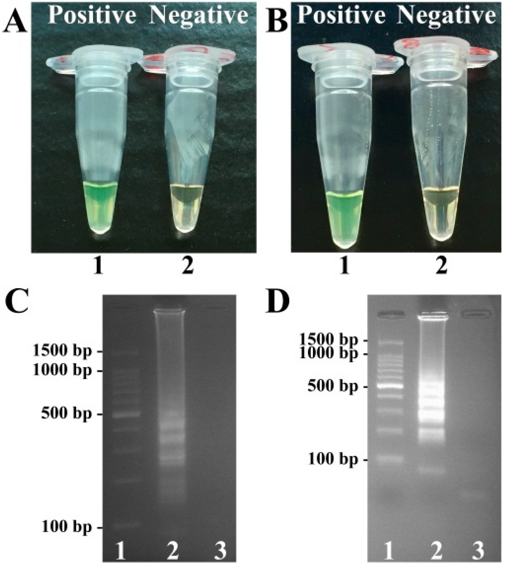
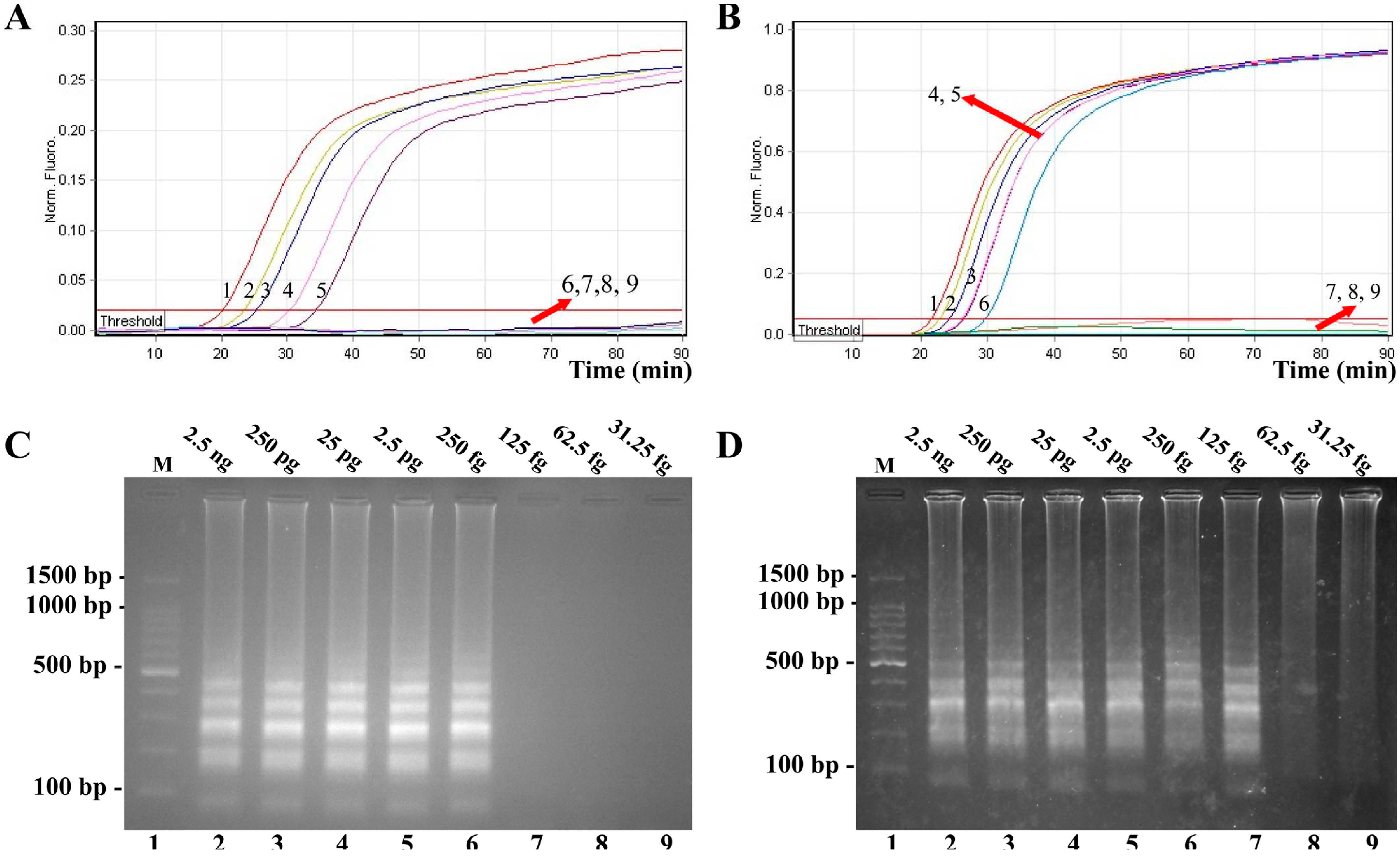
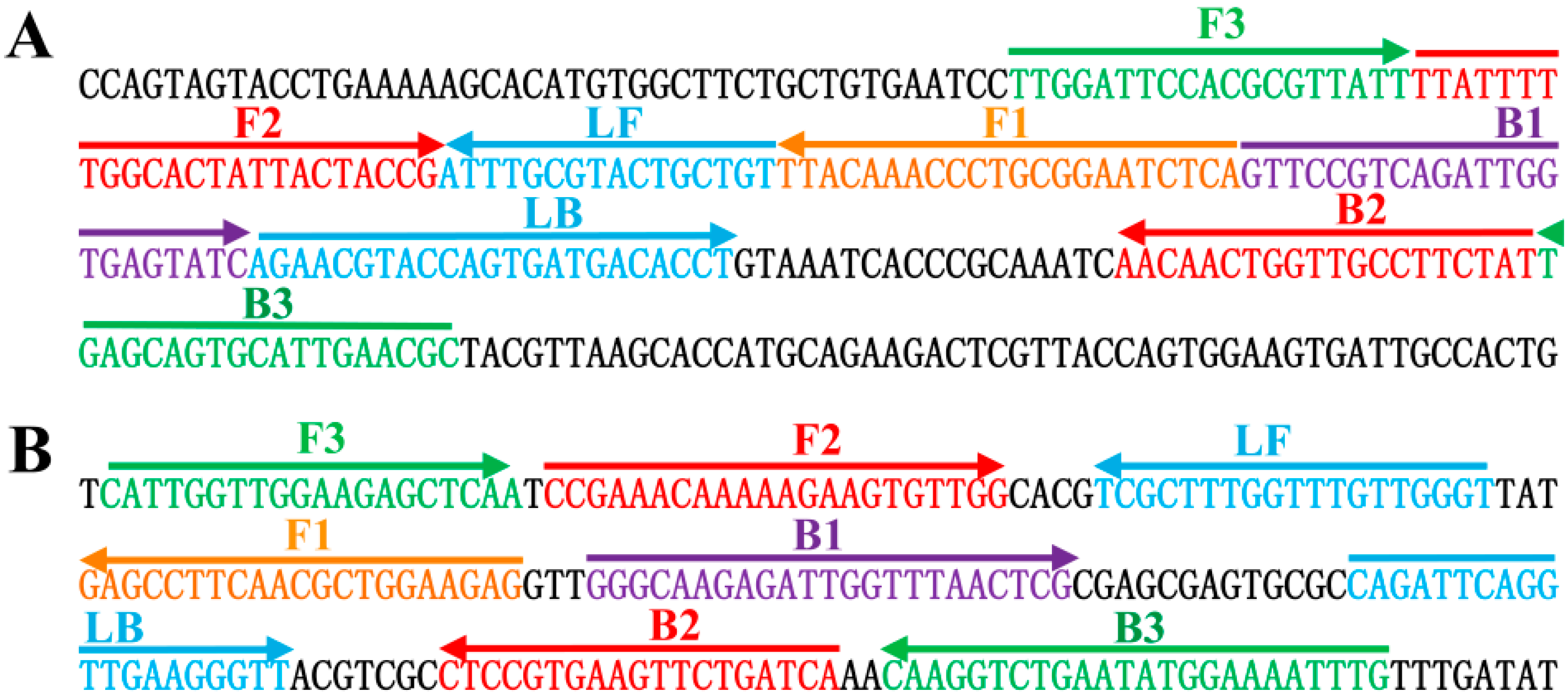
2.1. Detection of toxR- and rpoS-LAMP Products

To confirm the availability of toxR- and rpoS-LAMP primers, the LAMP reaction either for V. parahaemolyticus strains or V. vulnificus strains was processed in the absence or presence of genomic templates at 62 °C. The amplification products were analyzed by visual inspection using FD reagent, and the positive amplifications in V. parahaemolyticus- and V. vulnificus-LAMP tubes directly saw color change from light gray to green within 1h incubation periods (Figure 1). The conventional LAMP products were also analyzed by 2% agarose gel electrophoresis, and positive results showed the typical ladder-like patterns on gel electrophoresis (Figure 1). Thus, the toxR- and rpoS-LAMP primers for V. parahaemolyticus and V. vulnificus detection were found good candidates for developing the MERT-LAMP assays.
Figure 1. Confirmation and detection of toxR- and rpoS-LAMP products. (A,B), Color change of toxR- and rpoS-LAMP tubes; tube 1, positive reaction; tube 2, negative reaction; (C,D), 2% agarose gel electrophoresis applied to toxR- and rpoS-LAMP products; lane 1, DL 100-bp DNA marker; lane 2, positive LAMP reaction; lane 3, negative LAMP reaction.
Molecules 21 00111 g001

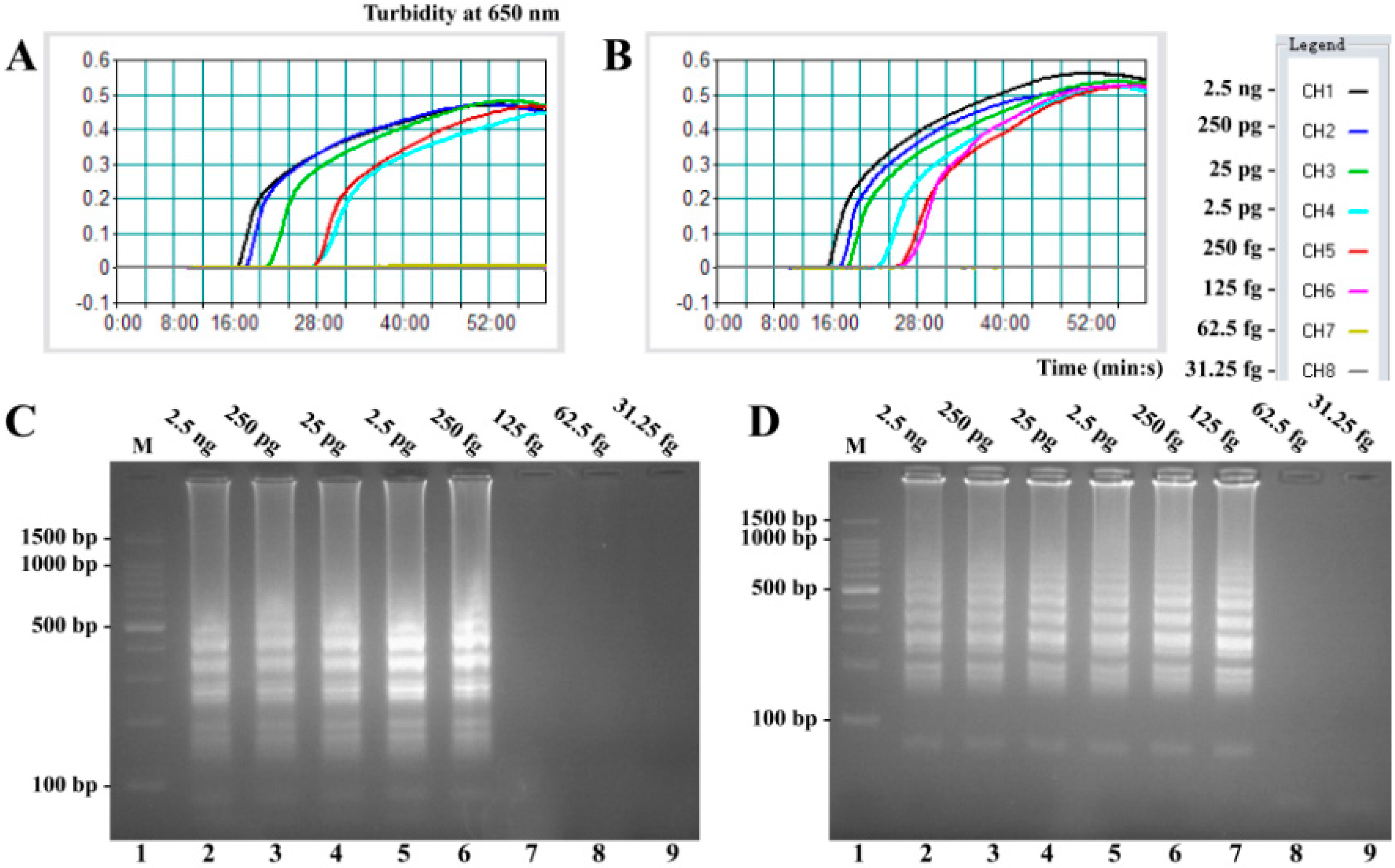
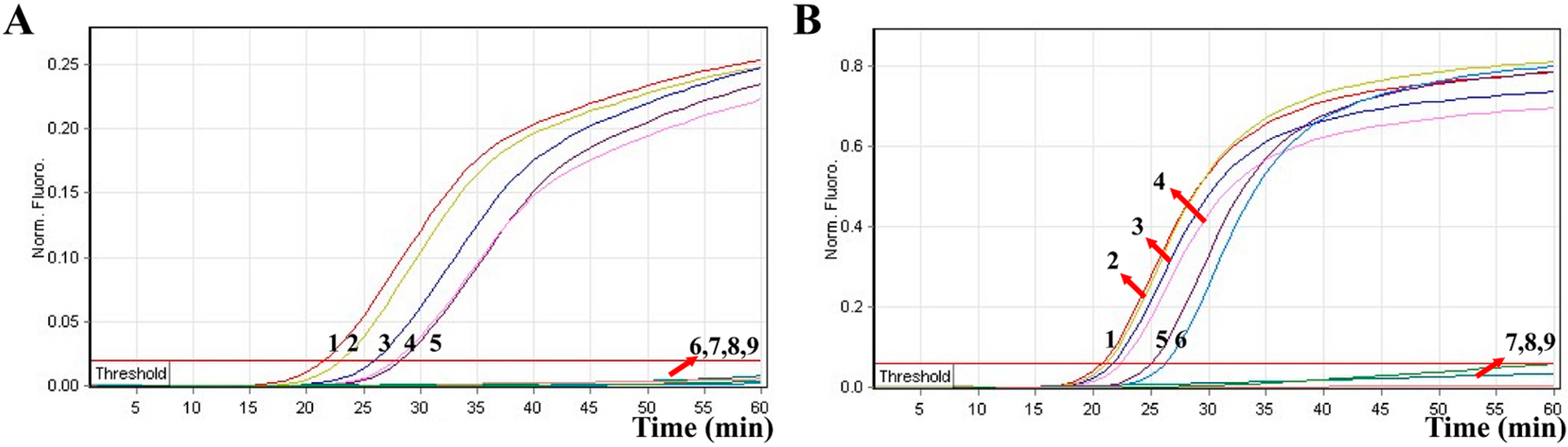
2.2. Sensitivity of V. parahaemolyticus- and V. vulnificus-LAMP Assay in Pure Culture

Sensitivity of conventional LAMP assays on V. parahaemolyticus and V. vulnificus were evaluated by analyzing products produced from the serial dilutions (2.5 ng, 250 pg, 25 pg, 2.5 pg, 250 fg, 125 fg, 62.5 fg, and 31.25 fg per microliter) of the V. parahaemolyticus and V. vulnificus genomic DNA templates in triplicate. As shown in Figure 2, the LAMP reactions were analyzed by real-time turbidity detection; the limit of detection (LoD) of V. parahaemolyticus-LAMP assay was 250 fg per tube (Figure 2A), and the V. vulnificus-LAMP assay for 125 fg per tube (Figure 2B). Moreover, the final LAMP products were also detected by 2% agarose gel electrophoresis, and positive amplifications were observed as a ladder-like pattern (Figure 2C,D). The LoD of the agarose gel electrophoresis detection for V. parahaemolyticus- and V. vulnificus-LAMP reactions was in complete conformity with turbidity measurements.

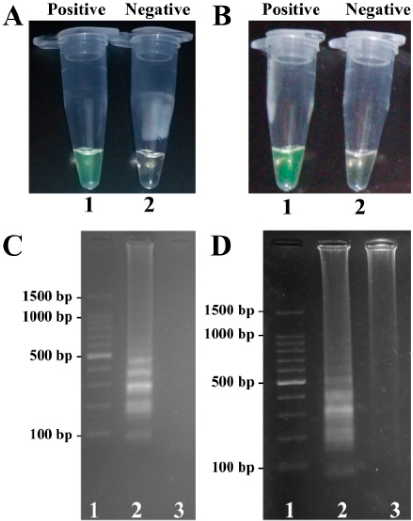
2.3. Detection of MERT-LAMP Products in Non-Real-Time Format

In order to demonstrate the utility of two MERT-LAMP primer sets, the MERT-LAMP assay either for V. parahaemolyticus strains or V. vulnificus strains was processed in the presence or absence of genomic DNA templates according to the standard MERT-LAMP condition. The MERT-LAMP products were detected by visual inspection using FD reagent, and the color change of positive MERT-LAMP reactions in V. parahaemolyticus- and V. vulnificus tubes from light gray to green were directly observed by naked eyes within 1 h incubation periods (Figure 3A). Furthermore, the MERT-LAMP products were also detected by 2% agarose gel electrophoresis, and the typical ladder-like patterns were observed but not in negative control (Figure 3).
Figure 2. Sensitivity of the toxR- and rpoS-LAMP assays using serially-genomic DNA with Vibrio parahaemolyticus isolates and Vibrio vulnificus isolates as templates. LoD of toxR-LAMP (A) and rpoS-LAMP (B) for V. parahaemolyticus and V. vulnificus detection was monitored by real-time measurement of turbidity and the corresponding curves of concentrations of genomic DNA were marked in the figure. The sensitivity of toxR-LAMP assay was 250 fg per reaction, and the rpoS-LAMP for 125 fg per reaction. Sensitivity of toxR-LAMP (C) and rpoS-LAMP (D) for V. parahaemolyticus and V. vulnificus detection was analyzed by 2% agarose gel electrophoresis, and the positive amplifications were observed as a ladder-like pattern on 2% agarose gel electrophoresis analysis. Lane 1, DL 100-bp DNA marker.
Molecules 21 00111 g002
Figure 3. Confirmation and detection of toxR- and rpoS-MERT-LAMP products. (A,B), color change of toxR- and rpoS-MERT-LAMP tubes; tube 1, positive amplification; tube 2, negative amplification; (C,D), 2% agarose gel electrophoresis applied to toxR- and rpoS-MERT-LAMP products; lane 1, DL 100-bp DNA marker; lane 2, positive LAMP reaction; lane 3, negative LAMP reaction.
Molecules 21 00111 g003

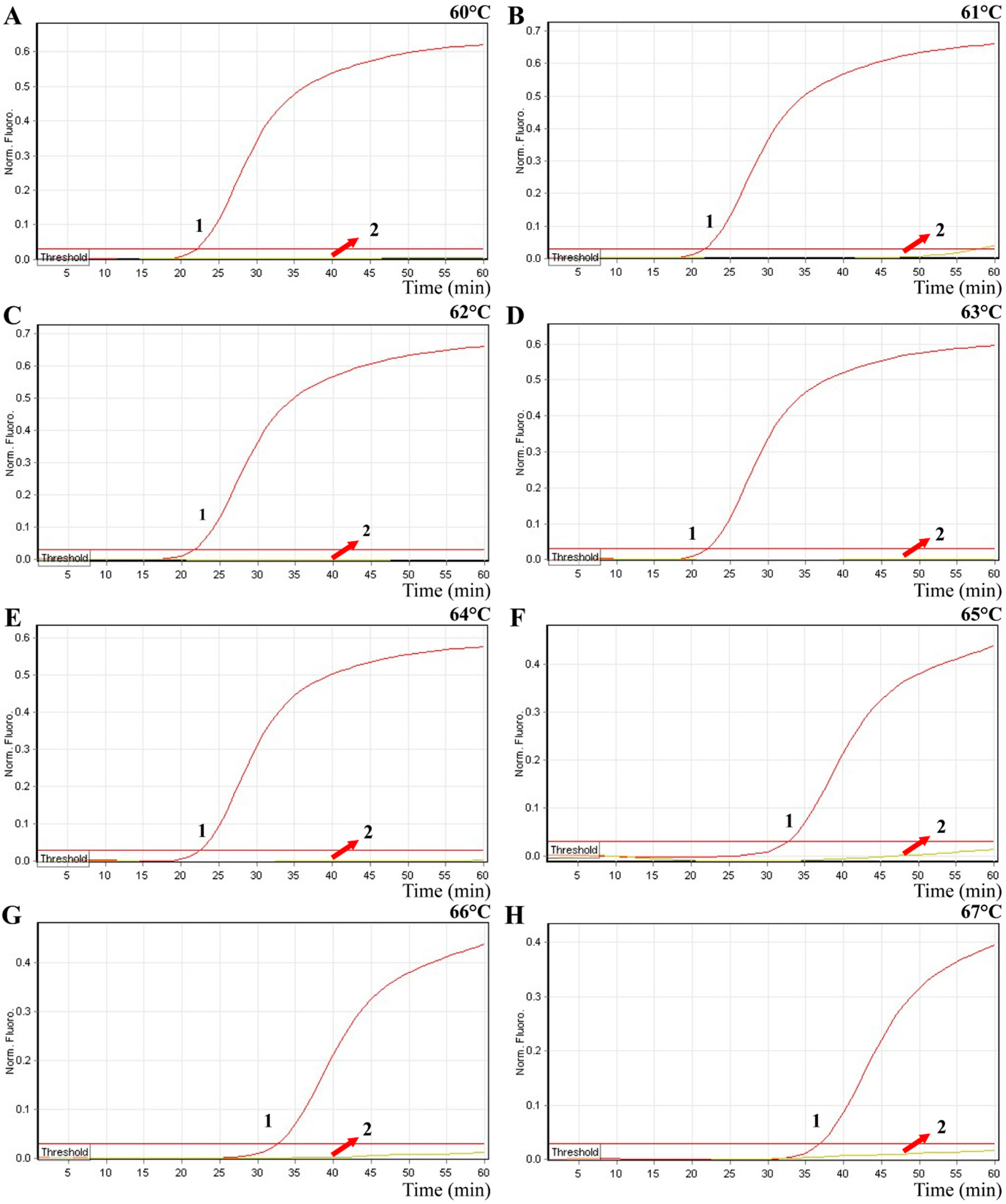
2.4. The Optimal Amplification Temperature of MERT-LAMP Approach

In order to test the optimal detection temperature, the MERT-LMAP amplifications were performed at distinct temperatures (60 to 67 °C) with V. vulnificus-MERT-LAMP primer set according to the standard MERT-LAMP reaction. The reference strain ATCC 27562 was selected as a positive control to examine the optimal temperature at the level of 250 pg of genomic DNA template per vessel. The results were detected by means of real-time format, and the typical kinetics graphs were yielded (Figure 4). Each amplification temperature produced a robust signal corresponding to Cy5 channel, with the faster amplifications were obtained from reaction temperatures of 60 to 64 °C, which were considered as the reference temperature for MERT-LAMP test. The detection temperature of 62 °C was applied for the rest of MERT-LAMP reactions conducted in this study.
Figure 4. The optimal temperature for MERT-LAMP method. The MERT-LAMP amplifications were analyzed by means of real-time format, and the corresponding curves of DNA concentrations were listed. Signal 1 indicates V. vulnificus strains of ATCC 27562, and signal 2 indicates negative control. Eight kinetic graphs (AH) were yielded at different amplification temperature (60–67 °C) with V. vulnificus genomic DNA at the level of 250 pg per tube. The graphs from 60 to 64 °C show robust amplification.
Molecules 21 00111 g004

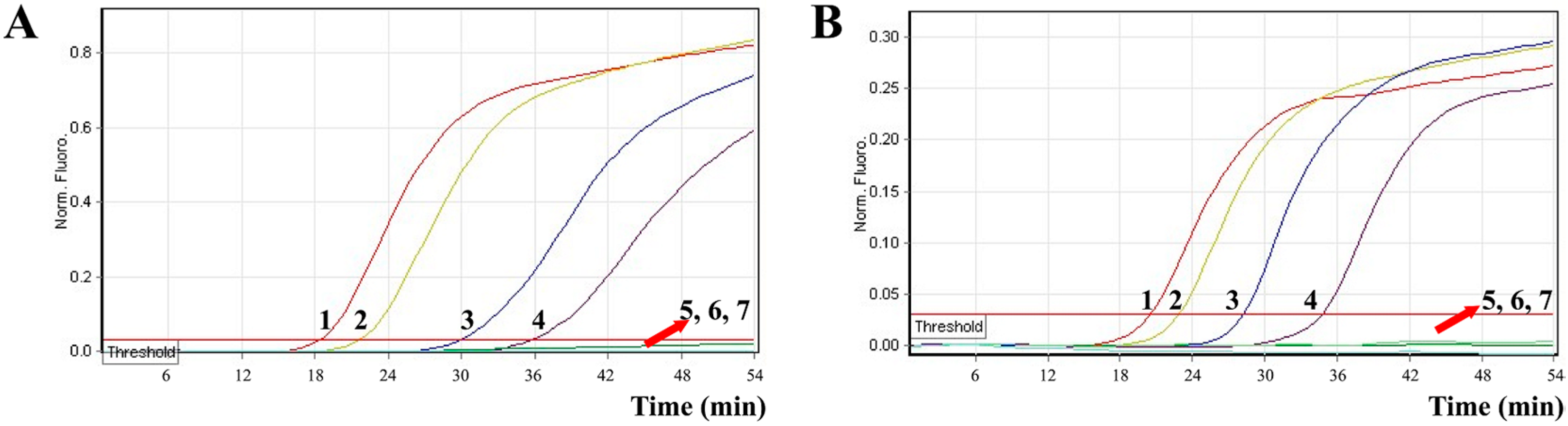
2.5. Sensitivity of MERT-LAMP Assay for a Single Target

In the present study, we tested the MERT-LAMP approach in a single target format by using separate amplification of toxR (V. parahaemolyticus-specific gene) and rpoS (V. vulnificus-specific gene) from an V. parahaemolyticus and V. vulnificus DNA templates in each reaction, respectively. The Hex and Cy5 fluorophores, which were used for yellow and red channels, were assigned to toxR- and rpoS-MERT-LAMP primer sets, respectively. As shown in Figure 5, the release of quenching was seen as a robust increase of Cy5 and Hex signals, and positive amplifications were generated in approximately 19 min. The LoD of MERT-LAMP technique for independently analyzing toxR and rpoS genes was 250 fg and 125 fg of genomic DNA templates per tube, respectively (Figure 5). Moreover, the final MERT-LAMP products were also analyzed by 2% agarose gel electrophoresis; the typical ladder-like patterns were visible in positive amplifications but not in negative reactions and control. The LoD of the real-time detection for V. parahaemolyticus- and V. vulnificus-MERT-LAMP amplifications was completed accordance with agarose gel electrophoresis measurement.
Figure 5. LoD of the toxR- and rpoS-MERT-LAMP approaches using serially-genomic DNA with V. parahaemolyticus strains and V. vulnificus strains as templates. Sensitivity of toxR-MERT-LAMP (A) and rpoS-MERT-LAMP (B) for V. parahaemolyticus and V. vulnificus detection was analyzed by real-time format, and signals 1, 2, 3, 4, 5, 6, 7, 8, and 9 represent DNA levels of 2.5 ng, 250 pg, 25 pg, 2.5 pg, 250 fg, 125 fg, 62.5 fg, and 31.25 fg per vessel and negative control. The LoD of toxR-MERT-LAMP assay was 250 fg per reaction, and the rpoS-MERT-LAMP for 125 fg per reaction. LoD of toxR-MERT-LAMP (C) and rpoS-MERT-LAMP (D) for V. parahaemolyticus and V. vulnificus detection was detected by 2% agarose gel electrophoresis, and the positive amplifications were seen as a ladder-like pattern on 2% agarose gel electrophoresis analysis. Lane 1, DL 100-bp DNA marker.
Molecules 21 00111 g005
The LoD of PCR, real-time PCR, conventional LAMP, and MERT-LAMP approaches on V. parahaemolyticus was 25 pg, 2.5 pg, 250 fg and 250 fg per reaction, and on V. vulnificus was 25 pg, 2.5 pg, 125 fg and 125 fg per reaction, respectively (Table 1, Figure 2 and Figure 5). These results suggested that the sensitivity of MERT-LAMP assay for detecting a single target was 10-fold and 100-fold more sensitive than that of qPCR and PCR assays, while was identical with conventional LAMP technique.
Table 1. The sensitivity and time for single MERT-LAMP technique targeting toxR and ropS genes compared with that of qPCR and PCR methods.
Approaches 1Isothermal Amplification 2Multiplex DetectionRegions RecognizedLoD for V. parahaemolyticus/V. vulnificus(No./Reaction) 3Fastest Time (min)LoD Time (min) 4
MERT-LAMP++8250 fg/125fg1935
LAMP+8250 fg/125 fg1935
qPCR+32.5 pg/2.5 pg3560
PCR+225pg/25pg150150
1 MERT-LMAP, multiple endonuclease restriction real-time loop-mediated isothermal amplification, qPCR, quantitative real-time PCR; 2 +, yes; –, no; 3 No., number; 4 LoD, limit of detection. The LoD values are the lowest gnomic DNA level that was positively amplified in triplicate. The positive amplifications of qPCR methods were generated as ct values, which were converted to time for detection.

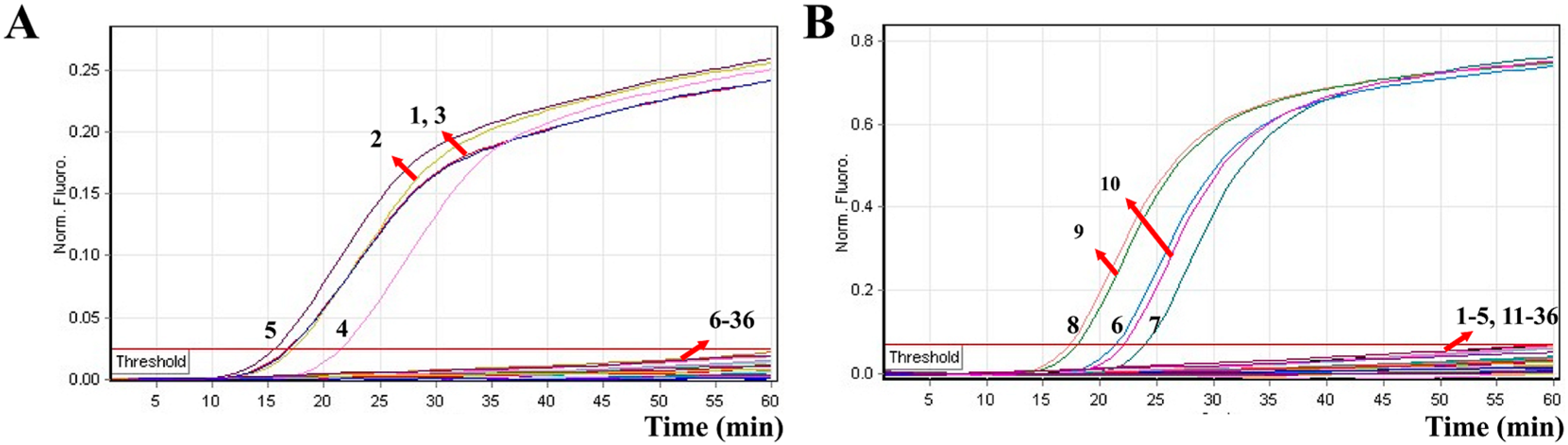
2.6. Sensitivity of MERT-LAMP Assay for Multiple Targets in a Reaction

To examine the capability of MERT-LAMP technique for simultaneously detecting V. parahaemolyticus and V. vulnificus in a single reaction, we slightly adjusted the amount of the primers on the base of standard MERT-LAMP reaction, and the multiplex MERT-LAMP amplifications were carried out at 62 °C for 60 min. Two different fluorescence curves were simultaneously obtained from multiplex MERT-LAMP reactions, which contained two complete primer sets and their corresponding genomic templates (Figure 6). The MERT-LAMP methodology successfully detected V. parahaemolyticus and V. vulnificus in a single reaction, and provided a robust signal for each target. The LoD of MERT-LAMP assay for simultaneously detecting toxR and rpoS genes was 250 fg and 125 fg of each genomic DAN template per tube, respectively (Figure 6). No difference of sensitivity was obtained between detecting multiple targets and a single target in a MERT-LAMP reaction.
Figure 6. The LoD of multiplex MERT-LAMP method for simultaneously detecting two target pathogens. Two MERT-LMAP primer sets targeting toxR and rpoS genes were simultaneously added to a reaction vessel. (A,B) were simultaneously yielded from Hex (labeling V. p-EFIP of toxR) and Cy5 (labeling V. v-EFIP of rpoS) channels, respectively. LoD of multiplex-MERT-LAMP for simultaneously detecting V. parahaemolyticus (A) and V. vulnificus (B) was analyzed by real-time format, and signals 1, 2, 3, 4, 5, 6, 7, 8, and 9 represent DNA levels of 2.5 ng, 250 pg, 25 pg, 2.5 pg, 250 fg, 125 fg, 62.5 fg, and 31.25 fg per reaction and negative control. respectively. The sensitivity of multiplex MERT-LAMP approached for V. parahaemolyticus detection was 250 fg per tube, and the LoD of multiplex MERT-LAMP for V. vulnificus detection was125 fg per tube.
Molecules 21 00111 g006

2.7. Analytical Specificity of the Multiplex MERT-LAMP Approach

In order to test the MERT-LAMP technique’s specificity, the multiplex MERT-LAMP amplifications were conducted under the multiplex MERT-LAMP conditions described above with the purely genomic DNA templates extracted from 19 V. parahaemolyticus, 15 V. vulnificus, and 20 non-V. parahaemolyticus and non-V. vulnificus strains. By observation, positive results could be obtained only when genomic DNAs of V. parahaemolyticus strains and V. vulnificus strains were used as templates in multiplex MERT-LAMP reactions, and the target pathogens were correctly identified (Figure 7). However, for the reaction tubes of non-V. parahaemolyticus stains, non-V. vulnificus strains and negative control, there was no positive amplification after 60-min incubation period. These results demonstrated that the multiplex MERT-LAMP technology developed here was specific to target sequence identification.
Figure 7. The specificity of multiplex MERT-LMAP technology for different strains. The multiplex MERT-LAMP reactions were carried out using different genomic DNA templates and were monitored by means of real-time format. (A,B) were simultaneously obtained from yellow (Hex) and red (Cy5) channels. Signals 1–5, Vibrio parahaemolyticus strains of ICDC-NVP001, ICDC-NVP002, ICDC-NVP003, ICDC-NVP004, ICDC-NVP005; signals 6–10, Vibrio vulnificus strains ATCC27562, ICDC-NVV001, ICDC-NVV002, ICDC-NVV003, ICDC-NVV004; signals 11–36, Vibrio cholerae, Vibrio mimicus, Vibrio fluvialis, Vibrio algindyticus, Shigella flexneri, Shigella boydii, Shigella sonnel, Shigella dysenteriae, Salmonella choleraesuis, Listeria monocytogenes (EGD-e), Listeria monocytogenes (ATCC19116), Enterobacter cloacae, Enterococcus faecalis, Enteropathogenic E. coli, Enteroaggregative E. coli, Enterotoxigenic E. coli, Enteroinvasive E. coli, Enterohemorrhagic E. coli, Plesiomonas shigelloides, Yersinia enterocolitica, Cronobacter sakazakii, Staphylococcus aureus, Campylobacter jejuni, Pseudomonas aeruginosa and Bacillus cereus, signal 36, negative control.
Molecules 21 00111 g007

2.8. Applicability of the MERT-LAMP Technology

In order to evaluate the practicability of MERT-LAMP approach as a surveillance tool for V. parahaemolyticus and V. vulnificus in seafood, the MERT-LAMP methodology was examined by artificially adding V. parahaemolyticus and V. vulnificus strains into oysters. The LoD of MERT-LAMP assay was 92 CFU/reaction of V. parahaemolyticus ICDC-NVP001 and 83 CFU/reaction of V. vulnificus ATCC 27562 in spiked oyster samples without enrichment, and the two target pathogens could be simultaneously detected in a single MERT-LAMP reaction (Figure 8, Table 2). The non-contaminated oyster sample was found to be negative.
Figure 8. The LoD of MERT-LAMP approach for simultaneously detecting two target pathogens in artificially contaminated oysters samples. Two MERT-LMAP primer sets targeting toxR and rpoS genes were added to a reaction tube. (A,B) were simultaneously yielded from Hex (labeling V. P-EFIP of toxR) and Cy5 (labeling V. v-EFIP of rpoS) channels, respectively. LoD of MERT-LAMP for simultaneously detecting V. parahaemolyticus (A) and V. vulnificus (B) in artificially contaminated oyster samples were monitored by real-time format, and signals 1, 2, 3, 4, 5, 6, and 7 represent V. parahaemolyticus DNA levels of 9200 CFU, 920 CFU, 92 CFU, 9.2 CFU, 0.92 CFU, and 0.092 CFU per reaction and negative control; V. vulnificus DNA levels for 9300 CFU, 830 CFU, 83 CFU, 8.3 CFU, 0.83 CFU, and 0.083 CFU per reaction and negative control. The sensitivity of MERT-LAMP method for V. parahaemolyticus detection in artificially contaminated oyster samples was 92 CFU per reaction, and the LoD of MERT-LAMP for V. vulnificus detection in artificially contaminated oyster samples was 83 CFU per vessel.
Molecules 21 00111 g008
The LoD of multiplex MERT-LAMP approach was in conformity with that of conventional LAMP assay only for V. parahaemolyticus or V. vulnificus, respectively (Table 2). Comparatively, the PCR and qPCR methods generated positive results when the contaminate numbers of V. parahaemolyticus ICDC-NVP001 amounted to more than 9200 and 920 CFU/reaction, V. vulnificus ATCC 27562 for 8300 and 830 CFU/reaction, respectively. The results suggested that the sensitivity of multiplex MERT-LAMP approaches was 100- and 10-fold more sensitive than that of PCR and qPCR technologies (Table 2).
Table 2. Comparison of MERT-LMAP, LAMP, qPCR, and PCR methods for detection of V. parahaemolyticus and V. vulnificus.in artificially contaminated oyster samples.
Detection Assays 1Multiplex Detection 2LoD (No./Reaction) 3
V. parahaemolyticus DetectionV. vulnificus Detection
MERT-LAMP+92 CFU83 CFU
LAMP92 CFU83 CFU
qPCR920 CFU830 CFU
PCR9200 CFU8300 CFU
1 MERT-LMAP, multiple endonuclease restriction real-time loop-mediated isothermal amplification; LAMP, loop-mediated isothermal amplification. 2 +, yes; –, no; 3 No., number.

3. Discussion

V. parahaemolyticus and V. vulnificus, as serious bacterial pathogens for aquatic animals and humans, are widely distributed in coastal and estuarine environments throughout the world [15]. In aquatic animals, the two pathogens are capable of causing serve illnesses in penaeid shrimp, fish, and shellfish, leading to significant losses in aquaculture industries [21]. In humans, most V. parahaemolyticus and V. vulnificus infections resulted from the consumption of raw, inadequately or improperly cooked seafood, particularly shellfish (such as oysters) [1]. Therefore, there is a need for rapid, specific, sensitive, and cost-effective diagnosis methodology that can be used effectively for simultaneous detection of V. parahaemolyticus and V. vulnificus in various samples.
In this report, the first MERT-LAMP assay targeting the toxR gene of V. parahaemolyticus and the rpoS gene of V. vulnificus has was successfully established. The new MERT-LAMP technique could simultaneously detect and distinguish toxR and rpoS genes in only one isothermal step, eliminating the use of temperature-regulating apparatus. Moreover, the MERT-LAMP did not require further processing, and the reaction tubes were not opened in the course of the experiment, which effectively alleviated any carryover contamination. The optimal MERT-LAMP amplification temperature was 60 °C to 64 °C, and 62 °C was selected as reaction temperature. The MERT-LAMP reactions were completed within 60 min. The detection speed of MERT-LAMP method was similar with traditional LAMP assays, while the MERT-LAMP methodology achieved multiplex detection in a single reaction; thus, V. parahaemolyticus and V. vulnificus strains in the same samples could be simultaneously detected and correctly identified (Figure 6 and Figure 8).
Sensitivity of the MERT-LAMP assay was examined using serial dilution of genomic DNA templates. First, we evaluated the MERT-LAMP technique in a single target format by using separate detection of toxR (V. parahaemolyticus-specific gene) and rpoS (V. vulnificus-specific gene) from an V. parahaemolyticus and V. vulnificus DNA templates in each reaction, and the LoD of MERT-LAMP methodology for independently amplifying toxR gene or rpoS gene was 250 fg or 125 fg of genomic DNA templates per reaction, respectively (Figure 5). The sensitivity of MERT-LAMP method for detecting a single target was consistent with conventional LAMP assay (Figure 2 and Figure 5, Table 1). Second, we extended the MERT-LAMP approach to simultaneously amplify toxR and rpoS genes in a single reaction, and the two target pathogens were successfully identified and detected. The LoD of MERT-LAMP approach for simultaneously detecting toxR and rpoS genes was 250 fg and 125 fg of genomic DNA templates per tube, respectively, and which is the same as that of singleplex MERT-LAMP detections (Figure 5 and Figure 6). The LoD of multiplex MERT-LAMP technology was identical with that the conventional LAMP technique described in this study or developed in other publications, and 10-fold and 100-fold more sensitive than conventional qPCR and PCR techniques (Figure 2 and Figure 6, Table 1) [19]. In addition, the multiplex MERT-LAMP assay was also successfully examined using artificially contaminated oyster samples, and the multiplex MERT-LAMP methodology performed better than PCR and qPCR techniques with respect to detection speed and LoD (Figure 8 and Table 2).
In addition to its sufficient sensitivity, the MERT-LAMP assay in identification of V. parahaemolyticus and V. vulnificus offered a high degree of specificity and the assay specificity was successfully ascertained. Each primer set contained six specific primers that recognized eight distinct regions on the toxR gene of V. parahaemolyticus and rpoS gene of V. vulnificus, which ensured high specificity of the DNA template detection [7,9,19]. In order to verify the specificity of multiplex MERT-LAMP for detection of V. parahaemolyticus and V. vulnificus, a variety of bacterial strains listed in Table 3 were determined in this study. The results obtained from our study showed that the MERT-LAMP technology indicated a high degree of specificity to V. parahaemolyticus and V. vulnificus by generating positive results to all examined isolates of the two bacteria while producing a negative result to non-V. parahaemolyticus and non-V. vulnificus bacteria strains. Moreover, the two target pathogens were correctly differentiated (Figure 7). These results suggested that the MERT-LAMP assay established here was specific to target sequence detection.
In conclusion, our study is the first report of the use of MERT-LAMP assay for simultaneous detection of V. parahaemolyticus and V. vulnificus, comparing with regular LAMP detection approaches, which provides advantages on real time and multiplex detection. Compared with PCR-based techniques, the MERT-LAMP method is advantageous on sensitivity, specificity, rapidity, time consumption, and ease in operation. Therefore, the developed MERT-LAMP assay can be used as a valuable tool for detection of V. parahaemolyticus and V. vulnificus in basic and field laboratories.

4. Materials and Methods

4.1. Design of the MERT-LAMP Primers

Based on the toxR gene of V. parahaemolyticus and rpoS gene of V. vulnificus, two sets of MERT-LAMP primers were designed by PrimerExplorer V4 (Eiken Chemical) according to the mechanism of MERT-LAMP methodology [7,9]. Blast analysis validated that two sets of MERT-LAMP primers were specific for V. parahaemolyticus and V. vulnificus. The dark quenchers used were Black Hole Quencher-1 and Black Hole Quencher-2, and the fluorophores used were HEX and Cy5, which are assigned to toxR and rpoS primer sets, respectively. All oligonucleotides described here were synthesized and purified by Tianyi Biotech (Beijing, China). The details of target sequences, primer design, primers sequences and locations were shown in Table 3 and Figure 9.
Figure 9. Location and sequence of toxR (Vibrio parahaemolyticus) and rpoS (Vibrio vulnificus) genes used to design MERT-LAMP primers. The nucleotide sequences of the sense strands of toxR (A) and rpoS (B) are showed. The sites of primer sequences were underlined. Right arrows and left arrows showed sense and complementary sequences that are used.
Molecules 21 00111 g009
Table 3. The primers used in this study.
Primers Name 1Sequences and ModificationsLengthGenes
V.p-F35′-TTGGATTCCACGCGTTATT-3′19 nttoxR
V.p-B35′-GCGTTCAATGCACTGCTCA-3′19 nt
V.p-FIP5′-TGAGATTCCGCAGGGTTTGTAATTATTTTTGGCACTATTACTACCG-3′46 mer
V.p-BIP5′-GTTCCGTCAGATTGGTGAGTATCATAGAAGGCAACCAGTTGTT-3′43 mer
V.p-EFIP5′-HEX-TGCAATG-TGAGAT(BHQ1)TCCGCAGGGTTTGTAATTATTTTTGGCACTATTACTACCG-3′53 mer
V.p-LF5′-ACAGCAGTACGCAAAT-3′16 nt
V.p-LB5′-AGAACGTACCAGTGATGACACCT-3′23 nt
V.v-F35′-CATTGGTTGGAAGAGCTCAA-3′20 ntrpoS
V.v-B35′-CAAATTTTCCATATTCAGACCTTG-3′24 nt
V.v-FIP5′-CTCTTCCAGCGTTGAAGGCTCTTTTCCGAAACAAAAAGAAGTGTTGG-3′47 mer
V.v-EFIP5′-Cy5-TGCAATG-CTCT(BHQ-2)TCCAGCGTTGAAGGCTCTTTTCCGAAACAAAAAGAAGTGTTGG-3′54 mer
V.v-BIP5′-GGGCAAGAGATTGGTTTAACTCGTTTTTGATCAGAACTTCACGGAG-3′46 mer
V.v-LF5′-ACCCAACAAACCAAAGCGA-3′19 nt
V.v-LB5′-CAGATTCAGGTTGAAGGGTT-3′20 nt
1 V. p, Vibrio parahaemolyticus; V. v, Vibrio vulnificus; F, forward; B, backward; FIP, forward inner primer; EFIP, the novel forward inner primer; BIP, backward inner primer; LF, loop forward primer; LB, loop backward primer.

4.2. Reagents

LoopampTM Fluorescent Detection Reagent (FD) and the Loopamp kits were purchase from Eiken Chemical (Tokyo, Japan, and Beijing, China). The DNA extraction kits (QIAamp DNA Mini Kits) were purchased from Qiagen (Beijing, China), and the Nb.BsrDI was purchased from New England Biolabs (Beijing, China).

4.3. Bacterial Strains

The bacterial strains tested in this study were listed in Table 4. All Vibrio strains were routinely cultured using thiosulfate citrate bile salt sucrose agar (TCBS agar, Eiken Chemical) at 35 °C overnight. Storage and culture conditions of non-Vibrio strains were followed as previously described [11].
Table 4. Bacterial strains used in this study.
BacteriaStrain No. (Source of Strain) 1No. of Strains
Vibrio parahaemolyticusICDC-NVP0011
Isolated strains26
Vibrio vulnificusATCC275621
Isolated strains11
Vibrio choleraeATCC140351
Isolated strains1
Vibrio mimicusIsolated strains1
Vibrio fluvialisIsolated strains1
Vibrio alginolyticusIsolated strains1
Enterohemorrhagic E. coliEDL9331
Enteropathogenic E. coliIsolated strains1
Enterotoxigenic E. coliIsolated strains1
Enteroaggregative E. coliIsolated strains1
Enteroinvasive E. coliIsolated strains1
Yersinia enterocoliticaATCC237151
Shigella dysenteriaeIsolated strains1
Shigella boydiiIsolated strains1
Shigella flexneriIsolated strains10
Shigella sonneiIsolated strains1
SalmonellaIsolated strains10
Plesiomonas shigelloidesATCC519031
Listeria monocytogenesEGD-e1
ATCC191161
Listeria ivanoviiATCCBAA-6781
Listeria grayiATCC254021
Enterococcus faecalisATCC356671
Enterobacter cloacaeIsolated strains1
Cronobacter sakazakiiIsolated strains1
Bacillus cereusIsolated strains5
Campylobacter jejuniATCC332911
Pseudomonas aeruginosaIsolated strains10
Staphylococcus aureusIsolated strains10
Klebsiella pneumoniaeIsolated strains5
1 ATCC, American Type Culture Collection; ICDC, National Institute for Communicable Disease Control Disease Control and Prevention, Chinese Center for Disease Control and Prevention.

4.4. DNA Templates Preparation

According to the manufacturer’s instructions, DNA templates used in this study were extracted from all culture strains using DNA extraction kits (QIAamp DNA minikits; Qiagen, Hilden, Germany). The extracted templates were tested with ultraviolet spectrophotometer at A260/280 and stored under at −20 °C until use.

4.5. The Conventional LAMP Assay

In order to examine the availability of two LAMP primer sets, the LAMP amplification either for V. parahaemolyticus strains or V. vulnificus strains was carried out as the following description. Briefly, the traditional LAMP reaction was performed with the Loopamp Kit in a final volume of 25 μL containing 1.6 μM each FIP and BIP primers, 0.8 μM each LF and LB primers, 0.4 μM each F3 and B3 primers, 12.5 μL 2× reaction mix, 1 μL of Bst DNA polymerase (8 U), 1 μL FD and 1 μL DNA template.
The amplification mixtures of convention LAMP were heated at 62 °C for 1 h then at 85 °C for 5 min to stop the amplification. Three methods were employed to monitor the conventional LAMP reaction. First, a real-time turbidimeter (LA-320C, Eiken Chemical Co., Ltd., Tokyo, Japan) was used to minor the LAMP reaction by recording the optical density (OD) at 650 nm every 6 s. The turbidity readings were produced in real-time format and a turbidity threshold value of 0.1 was defined. Second, the positive reactions could be directly seen color change by FD reagent. Moreover, the amplification products were also detected by electrophoresis on 2% agarose gels with ethidium bromide staining. Mixture without DNA template was used as a negative control.

4.6. The Standard MERT-LAMP Assay

In order to test utility of two MERT-LAMP primer sets, the reaction mixtures of MERT-LAMP were performed in a final volume of 25 μL containing 0.8 μM EFIP and FIP primers, 1.6 μM BIP primers, 0.8 μM each LF and LB primers, 0.4 μM each F3 and B3 primers, 12.5 μL 2× reaction mix, 1 μL (8 U) of Bst DNA polymerase, 1.5 μL (15 U) of Nb.BsrDI endonuclease and 1μL DNA template.
A real-time machine (Rotor-Gene Q, Qiagen, Hilden, Germany) was applied to run the MERT-LAMP reactions, and the assay was conducted at 62 °C for 1 h. Mixture without DNA template was used as a negative control. After amplification, the MERT-LAMP products were confirmed by electrophoresis on 2% agarose gels with ethidium bromide staining or directly observed the color change by FD reagent.
To assess the optimal reaction temperature of MERT-LAMP approach, we mixed the MERT-LAMP reaction mixtures at a constant temperature ranging from 60 °C to 67 °C for 60 min and then heated at 85 °C for 5 min to complete the reaction. Mixtures without DNA template were used as a negative control.

4.7. The Multiplex MERT-LAMP Reaction

For multiplex amplifications, the MERT-LAMP assay was performed as the following system: 25 μL containing 0.4 μMV.v-EFIP and V.v-FIP primers, 0.8 μMV.v-BIP primers, 0.4 μM each V.v-LF and V.v-LB primers, 0.4 μM each V.v-F3 and V.v-B3 primers,1.2 μMV.p-EFIP and V.p-FIP primers, 2.4 μMV.v-BIP primers, 1.2 μM each V.v-LF and V.v-LB primers, 0.4 μM each V.v-F3 and V.v-B3 primers, 12.5 μL 2× reaction mix, 1.5 μL (15 U) of Nb.BsrDI endonuclease,1 μL (8 U) of Bst DNA polymerase and 1 μL DNA template DNA each of V. parahaemolyticus strains and V. vulnificus strains. The MERT-LAMP reactions were conducted at 62 °C for 1 h in the real-time system, and amplification mixtures without the DNA template were used as a negative control. The lowest detectable template amount were determined in triplicate.

4.8. Determination of the Sensitivity of the MERT-LAMP in Pure Cultures

Strains of V. parahaemolyticus ICDC-NVP001 and V. vulnificus ATCC 27562 were used for the assay of sensitivity testing with pure culture, and the genomic DNA templates were serially diluted. Individual MERT-LAMP approaches, which target the toxR and rpoS genes individually, were carried out with serial dilutions (2.5 ng, 250 pg, 25 pg, 2.5 pg, 250 fg, 125 fg, 62.5 fg, and 31.25 fg per microliter) according to standard MERT-LAMP system. To make a comparative analysis of MERT-LAMP, LAMP, qPCR and PCR assays by using pure culture, the limit of detection (LoD) of LAMP, qPCR, and PCR assays was defined by genomic DNA amount of the template. The V. parahaemolyticus-qPCR, V. parahaemolyticus-PCR, V. vulnificus-qPCR, and V. vulnificus-PCR assays have been established in previous studies, which were selected for confirming the LoD of qPCR and PCR technologies [22,23,24,25].
To test the sensitivity of multiplex MERT-LAMP assay, the MERT-LAMP reaction was conducted under the conditions described above with serial dilutions (2.5 ng, 250 pg, 25 pg, 2.5 pg, 250 fg, 125 fg, 62.5 fg, and 31.25 fg per microliter) of V. parahaemolyticus and V. vulnificus, and 1μl DNA template DNA each of V. parahaemolyticus strains and V. vulnificus strains was simultaneously added into a MERT-LMAP reaction.

4.9. Evaluation of the Specificity of the MERT-LAMP Assay

To assess the MERT-LAMP technology’s specificity, the multiplex MERT-LAMP reactions were carried out under the conditions described above with the purely genomic DNA templates extracted from 111 strains (Table 2). Analysis of each sample was performed twice independently.

4.10. Practical Application of MERT-LAMP to V. parahaemolyticus and V. vulnificus Detection in Oyster Samples

In order to determine practicability of MERT-LAMP approach in identification of V. parahaemolyticus and V. vulnificus, V. parahaemolyticus (ICDC-NVP001), and V. vulnificus (ATCC27562) were simultaneously added to oyster samples, which were purchased at a local market in Beijing. The oyster samples were confirmed as being V. parahaemolyticus and V. vulnificus negative by traditional culture assay and PCR [23,25]. Only oyster samples, which were negative for V. parahaemolyticus and V. vulnificus, were applied to spiked oyster samples.
Firstly, to test the minimal detectable colony forming units (CFUs), the cultures with V. parahaemolyticus and V. vulnificus strains were serially diluted (10−1 to 10−9), and the aliquots of 100 μL appropriate dilution (10−6) was plated in triplicate on brain heart infusion (BHI). The CFUs were counted after 24 h at 37 °C [20]. The following procedures, one hundred microliters of appropriate dilutions of V. parahaemolyticus and V. vulnificus with known amounts was simultaneously spiked into 800 μL of each of the oyster homogenates and mixed well. The oyster homogenates was centrifuged at 200 g for 5 min and the supernatant was transferred to a new vessel, and then was centrifuged at 18,000 g for 5 min. After removal of the supernatant, the pellet was applied for DNA extraction. For MERT-LAMP, LAMP, real-time PCR, and PCR assays, 1 μL of each supernatants (2 μL) were used as templates. Non-contaminated oyster sample was chosen as negative control. This performance was carried out in triplicate independently.

Acknowledgments

We acknowledge the financial supports of the grants (Mega Project of Research on the Prevention and Control of HIV/AIDS, Viral Hepatitis Infectious Diseases 2011ZX10004-001, 2013ZX10004-101 to Changyun Ye) from the Ministry of Science and Technology, People’s Republic of China, and grant (2015SKLID507 to Changyun Ye) from State Key Laboratory of Infectious Disease Prevention and Control, China CDC.

Author Contributions

Yi Wang and Changyun Ye conceived and designed the experiments; Yi Wang, Dongxun Li, Kewei Li and Yan Wang performed the experiments; Yi Wang analyzed the data; Yi Wang, Dongxun Li and Yan Wang contributed reagents/materials/analysis tools; Yi Wang and Changyun Ye wrote the paper. Yi Wang, Yan Wang, and Changyun Ye have filed for a patent from the State Intellectual Property Office of the People’s Republic of China, which covers the novel method and sequences included in this manuscript (application number CN201610013308.9).

Conflicts of Interest

The authors declare no conflict of interest.

References

  1. Bauer, A.; Rorvik, L.M. A novel multiplex pcr for the identification of Vibrio parahaemolyticus, vibrio cholerae and Vibrio vulnificus. Lett. Appl. Microbiol. 2007, 45, 371–375. [Google Scholar] [CrossRef] [PubMed]
  2. Oliver, J.D. The biology of Vibrio vulnificus. Microbiol. Spectr. 2015, 3, 401–439. [Google Scholar] [CrossRef] [PubMed]
  3. Xu, D.; Wu, X.; Han, J.; Chen, L.; Ji, L.; Shen, Y. A cross-priming amplification assay coupled with vertical flow visualization for detection of Vibrio parahaemolyticus. Mol. Cell. Probes 2015, 29, 527–530. [Google Scholar] [CrossRef] [PubMed]
  4. Newton, A.; Kendall, M.; Vugia, D.J.; Henao, O.L.; Mahon, B.E. Increasing rates of vibriosis in the united states, 1996–2010: Review of surveillance data from 2 systems. Clin. Infect. Dis. 2012, 54, S391–S395. [Google Scholar] [CrossRef] [PubMed]
  5. Changchai, N.; Saunjit, S. Occurrence of Vibrio parahaemolyticus and Vibrio vulnificus in retail raw oysters from the eastern coast of thailand. Southeast Asian J. Trop. Med. Public Health 2014, 45, 662–669. [Google Scholar] [PubMed]
  6. Yamazaki, W.; Kumeda, Y.; Uemura, R.; Misawa, N. Evaluation of a loop-mediated isothermal amplification assay for rapid and simple detection of Vibrio parahaemolyticus in naturally contaminated seafood samples. Food Microbiol. 2011, 28, 1238–1241. [Google Scholar] [CrossRef] [PubMed]
  7. Chen, S.; Ge, B. Development of a toxr-based loop-mediated isothermal amplification assay for detecting Vibrio parahaemolyticus. BMC Microbiol. 2010, 10, 41. [Google Scholar] [CrossRef] [PubMed]
  8. Jones, J.L.; Noe, K.E.; Byars, R.; Depaola, A. Evaluation of DNA colony hybridization and real-time pcr for detection of Vibrio parahaemolyticus and Vibrio vulnificus in postharvest-processed oysters. J. Food Prot. 2009, 72, 2106–2109. [Google Scholar] [PubMed]
  9. Kim, D.G.; Ahn, S.H.; Kim, L.H.; Park, K.J.; Hong, Y.K.; Kong, I.S. Application of the rpos gene for species-specific detection of Vibrio vulnificus by real-time pcr. J. Microbiol. Biotechnol. 2008, 18, 1841–1847. [Google Scholar] [PubMed]
  10. Wang, Y.; Wang, Y.; Ma, A.J.; Li, D.X.; Luo, L.J.; Liu, D.X.; Jin, D.; Liu, K.; Ye, C.Y. Rapid and sensitive isothermal detection of nucleic-acid sequence by multiple cross displacement amplification. Sci. Rep. 2015, 5, 11902. [Google Scholar] [CrossRef] [PubMed]
  11. Wang, Y.; Wang, Y.; Ma, A.; Li, D.; Ye, C. Rapid and sensitive detection of listeria monocytogenes by cross-priming amplification of lmo0733 gene. FEMS Microbiol. Lett. 2014, 361, 43–51. [Google Scholar] [CrossRef] [PubMed]
  12. Wang, Y.; Wang, Y.; Xu, H.; Dai, H.; Meng, S.; Ye, C. Rapid and sensitive detection of listeria ivanovii by loop-mediated isothermal amplification of the smcl gene. PLoS ONE 2014, 9, e115868. [Google Scholar] [CrossRef] [PubMed]
  13. Zhang, X.; Lowe, S.B.; Gooding, J.J. Brief review of monitoring methods for loop-mediated isothermal amplification (LAMP). Biosens. Bioelectron. 2014, 61, 491–499. [Google Scholar] [CrossRef] [PubMed]
  14. Di, H.; Ye, L.; Neogi, S.B.; Meng, H.; Yan, H.; Yamasaki, S.; Shi, L. Development and evaluation of a loop-mediated isothermal amplification assay combined with enrichment culture for rapid detection of very low numbers of Vibrio parahaemolyticus in seafood samples. Biol. Pharm. Bull. 2015, 38, 82–87. [Google Scholar] [CrossRef] [PubMed]
  15. Ren, C.H.; Hu, C.Q.; Luo, P.; Wang, Q.B. Sensitive and rapid identification of Vibrio vulnificus by loop-mediated isothermal amplification. Microbiol. Res. 2009, 164, 514–521. [Google Scholar] [CrossRef] [PubMed]
  16. Khamlor, T.; Pongpiachan, P.; Parnpai, R.; Punyawai, K.; Sangsritavong, S.; Chokesajjawatee, N. Bovine embryo sex determination by multiplex loop-mediated isothermal amplification. Theriogenology 2015, 83, 891–896. [Google Scholar] [CrossRef] [PubMed]
  17. Wang, Y.; Wang, Y.; Ma, A.; Li, D.; Luo, L.; Liu, D.; Hu, S.; Jin, D.; Liu, K.; Ye, C. The novel multiple inner primers-loop-mediated isothermal amplification (MIP-LAMP) for rapid detection and differentiation of listeria monocytogenes. Molecules 2015, 20, 21515–21531. [Google Scholar] [CrossRef] [PubMed]
  18. Dhama, K.; Karthik, K.; Chakraborty, S.; Tiwari, R.; Kapoor, S.; Kumar, A.; Thomas, P. Loop-mediated isothermal amplification of DNA (LAMP): A new diagnostic tool lights the world of diagnosis of animal and human pathogens: A review. Pak. J. Biol. Sci. 2014, 17, 151–166. [Google Scholar] [CrossRef] [PubMed]
  19. Wang, Y.; Wang, Y.; Lan, R.; Xu, H.; Ma, A.; Li, D.; Dai, H.; Yuan, X.; Xu, J.; Ye, C. Multiple endonuclease restriction real-time loop-mediated isothermal amplification: A novel analytically rapid, sensitive, multiplex loop-mediated isothermal amplification detection technique. J. Mol. Diagn. JMD 2015, 17, 392–401. [Google Scholar] [CrossRef] [PubMed]
  20. Wang, Y.; Wang, Y.; Luo, L.; Liu, D.; Luo, X.; Xu, Y.; Xu, J.; Ye, C. Rapid and sensitive detection of Shigella spp. and Salmonella spp. By multiple endonuclease restriction real-time loop-mediated isothermal amplification technique. Front. Microbiol. 2015, 6, 1400. [Google Scholar] [CrossRef] [PubMed]
  21. Ren, T.; Su, Y.C. Effects of electrolyzed oxidizing water treatment on reducing Vibrio parahaemolyticus and Vibrio vulnificus in raw oysters. J. Food Prot. 2006, 69, 1829–1834. [Google Scholar] [PubMed]
  22. Liu, B.; He, X.; Chen, W.; Yu, S.; Shi, C.; Zhou, X.; Chen, J.; Wang, D.; Shi, X. Development of a real time pcr assay for rapid detection of Vibrio parahaemolyticus from seafood. Protein Cell 2012, 3, 204–212. [Google Scholar] [CrossRef] [PubMed]
  23. Kim, Y.B.; Okuda, J.; Matsumoto, C.; Takahashi, N.; Hashimoto, S.; Nishibuchi, M. Identification of Vibrio parahaemolyticus strains at the species level by pcr targeted to the toxr gene. J. Clin. Microbiol. 1999, 37, 1173–1177. [Google Scholar] [PubMed]
  24. Campbell, M.S.; Wright, A.C. Real-time pcr analysis of Vibrio vulnificus from oysters. Appl. Environ. Microbiol. 2003, 69, 7137–7144. [Google Scholar] [CrossRef] [PubMed]
  25. Hill, W.E.; Keasler, S.P.; Trucksess, M.W.; Feng, P.; Kaysner, C.A.; Lampel, K.A. Polymerase chain reaction identification of Vibrio vulnificus in artificially contaminated oysters. Appl. Environ. Microbiol. 1991, 57, 707–711. [Google Scholar] [PubMed]
  • Sample Availability: Samples of the compounds are available from the authors.

