


Diagnostics 2024, 14(7), 676; https://doi.org/10.3390/diagnostics14070676 - 22 Mar 2024
Viewed by 577
Abstract
►
Show Figures
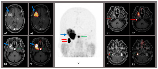
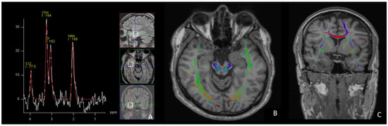
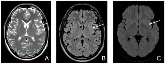
A 61-year-old patient was diagnosed with a left-sided falx meningioma. Histopathological analysis following extirpation showed a meningothelial meningioma ZNS WHO grade 1 with sparse mitoses. Over the course of 12 years, the patient received irradiation (54.0 Gy), peptide radio-receptor therapy (177Lu-DOMITATE)
[...] Read more.
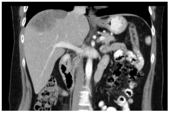
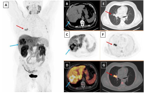
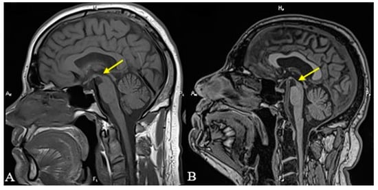
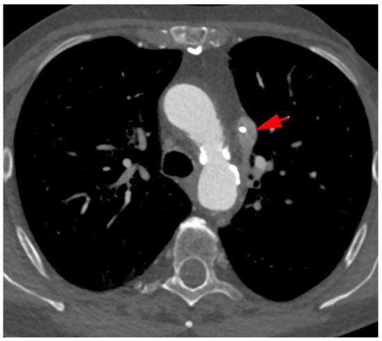
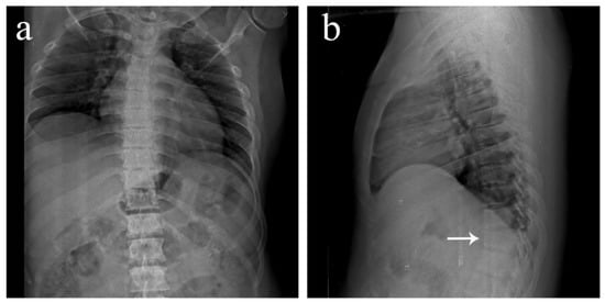
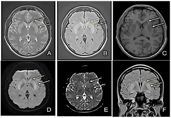
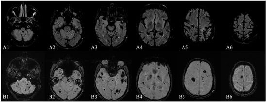
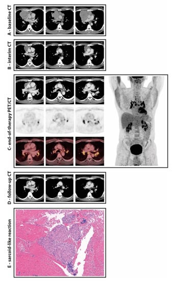
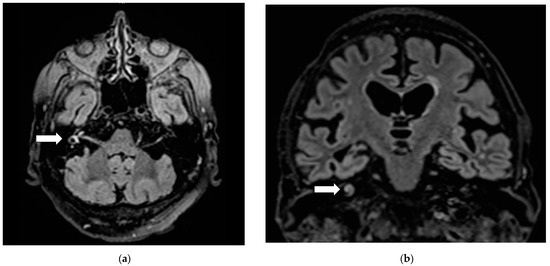
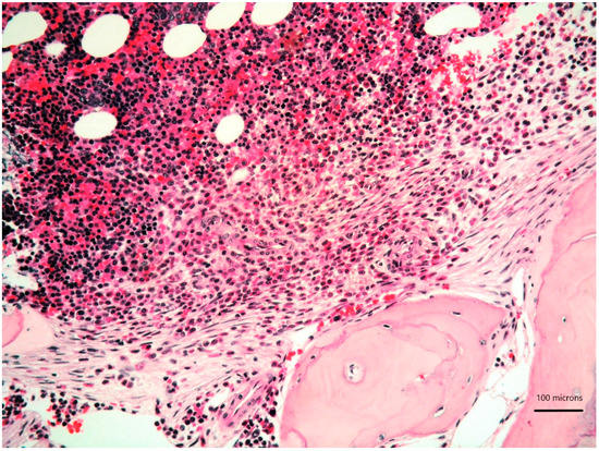
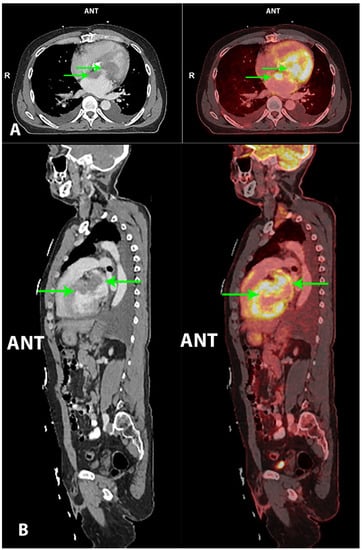
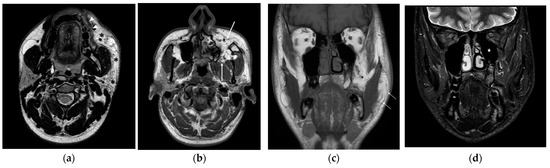
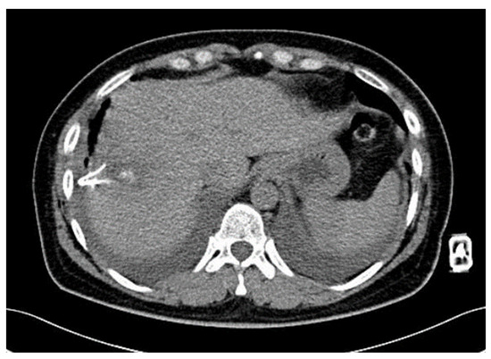
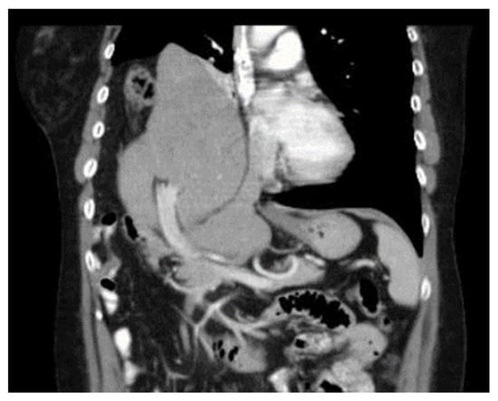
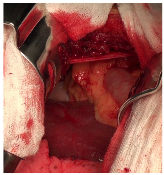
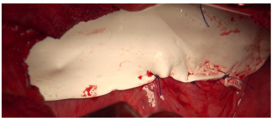
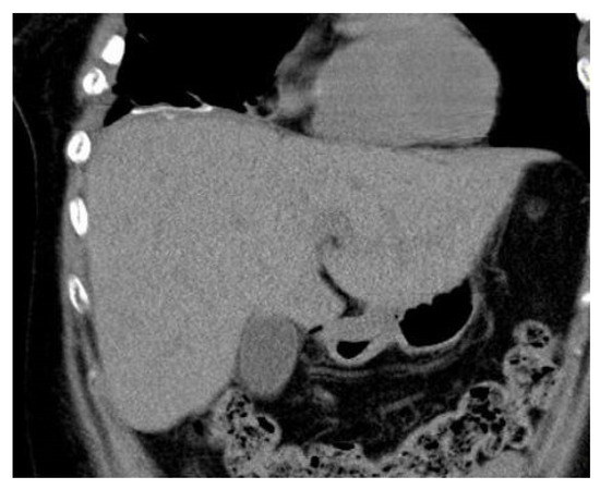
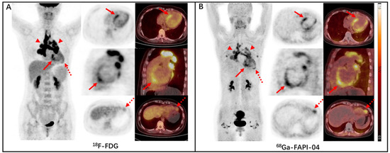
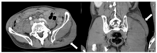
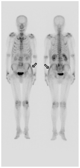
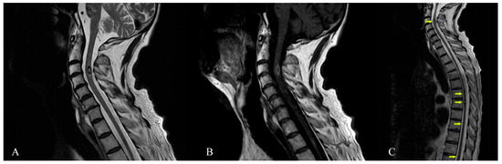
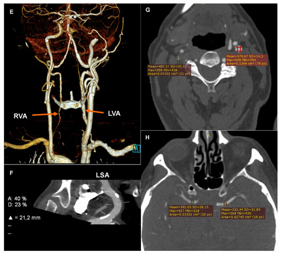
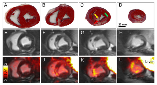
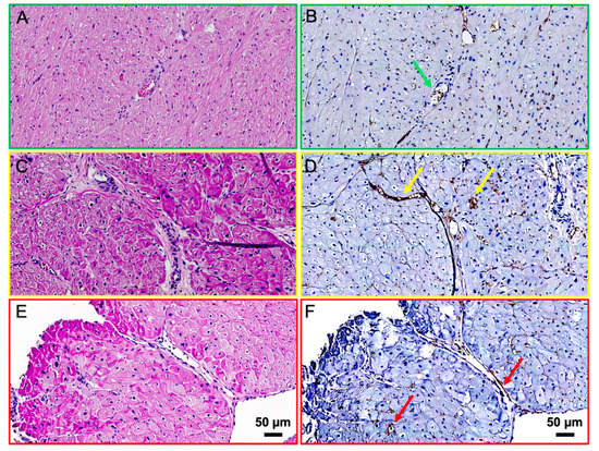
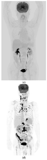
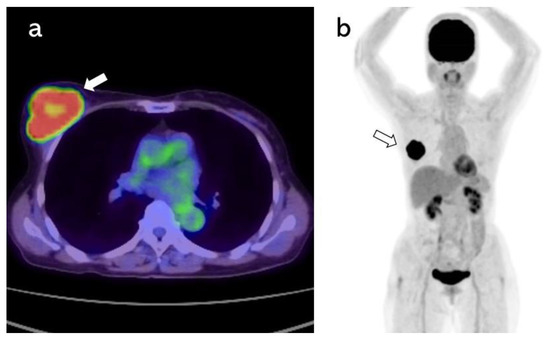
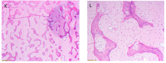
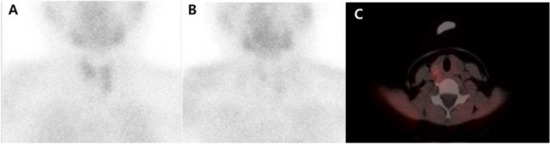
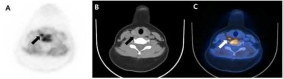
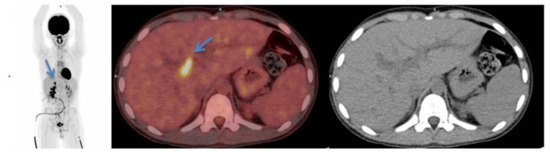
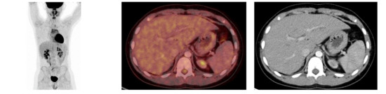
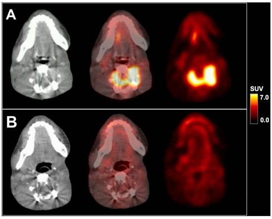
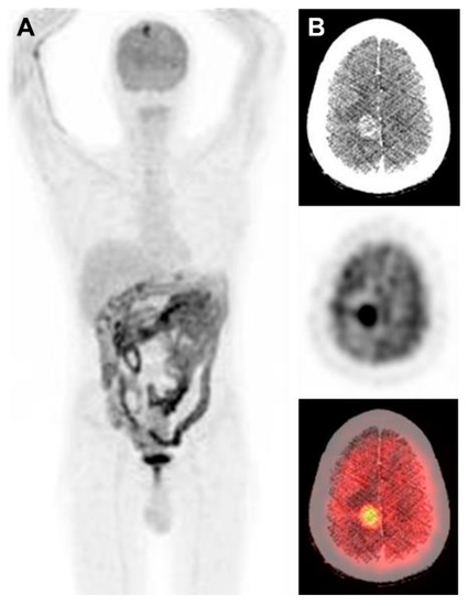
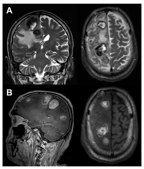
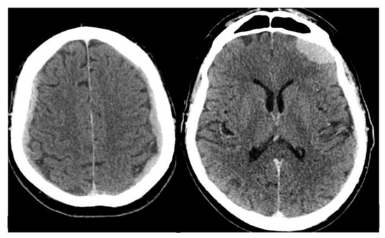
A 61-year-old patient was diagnosed with a left-sided falx meningioma. Histopathological analysis following extirpation showed a meningothelial meningioma ZNS WHO grade 1 with sparse mitoses. Over the course of 12 years, the patient received irradiation (54.0 Gy), peptide radio-receptor therapy (177Lu-DOMITATE) and targeted therapy (mTOR inhibitor). Follow-up imaging revealed an increased size of the residual tumor. Due to increased liver function parameters, imaging of the liver was performed, showing widespread space-occupying lesions with atypical appearance. Biopsy revealed metastasis of the meningioma, now with 2.7 mitoses/mm2, necrosis and homozygous CDKN2A/B deletion, corresponding to an anaplastic CNS meningioma WHO grade 3. A second small meningioma on the left petroclival side has been consistent in size over 12 years. Metastatic meningiomas pose a pertinent clinical challenge due to poor prognosis. The lung, bone, liver and cervical lymph nodes are the most common sites of extracranial metastasis. According to the World Health Organization criteria, the most important predictive factor for recurrence and metastasis is the tumor grade.
Full article
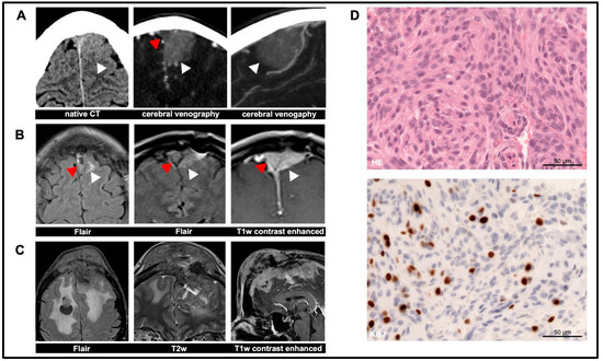
Figure 1


