Feature Papers in 'Biological and Bio- Materials' Section
Share This Topical Collection
Editor
 Dr. Bahman Anvari
Dr. Bahman Anvari
E-Mail
Website
Collection Editor
Department of Bioengineering, University of California Riverside, Riverside, CA 92521, USA
Interests: bioinspired materials; photonic materials; photomedicine; optical imaging; nanomedicine and nanobiotechnology; delivery systems; cell membrane mechanics
Special Issues, Collections and Topics in MDPI journals
Topical Collection Information
Dear Colleagues,
This Topical Collection, "Feature Papers in 'Biological and Bio- Materials' Section", aims to collect high-quality research articles, review articles, and communications on all aspects of biological and bio- materials. It is dedicated to recent advances in the research area of biological and bio- materials, and comprises a selection of exclusive papers from the Editorial Board Members (EBMs) of the 'Biological and Bio- Materials' Section, as well as invited papers from relevant experts. We also welcome established experts in the field to make contributions to this Topical Collection. Please note that all invited papers will be published online once accepted. We aim to represent our Section as an attractive open access publishing platform for biological and bio- materials research.
We invite submissions focusing on, but not restricted to:
- Molecular diagnostics, therapeutics, and theranostics.
- Biologically inspired materials and constructs.
- Biomimetics.
- Stem cells.
- Regenerative medicine.
- Self-assembling materials.
- Design, synthesis, and characterization of engineered materials.
- Delivery systems.
- Immunogenicity and toxicity.
- Smart, responsive, and bioactive materials.
Dr. Bahman Anvari
Collection Editor
Manuscript Submission Information
Manuscripts should be submitted online at www.mdpi.com by registering and logging in to this website. Once you are registered, click here to go to the submission form. Manuscripts can be submitted until the deadline. All submissions that pass pre-check are peer-reviewed. Accepted papers will be published continuously in the journal (as soon as accepted) and will be listed together on the collection website. Research articles, review articles as well as short communications are invited. For planned papers, a title and short abstract (about 100 words) can be sent to the Editorial Office for announcement on this website.
Submitted manuscripts should not have been published previously, nor be under consideration for publication elsewhere (except conference proceedings papers). All manuscripts are thoroughly refereed through a single-blind peer-review process. A guide for authors and other relevant information for submission of manuscripts is available on the Instructions for Authors page. Biomolecules is an international peer-reviewed open access monthly journal published by MDPI.
Please visit the Instructions for Authors page before submitting a manuscript.
The Article Processing Charge (APC) for publication in this open access journal is 2700 CHF (Swiss Francs).
Submitted papers should be well formatted and use good English. Authors may use MDPI's
English editing service prior to publication or during author revisions.
Published Papers (3 papers)
Open AccessArticle
Enhancing Osteoblastic Cell Cultures with Gelatin Methacryloyl, Bovine Lactoferrin, and Bioactive Mesoporous Glass Scaffolds Loaded with Distinct Parsley Extracts
by
Laura Isabel Arias-Rodríguez, Jesús L. Pablos, María Vallet-Regí, Martha A. Rodríguez-Mendiola, Carlos Arias-Castro, Sandra Sánchez-Salcedo and Antonio J. Salinas
Viewed by 907
Abstract
The increasing interest in innovative solutions for addressing bone defects has driven research into the use of Bioactive Mesoporous Glasses (MBGs). These materials, distinguished by their well-ordered mesoporous structure, possess the capability to accommodate plant extracts with well-established osteogenic properties, including bovine lactoferrin
[...] Read more.
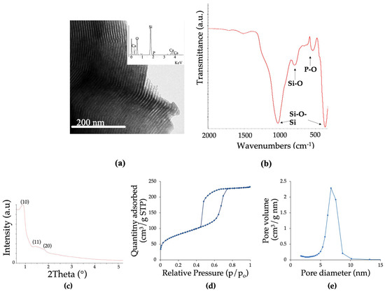
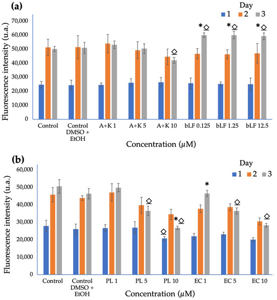
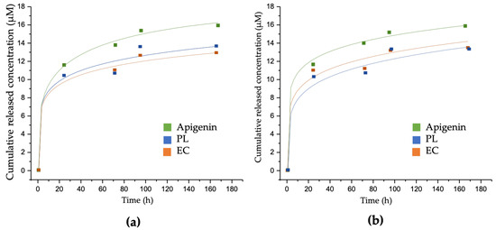
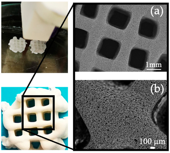
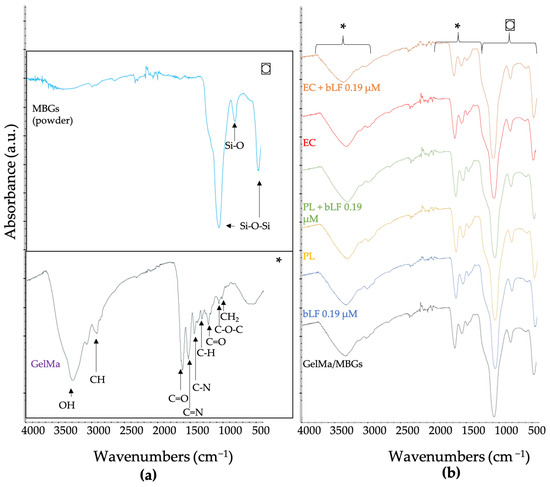
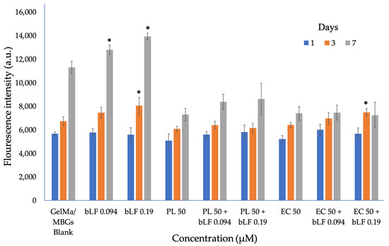
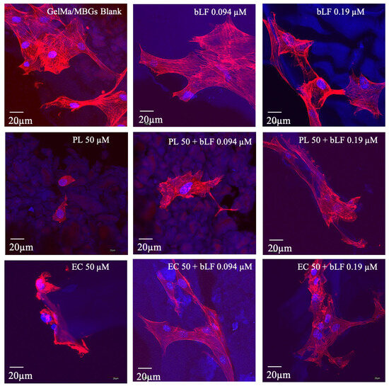
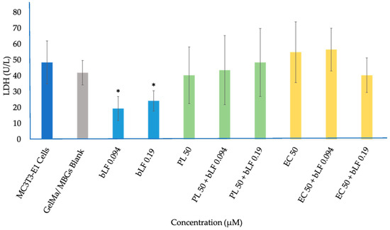
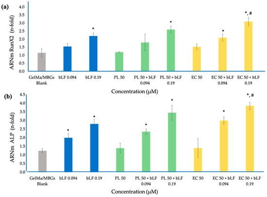
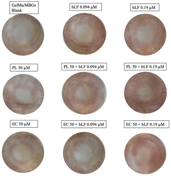
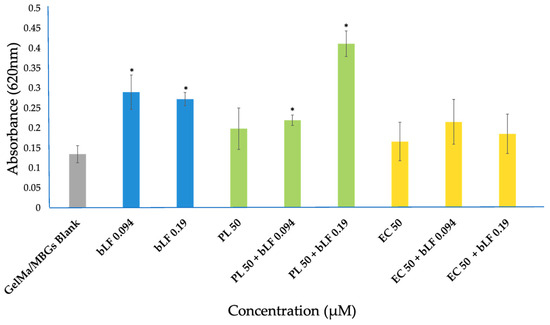
The increasing interest in innovative solutions for addressing bone defects has driven research into the use of Bioactive Mesoporous Glasses (MBGs). These materials, distinguished by their well-ordered mesoporous structure, possess the capability to accommodate plant extracts with well-established osteogenic properties, including bovine lactoferrin (bLF), as part of their 3D scaffold composition. This harmonizes seamlessly with the ongoing advancements in the field of biomedicine. In this study, we fabricated 3D scaffolds utilizing MBGs loaded with extracts from parsley leaves (PL) and embryogenic cultures (EC), rich in bioactive compounds such as apigenin and kaempferol, which hold potential benefits for bone metabolism. Gelatin Methacryloyl (GelMa) served as the polymer, and bLF was included in the formulation. Cytocompatibility, Runx2 gene expression, ALP enzyme activity, and biomineralization were assessed in preosteoblastic MC3T3-E1 cell cultures. MBGs effectively integrated PL and EC extracts with loadings between 22.6 ± 0.1 and 43.6 ± 0.3 µM for PL and 26.3 ± 0.3 and 46.8 ± 0.4 µM for EC, ensuring cell viability through a release percentage between 28.3% and 59.9%. The incorporation of bLF in the 3D scaffold formulation showed significant differences compared to the control in all assays, even at concentrations below 0.2 µM. Combinations, especially PL + bLF at 0.19 µM, demonstrated additive potential, with superior biomineralization compared to EC. In summary, this study highlights the effectiveness of MBGs in incorporating PL and EC extracts, along with bLF, into 3D scaffolds. The results underscore cytocompatibility, osteogenic activity, and biomineralization, offering exciting potential for future in vivo applications.
Full article
►▼
Show Figures
Open AccessArticle
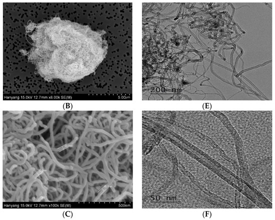
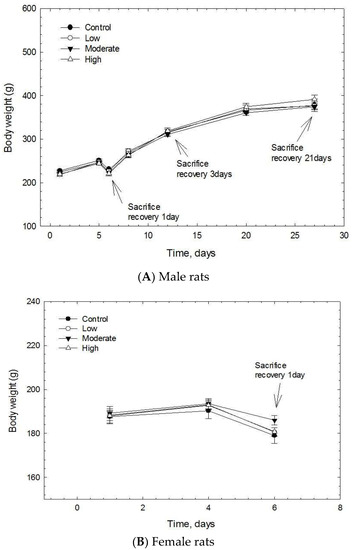
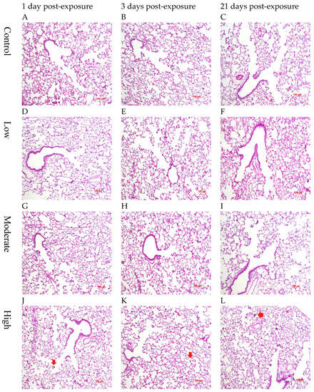
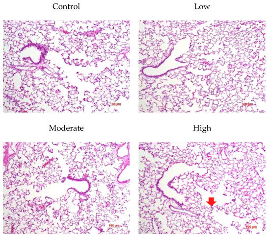
The Acute and Short-Term Inhalation of Carbon Nanofiber in Sprague-Dawley Rats
by
Mi Seong Jo, Boo Wook Kim, Young Hun Kim, Jin Kwon Kim, Hoi Pin Kim, Jae Hoon Shin, Gun Ho Lee, Kangho Ahn, Mary Gulumian and Il Je Yu
Cited by 1 | Viewed by 1329
Abstract
The inhalation toxicity of carbon nanofibers (CNFs) is not clearly known due to relatively few related studies reported. An acute inhalation study and short-term inhalation study (5 days) were therefore conducted using Sprague-Dawley rats. In the acute inhalation study, the rats were grouped
[...] Read more.
The inhalation toxicity of carbon nanofibers (CNFs) is not clearly known due to relatively few related studies reported. An acute inhalation study and short-term inhalation study (5 days) were therefore conducted using Sprague-Dawley rats. In the acute inhalation study, the rats were grouped and exposed to a fresh air control or to low (0.238 ± 0.197), moderate (1.935 ± 0.159), or high (24.696 ± 6.336 mg/m
3) CNF concentrations for 6 h and thereafter sacrificed at 14 days. For the short-term inhalation study, the rats were grouped and exposed to a fresh air control or low (0.593 ± 0.019), moderate (2.487 ± 0.213), or high (10.345 ± 0.541 mg/m
3) CNF concentrations for 6 h/day for 5 days and sacrificed at 1, 3, and 21 days post-exposure. No mortality was observed in the acute inhalation study. Thus, the CNF LC
50 was higher than 25 mg/m
3. No significant body or organ weight changes were noted during the 5 days short-term inhalation study or during the post-exposure period. No significant effects of toxicological importance were observed in the hematological, blood biochemical, and coagulation tests. In addition, the bronchoalveolar lavage (BAL) fluid cell differential counts and BAL inflammatory markers showed no CNF-exposure-relevant changes. The histopathological examination also found no CNF-exposure-relevant histopathological lesions. Thus, neither acute nor 5 days inhalation exposure to CNFs induced any noticeable toxicological responses.
Full article
►▼
Show Figures
Open AccessArticle
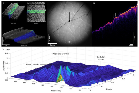
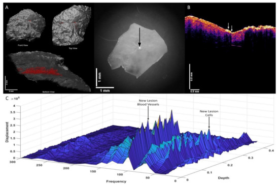
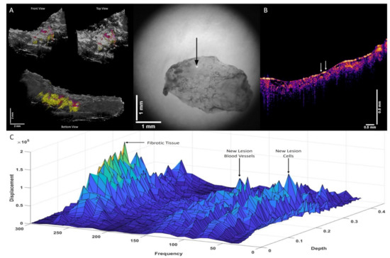
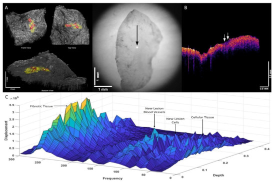
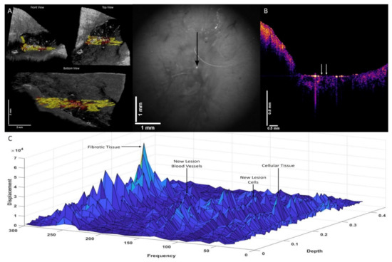
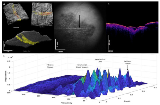
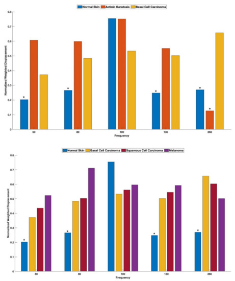
“Fingerprinting” Benign and Cancerous Skin Lesions Using Vibrational Optical Coherence Tomography: Differentiation among Cancerous Lesion Types Based on the Presence of New Cells, Blood Vessels, and Fibrosis
by
Frederick H. Silver, Tanmay Deshmukh, Nicole Ryan, Arielle Romm and Hari Nadiminti
Cited by 4 | Viewed by 1499
Abstract
In this study, we use vibrational optical coherence tomography (VOCT) to examine the morphology and stiffness of benign and cancerous lesions. Lesion images and 3D plots of weighted displacement versus frequency and depth were used to compare the cellular, dermal collagen, new blood
[...] Read more.
In this study, we use vibrational optical coherence tomography (VOCT) to examine the morphology and stiffness of benign and cancerous lesions. Lesion images and 3D plots of weighted displacement versus frequency and depth were used to compare the cellular, dermal collagen, new blood vessels, and fibrotic composition of normal skin, actinic keratoses (AK), nodular and superficial basal cell carcinomas (BCCs), squamous cell carcinomas (SCCs), and melanomas. The results of this study suggest that benign and cancerous lesions differ based on the addition of new cells with increased resonant frequency and stiffness (80 Hz, 1.8 MPa), new blood vessel peaks (130 Hz, 4.10 MPa) that appear to be less stiff than normal blood vessels, and new fibrous tissue peaks (260 Hz, 15–17 MPa) that are present in carcinomas but not in normal skin and only partially present (80 Hz and 130 Hz only) in AKs. Results obtained by creating images based on the location of the 80 Hz, 130 Hz, and 260 Hz peaks of cancerous skin lesions suggest that the fibrous tissue appears to surround the new cells and new lesion blood vessels. The results of this study suggest that the morphology and location of the fibrous tissues in relation to the new cancer-associated cells and lesion blood vessels may provide information on the invasiveness and metastatic potential of skin cancers. The invasiveness and metastatic potential of melanomas may be a result of the cancer-associated cells laying down fibrous tissue that is used as a pathway for migration. The new cancer-associated blood vessels in the vicinity of the new cancer-associated cells may promote this migration and eventual metastasis. The ratios of peak heights 50/130 Hz and 80/130 Hz of normal cells, new lesion cells, new lesion blood vessels, and fibrotic tissue may be used as a “fingerprint” for detecting melanoma and to differentiate it from other skin cancers non-invasively using VOCT.
Full article
►▼
Show Figures
Planned Papers
The below list represents only planned manuscripts. Some of these
manuscripts have not been received by the Editorial Office yet. Papers
submitted to MDPI journals are subject to peer-review.
Title: To be determined
Authors: Nathan Gallant; et al
Affiliation: Department of Mechanical Engineering , University of South Florida
Abstract: 1
Title: To be determined
Authors: Wei Seong Toh; et al
Affiliation: Faculty of Dentistry, National University of Singapore
Title: To be determined
Authors: Shinji Sakai; et al
Affiliation: Department of Materials Science and Engineering Osaka University, Japan


